Báo cáo khoa học: "Accelerated Partial Breast Irradiation (APBI): A review of available techniques" doc
Bạn đang xem bản rút gọn của tài liệu. Xem và tải ngay bản đầy đủ của tài liệu tại đây (1.91 MB, 28 trang )
Njeh et al. Radiation Oncology 2010, 5:90
/>
REVIEW
Open Access
Accelerated Partial Breast Irradiation (APBI):
A review of available techniques
Christopher F Njeh1*†, Mark W Saunders1†, Christian M Langton2†
Abstract
Breast conservation therapy (BCT) is the procedure of choice for the management of the early stage breast cancer.
However, its utilization has not been maximized because of logistics issues associated with the protracted
treatment involved with the radiation treatment. Accelerated Partial Breast Irradiation (APBI) is an approach that
treats only the lumpectomy bed plus a 1-2 cm margin, rather than the whole breast. Hence because of the small
volume of irradiation a higher dose can be delivered in a shorter period of time. There has been growing interest
for APBI and various approaches have been developed under phase I-III clinical studies; these include multicatheter
interstitial brachytherapy, balloon catheter brachytherapy, conformal external beam radiation therapy and intraoperative radiation therapy (IORT). Balloon-based brachytherapy approaches include Mammosite, Axxent electronic
brachytherapy and Contura, Hybrid brachytherapy devices include SAVI and ClearPath. This paper reviews the different techniques, identifying the weaknesses and strength of each approach and proposes a direction for future
research and development. It is evident that APBI will play a role in the management of a selected group of early
breast cancer. However, the relative role of the different techniques is yet to be clearly identified.
Introduction
Breast cancer is a worldwide problem, accounting for
10.4% of all cancer incidence among women, making it
the second most common type of non-skin cancer (after
lung cancer) and the fifth most common cause of cancer
death. In the USA, breast cancer has the highest incidence among all cancer types in females with one in
every eight to ten women being affected during her lifetime [1]; it is estimated that 192,370 women will be
diagnosed with, and 40,170 women will die of, cancer of
the breast in 2009 [2-4].
Breast cancer is the most common cancer in the UK
among women although it is rare in men. In 2006 there
were 45,822 new cases of breast cancer diagnosed in the
UK: 45,508 (over 99%) in women and 314 (less than 1%)
in men. Breast cancer is by far the commonest cancer in
women in the UK accounting for 31% of all cases. The
next most common cancer in women is lung cancer,
with 16,647 cases (11% of total) in 2006. So nearly, a
third of all new cancers in women are breast cancers. It
has been estimated that the lifetime risk of developing
breast cancer is 1 in 1,014 for men and 1 in 9 for
women in the UK. These were calculated using incidence and mortality data for 2001-2005 [5].
Early stage breast cancer is defined as stage II or less;
on the basis of the lack of lymph node, metastasis and
clinical lesion size of 2 cm or less [6]. The ‘surveillance,
epidemiology and end results’ (SEER) program reported
that in 2006, 60% of diagnosed breast cancers are early
stage [2,3]. Similarly in Japan, the fraction of early stage
breast cancer was reported to be 40.6% in 1996 [6].
With the increasing use of breast cancer screening by
mammography, more and more patients will have their
breast cancer diagnosed at the early stage. Hence, there
is a need for proper clinical management of early stage
breast cancer is required. Most women who are newly
diagnosed with early-stage breast cancer have a choice
of: breast-conserving surgery (such as lumpectomy), a
mastectomy (also called a modified radical mastectomy),
radiation therapy and systemic treatments.
* Correspondence:
† Contributed equally
1
Radiation Oncology Department, Texas Oncology Tyler, 910 East Houston
Street, Tyler, Texas, USA
Full list of author information is available at the end of the article
Rationale for Breast Conservation
Breast conservation therapy (BCT) is the procedure of
choice for the management of the early stage breast cancer. BCT consists of resection of the primary breast
© 2010 Njeh et al; licensee BioMed Central Ltd. This is an Open Access article distributed under the terms of the Creative Commons
Attribution License ( which permits unrestricted use, distribution, and reproduction in
any medium, provided the original work is properly cited.
Njeh et al. Radiation Oncology 2010, 5:90
/>
tumor (lumpectomy, segmental mastectomy or wide
local excision) followed by whole breast irradiation
(WBI). A total dose of 45-50 Gy is delivered to the
entire breast over 5 to 6 weeks (1.8 to 2 Gy per fraction). In most patients, a boost dose of 10-16 Gy to the
tumor bed is added. The establishment of BCT as the
standard of care resulted from many years of prospective studies such as the National Surgical Adjuvant
Breast and Bowel Project (NSABP) B-06 studies [7-9].
These studies found equivalent survival and local control rates among women treated with BCT compared to
those treated with mastectomy.
The value of radiation therapy as a breast conservation
component has been further validated by studies comparing lumpectomy alone to lumpectomy and radiation
therapy. These studies demonstrate a threefold reduction in recurrence with the use of radiation therapy following breast conserving surgery [7,10-13]. For patients
with ductal carcinoma in situ (DCIS), randomized studies comparing lumpectomy alone to lumpectomy plus
radiation therapy, conducted by the NSABP and
European organization for research and treatment of
cancer (EORTC) found a 55% and 47% reduction in the
ipsilateral breast cancer events respectively, with the
addition of radiation therapy [13,14]. These and other
studies have been recently pooled-analysed by Clarke et
al. [11] and Vinh-Hung et al. [12]. Vinh-Hung’s analysis
found that the relative risk of ipsilateral breast tumor
recurrence after breast-conserving surgery, comparing
patients treated with or without radiation therapy, was
3.00 (95% confidence interval [CI] = 2.65 to 3.40).
Further, the relative risk of mortality was 1.086 (95% CI
= 1.003 to 1.175), corresponding to an estimated 8.6%
(95% CI = 0.3% to 17.5%) relative excess mortality if
radiation therapy was omitted. BCT is well tolerated
with minimal long-term complications, favorable cosmetic outcome and reduced psychological trauma [7,9].
Radiation therapy therefore is an essential component of
BCT. It not only decreases local recurrence but
improves overall survival [11,12]. Because of these excellent results and the better cosmetic outcome, the United
States National Institute of Health released a consensus
statement, recommending breast conserving treatment
as the preferable option for women with early-stage
breast cancer [15].
Rationale for Accelerated Partial Breast
Irradiation (APBI)
Despite the advantages of BCT, its utilization remains a
problem [16]. It has been reported that many women
who are candidates for BCT do not receive it, only 10%
to 80% of patients actually receive it [17-19]. In addition
15% to 30% of patients who undergo lumpectomy do
not receive radiation therapy [20-22]. Similarly in Japan
Page 2 of 28
radiation therapy is performed in approximately 70% of
patients following breast conservation surgery [23]. The
under utilization of BCT has been associated with the
fact that some women cannot, or will not, commit to
the usual 6- 7 week course of adjunct conventional
radiation therapy that is part of the BCT package [24].
It has been further hypothesized that convenience,
access, cost and other logistical issues are major contributing factors. Other logistical issues include: distance
from the radiation therapy facility, lack of transportation, lack of social support structure and poor ambulatory status of the patient [18,25,26]. Other reasons that
may steer women away from BCT that have been identified include physician bias, patient age and fear of radiation treatments [22]. There has been a desire therefore
to identify a subset of women who may not benefit
from the addition of radiation therapy after lumpectomy
for early stage breast cancer; however, no such subset of
women has been identified [27].
Another criticism of BCT relates to consumption of
resources; while radiation therapy facilities in the USA
have largely kept up with demand for post-lumpectomy
radiation therapy, breast irradiation may constitute
25%-30% of patient visits and can stress a health-care
delivery system. However, not all countries have such
adequate resources. For example Palacios Eito et al. [28]
reported that the number of external irradiation units
available in Spain in 2004 (177) was clearly lower than
the number desirable (266-316). There is significant
shortage of radiation therapy equipment in most of Asia
and pacific regions [29], Latin America [30], Africa [31]
and Eastern Europe [32]. In Africa, the actual supply of
megavoltage radiation therapy machines (cobalt or linear
accelerator) was only 155 in 2002, 18% of the estimated
need [31]. In 12 Asia-Pacific countries with available
data, 1147 megavoltage machines were available for
an estimated demand of nearly 4000 megavoltage
machines [32].
The question that arises therefore is ‘can similar rates
of local control be achieved with radiation therapy delivered only to the area at highest risk for recurrence?’ If
so, radiation could be delivered in a significantly shortened period, thereby potentially making the BCT
option available and attractive to more women. This is
the concept of accelerated partial breast irradiation
(APBI) [26,33,34].
The stronger case for APBI has come from both retrospective and prospective studies; reporting that 44% to
86% of local recurrence occurs close to the tumor bed
[10,35-37]. Ipsilateral breast recurrences in areas other
than the tumor bed occurred rarely in 3% to 4% of the
cases [34]. An update of the NSABP B-06 trial also confirmed this pattern of local recurrence, with 75% of
recurrences at, or near, the lumpectomy site with other
Njeh et al. Radiation Oncology 2010, 5:90
/>
site ipsilateral breast recurrence rates similar to the
recurrence of contra-lateral second primary breast cancer [38]. Based upon this evidence, BCT, with whole
breast irradiation has been criticized as an overtreatment. Whole breast treatments incorporate the
entire breast (including the surgical cavity), overlying
skin, lower axilla and even small portions of the heart
and lung in the treatment fields; this may introduce
avoidable toxicity [39] whereas partial breast irradiation
spares more normal tissue.
An additional theoretical advantage of APBI is a
decreased dose to normal tissue. With a smaller target
volume, it may be expected that adjacent organs such as
the heart and lungs will receive less radiation. Radiation-induced lung injury after treatment for breast cancer, such as pneumonitis, lung fibrosis and pulmonary
function test changes, are well documented in the literature [40,41]. An increase in lung cancer incidence and
mortality after irradiation for breast cancer has also
been reported in large studies [42-45]. It worth noting
that the increase risk of long-term cardiac-related mortality after BCT may not be significant with modern
breast radiation therapy.
A number of pathology studies have also researched
local breast recurrence [46,47]. In the study by Holland
et al., mastectomy specimens from more than 300
women diagnosed with invasive breast carcinoma, who
fulfilled the criteria for breast conserving therapy, were
systematically investigated [47]. They found that of the
282 invasive cancers, 105 (37%) showed no tumor foci
in the mastectomy specimen around the reference mass.
In 56 cases (20%) tumor foci were present within 2 cm,
and in 121 cases (43%) the tumor was found more than
2 cm from the reference tumor [47]. This study justified
the concept that whole-breast treatment either with surgery or radiation therapy is necessary to achieve local
control. Supporters of APBI argue that this study was
flawed in its patient selection and that the quality of
mammography used at the time may have missed radiographic evidence of multicentric disease that would
today be detected [48]. Contrary to Holland’s data,
recent studies from women considered appropriate for
breast-conservation therapy reveal that the microscopic
extension of malignant cells is unlikely to be beyond
1 cm [49-51].
Accelerated Partial Breast Irradiation (APBI)
Techniques
APBI is an approach that treats only the lumpectomy
bed plus a 1-2 cm margin, rather than the whole breast.
By increasing the radiation fraction size and decreasing
the target volume, this technique allows the treatment
to be accomplished in a shorter period. APBI is generally defined as radiation therapy that uses daily fraction
Page 3 of 28
doses greater than 2 Gy delivered in less than 5 weeks.
There are a number of approaches now available for the
implementation of APBI, these include: multi-catheter
interstitial brachytherapy, balloon catheter brachytherapy, 3D-CRT (conformal radiation therapy) and intraoperative radiation therapy (IORT). Each of these techniques is vastly different from one another in terms of
degree of invasiveness, radiation delivery, operator proficiency, acceptance between radiation oncologist and
length of treatment. It is important to review the basic
principles of radiobiology, as well as critical aspects of
patient selection, surgical endpoints and radiotherapy
treatment planning. This paper reviews the different
techniques, identifying the weaknesses and strength of
each approach and proposes a direction for future
research and development.
Multi-catheter Interstitial Brachytherapy (MIB) Treatment
Technique
Multi-catheter interstitial brachytherapy is the APBI
technique that has been utilized the longest and has the
most extensive follow-up [24,33,52]. This technique was
initially developed to provide boost radiation after whole
breast radiation therapy. Flexible after-loading catheters
are placed through the breast tissues surrounding the
lumpectomy. The catheters are inserted at 1 to 1.5 cm
intervals in several planes; firstly to ensure adequate
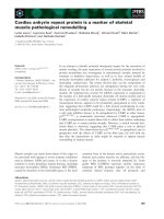
coverage of the lumpectomy cavity plus margins (Figure
1), and secondly, to avoid hot and cold spots. The procedure routinely requires between 14 to 20 catheters to
assure proper dose coverage; the exact number being
determined by the size and shape of the target, determined using established brachytherapy dosimetric guidelines [53,54].
Multiple catheters are placed in the breast using a
free-hand or template-guided approach. The configuration of the catheters and their relation to the tumor target volume are crucial for effective treatment. Catheter
insertion requires a high level of experience to produce
an implant of excellent quality. The incorporation computed tomography (CT) based 3D planning and imageguidance has made a significant impact on the quality of
the implants [55]. Determination of optimal catheter
configuration prior to the procedure (virtual planning)
would reduce the dependence of implant quality on the
expertise of the physician [56].
In MIB either low dose rate (LDR) or high dose rate
(HDR) brachytherapy may be used. With LDR, sources
of Ir-192 sources are implanted for approximately 2 to 5
days while the patient is admitted as an inpatient. HDR
however is an outpatient procedure, fractionated over
the course of a week, with each treatment varying
between seconds to minutes. The proposed dose of 34
Gy in 10 fractions BID (twice daily) for HDR was based
Njeh et al. Radiation Oncology 2010, 5:90
/>
Page 4 of 28
Figure 1 Diagrammatic illustration of multi-catheter interstitial brachytherapy.
on equivalence of the BED (biological effective dose) of
this schema to 45 Gy in 4.5 days of the LDR regimen
used in early APBI trials [57].
The majority of APBI patients treated with the longest follow-up have been treated with multi-catheter
interstitial brachytherapy. A systematic review of these
experiences have recently been presented by Offersen
et al. [52]. Polgar et al. [58] and Antonucci et al. [59]
have recently reported 12 year and 10 year follow-up
respectively. In the study of Antonucci et al., eight
ipsilateral breast tumor recurrences (IBTRs) were
observed in patients treated with MIB resulting in a 10
years cumulative incidence of 5% (95% confidence
interval [CI] 1.5-8.5%). The rate of incidence for WBI
was 4% (95% CI: 1.3-6.7%), which not statistically significantly different from MIB treated patients. Table 1
presents some of the reported MIB studies with more
than 5 years follow up.
Balloon-Based Brachytherapy Devices
The balloon based brachytherapy include Mammosite,
Axxent electronic brachytherapy, and Contura.
Table 1 Results of recent clinical experience with Interstitial brachytherapy with more than 5 years follow up
Author
No of
cases
Follow up interval
(years)
Dose rate/pt
no
Scheme
Total dose
(Gy)
5-year LR
(%)
Good/Excellent
cosmesis
Strnad et al.[60]
274
5.25
PDR/HDR
PDR = 0.6 Gy/hr
HDR = 4 Gy x8
PDR = 50 Gy
HDR = 32 Gy
2.9%
90%
Antonucci et al. [59]
199
9.6
LDR/HDR
LDR 0.52 Gy/h × 96
hours
HDR = 4 Gy x8
HDR = 3.4 Gyx10
LDR = 50 Gy
HDR = 32 Gy
HDR = 34 Gy
5%
99%
Johansson et al.[61]
50
7.2
PDR
50Gy/5
50
4%
56%
Arthur et al.[62]
99
7
LDR/HDR
LDR = 3.5 -5 days
HDR = 3.4 × 10
45 Gy (LDR)
34 Gy (HDR
4%
n/a
Polgar et al.[63]
128
6.8
HDR
5.2 × 7
36.4 Gy
4.7%
77%
King et al [64]
51
6.25
LDR/HDR
LDR = 4 days
4 Gyx8
45 Gy (LDR)
32 Gy (HDR)
3.9%
75%
Otto et al. [65]
274
5.25
PDR/HDR
PDR 5 days, 0.6 Gy/
hr
HDR = 4 Gyx8
49.8 Gy (PDR)
32 Gy (HDR)
2.9%
92%
Polgar et al.[58]
45
11.1
HDR
4.33 × 7
5.2 × 7
30.3 Gy
36.4 Gy
4.4%
78%
LR = local recurrence, HDR = high dose rate, LDR = low dose rate, PDR = pulsed dose rate, n/a = data not available
Njeh et al. Radiation Oncology 2010, 5:90
/>
1 MammoSite
Although MIB has had very encouraging results, the
technical challenges limit its widespread application.
The MammoSite® brachytherapy (MSB) system (Hologic,
Marlborough, MA) applicator was developed to be more
reproducible, easily applied and better tolerated. The
mammosite catheter consists of a silicone balloon connected to a 15 cm double-lumen catheter (Figure 2) that
is 6 mm in diameter. The catheter has both a small
inflation channel and a channel for the passage of an Ir192 high dose rate (HDR) brachytherapy source. The
source channel runs centrally through the length of the
balloon. The balloon is inflated with saline solution
mixed with a small amount of contrast material to aid
visualization. The balloon is inflated to a size that would
completely fill the lumpectomy cavity and ensures conformance of the tissue to the balloon. An Ir-192 radioactive source, connected to a computer-controlled HDR
remote after-loader, is inserted through the catheter into
the balloon to deliver the prescription radiation dose
[66,67].
The MammoSite applicator can be placed into the
lumpectomy cavity at the time of surgery or in a separate procedure after surgery. In the latter case, the applicator can be inserted under ultrasound guidance either
through the lumpectomy scar or via small separate incision. Following placement, a computed tomography
(CT) scan is performed to assess the quality of the
implant and for use in radiation planning. Implant
Page 5 of 28
quality is determined by examination of three parameters: balloon conformance to the lumpectomy cavity,
distance from the surface of the balloon to the skin surface, and the symmetry of the balloon in relationship to
the central catheter. Treatment planning parameters are:
the diameter of the inflated balloon, the planning target
volume, and the dose distribution [66-68]. While a minimum balloon-to-skin distance of 5 mm is required, a
threshold of at least 7 mm is strongly recommended
[69,70]. A longer skin distance is associated with greater
improvement in cosmesis [71]. Conformance of the balloon to the lumpectomy cavity is assessed by quantifying
the volume of the planning target volume (PTV) that is
filled by air or seroma fluid. Adequate conformance is
considered to have been achieved when less than 10% of
the PTV is composed of fluid or air. A symmetric
implant in relation to the source channel is also essential for adequate dosimetry. A non-symmetrical implant
can result in dose inhomogeneity in the surrounding tissues since the MSB device contains a single, central
source channel that does not allow for shaping of the
radiation isodose curves in the direction perpendicular
to the central channel [67]. The MSB may not be suitable in patients with small breast or for tumors located
in the upper-inner quadrant because of the requirement
for skin-to-cavity distances. Recently, Hologic has introduced a MammoSite Multi-lumen (4 lumen) device with
the potential to eliminate some of the drawbacks of the
single lumen device (see figure 3)
Figure 2 The MammoSite Balloon applicator (courtesy of Hologic, Marlborough).
Njeh et al. Radiation Oncology 2010, 5:90
/>
Page 6 of 28
Figure 3 The MammoSite Multilumen System (courtesy of Hologic, Marlborough).
The MSB radiation therapy device generally delivers
34 Gy over 10 fractions (3.4 Gy per fraction, twice daily
(BID)). The prescription point is 1 cm from the balloon
surface with a minimum of 6 hours between fractions
on the same day.
The MSB was approved by the USA food and drug
administration (FDA) in May of 2002 and September
2009, the multi-lumen device was also approved. Bensaleh et al [68] and Shah and Wazer [72] have recently
reviewed the MSB system. There are limited published
data regarding the long-term tumor control and cosmesis associated with MSB. However, the results thus far
are promising. Some of the studies with more than 12
months follow up are presented in Table 2, with the
longest follow up published by Benitez et al. [73]. In this
study, 43 patients were treated with MSB and had a
median follow up of 65 months. So far, no loco-regional
recurrences have been identified, with cosmetic outcomes of good to excellent achieved in 81.3% of the
patients. Toxicities were significantly less frequent in
patients with skin spacing of greater than 7 mm. The
American Society of Breast Surgeon (ASBS) [71] registry
trial recently reported 1440 patients treated, with a median follow up of 30.1 months. There have been 23 cases
(1.6%) of ipsilateral breast tumor recurrence for a twoyear actuarial rate of 1.04%. The cosmetic outcome of
good to excellent was 95% at 12 months. For a subset of
patients (n = 194) with DCIS in the ASBS registry, 6
patients (3.1%) had an ipsilateral breast recurrence, with
1 (0.5%) experiencing recurrence in the breast and axilla,
for a 5-year actuarial local recurrence rate of 3.39% [74].
The acute and late-term toxicity profiles of MSB have
been acceptable. Cosmetic outcome is improved by
proper patient selection and infection prevention [70].
Table 2 Results of some of the recent clinical experience
with Mammosite Brachytherapy System with more than a
year follow up
2. Axxent Electronic Brachytherapy
Author
No of
cases
Median follow up
interval (months)
IBF
Good/
Excellent
cosmesis
81.3%
Benitez et al.[73]
43
65
0%
Niehoff et al [69]
11
20
0%
n/a
Patel et al.[75]
26
48.5
0%
n/a
95%
Vicini et al.[71]
1440
30
1.6%
Chen et al.[76]
70
26.1
5.7%
n/a
Belkacemi et al. [77]
25
13
0%
84%
Voth et al.[78]
55
24
3.6%
n/a
Dragun et al. [70]
90
24
2.2%
90%
Vicini et al.[79]
1440
60
2.6%
90.6%
Jeruss et al. [74]
194$
54.4
3.1%
92%
n/a data not available, IBF = ipsilateral breast failure, $ these are ductal
carcinoma in situ (DCIS) patients recruited in the American Society of Breast
Surgeons APBI registry trial.
Since the MSB has shown promising results, other
forms of balloon-based brachytherapy have been developed. The novel Axxent electronic brachytherapy (eB)
system (Xoft, Fremont, CA) is a modified form of balloon-based brachytherapy [67,80] (Figures 4, 5, 6). It is
similar to the MammoSite system, consisting of a balloon catheter that is inserted into the lumpectomy cavity
by means of a percutaneous approach. The catheter
similarly has a central lumen through which the source
is inserted. A second port enables inflation of the balloon with saline and a third port may be attached for
drainage of seroma fluid or air surrounding the lumpectomy cavity. The wall of the balloon is covered in
radiolucent material that is visible on a plain x-ray film
or CT scan: addition of radiographic contrast is not
therefore required. The Axxent electronic brachytherapy
system is novel in that it uses an electronic 50 kilo-voltage x-ray source rather then an iridium-192 ( 192 Ir)
Njeh et al. Radiation Oncology 2010, 5:90
/>
Page 7 of 28
Figure 4 Axxent electronic brachytherapy, controller front view (courtesy of Xoft).
high-dose-rate (HDR) source. The X-ray source consists
of a miniature x-ray tube that is inserted into the balloon catheter and delivers the radiation therapy to the
patient. The eB controller is a portable unit, consisting
of a digital touch-screen for the Physician and Physicist
to input treatment data and monitor treatment progress
[67].
This approach implies that a specifically shielded
radiation room or an HDR afterloader unit are not
required, both of which are needed for treatment with
brachytherapy using Ir-192. The elimination of these
requirements potentially open-up this APBI approach to
a wider usage, particularly for patients who do not live
in close proximity to a radiation center with a HDR
Njeh et al. Radiation Oncology 2010, 5:90
/>
Page 8 of 28
Figure 5 Axxent electronic brachytherapy, HDR X-ray source (courtesy of Xoft).
after-loader unit. Since a shielded room is not required
for treatment and the eB device is very portable, the
number of setting in which the device can be used
increases. It has also been suggested to use the device
for intra-operative radiation therapy [81] and 11 patients
have successfully had IORT using the eB device [82]. eB
received FDA clearance for the treatment of breast cancer in January of 2006.
One inherent problem with MSB techniques is the
high skin dose when the excision cavity is near the skin
surface; that can result in late effect skin toxicity. The
Axxent source model S7500 has pronounced anisotropy
resulting in decreased dose at the proximal portion of
the balloon [83]; this can be used as an advantage to
optimize skin dose, particularly, if the cavity to skin distance is small. This anisotropy can also be accounted
Figure 6 Axxent electronic brachytherapy, balloon applicator (courtesy of Xoft).
Njeh et al. Radiation Oncology 2010, 5:90
/>
for by placing a dwell position outside the balloon surface along the proximal end of the catheter [67].
Being a relatively new device, there is a dearth in clinical experience and hence there are no clear recommendations on clinical use, for example, the surface-to-skin
distance using electronic brachytherapy. Chen et al.
recently reported a case report of radiation recall associated with the eB device and docetaxel administration
[84]. They argued that the prescription of 34 cGy at 1
cm may result in a higher skin dose (when the skin to
balloon distance is less than 1 cm) for eB because of the
relatively higher fall off rate of the 50 KVp photon compared to Ir-192. The patient that they reported had a
surface-to-skin distance of 7.5 mm, greater than the 7
mm MammoSite guideline. The calculated dose to the
skin was approximately 537 cGy per fraction. If an 192Ir
source had been used instead, the skin dose would have
been approximately 470 cGy per fraction, corresponding
to a relative dose increase for the electronic source of
approximately 14%.
Another potential contributing factor is the increase in
relative biologic effectiveness (RBE, the ratio of doses for
photons of differing energies required to produce the
same biologic effect) related to the lower energy of the
photons emitted by the electronic brachytherapy source.
It is well established that the biological effectiveness of
low-energy photons is large compared with higherenergy gamma rays, because of the dominance of photoelectric absorption at low energies [85]. The RBE for a
40 kVp source (very similar to the Axxent photon spectrum) has been calculated to be 1.28 greater than. an
192
Ir source [85]; hence, the dose from the 192-Ir source
must therefore be 1.28 times greater than that of the
low energy photon source to produce the same effect
(e.g., skin ulceration).
3. Contura
The balloon catheter of the Contura device (SenoRx,
Inc, Aliso Viejo, Ca) differs from the MSB and eB catheters in that it has multiple lumens for passage of an Ir192 HDR source (figure 7). In addition to a central
lumen, the Contura balloon has four surrounding channels to accommodate the HDR source. The positions of
the surrounding channels have a fixed 5-mm offset
around the central channel [67]. These channels provide
additional source positions and thus allow increased
dose flexibility compared with a single-catheter
approach. This approach has the potential to reduce the
dose to normal tissues (chest wall and skin) and organs
at risk such as the heart and lungs. In addition, multiple
catheters make it possible to account for asymmetric
balloon implant with respect to the central channel.
Like the eB catheter, Contura has a port for a vacuum
to remove fluid or air around the lumpectomy cavity;
the use of this vacuum port can improve tissue-balloon
Page 9 of 28
conformance. The Contura device received FDA clearance in May 2007.
MSB has the longest duration in follow up and new
APBI devices compare its clinical efficacy to that of
MammoSite. A recent study by Wilder et al. [86] evaluated one hundred and eighty-two women with early
breast carcinoma treated with post lumpectomy brachytherapy using Contura (n = 45) and MammoSite
(n = 137) devices with a median follow-up of 16
months. A Contura catheter did not require explantation in 16% (7 of 45) of patients where balloon-to-skin
spacing was only 3-6 mm and 11% (5 of 45) of patients
where there was an air/fluid pocket greater than 10% of
the planning target volume for plan evaluation. A MammoSite catheter was explanted in 10% of cases where
the minimum balloon-to-skin distance was <7 mm and
in 13% of cases where there was a large air/fluid pocket
next to the balloon. They observed incidence rates of
acute toxicity with a Contura device similar to those
with a MammoSite device [86]. Brown et al. [87] have
also reported similar improvements in dosimetric capabilities (i.e., reduced skin and rib doses and improved
PTV_EVAL coverage) with the Contura device.
Hybrid Brachytherapy Devices
Hybrid devices were developed to take advantages of the
versatility and dosimetric conformity of multicatheter
interstitial brachytherapy with the convenience and aesthetics of a single entry device. There are currently two
devices in this category namely the Struts Adjusted
Volume Implant (SAVI) and the ClearPath.
1. Strut Adjusted Volume Implant (SAVI)
The SAVI device (Cianna Medical, Aliso, Viejo, Ca)
(Figure 8) consists of a central strut surrounded by 6, 8
or 10 peripheral struts, depending on the size of the
device [67,88]. The peripheral struts can be differentially
loaded with a HDR source. The device is inserted in collapsed form through a small incision; once placed, it is
then expanded to fit the lumpectomy cavity by clockwise rotation of a knurled knob at the proximal end of
the expansion device, expanding the peripheral struts
and providing a pressure fit [89]. The outward pressure
exerted by the expanded struts pushes against the cavity
walls securing the struts in place. Some tissue invagination between the struts has been observed during the
course of the treatment. Radio-opaque markers are present on three of the peripheral struts (number 2, 4 and
6) for identification during the reconstruction process in
treatment planning.
The SAVI device is surgically implanted on an outpatient basis by the treatment radiation oncologist using
ultrasound guidance with the patient under local
anesthesia. A CT scan is acquired immediately following
the implant surgery, both for the verification of the
Njeh et al. Radiation Oncology 2010, 5:90
/>
Page 10 of 28
Figure 7 The Contura balloon applicator (courtesy of SenoRx).
proper deployment of the device, and for treatment planning. It was recommended by Scanderbeg et al [89], that
although the device does not move independently to the
body, one should always try to attain a position as close
to the planned patient position due to breast deformation. They found a breast board to be best for patient
setup because of its ease of setup and reproducibility.
2. ClearPath (CP)
Another hybrid device similar to the SAVI has also been
developed called ClearPath (CP; North American Scientific (Chatsworth, CA)). CP was developed to combine
the advantage of balloon brachytherapy and multicatheter brachytherapy. The CP consists of both inner and
outer catheters that expand by rotating a knob on the
base of the device (Figure 9) [67,90]. The CP device
contains six outer expandable plastic tubes to displace
the tissue. The radii of expansion of these tubes are
adjusted at the base of the device and can be expanded
to conform to a similar shape and size as a balloon
device. In the center of the expandable tubes is a central
catheter surrounded by six additional catheters that
allow the passage of an HDR Iridium-192 source. In
contrast to the SAVI device, the radiation source is not
in direct contact with the breast tissue. In addition, after
the device is placed in the patient, the rubber sleeve is
sutured to the patient, and the base of the device is cut
off. This leaves only the catheters exposed and visible
external to the patient’s skin [91]. Normally a cap is
placed over the HDR channels. This could potentially
lead to increased patient comfort by eliminating the
dangling external catheters.
CP is a relatively new device and hence no clinical outcome data have been reported. However, retrospective
dosimetric analysis has been reported [90,91]. Dickler
et al. [91] found that MSB and CP offered comparable
target volume coverage, but CP allowed significantly
more normal-tissue sparing. Similarly, Beriwal et al.
simulated a phantom study and the parameters of the CP
catheter were superimposed on the MSB planning CT
scans. The authors found that the median maximum skin
dose was 161% for MSB and 113% for CP of the prescription dose [90].
External Beam Radiation Therapy (EBRT)
Several techniques may be classified as ‘external beam
radiation therapy’ including 3D-conformal radiation
therapy (3D-CRT) with multiple static photons, and/or
electrons fields, intensity modulated radiation therapy
(IMRT) and proton beams [92]. The most widely used
3D-CRT approach was initially described by Baglan et al
[93]. This technique was adopted for use as one of the
allowed treatment modalities for patients randomized to
APBI in the National Surgical Adjuvant Breast and
Bowel Project B- 39/Radiation Therapy Oncology group
Njeh et al. Radiation Oncology 2010, 5:90
/>
Page 11 of 28
Figure 8 Different sizes of SAVI with peripheral struts expanded (courtesy of Cianna Medical).
(NSABP/RTOG) 0413 phase III trail [94]. The technique
uses four to five tangentially positioned non-coplanar
beams (Figure 10). The tumor bed is defined by the
computed tomography visualized seroma cavity, postoperative changes, and surgical clips, when available.
The clinical target volume (CTV) is defined as the
tumor bed with a 1.5 cm margin limited by 0.5 cm from
the skin and chest wall. The planning tumor volume
(PTV) is defined as the CTV with a 1.0 cm margin. The
prescription dose used for NSABP/RTOG protocol is
3.85 Gy twice daily (separated by at least 6 hours) to a
total dose of 38.5 Gy delivered within 1 week [94].
EBRT has many potential advantages, over the other
techniques [95].
1. The technique is non-invasive and the patient is
not subjected to a second invasive surgical procedure
or anesthesia, thereby reducing the potential risk of
complications. The treatment can wait until completion of pathological analysis about the original
tumor and the status of the resection margins are
available.
2. The technique has potential for widespread availability since most radiation therapy centers already
perform 3D-CRT for other cancers.
3. It is likely that an external beam approach will be
easier for radiation oncologists to adopt than brachytherapy techniques because the technical
demands and quality assurance issues are much
simpler.
4. Treatment results with external beam may be
more uniform between radiation oncologists because
the outcome depends less on the experience and
operative skills of the person performing the procedure than for brachytherapy (especially using interstitial implantation).
5. It seems less likely that technical issues arising
during external beam radiation therapy will require
the procedure to be aborted as is not infrequently
the case when brachytherapy techniques are used.
6. External beam is intrinsically likely to generate
better dose homogeneity and thus may results in a
better cosmetic outcome when compared with bracytherapy techniques.
Njeh et al. Radiation Oncology 2010, 5:90
/>
Page 12 of 28
Figure 9 ClearPath device (a) the base detached (b) a cap placed over the HDR channels (courtesy of North America Scientific).
Despite the above appeal of EBRT APBI, many issues
and unanswered question remain. These include breathing motion, treatment setups variation, and the fractionation scheme adopted. The target may move during
breathing and the patient may be positioned differently
for different fractions. To avoid missing the planned target, a large treatment volume is used. A prone patient
position has been suggested by Formenti et al.[96] to
minimize target tissue movement during breathing. The
prone position also provides exceptional sparing of the
heart and lung tissues. Unfortunately, the prone position
is not widely used because it requires a special immobilization device and is uncomfortable for some patients.
The use of multiple treatment fields in 3D-CRT/IMRT
can increase the volume of normal tissue irradiated to
low or moderate doses (i.e increase in integral dose).
Also, 3D-CRT delivers higher doses to normal breast
tissue since the PTV around the lumpectomy cavity is
increased to account to breathing and setup errors [97].
The identification and contouring of the lumpectomy
cavity (LC) is another issue with 3D-CRT APBI. LC
determination is critical because treatment delivery is
Njeh et al. Radiation Oncology 2010, 5:90
/>
Page 13 of 28
Figure 10 3D-CRT typical 4-field arrangement for right sided lesions and 5 field arrangement for left sided lesions (reprinted with
permission from Baglan et al.[93].
delayed after breast surgery. Furthermore, the GTV and
CTV are generally defined as the contouring of a seroma within the lumpectomy cavity, expanded by some
margin, usually 1 cm [93]. However, the delineation of
the seroma could vary among different observers and
even among experienced ones [98]. It has been suggested by Dzhugasvili et al. [99] that the use of surgical
clips as fudicial markers may reduce such observer
variability.
There is still the question of the appropriate dose and
fractional scheme for 3D-CRT APBI. As evident in
Table 3 different doses and fractionation schemes have
been reported in the literature. Rosenstein et al. [100]
assessed the biologically equivalent doses (BEDs) of several APBI schedules using a linear quadratic model.
Using an a/b ratio of 10, they found the Vicini fractionation scheme provided a BED of 53 Gy, the Formenti
fractionation scheme gave 48 Gy and the 32-Gy dose
used by Taghian et al. [101] gave a BED of 45. Livi et al.
[102] in randomized Phase III trial have used a dose of
30 Gy in five fractions (6 Gy/fraction) and argued that it
was equivalent to 54 Gy in a standard fractionation of 2
Gy fractionation. However, Cuttino et al. [103] utilizing
a wide range of established radiobiological parameters,
determined that the maximum fraction size needed to
deliver a biologically equivalent dose using 3D-CRT is
3.82 Gy, supporting the continued use of 3.85Gy BID in
the current national cooperative trial.
Intra-Operative Radiation Therapy Techniques
Intra-operative radiation therapy (IORT) refers to the
delivery of a single fractional dose of irradiation directly
to the tumor bed during surgery. These techniques have
been reviewed by Reitsamer et al. [112], Vaidya et al.
[113,114] and Orecchia and Veronesi [115]. Older intraoperative radiation therapy devices were technically
cumbersome, commonly relying on the transportation of
the patient from the operating theatre to the radiation
therapy unit during surgery, or require custom-built
intra-operative radiation therapy theatres [113]. These
technical and financial limitations to delivery of intraoperative radiation therapy have prevented widespread
use of the approach. Advances in miniaturization technology have enabled the development of mobile intraoperative radiation therapy devices. Intra-operative
radiation therapy was first used in 1998 with a device
called the Intrabeam, since then, two other mobile linear
accelerators have become available (the Mobetron and
Novac-7 systems). These systems either generate megavoltage electrons (Mobetron and Novac-7) or kilovoltage
photons (intrabeam).
The potential advantages of IORT include delivering of
the radiation before tumor cells have a chance to proliferate. Furthermore, tissues under surgical intervention
have a rich vascularization, with aerobic metabolism,
which makes them more sensitive to the action of the
radiation (oxygen effect). Also, the radiation is delivered
under direct visualization at the time of surgery. IORT
could minimize some potential side effects since skin and
the subcutaneous tissue can be displaced during the
IORT to decrease dose to these structures, and the
spread of irradiation to lung and heart is reduced significantly [116]. IORT eliminates the risk of patients not
completing the prescribed course of breast radiotherapy
(a well-recognized risk of conventional breast radiotherapy) and allows radiotherapy to be given without delaying
administration of chemotherapy or hormonal therapy
[117]. IORT has the potential for accurate dose delivery:
by permitting delivery of the radiation dose directly to
the surgical margins, IORT eliminates the risk of geographical miss in which the prescribed radiation dose is
inaccurately and incompletely delivered to the tumor
Njeh et al. Radiation Oncology 2010, 5:90
/>
Page 14 of 28
Table 3 Accelerated partial breast irradiation clinical studies using external beam radiation
Author
No of cases
Follow up (months)
Fractionation scheme
IBF
Vicini et al[104]
52
54
3.85 Gy × 10 (bid)
6%
Good/Excellent cosmesis
n/a
Vicini et al.[105]
91
24
3.85 Gy × 10 (bid)
0%
90%
89%
Chen et al. [106]
94
51
3.85 Gy × 10 (bid)
1.1%
Taghian et al.[107]
99
36
3.2 Gy × 4 (bid)$
2%
97%
Formenti et al.[108]
10
36 (minimum)
5.0, 5.5, 6.0 Gy × 5 (10 days)
0%
100%
n/a
Formenti et al.[96]
47
18
6.0 Gy x5 (10 days)
0%
Magee et al.[109]
353
96 (mean)
5.0 - 5.31 Gy × 8 (10 days)&
25%
n/a
Leonard et al. [110]
55
34 median
3.85 cGy x10 (bid)
0%
n/a
Hepel et al.[94]
60
15
3.85 Gy × 10 (bid)
n/a
81.7%
Jagsi et al.[111]
34
> 24
3.85 Gy × 10
n/a
79.5%
$
Technique used were: mixed photons and electrons (63 patients), photons alone (16 patients), and protons (20), &Technique was electron field with a beam
energy of 8-14 MeV, the majority being treated with 10 MeV, IBF = ipsilateral breast failure, n/a = data not available.
bed. Geographical miss may result from patient movement, inconsistent patient setup, and difficulty identifying
the tumor site weeks or months postoperatively and is
estimated to occur in up to 70% of patients receiving
conventional breast boost radiotherapy [118]. There is
potential for decreasing healthcare cost because it is one
fraction as opposed to 25 fractions.
With IORT the final pathology reports arrives days
post-festum. This has been one of the major criticisms
of the technique. So recently a novel handheld probe
(Dune Medical Devices, Caesarea, Israel) has been developed for intra-operative detection of positive margins
[119] Such a device can help reduce re-excision rate and
improve acceptance of IORT technique.
1. INTRABEAM (X-rays)
The mobile X-ray system Intrabeam™ is manufactured
by Carl Zeiss (Oberkochen, Germany) [120]. The system
is composed of a miniature, light-weight (1.6 kg) X-ray
source (PRS- 400), combined with a balanced floor
stand with six degrees of freedom to gain access to target sites throughout the body (Figure 11). The miniature
X-ray source has a probe of 10 cm length and 3.2 mm
diameter. Within this device, electrons are accelerated
to the desired energy level and focused down the probe
to strike a gold target. Various spherical applicators with
a diameter ranging from 1.5 to 5 cm are available to
match the size of the surgical cavity (Figure 12). They
are fixed to the end of the source and placed in the
excision cavity to obtain a homogeneous dose distribution on the surface of the applicator and consequently
on the surface of the tumor cavity. When mounted onto
the Intrabeam unit, each spherical applicator conforms
the breast tissue around the radiation source to permit
delivery of a uniform field of radiation to a prescribed
tissue depth. Accurate and uniform dose delivery is
further achieved by placement of “pursestring” sutures
within the breast to hold the pliable breast tissue against
the applicator surface [117].
The X-ray system produces low-energy photons (3050 KVp) with a steep dose fall-off in soft-tissue; no special shielding is therefore required in the room [120].
Dosimetry varies by applicator tip size with the commonly used 3.5 cm applicator sphere delivering 20 Gy at
a radius of 1 mm from the surface, 5 Gy at 10 mm and
1 Gy at 27 mm in about 20 minutes [113]. Treatment
time lasts for approximately 20 to 45 minutes, depending on the size of the lumpectomy cavity, the size of the
selected applicator, and the prescribed dose.
Treatment can be carried out in unmodified operating
rooms with minimal exposure to the staff and patient;
rapid dose fall-off in the tissue around the applicator
guarantees minimal exposure of the surrounding tissue
such as the lung and cardiac tissue in the patient.
The physics, radiobiology, dosimetry, and early clinical
applications of this low energy x-ray device have been
fully evaluated, and the device has received Federal
Drug Administration approval for use in any part of the
body since 1999 [121]. The RBE for this low-energy xrays have been estimated to be 1.5 [85]. It has been suggested that the biologically weighted dose (physical dose
× RBE) decreases with depth less quickly than physical
dose [85] Therefore despite the steep gradient in physical dose, an effective uniform biological dose is distributed inside a rim of about 15 mm around the most often
used intrabeam applicator [122]. Another potential
advantage of Intrabeam is that, because normal tissues
can repair their damaged DNA within a few minutes
but cancer cells with poor DNA- repair machinery may
be unable to repair quickly. So treatment given over a
long time (intrabeam is between 25-35 minutes) may
have a higher therapeutic index than giving similar
doses over 2 to 3 minutes [114].
Njeh et al. Radiation Oncology 2010, 5:90
/>
Page 15 of 28
Figure 11 The mobile X-ray intraoperative radiation therapy device: The Intrabeam device intraoperative photon device.
Encouraged by initial pilot studies (see Table 4), a
phase III, prospective randomized non-inferiority trial
called TARGIT (targeted intraoperative radiation therapy) began in March 2000. This trial compares single
dose intraoperative radiation therapy targeted to the
tumor bed to conventional whole breast external beam
radiation therapy in early breast cancer.[114,117].
Patients were enrolled from 28 centers in nine countries
including UK, Germany, Italy, USA and Australia. Data
accrual was closed in May 2010 and the results of this
trial have recently been published by Vaidya et al. [123].
In this trial 1113 patients were randomly assigned to the
targeted intraoperative radiotherapy group and 1119
allocated to the whole breast external beam radiation
therapy group. From this, 854 patients received targeted
intraoperative radiotherapy, only 142 received targeted
intraoperative radiotherapy with external beam radiotherapy and 1025 patients in the external beam radiotherapy group receiving the allocated treatment. They
observed at 4 years follow up, there were six local recurrences in the intraoperative radiotherapy group and five
in the external beam radiotherapy group. The KaplanMeier estimate of local recurrence in the conserved
breast at 4 years was 1.20% (95% CI 0.53-2.71) in the
targeted intraoperative radiotherapy and 0.95% (0.392.31) in the external beam radiotherapy group. The rate
Njeh et al. Radiation Oncology 2010, 5:90
/>
Page 16 of 28
Figure 12 Various spherical applicators with diameters ranging from 1.5 to 5 cm used in the intrabeam device (reprinted with
permission Holmes et al: [117].
of recurrence between the two groups was not statistically significant. Similarly the total rate of major toxicities was similar in the two groups[123]. This study
presents the first level 1 evidence of the equivalence of
APBI using IORT to WBI and confirms that targeted
IORT allows the entire dose of radiation therapy to be
administered in a single fraction at the time of breastconserving surgery, thus avoiding the need for repeated
radiation therapy treatments or placement of in dwelling
radiation therapy devices.
Table 4 Some clinical studies using Intra-operative radiation therapy (IORT)
Author
No of cases
Median follow up interval(months)
Technique
IBF
Good/Excellent cosmesis
Lemanski et al. [131]
42
30
Electrons
4.8%
100%
Veronesi et al.[130]
590
20
Electrons
0.5%
n/a
Mussari et al.[132]
47
48
Electrons
0%
92%
Vaidya et al.[121]
25
24
Photons
0%
Vaidya et al. [123]
$
854
48
photons
1.2% (95%CI = 0.53-2.71)
n/a
$
n/a
TARGIT phase III trial, at 4 years there 6 local recurrences in the target treated group and 5 in the whole breast treated group, giving the Kaplan-Meier estimates
(not crude estimates), n/a not available, IBF = ipsilateral breast failure.
Njeh et al. Radiation Oncology 2010, 5:90
/>
Page 17 of 28
2. MOBETRON (electrons)
3. NOVAC-7 (electrons)
The Mobetron (IntraOP Medical Inc, Santa Clara, CA),
is a mobile electron beam intraoperative treatment system. The Mobetron system (Figure 13a) is composed of
three separate units: the control console, the modulator
and the therapy module [124]. The control console
which operates the accelerator during radiation treatment delivery is placed outside the OR so that the radiation treatment delivery is controlled remotely. The
modulator houses the electronic systems of the accelerator and energizes the accelerator to produce the electron. The therapy module houses the accelerator guide
and control systems that generate and deliver radiation
[125]. The Mobetron uses two X-band (3 cm wavelength, 10 GHz frequency) collinear accelerators. This
design eliminates the need for a bending magnet thus
affecting a reduction in photon leakage [126]. The
Mobetron system produces electrons of nominal energies of 4 MeV, 6 MeV, 9 MeV and 12 MeV with therapeutic ranges up to 4 cm. The system is designed to
deliver a very large uniform dose of 10 to 25 Gy in a
single fraction at a dose rate of 10 Gy/min [124].
The NOVAC-7 system (Figure 13b) (Hitesys, Latina,
Italy) delivers electrons with the use of a mobile dedicated linear accelerator; its radiating head can be moved
by an articulated arm that can work in an existing operating room. It is based on a compact S-band standing
wave electron beam linear accelerator utilizing a
patented auto-focusing structure which eliminates the
need of focusing solenoids. The accelerator is moved by
six axis robotic arm. It delivers electron beams at four
different nominal energies (3, 5, 7 and 9 Mev) [113].
Beam are collimated by means of a hard docking system, consisting of cylindrical perspex applicators available in different diameters (4 to 10 cm) and angles of
the head (perpendicular or oblique 15° to 45° with
respect to their axis).
A phase III, prospective randomized trial called
ELIOT (electron intraoperative therapy) began in 2000
in Italy. A single dose of 21 Gy with energies up to 9
MeV, biologically equivalent to 58-60 Gy in standard
fractionation is applied to the tumor bed. The dose of
21 Gy was established from a dose-escalating phaseI/II
Figure 13 The mobile electron intraoperative radiation therapy devices: (a) Novac7 (b) Mobetron intra-operative electron device
(reprinted with permission Beddar el al. [124]).
Njeh et al. Radiation Oncology 2010, 5:90
/>
study [127]. The electron energy used is determined
from the depth of the tissue to be irradiated. The accelerators used have been designed to have a dose rate
(15-20 Gy per minute) higher than conventional and
can deliver 21 Gy in less than 2 min [115]. The entire
procedure last for about 15 to 20 minutes. Unnecessary
radiation to the underlying normal tissue can be avoided
by mobilizing the mammary gland during surgery and
placing a lead plate for shielding on its dorsal surface.
The costs of the mobile linear accelerator with a
robotic arm, used in intra-operative radiation therapy,
are prohibitive for poor countries. Frasson et al. [128]
evaluated the feasibility of ELIOT in the accelerator
room of the radiation therapy service for early breast
cancer treatment; demonstrating that intra-operative
radiation therapy with electrons can be safely performed
in an accelerator room with a conventional machine.
A systematic review by Cuncins-Hearn et al. [129]
concluded that the short-term results were similar for
both BCT and IORT in terms of local recurrence, disease-free and overall survival. The current evidence base
is however poor, making definitive assessment on IORT
very difficult. They suggested that further research is
required to clarify several issues such as identification of
the most appropriate subgroups of patients for IORT, a
comparison of the currently available mobile IORT technologies, establishing whether IORT is most appropriate
as a boost replacement dose or replacement for all postoperative radiation therapy, the examination of how biological repair processes may differ between the two
treatment modalities and determining precisely where
local recurrences originate with respect to the original
tumor site.
The IORT approach has the advantage of shortening
the treatment course further, conveniently delivering the
entire course of local therapy at the time of initial excision. However, IORT also presents significant technical
challenges, not the least due to the need for accuracy of
target definition and treatment delivery inherent in a
single dose radiotherapy delivery. One issue is the accuracy of tumor bed definition when tissues are reapproximated following excision. Another is the variable
margin of normal tissue irradiated in the re-opposed tissues. IORT may be complicated in patients who are
determined to have positive surgical margins and need
re-excision. This may not have been a significant issue
in the Versonesi studies [130], as all patients received
generous resections with quadrantectomies.
Discussion
The issue of the need and utility of APBI is highly
debated within the medical community. There are those
of the school of thought that the current standard of
care for early breast cancer works well. So, the frame of
Page 18 of 28
mind is “if it is not broken why fix it?”, evidenced by
the commentary in medical journals such as “Is APBI a
step backward?” [133]. On the other hand, there are
those who belief that APBI has a role to play in the clinical management of early stage breast cancer [134]. If
the proliferation of APBI techniques is anything to go
by, there is indeed a high level of interest. As reviewed
in this paper, there are several different approaches to
APBI, each with their merits and limitations (Table 5,
[135]). For those who believe in the role of APBI, there
is a general consensus that a few questions remain to be
satisfactorily addressed including the appropriate fractionation scheme, the appropriate patient selection criteria,
the need for phase II/III clinical trials establishing
equivalence or improvement to WBI and the appropriate technique.
Patient Selection
Patient selection is critical to the successful application
of APBI[136]. In a recent review, Polgar et al. [137]
argued that the relatively poorer results of early APBI
studies, with high local recurrence rates exceeding 1%
per year could be attributed to inadequate patient selection criteria and/or suboptimal treatment technique and
lack of appropriate QA procedures. Similarly in a recent
study by Chen et al. [76], 70 patients were treated with
MammoSite at the median follow up of 26.1 months,
four local failures were observed of which two did not
meet the ABS and ASBS selection criteria. These failures
highlight the need to better define the subset of patients
for whom APBI is most appropriate. Various societies
have now published recommendations of patient selection criteria for APBI. These include, the American
Society of Breast surgeons (ASBS), the American Brachytherapy Society (ABS), American Society for Radiation Oncology (ASTRO) and European Society for
therapeutic Radiology and Oncology (ESTRO)
[48,137,138]. The recent GEC-ESTRO recommendations
([137] have stratified the patients into three groups: low
risk, intermediate and high risk (contraindication for
APBI); similarly, ASTRO [138] has stratified them into
suitable, cautionary and unsuitable. The low risk (suitable) group describes patients where APBI outside of a
clinical trial would be considered acceptable (see Table
6); these criteria are stricter than those recommended
by the ASBS or ABS. However, less restrictive criteria
could be applied to patients who enrolled in a clinical
trial. Generally young patients (< 50 years) and those
who may harbor disease a significant distance from the
edge of the excision cavity or potentially have multi-centric disease should not be treated with APBI off protocol. It also worth noting that these recommendations
were determined from a systematic review of the APBI
literature. The groupings were based primarily on an
Variable
Average
Not suitable for
large/irregular
cavities or at the
periphery
Fair
Good
Least
Dose
Homogeneity
Sparing of OAR
Skin Dose
Expertise required High
Suitability for
various tumor
size, location and
shape
Better
Limited
Cavity
Cavity
shape and shape
size
and size
High expertise
required and
QA
Main drawback
Stringent QA is
required
Cavity, shape and
size
5 years case studies None
11 years case
studies
Clinical outcome
data
Not
suitable
Large
cavities
Average
variable
Better
Fair
Good
1 cm
Very good Very
good
Not
suitable
Large
cavities
Average
variable
Very good
Good
Fair
Good
1 cm
Not
suitable
Large
cavities
Average
variable
Better
Fair
Good
1 cm
Treatment
planning
complex
Limited
Treatment
planning
complex
None
Least
maximum
Varies
Fair
Good
1.5 - 2 cm
Electrons
External beam
Setup and
breathing
errors
High skin Dose
4.5 years
8 years case
case studies studies
Very good
May not be Not suited for
suitable for deep seated
small breast cavities in large
breast
Average
Least
Least
Best
Best
1.5 - 2 cm
Photons
Very good Very good Very good
Not
suitable
Large
cavities
Average
variable
Better
Fair
Good
1 cm
ClearPath
Hybrid based
brachytherapy
Axxent
Contura SAVI
Electronic
Potential for wide Fair
spread use
Not suitable if
inadequate
tissue or near
axilla
Good
Fair
1 cm
Variable
Coverage of
target volume
Mammosite
Balloon based brachytherapy
Prescription point 1.5 - 2 cm
MIB
Expensive
and 2nd
neutrons
Limited
Limited
Superficial
tumor
High
Least
Good
Best
Best
1.5-2 cm
Protons
4 years RCT
Pathology not
available
Pathology not
available
fair
Not suitable for large
irregular cavities or at
the periphery of breast
High
Least
Best
Fair
Good
2 mm
Photons
4 years Case
studies
limited
Not suitable for
tumors near
brachial plexus/
axilla or skin
Very High
Least
Good
Fair
Good
10- 30 mm
electrons
IORT
Table 5 Comparison of the current available APBI techniques (adapted from Sarin [135]), MIB = multicatheter Interstitial brachytherapy, IORT =
intraoperative radiation therapy, RCT = randomized Clinical trials, OAR organ at risk
Njeh et al. Radiation Oncology 2010, 5:90
/>Page 19 of 28
Njeh et al. Radiation Oncology 2010, 5:90
/>
Page 20 of 28
Table 6 ASTRO and GEC-ESTRO suitable patient recommendation selections for APBI outside of clinical trials
Suitable group by ASTRO [138]
Low Risk group by GEC-ESTRO [137]
Factors
Criterion
Criterion
Age
> 60 y
> 50
BRCA 1, 2 Mutation
Not present
na
Tumor Size
< 2 cm
< 3 cm
T stage
T1
T1-2
Margins
Negative by at least 2 mm
Negative by at least 2 mm
Grade
any
any
LVSI
Not allowed
Not allowed
ER status
positive
any
Multicentricity
unicentric
unicentric
Multifocality
Unifocal with total size of < 2 cm
unifocal
Histology
IDC, mucinous, tubular and colloid
IDC, mucinous, medullary, colloid
DCIS
Not allowed
Not allowed
EIC
Not allowed
Not allowed
Associated LCIS
Allowed
Allowed
Nodal status
pN0 (by SN Bx or ALND
pN0 (by SLNB or ALND)
Neoadjuvant Therapy
Not allowed
Not allowed
APBI = accelerated partial breast irradiation, IDC = invasive ductal carcinoma, ILC = invasive lobular carcinoma, LCIS = lobular carcinoma in situ; DCIS = ductal
carcinoma in situ; EIC = extensive intraductal component; LVI = lympho-vascular invasion; ER = estrogen receptor; SLNB = sentinel lymph node biopsy; ALND =
axillary lymph node dissection
analysis of the characteristics of patients most frequently
included in trials of APBI and not on data that identified subsets of patients with higher rates of ipsilateral
breast tumor recurrence (IBTR) when treated with
APBI. Recent analysis using ASBS registry trial [139,140]
and using data from using of University of Wisconsin
[141] show that the ASTRO consensus groupings may
not be optimal in identifying patients for APBI.
Fractionation Scheme
One concern regarding APBI is the proliferation of
approaches; this inherently makes it difficult to elucidate
the generic effect of APBI from the specific effect of a
particular technique. As described within this review,
many dosing schemes have been used; the different fractionation schemes and different BED make it possible
for a failure to occur due to inappropriate dosing rather
than the fact that only a partial region of the breast had
been irradiated using APBI. Taking the 3D-CRT
approach as an example, (see Table 3) many dosing
schemes have been reported; for example, in the ELIOT
studies, three different dose levels were used: 20 Gy
(seven patients), 22 Gy (20 patients), and 24 Gy (20
patients) [132]. Further, the use of soft x-rays as in
Intrabeam and Xoft approaches introduce another concept of relative biological effectiveness; thereby introducing another variable when trying to determine the
effectiveness of the dosing.
Target Definition
The basic tenet of radiation therapy is the delivery of a
tumorcidal dose to the clinical target volume. In terms
of applying APBI, there are questions of the appropriate
target volume; is 1 cm or 2 cm enough margin for the
irradiation of residual tumor? Depending on the particular technique, the delineation of this target can be problematic. It has been well documented that
inappropriate target delineation will result in under dosing of the tumor or irradiating excessive volumes of
normal tissues and organ at risk [142]. For non-brachytherapy techniques, substantial differences in delineation of the lumpectomy cavity have been observed, even
by dedicated breast radiation oncologists [98]. The definition of the CTV is influenced by clinical features in
the breast such as dense breast parenchyma, benign calcifications, low seroma clarity score, small volume and
proximity to the pectoralis muscles [143]. To facilitate
the contouring, surgically placed clips after lumpectomy
have demonstrated strong radiographic surrogates of the
lumpectomy cavity [99,144]. Also, written guidelines for
contouring CTV have been shown to significantly
reduce the inter-observer variability and minimize the
volumes for radiation [145].
Clinical Trial Evidence
As seen in tables 1, 2, 3, 4, the longest follow-up for
APBI is with multi-catheter interstitial brachytherapy
Njeh et al. Radiation Oncology 2010, 5:90
/>
(MIB). Vicini et al. [146] have shown local recurrence
rates of 3.6% at 10 years. More recently, Polgar et al.
[58] reported a 12 year prospective study using MIB;
four (8.9%) ipsilateral breast tumor recurrences were
observed, for a 5-, 10- and 12 year actuarial rate of
4.4%, 9.3% and 9.3% respectively. The other techniques
have shorter follow-up, with no local recurrence rates at
5 years follow up for MammoSite brachytherapy (MSB)
[73], no reported recurrences in 10 to 28 months with
single institution studies of 3D-CRT and 1.3% at 19
months in a single-institution study of intra-operative
radiation therapy (IORT) [147].
It is now accepted that critical evaluation of clinical
studies is appropriately done in terms of evidence based
medicine. There are a few methodologies for reviewing
the quality of the evidence including SORT (strength of
recommendation taxonomy)[148], Grade (grades of
recommendation, assessment, development and evaluation)[149] and CEBM (center for evidence based medicine). This critical evaluation is usually done under the
umbrella of a systematic review and meta-analyses. The
present review was not designed as a systematic review,
but as a detailed analysis focussed towards providing the
details and nuances of the different techniques. Nonetheless, an evaluation of a particular technique will not
be complete without some assessment of its clinical
validity. In terms of the SORT approach of clinical evidence, RCT provides level 1 clinical evidence of efficacy
and validity. There are currently four reported APBI
RCT [63,123,150,151]. However, not all of these studies
met the SORT recommendation of quality, quantity and
consistency. The YBCG (Yorkshire Breast Cancer
Group)[151] and Christie Hospital[150] trials lack consistency in terms of patient selection and appropriate
target definition, So, these two trials only provides level
3 evidence of efficacy. The Hungary trial [63] lacks the
sample size to detect a difference. Hence, the only RCT
that provides level 1 evidence is the recently published
TARGIT study[123]. Case control studies provide level
3 evidence of efficacy and validity. Hence, for the other
APBI techniques (MIB, Mammosite, 3DCT, and electron
IORT) there is currently only level 3 evidence of
efficacy.
Hence clinical community awaits the results of the
other ongoing trials for more data on the long-term
effectiveness of these techniques. Seven current phase
III randomized clinical trials are currently evaluating the
clinical efficacy of these APBI techniques (see Table 7);
these studies include the National Surgical Adjuvant
Breast and Bowel Project (NSABP) B- 39/Radiation therapy oncology group (RTOG) 0413 trial, RAPID(randomized trial of accelerated partial breast irradiation)/
Ontario clinical oncology group, GEC-ESTRO,
IMPORT-LOW (intensity modulated and partial organ
Page 21 of 28
radiotherapy) trial in the UK, electron intra-operative
therapy (ELIOT) trial and targeted intra-operative radiotherapy (TARGIT) [52]. These trials have been examined in great details recently by Mannino and Yarnold
[27] identifying the differences between them. These
trials differ in patient selection criteria, radiotherapy
technique used in the experimental (APBI) arm, radiation dose and fractionation scheme [27,152]. The sample
size also varies with the different trials. If one was to
assume an annual recurrence rate of 1% or less and a
randomization ratio of 1:1, one will need about 810
patients per arm, assuming no attrition in three years
(80% power and 95% confidence). All the trials met this
requirement apart from the ELIOT trial. A larger sample size as required in the NSABP trial increases the
power to detect smaller differences. In addition, a larger
sample size makes it possible to study subgroups of
patients with statistical power to detect a difference. If
these trials accrue to target, almost 16000 women will
be followed, hence providing level I evidence for or
against the application of APBI in women with early
stage breast cancer.
APBI in Asia
Breast Conservation Therapy (BCT) in the Asia region
has not observed the level of interest and growth
observed in the western countries. In Hong Kong, the
limited usage of BCT has been associated with limited
number of radiation therapy facilities [154]. However,
because of the increasing local experience in the administration of BCT, increasing numbers of young patients
in the population and increasing efforts to promote
breast cancer awareness in recent years, the use of BCT
is steadily increasing [154]. For example, in Western
Australia the proportion of women under going initial
BCT doubled from 33% in 1982-1985 to 72% in 19982000 [155]. One will further expect that APBI to
increase the use of BCT in the management of early
breast cancer. However, there is another issue in the
application of APBI to the Asian population which is
breast size. Asian women generally have smaller breast
compare to European. Some of the APBI techniques
might be challenging to apply to this patient group. In
Japan for example, excision involving 2 cm free margin
from the tumor is most commonly performed. In many
cases mammary gland tissue does not remain on the
dermal or pectoralis muscle sides of the tumor. The target of irradiation is only the lateral stump [23]. Hence
APBI techniques like the Mammosite will not be very
applicable in the Asian population, because of potential
excessive radiation dose to the skin. Maybe more conformal techniques like the SAVI or Clearpath might be
appropriate. However, treatment results have not yet
been published.
Njeh et al. Radiation Oncology 2010, 5:90
/>
Page 22 of 28
Table 7 Prospective randomized phase II clinical APBI adapted from Offersen et al. [52] and Lehman and Hickey [152]
WBI = whole breast irradiation, RAPID = randomized trial of accelerated partial breast irradiation
Trial
Trial Design
N
Control Arm
APBI technique (Experimental Status
Arm)
TARGIT
[123]
Equivalence
2232 ≥ 45 years
T1 small T2, N0, 1, Ductal
WBI as per institutional
guidelines
IORT, Low energy X-rays 50 KV,
20 Gy/1 fraction
Started March 2000,
completed
enrollment march
2010
ELIOT
[115]
Equivalence
824
≥ 48 years
invasive carcinoma
T ≤ 2.5 cm, pN0,
Quadrantectomy
WBI, 50 Gy/25 fractions +
optional 10 Gy Boost
IORT 21 Gy/1 fraction, electrons
up to 9 MeV
Started in Dec 2000
GECESTRO
Non-inferiority,
non-irrelevant,
3% difference
1170 ≥ 40 years
stages O-II ductal/lobular
carcinoma
T ≤ 3 cm, pNO-pNmi,
margin ≥ 2 mm
WBI 50- 50.4 Gy/25-28
fractions + optional 10 Gy
boost
MIB, 32 Gy/8 fractions HDR, 30.3 Started 2004
Gy/7 fractions HDR, 50 Gy PDR
NSABP/
RTOG
0413
Equivalence
4300 ≥ 18 years
stage 0, I, II (T < 3 cm)
DCIS or invasive
adenocarcinoma, ≤ 3 nodes
positive, Margin negative
WBI
50-50.4 Gy/25-28 fractions,
optional 10- 16 Gy boost
MIB
Mammosite 34 Gy/10 fractions
(5-10 days)
3D EBCRT 38.5 Gy/10 fractions
(5-10 days)
RAPID
Equivalence
2128 ≥ 40 years
DCIS or invasive carcinoma
T< 3 cm, margin negative,
node negative, not BRCA 1/
BRCA 2
WBI 42.5 Gy/16 fractions/22
days (small breast)
50 Gy/25 fractions/35 days
(large breast plus optional
boost 10 Gy/4-5 fractions
3D CRT 38.5 Gy/10 fractions (5-8 Started in January
days)
2006
Minimum daily fraction
separation 6 -8 hours
IMPORT- Non-inferiority
LOW
1935 ≥ 50 years
invasive adenocarcinoma
(not lobular) T ≤ 3 cm,
margin ≥ 2 mm, node
negative
WBI
40 Gy/15 fractions/21 days
EBRT (IMRT)
Started in 2006
Arm 1 40 Gy/15 fractions to
primary tumour region + 36 Gy/
15 fractions to low risk region
Arm 2 40 Gy/15 fractions to
primary tumour region
IRMA
n/a
≥ 49 years
pT1-2 (< 3 cm)
invasive carcinoma
pN0- N1
Margins ≥ 2 mm
WBI
45 Gy/18 fractions, or 50
Gy/25 fractions, or 50,4 Gy/
28
fractions
3D CRT
38.5 Gy total in 10 fractions
(3.85 Gy per fraction), twice a
day
with an interval of at least 6
hours
Non-inferiority
Inclusion
3D-CRT APBI also has similar limitations in the Asia
region. When irradiation is performed in the supine
position, flat extension of the breast reduces the distance between the target of the irradiation and the skin,
leading to excessive exposure of the skin. However,
using the 4field technique of 3D-CRT, Kosata et al.
[156] demonstrated that in Japanese women, patients
with a laterally located small tumor can be candidates
for APBI, although patients with medially located tumor
cannot. They also noted that a new beam arrangement
using a combination of photons and electrons (a threefield technique that consisted of opposed, conformal
tangential photons and enface electrons) recently proposed by Massachusetts General Hospital [101] may be
more suited to Japanese women than that of the NSABP
B-39/RTOG 0413 protocol [156].
IORT is also been explored as a way to provide APBI
to the Japanese population. A phase I study designed
Started in 2005
(accrual now closed
to low risk patients)
Started in 2007
using a scheme of dose-escalation from 19, 20, and 21
Gy at 90% isodose has been reported by Sawaki et al.
[157]. The IORT treatment was well tolerated in Japanese women, with a prescription dose of 21 Gy was
recommended.
Dose Coverage
The coverage of the target varies depending on the technique. There are limited studies evaluating multiple
techniques [158]. Weed and colleagues compared 3DCRT, mammosite and interstitial brachytherapy; they
found that at the coverage at 90% of the prescribed
dose, no difference was observed between 3D-CRT and
MammoSite (which were both better than interstitial)
[158]. 3D-CRT resulted in better coverage of the PTV
compared with MammoSite or interstitial brachytherapy
techniques. Better PTV coverage with 3D-CRT came at
the cost of a higher integral dose to the remaining
Njeh et al. Radiation Oncology 2010, 5:90
/>
normal breast. Dosimetrically, the best partial breast
irradiation technique appears to depend on the clinical
situation.
Quality of Life (QOL)
In addition to local control, improved survival and better cosmesis, quality of life is also an important variable
in evaluating treatment technique for breast cancer
patients; with limited studies evaluating QOL aspects of
breast cancer treatment. In a study by Wadasadawala et
al [159], comparing APBI and WBI they found that the
scores for social functioning and financial difficulties
showed a trend towards a better outcome in the APBI
group (p = 0.025 and p = 0.019 respectively). However,
body image was significantly better in the APBI group
as compared with the WBRT group (p = 0.005). Reports
evaluating QOL for the different APBI have not been
reported to date; although patients undergoing Mammosite have been reported to be very satisfied with their
outcome [160].
Cost Effectiveness
In the age of rapidly increasing health care costs, evaluation of techniques has to include cost effectiveness.
Cost comparisons have been reported by Suh et al.
[161,162] and Sher et al. [163] modeled treatment planning and delivery for different WBI fractionation
schemes, Mammosite, MIB, APBI - 3DCRT and APBIIMRT. They found that the least expensive partial
breast-based radiation therapy approaches were the
external beam techniques (APBI-3D-CRT and APBIIMRT); any reduced cost to patients for the HDR brachytherapy-based APBI regimens were overshadowed by
substantial increases in cost to payers, resulting in
higher total societal costs. The cost of HDR treatment
delivery was primarily responsible for the increased
direct medical cost. APBI approaches in general were
favored over whole-breast techniques when only considering costs to patients. However, If one were to pursue
a partial-breast radiation therapy regimen to minimize
patient costs, it would be more advantageous from a
societal perspective to pursue external beam-based
approaches such as APBI-3D-CRT or APBI-IMRT in
lieu of the brachytherapy-based regimens [162]. Similarly, Sher et al. [163] reported that APBI-3DCRT was
the most cost-effective strategy for postmenopausal
women with early-stage breast cancer. Unless the quality of life after MSB proves to be superior, it is unlikely
to be cost-effective [163].
Further Research
As eluded in this review, there are still a few unanswered questions including optimal technique, patient
selection and target volume definition.
Page 23 of 28
1 Optimal Technique
As reviewed herein, there are quite a variety of techniques available for APBI, but with insufficient clinical
and dosimetric data to determine the optimal technique.
It is worth noting that none of the current RCT will
address this issue since a direct comparison of the technique is not part of any of the current trials. So research
is required to determine (a) what is the optimal technique? (b) what technique is best for which patient? Breast
size and location of the lumpectomy cavity might dictate
which technique to use. For example, small breasted
patients might be best suited for IORT, while larger
breasted are best served by balloon based brachytherapy
techniques such as the MammoSite.
2 Patient Selection
There is yet to be a consensus in terms of which
patients characteristics are suitable for APBI. Different
societies have come up with varying patient selection
criteria. Current data analysis shows that these recommendations might not be optimal. Therefore there is a
need for a definitive clinical and pathological criteria for
APBI patient selection.
3. Target Volume Definition
As reviewed herein, the volume of breast tissue irradiated varies with the technique used. Empirical and
pathological studies are required to determine the level
and degree of spread of micro-calcification. This will
give a definitive guidance on how much tissue needs to
be irradiated.
4 Optimal Dose and Fractionation Scheme
There is growing evidence that the linear quadratic
model (LQM) may not be appropriate for modeling
high dose per treatment[164,165]. It has been suggested
that LQM consistently overestimates cell killing at high
single doses because it predicts a survival curve that
continuously bends downward, whereas the experimental data are consistent with a constant slope (D0) at
high doses. Furthermore, high-dose radiotherapy is
achieving higher local control than could be explained
by our current knowledge of radiation killing of cancer
cells in a tumor. Proper radiobiological modeling is
required to determine the optimal dose for APBI and
fractionation scheme for the different techniques. The
impact of the radiation energy used in determining the
dosing also has to be investigated. For example,
the dose for low energy x-rays will be different from the
dose needed for high energy x-rays.
5. Imaging and Pathology
The role of imaging in the management of most diseases
is unquestionable. This is also true for breast cancer
management. The success of APBI depends highly on
the ability to identify patients at low risk of multi-centric disease. Hence, the appropriate imaging technique
has to be determined. For example, the value of adding
Njeh et al. Radiation Oncology 2010, 5:90
/>
a preoperative breast MRI to conventional mammography remains controversial[166]. Hence, an imaging technique is required to increase the specificity and
sensitivity of multi-centric disease diagnosis.
Conclusions
The interest in APBI is evident from the proliferation of
approaches and devices. However, studies are required,
not only to evaluate the efficacy of APBI, but also to
assess the safety and toxicity of the various techniques
and dosing schedules. Furthermore, it is hoped that
more research will be carried out to determine the
strengths and weaknesses of the different techniques;
thereby creating a consensus and identifying where each
technique may be best applied. Whole Breast Irradiation
(WBI) as part of Breast Conservation Therapy has well
established results in terms of disease control, good
cosmesis, and low toxicity. The acceptance of APBI as a
standard of care therefore rides on its ability to match
or better WBI in terms of efficacy, quality of life outcomes, and cost-effectiveness.
Author details
1
Radiation Oncology Department, Texas Oncology Tyler, 910 East Houston
Street, Tyler, Texas, USA. 2Physics, Faculty of Science and Technology,
Queensland University of Technology, Brisbane, Australia.
Page 24 of 28
9.
10.
11.
12.
13.
14.
15.
16.
Authors’ contributions
CFN, MWS, CML: conception and design. CFN drafted the manuscript, MWS
and CML critiqued the manuscript. CFN, MWS and CML read and approved
the final manuscript.
17.
18.
Competing interests
The authors declare that they have no competing interests.
19.
Received: 15 June 2010 Accepted: 4 October 2010
Published: 4 October 2010
20.
References
1. Feuer EJ, Wun LM, Boring CC, Flanders WD, Timmel MJ, Tong T: The
lifetime risk of developing breast cancer. J Natl Cancer Inst 1993,
85:892-897.
2. SEER Cancer Statistics Review, 1975-2006. [ />1975_2006/].
3. Hayat MJ, Howlader N, Reichman ME, Edwards BK: Cancer statistics, trends,
and multiple primary cancer analyses from the Surveillance,
Epidemiology, and End Results (SEER) Program. Oncologist 2007, 12:20-37.
4. U.S. Cancer Statistics Working Group: United States Cancer Statistics: 19992006 Incidence and Mortality Web-based Report. Atlanta: U.S.
Department of Health and Human Services, Centers for Disease Control and
Prevention and National Cancer Institute 2010 [ />5. anon: Cancer Statistics registrations: Registrations of cancer diagnosed in
2006, England. Book Cancer Statistics registrations: Registrations of cancer
diagnosed in 2006, England. (Editor ed.^eds.) City: National Statistics 2009.
6. Suzuki T, Toi M, Saji S, Horiguchi K, Aruga T, Suzuki E, Horiguchi S, Funata N,
Karasawa K, Kamata N: Early breast cancer. Int J Clin Oncol 2006, 11:108-119.
7. Fisher B, Anderson S, Bryant J, Margolese RG, Deutsch M, Fisher ER,
Jeong JH, Wolmark N: Twenty-year follow-up of a randomized trial
comparing total mastectomy, lumpectomy, and lumpectomy plus
irradiation for the treatment of invasive breast cancer. N Engl J Med
2002, 347:1233-1241.
8. Poggi MM, Danforth DN, Sciuto LC, Smith SL, Steinberg SM, Liewehr DJ,
Menard C, Lippman ME, Lichter AS, Altemus RM: Eighteen-year results in
21.
22.
23.
24.
25.
26.
27.
28.
29.
the treatment of early breast carcinoma with mastectomy versus breast
conservation therapy: the National Cancer Institute Randomized Trial.
Cancer 2003, 98:697-702.
Veronesi U, Cascinelli N, Mariani L, Greco M, Saccozzi R, Luini A, Aguilar M,
Marubini E: Twenty-year follow-up of a randomized study comparing
breast-conserving surgery with radical mastectomy for early breast
cancer. N Engl J Med 2002, 347:1227-1232.
Veronesi U, Marubini E, Mariani L, Galimberti V, Luini A, Veronesi P,
Salvadori B, Zucali R: Radiotherapy after breast-conserving surgery in
small breast carcinoma: long-term results of a randomized trial. Ann
Oncol 2001, 12:997-1003.
Clarke M, Collins R, Darby S, Davies C, Elphinstone P, Evans E, Godwin J,
Gray R, Hicks C, James S, et al: Effects of radiotherapy and of differences
in the extent of surgery for early breast cancer on local recurrence and
15-year survival: an overview of the randomised trials. Lancet 2005,
366:2087-2106.
Vinh-Hung V, Verschraegen C: Breast-conserving surgery with or without
radiotherapy: pooled-analysis for risks of ipsilateral breast tumor
recurrence and mortality. J Natl Cancer Inst 2004, 96:115-121.
Fisher B, Dignam J, Wolmark N, Mamounas E, Costantino J, Poller W,
Fisher ER, Wickerham DL, Deutsch M, Margolese R, et al: Lumpectomy and
radiation therapy for the treatment of intraductal breast cancer: findings
from National Surgical Adjuvant Breast and Bowel Project B-17. J Clin
Oncol 1998, 16:441-452.
Bijker N, Meijnen P, Peterse JL, Bogaerts J, Van Hoorebeeck I, Julien JP,
Gennaro M, Rouanet P, Avril A, Fentiman IS, et al: Breast-conserving
treatment with or without radiotherapy in ductal carcinoma-in-situ: tenyear results of European Organisation for Research and Treatment of
Cancer randomized phase III trial 10853–a study by the EORTC Breast
Cancer Cooperative Group and EORTC Radiotherapy Group. J Clin Oncol
2006, 24:3381-3387.
Consensus statement: treatment of early-stage breast cancer. National
Institutes of Health Consensus Development Panel. J Natl Cancer Inst
Monogr 1992, 1-5.
Morrow M, White J, Moughan J, Owen J, Pajack T, Sylvester J, Wilson JF,
Winchester D: Factors predicting the use of breast-conserving therapy in
stage I and II breast carcinoma. J Clin Oncol 2001, 19:2254-2262.
Hiotis K, Ye W, Sposto R, Skinner KA: Predictors of breast conservation
therapy: size is not all that matters. Cancer 2005, 103:892-899.
Nattinger AB, Gottlieb MS, Veum J, Yahnke D, Goodwin JS: Geographic
variation in the use of breast-conserving treatment for breast cancer. N
Engl J Med 1992, 326:1102-1107.
Legorreta AP, Liu X, Parker RG: Examining the use of breast-conserving
treatment for women with breast cancer in a managed care
environment. Am J Clin Oncol 2000, 23:438-441.
Farrow DC, Hunt WC, Samet JM: Geographic variation in the treatment of
localized breast cancer. N Engl J Med 1992, 326:1097-1101.
Lazovich DA, White E, Thomas DB, Moe RE: Underutilization of breastconserving surgery and radiation therapy among women with stage I or
II breast cancer. JAMA 1991, 266:3433-3438.
Ballard-Barbash R, Potosky AL, Harlan LC, Nayfield SG, Kessler LG: Factors
associated with surgical and radiation therapy for early stage breast
cancer in older women. J Natl Cancer Inst 1996, 88:716-726.
Mitsumori M, Hiraoka M: Current status of accelerated partial breast
irradiation. Breast Cancer 2008, 15:101-107.
Arthur DW, Vicini FA: Accelerated partial breast irradiation as a part of
breast conservation therapy. J Clin Oncol 2005, 23:1726-1735.
Kuerer HM, Julian TB, Strom EA, Lyerly HK, Giuliano AE, Mamounas EP,
Vicini FA: Accelerated partial breast irradiation after conservative surgery
for breast cancer. Ann Surg 2004, 239:338-351.
Swanson TA, Vicini FA: Overview of accelerated partial breast irradiation.
Curr Oncol Rep 2008, 10:54-60.
Mannino M, Yarnold J: Accelerated partial breast irradiation trials:
diversity in rationale and design. Radiother Oncol 2009, 91:16-22.
Palacios Eito A, Espinosa Calvo M, Manas Rueda A, de Las Heras M:
Radiation oncology: future needs and equipment. Current situation in
Spain. Clin Transl Oncol 2008, 10:478-485.
Tatsuzaki H, Levin CV: Quantitative status of resources for radiation
therapy in Asia and Pacific region. Radiother Oncol 2001, 60:81-89.
Njeh et al. Radiation Oncology 2010, 5:90
/>
30. Zubizarreta EH, Poitevin A, Levin CV: Overview of radiotherapy resources
in Latin America: a survey by the International Atomic Energy Agency
(IAEA). Radiother Oncol 2004, 73:97-100.
31. Levin CV, El Gueddari B, Meghzifene A: Radiation therapy in Africa:
distribution and equipment. Radiother Oncol 1999, 52:79-84.
32. Barton MB, Frommer M, Shafiq J: Role of radiotherapy in cancer control in
low-income and middle-income countries. Lancet Oncol 2006, 7:584-595.
33. Goyal S, Kearney T, Haffty BG: Current application and research directions
for partial-breast irradiation. Oncology (Williston Park) 2007, 21:449-461;
discussion 461-442, 464, 470.
34. Sanders ME, Scroggins T, Ampil FL, Li BD: Accelerated partial breast
irradiation in early-stage breast cancer. J Clin Oncol 2007, 25:996-1002.
35. Fisher B, Anderson S: Conservative surgery for the management of
invasive and noninvasive carcinoma of the breast: NSABP trials. National
Surgical Adjuvant Breast and Bowel Project. World J Surg 1994, 18:63-69.
36. Holli K, Saaristo R, Isola J, Joensuu H, Hakama M: Lumpectomy with or
without postoperative radiotherapy for breast cancer with favourable
prognostic features: results of a randomized study. Br J Cancer 2001,
84:164-169.
37. Malmstrom P, Holmberg L, Anderson H, Mattsson J, Jonsson PE, TennvallNittby L, Balldin G, Loven L, Svensson JH, Ingvar C, et al: Breast
conservation surgery, with and without radiotherapy, in women with
lymph node-negative breast cancer: a randomised clinical trial in a
population with access to public mammography screening. Eur J Cancer
2003, 39:1690-1697.
38. Fisher ER, Anderson S, Tan-Chiu E, Fisher B, Eaton L, Wolmark N: Fifteenyear prognostic discriminants for invasive breast carcinoma: National
Surgical Adjuvant Breast and Bowel Project Protocol-06. Cancer 2001,
91:1679-1687.
39. Vallis KA, Pintilie M, Chong N, Holowaty E, Douglas PS, Kirkbride P,
Wielgosz A: Assessment of coronary heart disease morbidity and
mortality after radiation therapy for early breast cancer. J Clin Oncol
2002, 20:1036-1042.
40. Kahan Z, Csenki M, Varga Z, Szil E, Cserhati A, Balogh A, Gyulai Z, Mandi Y,
Boda K, Thurzo L: The risk of early and late lung sequelae after
conformal radiotherapy in breast cancer patients. Int J Radiat Oncol Biol
Phys 2007, 68:673-681.
41. Kwa SL, Lebesque JV, Theuws JC, Marks LB, Munley MT, Bentel G, Oetzel D,
Spahn U, Graham MV, Drzymala RE, et al: Radiation pneumonitis as a
function of mean lung dose: an analysis of pooled data of 540 patients.
Int J Radiat Oncol Biol Phys 1998, 42:1-9.
42. Darby SC, McGale P, Taylor CW, Peto R: Long-term mortality from heart
disease and lung cancer after radiotherapy for early breast cancer:
prospective cohort study of about 300,000 women in US SEER cancer
registries. Lancet Oncol 2005, 6:557-565.
43. Kirova YM, Gambotti L, De Rycke Y, Vilcoq JR, Asselain B, Fourquet A: Risk
of second malignancies after adjuvant radiotherapy for breast cancer: a
large-scale, single-institution review. Int J Radiat Oncol Biol Phys 2007,
68:359-363.
44. Schaapveld M, Visser O, Louwman MJ, de Vries EG, Willemse PH, Otter R,
van der Graaf WT, Coebergh JW, van Leeuwen FE: Risk of new primary
nonbreast cancers after breast cancer treatment: a Dutch populationbased study. J Clin Oncol 2008, 26:1239-1246.
45. Schaapveld M, Visser O, Louwman WJ, Willemse PH, de Vries EG, van der
Graaf WT, Otter R, Coebergh JW, van Leeuwen FE: The impact of adjuvant
therapy on contralateral breast cancer risk and the prognostic
significance of contralateral breast cancer: a population based study in
the Netherlands. Breast Cancer Res Treat 2008, 110:189-197.
46. Vaidya JS, Vyas JJ, Chinoy RF, Merchant N, Sharma OP, Mittra I:
Multicentricity of breast cancer: whole-organ analysis and clinical
implications. Br J Cancer 1996, 74:820-824.
47. Holland R, Veling SH, Mravunac M, Hendriks JH: Histologic multifocality of
Tis, T1-2 breast carcinomas. Implications for clinical trials of breastconserving surgery. Cancer 1985, 56:979-990.
48. Arthur DW, Vicini FA, Kuske RR, Wazer DE, Nag S: Accelerated partial breast
irradiation: an updated report from the American Brachytherapy Society.
Brachytherapy 2003, 2:124-130.
49. Faverly D, Holland R, Burgers L: An original stereomicroscopic analysis of
the mammary glandular tree. Virchows Arch A Pathol Anat Histopathol
1992, 421:115-119.
Page 25 of 28
50. Faverly DR, Burgers L, Bult P, Holland R: Three dimensional imaging of
mammary ductal carcinoma in situ: clinical implications. Semin Diagn
Pathol 1994, 11:193-198.
51. Imamura H, Haga S, Shimizu T, Watanabe O, Kinoshita J, Nagumo H,
Kajiwara T, Aiba M: Relationship between the morphological and
biological characteristics of intraductal components accompanying
invasive ductal breast carcinoma and patient age. Breast Cancer Res Treat
2000, 62:177-184.
52. Offersen BV, Overgaard M, Kroman N, Overgaard J: Accelerated partial
breast irradiation as part of breast conserving therapy of early breast
carcinoma: a systematic review. Radiother Oncol 2009, 90:1-13.
53. Zwicker RD, Arthur DW, Kavanagh BD, Mohan R, Schmidt-Ullrich RK:
Optimization of planar high-dose-rate implants. Int J Radiat Oncol Biol
Phys 1999, 44:1171-1177.
54. Zwicker RD, Schmidt-Ullrich R: Dose uniformity in a planar interstitial
implant system. Int J Radiat Oncol Biol Phys 1995, 31:149-155.
55. Kuske RR Jr: Breast brachytherapy. Hematol Oncol Clin North Am 1999,
13:543-558, vi-vii.
56. Schmidt-Ullrich PN, Todor DA, Cuttino LW, Arthur DW: Virtual planning of
multicatheter brachytherapy implants for accelerated partial breast
irradiation. Conf Proc IEEE Eng Med Biol Soc 2004, 5:3124-3127.
57. Arthur DW, Koo D, Zwicker RD, Tong S, Bear HD, Kaplan BJ, Kavanagh BD,
Warwicke LA, Holdford D, Amir C, et al: Partial breast brachytherapy after
lumpectomy: low-dose-rate and high-dose-rate experience. Int J Radiat
Oncol Biol Phys 2003, 56:681-689.
58. Polgar C, Major T, Fodor J, Sulyok Z, Somogyi A, Lovey K, Nemeth G,
Kasler M: Accelerated partial-breast irradiation using high-dose-rate
interstitial brachytherapy: 12-year update of a prospective clinical study.
Radiother Oncol 2010, 94:274-279.
59. Antonucci JV, Wallace M, Goldstein NS, Kestin L, Chen P, Benitez P,
Dekhne N, Martinez A, Vicini F: Differences in patterns of failure in
patients treated with accelerated partial breast irradiation versus wholebreast irradiation: a matched-pair analysis with 10-year follow-up. Int J
Radiat Oncol Biol Phys 2009, 74:447-452.
60. Strnad V, Hildebrandt G, Potter R, Hammer J, Hindemith M, Resch A,
Spiegl K, Lotter M, Uter W, Bani M, et al: Accelerated Partial Breast
Irradiation: 5-Year Results of the German-Austrian Multicenter Phase II
Trial Using Interstitial Multicatheter Brachytherapy Alone After BreastConserving Surgery. Int J Radiat Oncol Biol Phys 2010.
61. Johansson B, Karlsson L, Liljegren G, Hardell L, Persliden J: Pulsed dose rate
brachytherapy as the sole adjuvant radiotherapy after breast-conserving
surgery of T1-T2 breast cancer: first long time results from a clinical
study. Radiother Oncol 2009, 90:30-35.
62. Arthur DW, Winter K, Kuske RR, Bolton J, Rabinovitch R, White J, Hanson WF,
Wilenzick RM, McCormick B: A Phase II trial of brachytherapy alone after
lumpectomy for select breast cancer: tumor control and survival
outcomes of RTOG 95-17. Int J Radiat Oncol Biol Phys 2008, 72:467-473.
63. Polgar C, Fodor J, Major T, Nemeth G, Lovey K, Orosz Z, Sulyok Z, TakacsiNagy Z, Kasler M: Breast-conserving treatment with partial or whole
breast irradiation for low-risk invasive breast carcinoma–5-year results of
a randomized trial. Int J Radiat Oncol Biol Phys 2007, 69:694-702.
64. King TA, Bolton JS, Kuske RR, Fuhrman GM, Scroggins TG, Jiang XZ: Longterm results of wide-field brachytherapy as the sole method of radiation
therapy after segmental mastectomy for T(is,1,2) breast cancer. Am J
Surg 2000, 180:299-304.
65. Ott OJ, Hildebrandt G, Potter R, Hammer J, Lotter M, Resch A, Sauer R,
Strnad V: Accelerated partial breast irradiation with multi-catheter
brachytherapy: Local control, side effects and cosmetic outcome for 274
patients. Results of the German-Austrian multi-centre trial. Radiother
Oncol 2007, 82:281-286.
66. Edmundson GK, Vicini FA, Chen PY, Mitchell C, Martinez AA: Dosimetric
characteristics of the MammoSite RTS, a new breast brachytherapy
applicator. Int J Radiat Oncol Biol Phys 2002, 52:1132-1139.
67. Dickler A, Patel RR, Wazer D: Breast brachytherapy devices. Expert Rev Med
Devices 2009, 6:325-333.
68. Bensaleh S, Bezak E, Borg M: Review of MammoSite brachytherapy:
advantages, disadvantages and clinical outcomes. Acta Oncol 2009,
48:487-494.
69. Niehoff P, Polgar C, Ostertag H, Major T, Sulyok Z, Kimmig B, Kovacs G:
Clinical experience with the MammoSite radiation therapy system for


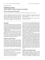
![Báo cáo khoa học: Calix[4]arene methylenebisphosphonic acids as inhibitors of fibrin polymerization doc](https://media.store123doc.com/images/document/14/rc/tp/medium_tpb1394040066.jpg)