Báo cáo khoa học: "Reduction of radiation pneumonitis by V20-constraints in breast cancer" pps
Bạn đang xem bản rút gọn của tài liệu. Xem và tải ngay bản đầy đủ của tài liệu tại đây (563.09 KB, 6 trang )
RESEARC H Open Access
Reduction of radiation pneumonitis by
V20-constraints in breast cancer
Ulla Blom Goldman
1,5*
, Berit Wennberg
2
, Gunilla Svane
3
, Håkan Bylund
4
, Pehr Lind
5
Abstract
Introduction: Adjuvant local-regional radiotherapy (LRRT) is routinely recommended for breast cancer patients. It is
well known being related to pulmonary side-effects. We studied post-RT radiological changes on X-ray and CT, and
correlated the findings with Quality of Life (QoL), common dosimetric factors and co-variates. The results were
compared with a previou sly reported cohort of 137 irradiated women.
Methods: 88 women underwent chest X-ray and CT pre-and 4-5 months after 3-D planned LRRT, minimizing the
dose to the ipsilateral lung to V
20
< 30%. The lung field was divided into 3 regions and the development of post-
RT density changes were graded (0-3). Patients with radiological changes were compared with non-responders.
Clinical symptoms were registered and data on patient and treatment related co-variates were gathered
prospectively. The ipsilateral lung dosimetric factors V
13
,V
20
,V
30
and mean dose were calculated and QoL was
assessed before and 4 months after RT.
Results: The use of dose-volume constraints significally reduced moderate-severe radiological changes on chest X-ray
compared with our earlier study (Chi square trend test: p < 0.001). Symptomatic pneumonitis was also rare in the
present study. No agreement was found between CT and chest X-ray as diagnostic tools for post-RT pneumonitis. V
13
correlated independently with radiological changes on CT (logistic regression: p = 0.04; ROC area: 0.7). The Co-variates
smoking habits, age, chemotherapy, endocrine or trastuzumab therapy did not influence the outcome on multivariate
analysis. QoL changes in physical function, i.e. fatigue, dyspnoea were not detected but there was a trend for a worse
recovery after chemotherapy in patients with high V
13
(Spearman Rank Correlation: p < 0.05).
Conclusions: The use of dose-volume constraints significantly reduced post-RT radiological cha nges on chest X-ray
in LRRT for BC. The lung changes on CT were also generally limited when we used this strategy and was not
always picked up on chest X-ray. Variation in V
13
alone was correlated with occurrence of lung changes on CT.
Introduction
Postoperative radiotherapy (RT) for breast cancer (BC)
plays an important role for reducing the rates of local
recurrence and death [1-3]. The treatment, however, deli-
vers some unwanted irradiation to the lung and heart.
Side-effects to the lungs are in the form of acute pneumo-
nitis and sub acute/late lung fibrosis. The risk for acute
and chronic RT-induced lung morbidity is influenced by
total dose, dose per fraction and irradiated lung volume.
When a 3-D RT-planning technique is used, it is possible
to quantify and limit the amount of individually irradiated
lung volume. Clinical data suggest that a total lung dose of
more than 20 Gy given with conventional fractionation
should be avoided if the unirradiate d lung volume is not
sufficient to guarantee essential breathing function [4]. In
our previous work, we found no case of moderate sympto-
matic radiation pneumonitis (RP) in patients who received
doses ≥ 20 Gy (V
20
) to less than 30% of the ipsilateral lung
volume [5]. We therefore used this cut-off level in the pre-
sent trial. Other groups have found relations between che-
motherapy [6,7] and tamoxifen intake [8] and RT-induced
lung toxicity. In previous studies we have also found an
association with age [5,9]. Individual sensitivity to irradia-
tion is also known but a rare genetic condition in the
population[10]. However it is shown that possessions of
specific genes variants is predictive for the development of
adverse effects after radiotherapy [11-13]. In contrast
smoking has been reported to reduce the risk of RT-
induced pneumonitis [14]. Side-effects to the normal lung
* Correspondence:
1
Department of Oncology, Karolinska University Hospital, Stockholm, Sweden
Full list of author information is available at the end of the article
Goldman et al. Radiation Oncology 2010, 5:99
/>© 2010 Goldman et al; licensee BioMed Central Ltd. This is an Open Access article distributed under the terms of the Creative
Commons Attribution License (http:/ /creativecommons.org/licenses/by/2.0), which permits unrestricted use, distribution, and
reproduction in any medium, provided the original work is properly cited.
tis sue can occu r as early as 6 weeks from the start of RT
with symptoms of fever, dyspnoea and cough [15]. Signs
of interstitial pulmonary inflammation can be detected on
chest radiography (X-ray) in the irradiated lung. A later
phase with fibrosis can be detected from 20 weeks and
after about 36 weeks stationary fibrosis is obtained [16,17].
This study was performed to evaluate radiological
pneumonitis (RP) on X-ray and CT in irradiated breast
cancer women when the lung dose-volume constraints
of V
20
< 30% was used and to correlate the findings
with common dosimetric factors (ipsilateral V
13
,V
20
,
V
30
, MLD), Quality of life (QoL)-effects, symptoms and
co-variates and compare the outcome to a previously
reported study of 137 irradiated women [9].
Methods
This study was approved by the local ethics committee.
Participating women gave informed consent before
study enrolment.
Study population
All women who were referred to the Radiotherapy
Department at Stockholm Söder Hospital during 2003-
2005 for adjuvant LRRT after surgery for early breast
cancer were asked to participate in this trial. Ninety-five
patients were included, but seven patients withdrew
their consent due to early relapse and were not evalu-
able. Eighty-eight patients were thus followed for seven
months after RT for symptoms of acute/subacute radia-
tion induced pulmonary complication. Mastectomy was
done in 69 patients, while 19 patients were operated
with conservative breast surgery. Seventy-two patients
were irradiated with LRRT to the chest wall or breast,
axilla and supra clavicular region a nd in these patients
the internal mammary lymph nodes (IMN) were
included. A total of 16 patients received RT excluding
the IMN, i.e. 9 patients were given irradiation to t he
breast, axilla and supraclavicular region and 7 patients
were referred for RT to the axilla and supraclavicular
fossa only.
The mean age of the patients was 56 years (range 32-81).
Data on potential confounding factors were collected
prospectively, i.e. history of cardio vascular or pulmonary
co-morbidity, smoking habits, functional level (i.e. not
being able to climb three flights of stairs without a rest
due to shortness of breath) and adjuvant hormonal-, tras-
tuzumab- and chemotherapy treatment.
The chemotherapy was concluded 3-4 weeks prior
to RT. Concurrent chemotherapy was never given. The
typically regime consisted of doxorubicin, cyclophospha-
mide and 5-fluorouracil, but in 28 patients the therapy
included docetaxel. Five patients received trastuzumab
during RT. Intake of tamoxifen and anastrozol during
RT was evenly split among the women.
To asses and evaluate Quality of life (QoL) before and
after RT we used the EORTC QLQ-C30 version 3.0 and
the EORTC QLQ-BR23 questionnaires.
Radiotherapy treatment techniques
The used RT treatment techniques are described in
detail in an earl ier publication [18]. LRRT after mastect-
omy was delivered with an anterior electron bea m cov-
ering the chest wall and the IMN (range 6-12 MeV) and
with a 6 MV photon beam covering the supraclavicular
region. LRRT after partial mastectomy consisted of two
tangential photon beams of 4 o r 6 MV inc luding the
breast parenchyma (50 Gy) and the regional lymph
nodes were treated in a similar way as described above
(46 Gy). An additional oblique electron beam was added
to include the IMN (46 Gy) in four cases. The pre-
scribed dose was given in daily fract ions of 2 Gy, five
days a week. In the present study all patients underwent
3-D dose treatment planning (Pinnacle; version 6.2b)
with avoidance of a dose exciding 20 Gy to more than
30% of the ipsilateral lung volume but with a good cov-
erage of the clinical target volume (CTV). The cumula-
tive dose-volume histograms were calculated and the
ipsilateral lung volume receiving ≥ 13 Gy (V
13
), >20 Gy
(V
20
), >30 Gy (V
30
) and mean lung dose were defined.
Monitoring for symptomatic pneumonitis and evaluation
of radiological pneumonitis on X-ray and CT with the
Arrigada’s classification
All patients were followed for respiratory symptoms, i.e.
cough, dyspnoea with or without fever, 1, 4 and 7 months
after the termination of RT. The patients were classified
into three groups according to CTC-criteria (version 2.0)
[19].
0. N o complications: no registered respiratory symp-
toms monitored by the clinician.
1. Mild reaction: cough and/or dyspnoea with our
without fever judged to be radiation induced.
2. Moderate reaction: same as 1 but with impaired
daily functions and treated with corticosteroids.
CT of the thorax was performed before and 4 months
after RT and standardized chest X-ray was conducted
after 5 months and evaluated by the same specialist in
diagnostic radiology (HB) as in our previous trial [5].
The reproducibility of this scoring system was validated
in our earlier publication [5].
On the frontal chest radiograph the lung was divided
into the three regions suggested by Arrigad a, i.e. the apical-
lateral (A-L), central-parahilar (C-P) and basal-lateral (B-L)
regions [19]. The border between the A-L and B-L regions
was set at the level of the pulmonary artery. The width of
the C-P region was set to 5 cm and the upper and lower
border were set two vertebrae above and below the level of
pulmonary artery, respectively. Radiological pneumonitis
Goldman et al. Radiation Oncology 2010, 5:99
/>Page 2 of 6
(RP) was quantified according to Arrigada’s classification.
The highest-density grade in each region, i.e. 0 = no evi-
dence of fibrosis, 1 = linear streaks, 2 = moderate opac ifica-
tion, 3 = complete opacification were added together to
form total scores ranging from 0 to 9. Total scores of 1-3
were considered to represent slight radiological RP and
score of 4-9 moderate to severe RP. This method has been
described in detail in our earlier s tudy [5].
Evaluation of Quality of life
We used the European Organisation for Research and
Treatment of Cancer (EORTC) form QLQ-30 (version
3.0) [20] and the EORTC QLQ-BR-23 to asses QoL [21].
The forms were completed at baseline prior to and 4
months after RT. The QLQ-30 questionnaires consist of
a total of 30 items. Five functional scales (physical, role,
cognitive, emotional and social); nine symptom scales
(fatigu e, nausea/vomiting, pain, dyspnea, insomnia, appe-
tite loss, constipation, diarrhea and financ ial difficulties)
and one Global health status. QLQ-BR23 includes 23
items assessing disease symptoms,therapysideeffects
such as breast , arm symptom, hair loss, body image, sex-
ual functioning, sexual enjoyment and future perspective.
Eighty-one patients completed both measurements. In
seven cases the pts did not receive the 2
nd
form. The
form was double-sided and in some cases not completed
on the back page. Scoring was performed in according
with the EORTC scoring manual. Statistics and missing
data were handled according to the manual. A four-
point response scale was used to asses each item con-
cerning functions or symptoms from 1 (not at all) to 4
(very much), and a seven-point scale was used for global
health status/QoL from 1 (very poor) to 7 (excellent).
The scale scores were li nearly transformed into scores
of 0-100 according to the EORTC manual. A high score
on the global health status/functional scale represents a
high/healthy level of functioning. In contrast a high
score on the symptom scale represents a high level of
symptomatology/problems. A study of the subjective sig-
nificance of changes in QoL scores has suggested that a
mean change of 5 to 10 on the multi -item scales is per-
ceived as little change, 10 to 20 as moderate change and
greater than 20 as very much change. Greater than ten
points on the transformed questionnaire scale were con-
sidered clinically meaningful [22,23].
In the present paper, three functional scales (physical
functioning, role functioning and social functio ning) and
four symptom scales (fatigue, pain, dyspnoea and insomnia)
were included from QLQ C-30. In the EORTC QLQ-BR23
form we included functional scales (future perspective).
Statistical methods
The relation between symptomatic and radiological RP
and the relation between radiological RP and the
dosimetric factors and co-variates was analyzed with uni-
variate and multivariate logistic regression (Wald-Enter
method). Chi square trend test was used for test of corre-
lation between radiological RP on X-ray in the present and
earlier studies. To test agreeme nt between CT-and X-ray
for the diagnosis of radiological RP, Kappa-statistics was
used.
Receiver operating characteristics curves (ROC) were
used to predict radiological RP with V
13
[24]. Changes
in QoL-scores in relation with V
13
were evaluated with
Spearman Rank Correlation. All reported results were
based on two-sided tests and p-values < 0.05 were con-
sidered statistically significant.
Results
Radiological and symptomatic radiation pneumonitis
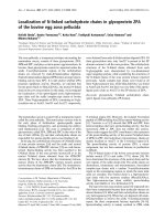
Figure 1 shows an example of post-RT radiological RP
of grade 3 in the apical-lateral region of the left lung on
chest X-ray (= total score 3). Symptomatic pneumonitis
was very rare in this study. Only one patient develope d
a moderate reaction and was treated with corticosteroids
and antibiotics, mild reactions were detected in 6
patients. There was, furthermore, no relation between
symptomatic RP and radiological RP on chest X-ray or
CT. Minor changes are not seen on chest X-ray, in due
to that CT is a more sensitive method than X-ray to
detect small effected areas of pulmonary changes.
The use of dose-volume constraints significall y
reduced moderate-severe radiological RP on X-ray co m-
pared with the earlier treatment series for the technique
with LRRT + IMN (Chi square trend test p < 0 .001)
(Table 1). There was, however, no difference when we
comp ared the o utcome for the technique LRRT-IMN in
Figure 1 Example of grade 3 RP in the apical-later al region of
the left lung on chest x-ray. (= total score 3).
Goldman et al. Radiation Oncology 2010, 5:99
/>Page 3 of 6
the present series with the previous trial (Table 1). The
mean V
20
for responders and non-responding patients
areshowninFigure2.TheaverageV
20
and MLD in
our previous study was 35% and 16 Gy, respectively
[25]. We found no correlation between any dosimetric
factor or the studied co-variates and RP on ch est X-ray
(score 0 vs score 1-9) (logistic regression). In the pre-
ceding univariate analysis there was a borderline relation
with radiological RP and anastrazol but this relation was
thus not detected on the subsequen t multivariate analy-
sis which included the dosimetric factors and other
co-variates.
There was no agreement between X-ray and CT as
diagnostic tools for post -radiological RP, (Kappa test)
(Table 2). V
13
was most strongly and independently
related with radiological changes on CT (score 0 vs 1-9)
(logistic regression: p = 0.04; ROC-area: 0.7) [24]. No
other factor was related to RP on CT. Table 3 shows
the correlation between the do simetric factors in this
study. V
13
was stronger correlated to MLD than V
20.
Quality of life
Most of the side effects from RT appeared to have little
effect on QoL in the present trial. Chemotherapy was
concluded 3-4 weeks prior to RT and the patients started
with a higher score on fatigue at baseline due to this.
The variables role functioning, social functioning and
future perspective, were improved 4 months after RT
compared to baseline (Table 4). Physical functioning,
appeared not to be affected by RT. There were no
changes for pain and dyspnoea after RT in this series.
Patients with high V
13
appeared however not recover
equally well. However, insomnia showed a trend to
increase after RT (Table 4). When changes in the indivi-
dual QLQ-variates fatique and dyspnoea were related to
V
13
(Spearma n correlation) there was thus a negative
correlation. There was a signif icant correlation between
high V
13
and difficulties to take short walks, which
could be of clinical significance, and the correlation was
reported also when patients rated there overall total
quality of life during the last week.
Discussion
Clinically significant radiological and symptomatic RP
was rare in this study when 3-D tre atment planning,
aiming at minimizing V
20
to the ipsilateral lung to <30%
was used for LRRT in early breast cancer. The result
indicates that the used dose-volume constraints s ignifi-
cantly reduced moderate-severe radiological RP on chest
X-ray, in the present series, compared to our previous
study [25]. The lung changes could not always be
detected on chest X-ray and were also infrequent and
generally limited on CT when this strategy was used.
Variation in dosimetry alone (V
13
) was correlated with
occurrence of radiological RP on CT. ROC analys es was
performed, yet the area under the curve was only 0.7
which is not an ideal predictive value [24]. Co-variates
Table 1 Relation between radiological changes and
treatment techniques in the present and previous studies
Technique LRRT+ IMN; n LRRT-IMN; n
Arrigada’s classification scores 0 1-3 4-9 0 1-3 4-9
Present study 60 11 1 5 4 0
Previous study 58 38 20 12 9 0
Chi square trend
test p < 0.001
Chi square trend
test p = 0.9
Figure 2 Mean lung dos volume histograms (DVH) in patients
with or without RP on chest X-ray.
Table 2 Relation between radiological scores on X-ray
and CT in the present series
CT score
X-ray score 0 1-3 4-9 Total
013251250
1-3 1 7 3 11
4-9 0 1 0 1
Total 14 33 15 62
Kappa statistics: p = 0.3
Table 3 Correlation between lung dosimetric factors in
breast cancer irradiation
V13 V20 V30 Mean
V13 Pearsson Correlation 1 .925** .619** .975**
Sig. (2-tailed) .000 .000 .000
V20 Pearsson Correlation .925** 1 .820** .926**
Sig. (2-tailed) .000 .000 .000
V30 Pearsson Correlation .619** .820** 1 .687**
Sig. (2-tailed) .000 .000 .000
Mean Pearsson Correlation .975** .926** .687** 1
Sig. (2-tailed) .000 .000 .000
** Correlation is significant at the 0.01 level (2-tailed)
Goldman et al. Radiation Oncology 2010, 5:99
/>Page 4 of 6
as smoking habits, age, exposure to chemotherapy,
endocrine- or trastuzumab therapy did not influence the
outcome, but the few events may have hampered the
possibility to evaluate this. In the present study, some
women received radiation to the internal mammary
nodes (IMN). Whether the IMN need to be included in
the CTV is not fully known. In the last years, many RT
centers have excluded RT to the lower IMN, in order to
avoid cardiac and lung toxicity. The meta-analysis in
Lancet, 2005, howeve r demonstrated a benefit for post
mastectomy RT in women with positive LN and the
majority of these women received RT t o the lower IMN
(21 of 23 studies) [1].
The LRRT-IMN group of our present series included
only nine patients. We used the same RT-technique
in both this and the previous study, and as could be
expected, there was no difference in radiological RP
(Table 1). CTV volumes minus IMN usually give lower
doses to the lung. Limiting the IMN irradiation to the
three upper intercostals spaces also lower the dose to
the heart. It is probably of great importance to reduce
radiation to organs at risk like h eart and lung, when
adjuvant treatment is given. The average patient has a
long expected survival, b ut as there are many new sys-
temic therapies which may interact with RT this can
lead to additional side-effects. Aromatase Inhibitors (AI)
have replaced tamoxifen in many postmenopausal
patients.
The AI treatment in combination with RT is investi-
gated i n a randomised trial presented in Lancet Oncol.
2010. The results suggested that AI can be used early,
but there still are doubts on potential long-term toxic
effects, mainly cardiac in combination with RT [26,27].
Genetic factors may also play a vitale role in treat-
ment. By identifying genetic factors associated with
radiosensitivity it will be easier to predict which patients
are at increased risk for c omplications secondary to
radiation treatment [11-13].
Even though symptomatic and radiological RP were
rare in our trial, they still could be of importance if they
prevail, as late changes could increase the risk of sec-
ondary lung cancer. This increased risk is seen in smo-
kers five years after RT [28].
To improve radiotherapy techniques and continue to
study pulmonary morbidit yandQoLafterRT,isof
great importance as breast can cer is a common disease
among women.
In conclusion, V20-constraints significantly reduced
post-RT radiological changes on chest X-ray in LRRT
for breast cancer. Symptomatic pneumonitis was,
furthermore, rare in the present study when this strategy
was used. There was no agreement b etween X-ray and
CT as diagnostic tools for post-RT in this trial, as the
lung changes typicall y were too limited for detection on
X-ray. V
13
was most strongly related to radiological RP
on CT. V
13
was stronger co rrelated to MLD than V
20
,
and may be an important metric in future trials on RT-
induced lung toxicity.
Acknowledgements
Presented at ASTRO 2009
This work was supported by The Swedish Cancer Foundation
(Cancerfonden). We are grateful for the work that the staff of the
Radiotherapy Department and the Breast Cancer Outpatient Ward at
Stockholm Söder Hospital has put into this study.
Author details
1
Department of Oncology, Karolinska University Hospital, Stockholm,
Sweden.
2
Department of Hospital Physics, Karolinska University Hospital,
Stockholm, Sweden.
3
Department of Radiology, Karolinska University
Hospital, Stockholm, Sweden.
4
Department of Radiology, Ersta Hospital,
Stockholm, Sweden.
5
Karolinska Institutet Stockholm, Sweden.
Authors’ contributions
UBG coordinated the study, collected the data and drafted the manuscript.
Table 4 Pre and post RT Quality of Life EORTC scores in breast cancer irradiation
Mean QLQ-30 scale values before and after RT and for paired difference
n Before RT After RT Difference 95% CI P-value
Functional scales
Physical functioning 81 78.5 80.6 2.1 -1.2-5.4 0.20
Role functioning 80 62.3 70.6 8.3 1.3-15.4 0.021
Social functioning 73 70.6 76.3 5.7 0.7-10.7 0.026
Symptom scales
Fatique 73 37.1 31.7 -5.5 -10.0 - -1.0 0.018
Pain 81 24.7 24.3 -0.4 -5.3 - 4.4 0.87
Dyspnoea 79 29.1 28.3 -0.8 -6.9 - 5.3 0.78
Insomnia 81 36.6 41.6 4.9 -1.5 - 11.3 0.13
Mean QLQ-BR23 scale values before and after RT and for paired difference
n Before RT After RT Difference 95% CI P-value
Functional scales
Future perspective 77 44.2 53.7 9.5 3.4-15.6 0.003
Goldman et al. Radiation Oncology 2010, 5:99
/>Page 5 of 6
UBG, BW, GS and PL were involved with the design of the study.
HB and GS analysed X-ray and CT diagnostics. BW analysed RT-doses. PL
supported with the statistics. All authors read and approved the final
manuscript.
Competing interests
The authors declare that the y have no competing interests.
Received: 2 June 2010 Accepted: 29 October 2010
Published: 29 October 2010
References
1. Clarke M, Collins R, Darby S, Davies C, Elphinstone P, Evans E, Godwin J,
Gray R, Hicks C, James S, et al: Effects of radiotherapy and of differences
in the extent of surgery for early breast cancer on local recurrence and
15-year survival: an overview of the randomised trials. Lancet 2005,
366:2087-2106.
2. Overgaard M, Hansen PS, Overgaard J, Rose C, Andersson M, Bach F,
Kjaer M, Gadeberg CC, Mouridsen HT, Jensen MB, Zedeler K: Postoperative
radiotherapy in high-risk premenopausal women with breast cancer
who receive adjuvant chemotherapy. Danish Breast Cancer Cooperative
Group 82b Trial. N Engl J Med 1997, 337:949-955.
3. Overgaard M, Jensen MB, Overgaard J, Hansen PS, Rose C, Andersson M,
Kamby C, Kjaer M, Gadeberg CC, Rasmussen BB, et al: Postoperative
radiotherapy in high-risk postmenopausal breast-cancer patients given
adjuvant tamoxifen: Danish Breast Cancer Cooperative Group DBCG 82c
randomised trial. Lancet 1999, 353:1641-1648.
4. Herrman T, Schorcht J, V M: Late sequelae in oncology. In Berlin Edited by:
Dunst J, Saueer R 1995.
5. Lind PA, Svane G, Gagliardi G, Svensson C: Abnormalities by pulmonary
regions studied with computer tomography following local or local-
regional radiotherapy for breast cancer. Int J Radiat Oncol Biol Phys 1999,
43:489-496.
6. Lind PA, Marks LB, Jamieson TA, Carter DL, Vredenburgh JJ, Folz RJ,
Prosnitz LR: Predictors for pneumonitis during locoregional radiotherapy
in high-risk patients with breast carcinoma treated with high-dose
chemotherapy and stem-cell rescue. Cancer 2002, 94:2821-2829.
7. Taghian AG, Assaad SI, Niemierko A, Kuter I, Younger J, Schoenthaler R,
Roche M, Powell SN: Risk of pneumonitis in breast cancer patients
treated with radiation therapy and combination chemotherapy with
paclitaxel. J Natl Cancer Inst 2001, 93:1806-1811.
8. Bentzen SM, Skoczylas JZ, Overgaard M, Overgaard J: Radiotherapy-related
lung fibrosis enhanced by tamoxifen. J Natl Cancer Inst 1996, 88:918-922.
9. Lind PA, Bylund H, Wennberg B, Svensson C, Svane G: Abnormalities on
chest radiographs following radiation therapy for breast cancer. Eur
Radiol 2000, 10:484-489.
10. Baumann M: Impact of endogenous and exogenous factors on radiation
sequelae Berlin: Springer-Verlag; 1995.
11. Ho AY, Atencio DP, Peters S, Stock RG, Formenti SC, Cesaretti JA, Green S,
Haffty B, Drumea K, Leitzin L, et al: Genetic predictors of adverse
radiotherapy effects: the Gene-PARE project. Int J Radiat Oncol Biol Phys
2006, 65:646-655.
12. Ozsahin M, Crompton NE, Gourgou S, Kramar A, Li L, Shi Y, Sozzi WJ,
Zouhair A, Mirimanoff RO, Azria D: CD4 and CD8 T-lymphocyte apoptosis
can predict radiation-induced late toxicity: a prospective study in 399
patients. Clin Cancer Res 2005, 11:7426-7433.
13. Azria D, Ozsahin M, Kramar A, Peters S, Atencio DP, Crompton NE,
Mornex F, Pelegrin A, Dubois JB, Mirimanoff RO, Rosenstein BS: Single
nucleotide polymorphisms, apoptosis, and the development of severe
late adverse effects after radiotherapy. Clin Cancer Res 2008, 14:6284-6288.
14. Bjermer L, Franzen L, Littbrand B, Nilsson K, Angstrom T, Henriksson R:
Effects of smoking and irradiated volume on inflammatory response in
the lung of irradiated breast cancer patients evaluated with
bronchoalveolar lavage. Cancer Res 1990, 50:2027-2030.
15. Davis SD, Yankelevitz DF, Henschke CI: Radiation effects on the lung:
clinical features, pathology, and imaging findings. AJR Am J Roentgenol
1992, 159:1157-1164.
16. Slanina J, Sigmund G, Hinkelbein W, Wenz W, Wannenmacher M:
[Pulmonary reaction to radiation following mantle-field irradiation.
Comparison of follow-up by conventional x-ray and by computed
tomography]. Radiologe 1988, 28:20-28.
17. Lind P: Clinical relevance of pulmonary toxicity in adjuvant breast cancer
irradiation. Acta Oncol 2006, 45:13-15.
18. Lind PA, Rosfors S, Wennberg B, Glas U, Bevegard S, Fornander T:
Pulmonary function following adjuvant chemotherapy and radiotherapy
for breast cancer and the issue of three-dimensional treatment
planning. Radiother Oncol 1998, 49:245-254.
19. Arriagada R, de Guevara JC, Mouriesse H, Hanzen C, Couanet D, Ruffie P,
Baldeyrou P, Dewar J, Lusinchi A, Martin M, et al: Limited small cell lung
cancer treated by combined radiotherapy and chemotherapy:
evaluation of a grading system of lung fibrosis. Radiother Oncol 1989,
14:1-8.
20. Aaronson NK, Ahmedzai S, Bergman B, Bullinger M, Cull A, Duez NJ,
Filiberti A, Flechtner H, Fleishman SB, de Haes JC, et al: The European
Organization for Research and Treatment of Cancer QLQ-C30: a quality-
of-life instrument for use in international clinical trials in oncology. J Natl
Cancer Inst 1993, 85:365-376.
21. Sprangers MA, Groenvold M, Arraras JI, Franklin J, te Velde A, Muller M,
Franzini L, Williams A, de Haes HC, Hopwood P, et al: The European
Organization for Research and Treatment of Cancer breast cancer-
specific quality-of-life questionnaire module: first results from a three-
country field study. J Clin Oncol 1996, 14:2756-2768.
22. Osoba D, Rodrigues G, Myles J, Zee B, Pater J: Interpreting the significance
of changes in health-related quality-of-life scores. J Clin Oncol 1998,
16:139-144.
23. Velikova G, Stark D, Selby P: Quality of life instruments in oncology. Eur J
Cancer 1999, 35:1571-1580.
24. Lind PA, Wennberg B, Gagliardi G, Rosfors S, Blom-Goldman U, Lidestahl A,
Svane G: ROC curves and evaluation of radiation-induced pulmonary
toxicity in breast cancer. Int J Radiat Oncol Biol Phys 2006, 64:765-770.
25. Lind PA, Wennberg B, Gagliardi G, Fornander T: Pulmonary complications
following different radiotherapy techniques for breast cancer, and the
association to irradiated lung volume and dose. Breast Cancer Res Treat
2001, 68:199-210.
26. Azria D, Belkacemi Y, Romieu G, Gourgou S, Gutowski M, Zaman K,
Moscardo CL, Lemanski C, Coelho M, Rosenstein B,
et al: Concurrent or
sequential adjuvant letrozole and radiotherapy after conservative
surgery for early-stage breast cancer (CO-HO-RT): a phase 2 randomised
trial. Lancet Oncol 2010, 11:258-265.
27. Recht A: Radiotherapy, antihormonal therapy, and personalised
medicine. Lancet Oncol 2010, 11:215-216.
28. Prochazka M, Hall P, Gagliardi G, Granath F, Nilsson BN, Shields PG,
Tennis M, Czene K: Ionizing radiation and tobacco use increases the risk
of a subsequent lung carcinoma in women with breast cancer: case-only
design. J Clin Oncol 2005, 23:7467-7474.
doi:10.1186/1748-717X-5-99
Cite this article as: Goldman et al.: Reduction of radiation pneumonitis
by V20-constraints in breast cancer. Radiation Oncology 2010 5:99.
Submit your next manuscript to BioMed Central
and take full advantage of:
• Convenient online submission
• Thorough peer review
• No space constraints or color figure charges
• Immediate publication on acceptance
• Inclusion in PubMed, CAS, Scopus and Google Scholar
• Research which is freely available for redistribution
Submit your manuscript at
www.biomedcentral.com/submit
Goldman et al. Radiation Oncology 2010, 5:99
/>Page 6 of 6