Báo cáo khoa học: " RadioImmunotherapy for adenoid cystic carcinoma: a single-institution series of combined treatment with cetuximab" docx
Bạn đang xem bản rút gọn của tài liệu. Xem và tải ngay bản đầy đủ của tài liệu tại đây (3.23 MB, 8 trang )
RESEARC H Open Access
RadioImmunotherapy for adenoid cystic
carcinoma: a single-institution series of
combined treatment with cetuximab
Alexandra D Jensen
1*
, Jürgen Krauss
2
, Wilko Weichert
3
, Jürgen Debus
1
, Marc W Münter
1
Abstract
Background: Local control in adjuvant/definitive RT of adenoid cystic carcinoma (ACC) is largely dose-dependent.
However, some clinical situations do not allow applica tion of tumouricidal doses (i.e. re-irradiation) hence radiation
sensitization by exploitation of high endothelial growth factor receptor (EGFR)-expression in ACC seems beneficial.
This is a single-institution experience of combined radioimmunotherapy (RIT) with the EGFR-inhibitor cetuximab.
Methods: Between 2006 and 2010, 9 pts received RIT for advanced/recurrent ACC, 5/9 pts as re-irradiation.
Baseline characteristics as well as treatment parameters were retrieved to evaluate efficacy and toxicity of the
combination regimen were evaluated. Control rates (local/distant) and overall survival were calculated using
Kaplan-Meier estimation.
Results: Median dose was 65 Gy, pts received a median of 6 cycles cetuximab. RIT was tolerated well with only
one °III mucositis/dysphagia. Overall response/remission rates were high (77,8%); 2-year estimate of local control
was 80% hence reaching local control levels comparable to high-dose RT. Progression-free survival (PFS) at 2 years
and median overall survival were only 62,5% and 22,2 mo respectively.
Conclusion: While local control and treatment response in RIT seems promising, PFS and overall survival are still
hampered by distant failure. The potential benefit of RIT with cetuximab warrants exploration in a prospective
controlled clinical trial.
Introduction
Adenoid cystic carcinomas are rare tumours mostly
of the head and neck and account for approximately
10-15% of malignant salivary gland tumours [1]. They are
characterised by a rather slow growth pattern but also
perineural spread and a high propensity for haematoge n-
ous metastases. Standard treatment so far consists of
complete surgical resec tion followed by adjuvant irradia-
tion in case of risk factors (i.e. close margins, perineural
invasion, extensive primary tumor (T3, T4) or high-grade
histology) [2-4].
Local control in this disease could already be improved
by adjuvant radiation, the introduction of high-precision
RT techniques (i.e. FSRT and/or IMRT) with consecutive
dose escalation, and last but not least high-LET RT. To
achieve local control, radiation doses of >60 Gy o r even
66 Gy are recommended [5-8].
Initial local control rates combined IMRT plus C12
heavy ion boost to a total dose of 72 GyE were 78% at 4
years [9]. Re cent updates including all patients treated
at the Gesellschaft für Schwerionenforschung (GSI)
Darmstadt between 1997 and 200 8 even yielded a local
control rate of 82% at 5 years [10,11]. Therefore the
combination of IMRT and carbon ion boost shows com-
parable or even superior control rates to neutron RT
[12,13] without increase of late toxicity and subsequent
morbidity consistent w ith outcomes reported by Mizoe
et al [14]. Therefore, IMRT plus C12 boost has been
accepted as a standard in Germany whenever available.
Albeit progress has been made by the introduction of
particle therapy in the treatment concept of adenoid cys-
tic carcinoma, local control rates still leave room for
improvement. With the successful introduction of com-
bination regimen in squamous cell carcinoma of the head
* Correspondence:
1
Dept of Radiation Oncology, INF 400, 69120 Heidelberg, Germany
Full list of author information is available at the end of the article
Jensen et al. Radiation Oncology 2010, 5:102
/>© 2010 Jensen et al; licensee BioMed Central Ltd. This is a n Open Access article distributed under the terms of the Creative Commons
Attribu tion License ( which permits unrestricted use, distribution, and repro duction in
any medium , provided the original work is properly cited.
and neck (SCCHN), leading to a significant improvement
not only in local control but also in overall survival,
investigation of this approach was obvious in adenoid
cystic carcinoma hoping for further improvement of local
control and higher response rates of bulky tumours.
Radiochemotherapy in the treatment of malignant sali-
vary gland tumors (MSGT) however, has not evolved
beyond the phase II-stage or retrospective analysis of
very heterogeneous treatment regimen [15-18] into a
treatment standard so far as results have been more or
less inconclusive.
Immunostaining of surgical specimen however [19],
could show over-expression of EGFR in adenoid cystic
carcinoma in high percentages hence implying use of
targeted therapies as potential alternative [19,20] to
comparatively toxic chemotherapy regimen commonly
used in recurrent or metastatic adenoid cystic carci-
noma since the mid 80-ies [21-24]. Despite the initial
euphoria, treatment results have so far failed to impress:
no objective response in recurrent or metastatic adenoid
cystic carcinoma could be shown in any of the trials
[25-27] although prolonged disease stabilization was
observed in the reported series [26,27].
Since t he publication of combined radioimmunother-
apy with the EGF receptor antibody cetuximab in
SCCHN of the Bonner trial in 2006 [28,29] though,
application of these drugs in adenoid cystic carcinoma
seemed feasible in view of potential increase of radiation
sensitivity and - albeit modest - systemic activity given
the relatively mild toxicity profile of EGFR antibodies.
Hence, we would like to present our experiences in
combined radioimmunotherapy of adenoid cystic carci-
noma with cetuximab.
Methods
In an individual approach patients received radioimmu-
notherapy with cetuximab for a dvanced or recurrent
adenoid cystic carcinoma between 01/2006 and 06/2010.
Baseline characteristics as well as treatment parameters
were retrieved to evaluate efficacy and toxicity of the
combination regimen were evaluated.
Indication
Radioimmunotherapy for adenoidcystic carcinoma not
representing a therapeutic standard, medical indication
was highly individual and made in interdisciplinary con-
sensus only in cases where applicable radiation doses
were deemed insufficient for reason able tumour control.
Given the fact carbon ion treatment was only available
three times a year for a very limited number of patients,
this series includes patients in need o f immediate treat-
ment due to rapid tumour progression or locoregional
relapse after prior RT. Sufficient dose prescription (>70
Gy) was not possible in al l of these cases either because
of proximity/involvement of critical structures or prior
RT, he nce the idea was to increase eff icacy of radiation
therapy by combined radioimmunotherapy usually
accompanied with only mild toxicity. Rationale for the
proposed t reatment was extensively discussed and deci-
sions made in accordance with the patients.
5/9 pts hade undergone prior RT (median dose: 58 Gy,
range 50,4 - 62 Gy) with a median time interval of
57 months for treatment for adenoidcystic carcinoma.
Four pts received radioimmunotherapy as part of their
primary treatment but showed extensive tumour mass
directly adjacent to or involving critical structures. 8/9
pts received IMRT ( 2 pts as tomotherapy, 6 pts in step
and shoot technique), 1 pt received combined IMR T plus
C12-boost due to rapid postoperative local progression.
Histomorphologic evaluation and immunohistochemistry
The histomorphological diagnosis of adenoid cyst ic car-
cinoma was confirmed in all cases by a board certified
pathologist with a special expertise in head and neck
pathology. For immunohistochemistry 5 μm paraffin
sections were cut. Detection of EGFR was performed
with the EGFR pharm Dx kit by DAKO (K1492) accord-
ing to the manufacturer’s instructions. Staining was
scored as positive if any membranous positivity was
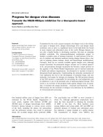
observed, h owever, all investigated cases showed strong
membranous positivity in a considerable number of
tumor cells (Figure 1a/b).
Radiation therapy
Immobilization/planning examinations
Patients were immobilized using individual scotch cast
or thermoplastic head masks with thermoplastic
shoulder fixation. Planning examinations consisted of a
planning CT scan (3 mm slice thickness) with the
patient positioned in the individual fixation device and
contrast-enhanced MRI for 3 D image correlation.
Target volumes: primary (photon) RT
Target delineation was carried out based on planning
CT and MRI scan. CTV1 included the macroscopic
tumor/prior tumor bed with a margin of 2 mm with
special focus on the R2/R1-area as well as respective
neural pathways to the base of skull (cave: perineural
invasion and skip lesions).
CTV2 included CTV1 with generous safety margin
along typical pathways of spread (if possible safety mar-
gin of about 5 cm) depending on the anatomical relation-
ship of adjacent structures. In particular, neurovascular
sheaths and locoregional ipsilateral nodal levels were also
included in the CTV2.
Target volumes: re-irradiation
For patients who had already undergone a course of
prior radiotherapy, the treatment volume was strictly
limited to the gross tumour volume and did not include
Jensen et al. Radiation Oncology 2010, 5:102
/>Page 2 of 8
elective nodal levels. Dos es of were highly individualised
but aimed at 50 - 60 Gy re-irradiation in 2 Gy/fraction
[30] depending on elapsed time since the first course of
RT and prior RT-dose.
Target volumes: combined IMRT + carbon ion boost
CTV1 (carbon ion boost) included the macroscopic
tumor/prior tumor bed with special focus on the R2/R1-
area as well as respective neural pathways to the base of
skull (cave: perineural invasion and skip lesions). PTV1
consists of a 3 mm margin around the CTV1 but does
not extend i nto critical orga ns at risk (i.e. brain stem,
spinal cord).
Treatment is given at the HIT (Heidelberg ion therapy
centre) after inverse treatment planning in active beam
application (raster-scanning method).
CTV2 included CTV1 with safety margins along typi-
cal pathways of spread. Only ipsilateral nodal levels (II
and III) are included, however, in case the primary
tumor is/was located at midline or crossing midline,
bilateral nodal levels II and III are covered. In case there
is pathological lymph node inv olvement, additional
nodal levels were covered as indicated. CTV2 also
encompassed the complete surgical operational area.
The CTV2 also takes account for set-up variations,
hence corresponds to the PTV2 (CTV2 = PTV2).
Immunotherapy
Cetuximab was administered as 400 mg/m2 body sur-
face loading dose 7 days prior to RT-treatment start
after administration of anti-histamines (dimetindene)
and corticosteroids (dexamethasone).
Weekly administrations of Cetuximab 250 mg/m2
body surface followed for the duration of radiotherapy.
Analysis
Treatment response was analysed 6 wks post completion
of RIT (first follow-up) and at each available follow-up
(best response) according to RECIST criteria [31] based
on available follow-up scans ( CT or MRI) and clinical
examinations. Treatment outcome (locore gion al, distant
and overall progression-free survival as well as overall
survival) was evaluated using higher non-parametric sta-
tistics (Kaplan-Meyer survival analysis) with the software
xlstat 2010. Progression-free survival was defined as the
time from start o f combined radioimmunotherapy until
the first event (i.e. locoregional relapse, distant metas-
tases, death). S imilarly, overall survival was calculated
from start of radioimmunotherapy until death from any
cause.
Results
Nine pts with aden oid cystic carcinoma receiving com-
bined radioimmunotherapy with cetuximab were identi-
fied. Median follow-up is 12,5 months [1,2 - 29,6 mo].
Tumours were mostly located near or at the base of
skull (epipharynx, pterygopalatine fossa, skull base). All
patients had a macroscopica lly visible tumour mass, all
but one T4 tumours (patient characteristics see table 1).
EGFR expression analysis was available in 8/9 pts (all of
them with at least moderate or high e xpression rates),
specimen for one pt were unfit for EGFR analysis.
Median dose applied was 65 Gy (total) and 50,4 Gy in
pts receiving the second course of radia tion (median
cumulative dose 111,2 Gy) after a median interval of
63,7 months between the two courses (table 2). All pts
Figure 1 ACC and EGFR expression. (A) Histology of an adenoid cystic carcinoma (H&E stain). (B) Strong membranous expression of EGFR in
the majority of tumor cells of the same tumor (EGFR immunohistochemistry). Magnification ×200.
Table 1 patient characteristics
patient characteristics
median age 56 a [40 - 77]
tumour localisation
Epipharynx 2 pts
base of skull 2 pts
Fossa pterygopalatina 3 pts
Maxilla 1 pt
tuba auditiva 1 pt
tumour stage T4 8 pts
T3 1 pt
no nodal involvement 9 pts
Jensen et al. Radiation Oncology 2010, 5:102
/>Page 3 of 8
in this series received IMRT either as single modality
(8/9pts) or as combined treatment with carbon ion
boost ( 1/9 pts). Prior RT in the 5 cases with re-irradia-
tion was carried out using IMRT (3 pts), stereotactic
single fraction only (1 pt) and 3 D conventional (1 pt).
All patients completed the treatment as planned; there
were no treatment interruptions. A median of 6 cycles
cetuximab [4 - 8 cycles] (excluding loading dose) were
applied. Treatment was tolerated well without any cases
of allergic reactions and only one case of CTC °III toxi-
city (mucositis °III leading to temporary feeding tube
dependence). 8/9pts developed acneif orme skin reac-
tions °I/II, 7/9 pts radiodermatitis °I/II a nd mucositis
°I/II (table 3). In the 7 pts with follow-up available,
acute reactions were completely resolved at first follow-
up, there were no treatment-related late effects.
At first follow-up 8/9 pts (88,9%) showed good partial
remissions. 1 pt died prior to the first f/u (6 wks post
RT) due to tumor bleeding. (table 4). On further f/u,
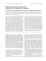
6/7pts stayed locally controlled. Figure 2(a/b) shows the
pretherapeutic MRI s can for a patient with large ACC
extending from the optic canal to the pterygoid muscles.
The applied IMRT treatment plan using a simultaneous
integrated boost concept and the corresponding DVH
are shown in Figures 3a/b and 4. 66 Gy were prescribed
to the median of CTV1. However, impaired coverage
needed to be accepted in order to spare the left optic
nerve. This patient is still locally controlled 21/2 years
post treatment, one f/u scan showing good PR is
depicted on Figure 5 (a/b). Another pt actually showed
a complete remission on further f/u corresponding to a
2-year local control rate of 80% (Figure 6). 1 pt
developed a regional relapse outside the re-RT field 10
mo post re-RT, 1 pt d eveloped local, locoregiona l, and
distant relapse (pulmonary and hepatic metastases).
Another patient has so far stayed locally controlled but
did develop bone metastases, distant control and PFS at
2 years are 62,5% (Figures 7+8). Four pts received
further treatment (2 pts with chemotherapy for local
progression (carboplatin/vinorelbine, 1 pt) and dist ant
failure (paclitaxel, 1 pt), 1 pt received photodynamic
therapy for contralate ral progression (out-of field), 1 pt
received further radiation for bone metastases). Four
patients are deceased as of July 2010 due to disease pro-
gression, hence overall survival at 2 years is only 25%
and corresponding to disease-specific survival (Figure 9).
Discussion
In SCCHN, combination therapy with the EGFR-anti-
body cetuximab yielded comparable results to chemor a-
diation regimen on retrospective comparisons [29,32]
without increase of toxicity - except acneiforme skin
reactions - and has therefore raised interest for this
combination also in other histologies. Analyses on ade-
noid cystic carcinoma surgi cal specimen showed high
rates of EGFR and c-kit expression, hence potential tar-
gets for biological agents [19,20]. Faced with limitations
in dose prescription in the presented cases, we saw the
chance to exploit radio-sensitizing potential of the
EGFR- antibody cetuximab to potentially improve these
patients’ outcome despite necessary compromises on
dose and target volume coverage at the cost of very low
toxicity. Consistent with the data by Bonner et al
[28,29,32], combined radioimmun otherapy with cetuxi-
mab was tolerated w ell and without a ny major, treat-
ment-related side effects despite one case of °III
mucositis and consecutive dysphagia. Considering the
extent and localisation of the target volume, this would
also have been expected in single-modality RT.
Treatment response with approx. 78% in all pts (7/9
pts) and 100% in pts with follow-up available as well as
local control (80% @2a) were comparatively high despite
the fact 5/9 pts (55,6%) received therapy as re-irradia-
tion. So far, published data report local control rates in
Table 2 treatment
radiotherapy median min max
(overall)
median dose 65 Gy 39,6 Gy 72,8 Gy
(re-RT)
median dose 50,4 Gy 39,6 Gy 69,9 Gy
median cumulative dose 111,2 Gy 97,6 Gy 130,5 Gy
median interval between RTs 63,7 mo 11,3 mo 91,1 mo
Table 3 observed toxicity
acute toxicity CTC v3.0
I/pts II/pts III/pts
acneiforme dermatitis 44
radiogenic erythema 52
mucositis 251
dysphagia 301
xerostomia 2
Table 4 treatment response
treatment response @ 6-8 wks post Rt further f/u
total PR 7 pts 6 pts
CR 1 pt
dna 2 pts 2 pts
re-RT PR 4 pts 2 pts
CR 0 1 pt
PD 0 1 pt
dna 1 pt 1 pt
Jensen et al. Radiation Oncology 2010, 5:102
/>Page 4 of 8
adenoid cystic carcinoma of 78% at 2 and 4 years with
IMRT and carbon ion boost [9], between 75 - 100% for
particle therapy including neutrons [12,13,33], and
between 24,5% and 82% in at least R1 resected tumours
with up to 62% in large primary or R2-resected disease
[4,7,13,34]. While our median local control has not been
reached, the actuarial local control in our series was
80% at 2 years, hence slightly higher than in the pre-
viously published IMRT-series (local control of 75% at 2
years and 38% at 4 years [34]) and comparable to the
above mentioned carbon ion results [9]. Considering a
median overall survival of 20 months, it is, however, not
possible to extr apolate expected local control rates. As
reported by the groups of Chen and Garden [6-8], local
control was largely dependent on applied dose, hence
doses of >60-66 Gy are recommended. Despite neces-
sary compromises on target volume coverage and or
dosage, all of our patients showed at least a partial
remission on their follow-up scans, hence, response
rates of the combined approach are encouraging.
Whilewehaveonlyseenonecaseoflocalfailurein
this cohort, distant failure seemed much higher (distant
ctrl @2a: 62,5%), hence the progression-free survival
rate is largely influenced by the rate of distant failure.
Overall survival in this series with 25% at 2 years was
disa ppointing, however, none of the pts in previous ser-
ies had undergone re-RT or been treated for disease
recurrence, therefore worse outcomes might be expected
Figure 2 Initial, contrast-enhanced MRI of a large adenoid cystic carcinoma extending from the left pterygoid muscles (a) into the
cavernous sinus and left orbit (b).
Figure 3 IMRT treatment plan applying an integrated boost concept; dose distribution pterygoid muscles (a) and cavernous sinus/left
orbit (b); 100% corresponding to 66 Gy; CTV1 receives a median of 66 Gy, CTV2 was prescribed 54 Gy.
Jensen et al. Radiation Oncology 2010, 5:102
/>Page 5 of 8
with patients already having a long history of their dis-
ease. Overall survival being a function of distant f ailure
is a finding consistent with ou r previo us experience and
other groups [9,12,13,34-36], therefore, a lot of research
has bee n directed at improvement of systemic control in
adenoidcystic carcinoma. Faced with the sometimes very
slow progression, interest in targeted therapies for sys-
temic treatment of MSGT and accompanying mild toxi-
city profile arose very early on. Based on pathological
findings of high EGFR -expression in adenoid cystic car-
cinoma surgical specimen [19], there have been various
phase I-II trials using EGFR antibodies or tyrosine
kinase inhibitors in locally recurrent/metastatic pts
[25-27]. However, though prolonged disease stabilization
was o bserved, no objective response could be shown. It
might be worth noting that initial experiments with
cetuximab and radiation in various cell lines were in
fact able to establish a synergistic effect of the two treat-
ment components resulting in increased efficiency of the
combin ation than either single modality [37,38]. Hence,
the fact cetuximab might not have vast impact in meta-
static disease does not preclude efficiency of the combi-
nation regimen. Further individualization of treatment
will require identification of predictors for metastatic
spread in adenoid cystic carcinoma in order to intensify
the systemic treatment component for these patients.
Patient s with localized disease however, may profit from
more aggressive local treatment procedures such as
combined with carbon ion therapy as proposed here.
Conclusion
In Summary, radioimmunotherapy with cetuximab was
tolerated well and yielded promising response and local
control rates. Overall survival in this series was com-
paratively low and it remains unclear whether cetuximab
or any other EGFR-antibody/tyrosine kinase inhibitor
may reduce the rate of distant metastases.
Hence, a prospective controlled trial is needed to
investigate the potential significance of targeted thera-
pies/combined radioimmunotherapy for EGFR positive
adenoid cystic carcinomas in a representative, homoge-
neous and untreated patient cohort. The ACCEPT
trial (Adenoidcystic carcinoma, Erbit ux® and particle
therapy) is currently in preparation to answer these
questions.
Figure 4 Corresponding DVH; dose prescribed to the median
of CTV1; 100% := 66 Gy.
Figure 5 Follow-up contrast enhanced MRI 14 months post radioimmunotherapy: therapy-related changes: pterygoid muscles (a) and
cavernous sinus/left orbit (b).
Jensen et al. Radiation Oncology 2010, 5:102
/>Page 6 of 8
Conflict of interest
JD is am member of Merck KGa advisory board.
Author details
1
Dept of Radiation Oncology, INF 400, 69120 Heidelberg, Germany.
2
National
Centre for Tumour Disease (NCT), INF 460, 69120 Heidelberg, Germany.
3
Institute of Pathology, INF 220/221, 69120 Heidelberg, Germany.
Authors’ contributions
ADJ, JK, WW, JD, and MWM were responsible for individual treatments,
concepts, and decisions; WW performed histopathological investigation of
tissue samples.
All authors read and approved the final manuscript.
Received: 2 September 2010 Accepted: 3 November 2010
Published: 3 November 2010
References
1. Spiro RH: Salivary neoplasms: overview of a 35-year experience with
2,807 patients. Head Neck Surg 1986, 8(3):177-84.
2. Chen AM, Granchi PJ, Garcia J, Bucci MK, Fu KK, Eisele DW: Local-regional
recurrence after surgery without postoperative irradiation for carciomas
of the major salivary glands: implications for adjuvant therapy. Int J
Radiat Oncol Biol Phys 2007, 67:982-987.
3. Gurney TA, Eisele DW, Weinberg V, Shin E, Lee N: Adenoid cystic
carcinoma of the major salivary glands treated with surgery and
radiation. Laryngoscope 2005, 115(7):1278-82.
4. Mendenhall WM, Morris CG, Amdur RJ, Werning JW, Hinerman RW,
Villaret DB: Radiotherapy alone or combined with surgery for adenoid
cystic carcinoma of the head and neck. Head Neck 2004, 26(2):154-62.
5. Chen AM, Bucci MK, Weinberg V, Garcia J, Quivey JM, Schechter NR,
Phillips TL, Fu KK, Eisele DW: Adenoid cystic carcinoma of the head and
neck treated by surgery with or without postoperative radiation therapy:
prognostic features of recurrence. Int J Radiat Oncol Biol Phys 2006,
66(1):152-9.
6. Garden AS, Weber RS, Ang KK, Morrison WH, Matre J, Peters LJ:
Postoperative radiation therapy for malignant tumors of minor salivary
glands. Outcome and patterns of failure. Cancer 1994, 73(10):2563-9.
7. Chen AM, Bucci MK, Quivey JM, Garcia J, Eisele DW, Fu KK: Long-term
outcome of patients treated by radiation therapy alone for salivary
gland carcinomas. Int J Radiat Oncol Biol Phys 2006, 66:1044-1050.
8. Terhaard CH, Lubsen H, Rasch CR, Levendag PC, Kaanders HH, Tjho-
Heslinga RE, van Den Ende PL, Burlage F: Dutch Head and Neck Oncology
Cooperative Group. The role of radiotherapy in the treatment of
malignant salivary gland tumors. Int J Radiat Oncol Biol Phys 2005,
61:103-111.
Figure 6 Local control; mean local ctrl: 13,5 m o; local ctrl
@2a:80%.
Figure 7 Distant control; mean distant ctrl.22,1mo;median
distant ctrl: 29,6 mo [95% CI: 12,1 - 29,6 mo].
Figure 8 overall progression-free survival (PFS); mean PFS: 22,1
mo, median PFS: 29,6 mo [95% CI: 11,9 - 29,6 mo]; PFS @ 2a:
62,5%.
Figure 9 Overall survival; mean OS: 16,8 mo; median OS: 22,2
mo; OS @2a: 25%.
Jensen et al. Radiation Oncology 2010, 5:102
/>Page 7 of 8
9. Schulz-Ertner D, Nikoghosyan A, Didinger B, Münter M, Jäkel O, Karger CP,
Debus J: Therapy strategies for locally advanced adenoid cystic
carcinomas using modern radiation therapy techniques. Cancer 2005,
104(2):338-44.
10. Münter M, Umathum V, Nikoghosyan A, Jensen A, Hof H, Jaekel O, Debus J:
Combination of intensity modulated radiation therapy (IMRT) and a
carbon ion boost for subtotal resected or inoperable adenoid cystic
carcinomas (ACC’s) of the head and neck. PTCOG meeting 2009, abstract
FC84.
11. Umathum V, Jensen A, Nikoghosyan A, Hof H, Jaekel O, Debus J,
Münter MW: Intensitätsmodulierte Radiotherapie (IMRT) in Kombination
mit einem Kohlenstoffionenbosst (C-12) bei adenoidzystischen
Karzinomen (ACCs) der Kopf-/Halsregionen: Prognostischer Vergleich
zwischen resezierten und nicht-resezierten Patienten. DEGRO meeting
2010, abstract W19-04. />eposter/W19-04.pdf as of 30.06.2010.
12. Huber PE, Debus J, Latz D, Zierhut D, Bischof M, Wannenmacher M,
Engenhart-Cabillic R: Radiotherapy for advanced adenoid cystic
carcinoma: neutrons, photons or mixed beam? Radiother Oncol 2001,
59(2):161-7.
13. Douglas JG, Koh WJ, Austin-Seymour M, Laramore GE: Treatment of
salivary gland neoplasms with fast neutron radiotherapy. Arch
Otolaryngol Head Neck Surg 2003, 129(9):944-8.
14. Mizoe JE, Tsujii H, Kamada T, Matsuoka Y, Tsuji H, Osaka Y, Hasegawa A,
Yamamoto N, Ebihara S, Konno A: Organizing Committee for the Working
Group for Head-And-Neck Cancer. Dose escalation study of carbon ion
radiotherapy for locally advanced head-and-neck cancer. Int J Radiat
Oncol Biol Phys 2004, 60(2):358-64.
15. Haddad RI, Posner MR, Busse PM, Norris CM, Goguen LA, Wirth LJ, Blinder R,
Krane JF, Tishler RB: Chemoradiotherapy for adenoid cystic carcinoma:
preliminary results of an organ sparing approach. Am J Clin Oncol 2006,
29:153-157.
16. Airoldi M, Pedani F, Marchionatti S, Gabriele AM, Succo G, Gabriele P,
Bumma C: Concomitant chemoradiotherapy followed by adjuvant
chemotherapy in parotid gland undifferentiated carcinoma. Tumori 2001,
87:14-17.
17. Tanvetyanon T, Qin D, Padhya T, McCaffrey J, Zhu W, Boulware D,
DeConti R, Trotti A: Outcomes of postoperative concurrent
chemoradiotherapy for locally advanced major salivary gland carcinoma.
Arch Otolaryngol Head Neck Surg 2009, 135:687-692.
18. Pederson AW, Haraf DJ, Blair EA, Stenson KM, Witt ME, Vokes EE, Salama JK:
Chemoreirradiation for recurrent salivary gland malignancies. Radiother
Oncol 2010, 95:308-311.
19. Vered M, Braunstein E, Buchner A: Immunhistochemical study of
epidermal growth factor receptor in adenoid cystic carcinoma of salivary
gland origin. Head Neck 2002, 24:632-636.
20. Younes MN, Park YW, Yazici YD, Gu M, Santillan AA, Nong X, Kim S,
Jasser SA, El-Naggar AK, Myers JN: Concomitant inhibition of epidermal
growth factor receptor tyrosine kinases reduces growth and metastasis
of human salivary adenoid cystic carcinoma on an orthotopic nude
mouse model. Mol Cancer Ther 2006, 5:2696-2705.
21. De Haan LD, de Mulder PH, Vermorken JB, Schornagel JH, Vermey A,
Verweij J: Cisplatin-based chemotherapy in advanced adenoid cystic
carcinoma of the head and neck. Head Neck 1992, 14
:273-277.
22. Creagan ET, Woods JE, Rubin J, Schaid DJ: Cisplatin-based chemotherapy
for neoplasms arising from salivary glands and contiguous structures in
the head and neck. Cancer 1988, 62:2313-2319.
23. Dreyfuss AI, Clark JR, Fallon BG, Posner MR, Norris CM, Miller D:
Cyclophosphamide, doxorubicin, and cisplatin combination
chemotherapy for advanced carcinomas of salivary gland origin. Cancer
1987, 60:2869-2872.
24. Venook AP, Tseng A, Meyers FJ, Silverberg I, Boles R, Fu KK, et al: Cisplatin,
doxorubicin, and 5-fluorouracil chemotherapy for salivary gland
malignancies: a pilot study of the Northern California Oncology Group. J
Clin Oncol 1987, 5:951-955.
25. Hotte SJ, Winquist EW, Lamont E, MacKenzie M, Vokes E, Chen EX, Brown S,
Pond GR, Murgo A, Siu LL: Imatinib mesylate in patients with adenoid
cystic cancers of the salivary glands expressing c-kit: a Princess Margaret
Hospital Phase II Consortium Study. J Clin Oncol 2005, 23:585-590.
26. Locati LD, Bossi P, Perrone F, Potepan P, Crippa F, Mariani L, Casieri P,
Orsenigo M, Losa M, Bergamini C, Liberatoscioli C, Quattrone P,
Calderone RG, Rinaldi G, Pilotti S, Licitra L: Cetuximab in recurrent and/or
metastatic salivary gland carcinomas: a phase II study. Oral Oncology
2009, 45:574-578.
27. Agulnik M, Cohen EW, Cohen RB, Chen EX, Vokes EE, Hotte SJ, Winquist E,
Laurie S, Hayes DN, Dancey JE, Brown S, Pond GR, Lorimer I,
Daneshmand M, Ho J, Tsao MS, Siu LL: Phase II study of lapatinib in
recurrent or metastatic epidermal growth fator receptor and/or erbB2
expressing adenoid cystic carcinoma and non-adenoid cystic carcinoma
malignant tumors of the salivary glands. J Clin Oncol 2007, 25:3978-3984.
28. Bonner JA, Harari PM, Giralt J, Azarnia N, Shin DM, Cohen RB, Jones CU,
Sur R, Raben D, Jassem J, Ove R, Kies MS, Baselga J, Youssoufian H,
Amellal N, Rowinsky EK, Ang KK: Radiotherapy plus cetuximab for
squamous-cell carcinoma of the head and neck. N Engl J Med 2006,
354:567-578.
29. Bonner JA, Harari PM, Giralt J, Cohen RB, Jones CU, Sur RK, Raben D,
Baselga J, Spencer SA, Zhu J, Youssoufian H, Rowinsky EK, Ang KK:
Radiotherapy plus cetuximab for locoregionally advanced head and
neck cancer: 5-year survival data from a phase 3 randomised trial, and
relation between cetuximab-induced rash and survival. Lancet Oncol
2010, 11:21-28.
30. Janot F, de Raucourt D, Benhamou E, Ferron C, Dolivet G, Bensadoun RJ,
Hamoir M, Géry B, Julieron M, Castaing M, Bardet E, Grégoire V, Bourhis J:
Randomized trial of postoperative reirradiation combined with
chemotherapy after salvage surgery compared with salvage surgery
alone in head and neck carcinoma. J Clin Oncol 2008, 26:5518-5523.
31. Therasse P, Arbuck SG, Eisenhauer EA, Wanders J, Kaplan RS, Rubinstein L,
Verweij J, Van Glabbeke M, van Oosterom AT, Christian MC, Gwyther SG:
New guidelines to evaluate the response to treatment in solid tumors.
J Natl Cancer Inst 2000, 92:205-216.
32. Curran D Giralt J, Harari PM, Ang KK, Cohen RB, Kies MS, Jassem J, Baselga J,
Rowinsky EK, Amellal N, Comte S, Bonner JA: Quality of life in head and
neck cancer patients after treatment with high-dose radiotherapy alone
or in combination with cetuximab. J Clin Oncol 2007, 25:2191-2197.
33. Pommier P, Liebsch NJ, Deschler DG, Lin DT, McIntyre JF, Barker FG,
Adams JA, Lopes VV, Varvares M, Loeffler JS, Chan AW: Proton beam
radiation therapy for skull base adenoid cystic carcinoma. Arch
Otolaryngol Head Neck Surg 2006, 132(11):1242-9.
34. Münter MW, Schulz-Ertner D, Hof H, Nikoghosyan A, Jensen A, Nill S,
Huber P, Debus J: Inverse planned stereotactic intensity modulated
radiotherapy (IMRT) in the treatment of incompletely and completely
resected adenoid cystic carcinomas of the head and neck: initial clinical
results and toxicity of treatment. Radiat Oncol 2006, 1:17.
35. Gomez DR, Hoppe BS, Wolden SL, Zhung JE, Patel SG, Kraus DH, Shah JP,
Ghossein RA, Lee NY: Outcomes and prognostic variables in adenoid
cystic carcinoma of the head and neck: a recent experience. Int J Radiat
Oncol Biol Phys 2008, 70:1365-1372.
36. Lloyd S, Yu JB, Wilson LD, Decker RH: Determinants and patterns of
survival in adenoid cystic carcinoma of the head and neck, including
analysis of adjuvant radiation therapy. Am J Clin Oncol 2010.
37. Huang SM, Bock JM, Harari PM: Epidermal growth factor receptor
blockade with C225 modulates proliferation, apoptosis, and
radiosensitivity in squamous cell carcinoma of the head and neck.
Cancer Res 1999, 59:1935-1940.
38. Saleh MN, Raisch KP, Stackhouse MA, Grizzle WE, Bonner JA, Mayo MS,
Kim HG, Meredith RF, Wheeler RH, Buchsbaum DJ: Combined modality
therapy of A431 human epidermoid cancer using anti-EGFR antibody
C225 and radiation. Cancer Biother Radiopharm 1999, 14:451-463.
doi:10.1186/1748-717X-5-102
Cite this article as: Jensen et al.: RadioImmunotherapy for adenoid
cystic carcinoma: a single-institution series of combined treatment with
cetuximab. Radiation Oncology 2010 5:102.
Jensen et al. Radiation Oncology 2010, 5:102
/>Page 8 of 8