Báo cáo khoa học: "Dysphagia in head and neck cancer patients following intensity modulated radiotherapy (IMRT)" pptx
Bạn đang xem bản rút gọn của tài liệu. Xem và tải ngay bản đầy đủ của tài liệu tại đây (304 KB, 8 trang )
RESEARCH Open Access
Dysphagia in head and neck cancer
patients following intensity modulated
radiotherapy (IMRT)
Evangelia Peponi
1
, Christoph Glanzmann
1
, Bettina Willi
2
, Gerhard Huber
3
, Gabriela Studer
1*
Abstract
Background: To evaluate the objective and subjective long term swallowing function, and to relate dysphagia to
the radiation dose delivered to the critical anatomical structures in head and neck cancer patients treated with
intensity modulated radiation therapy (IMRT, +/- chemotherapy), using a midline prot ection contour (below hyoid,
~level of vertebra 2/3).
Methods: 82 patients with stage III/IV squamous cell carcinoma of the larynx, oropharynx, or hypopharynx, who
underwent successful definitive (n = 63, mean dose 68.9Gy) or postoperative (n = 19, mean dose 64.2Gy)
simultaneous integrated boost (SIB) -IMRT either alone or in combination with chemotherapy (85%) with curative
intent between January 2002 and November 2005, were evaluated retrospectively. 13/63 definitively irradiated
patients (21%) presented with a total gross tumor volume (tGTV) >70cc (82-173cc; mean 106cc). In all patients, a
laryngo-pharyngeal midline sparing contour outside of the PTV was drawn. Dysphagia was graded according
subjective patient-reported and objective observer-asse ssed instruments. All patients were re-assessed 12 months
later. Dose distribution to the swallowing structures was calculated.
Results: At the re-assessment, 32-month mean post treatment follow-up (range 16-60), grade 3/4 objective toxicity
was assessed in 10%. At the 32-month evaluation as well as at the last follow up assessment mean 50 months
(16-85) post-treatment, persisting swallowing dysfunction grade 3 was subjectively and objectively observed in
1 patient (1%). The 5-year local control rate of the cohort was 75%; no medial marginal failure s were observed.
Conclusions: Our results show that sparing the swallowing structures by IMRT seems effective and relatively safe
in terms of avoidance of persistent grade 3/4 late dysphagia and local disease control.
Background
Limited data are available on the long term swallowing
function in intensity modulated radiotherapy (IMRT)
treated patients at risk for dysphagia [1-3].
We aimed to evaluate the objective and subjective long
term swallowing function, and to relate dysphagia to the
radiation dose delivered to the critical anatomical struc-
tures in our consecutively IMRT (+/- chemotherapy)
treated head and neck cancer patients.
We focused on serious subj ective as well as obj ective
symptoms (grade 3/4 late effects).
Methods
Patient, disease and staging characteristics
A total of 82 out of 96 eligible patients ‘at risk’ for dys-
phagia due to a stage III/IV squamous cell carcinoma of
the l arynx, oropharynx or hypopharynx agreed to parti-
cipate in our retrospective assessment. All included
patients were successfully treated with curative intent by
simultaneous integrated boost (SIB)-IMRT either alone
or in combination with chemothe rapy or surgery at our
department between January 2002 and November 2005.
Seventy patients (85%) received concurrent cisplatin
chemotherapy (40mg/m2 i. v. weekly).
Exclusion criteria included loco-regional recurrence
at the time of assessment of swallowing dysfunction, a
follow-upperiod<4monthsatthefirstassessment,
* Correspondence:
1
Department of Radiation Oncology, University Hospital Zurich, Zurich,
Switzerland
Full list of author information is available at the end of the article
Peponi et al. Radiation Oncology 2011, 6:1
/>© 2011 Peponi et al; licensee BioMed Central Ltd. This is an Open Access article distributed under the terms of th e Creative Commons
Attribution License ( which permits unre stricted use, distri bution, and reproduction in
any medium, provided the original work is properly cited.
patients having tracheostomy tubes and/or laryngect-
omy, and loco-regional tumor stage ≤T1/2 N0.
Analysis has been performed after institutional
research ethics board approval. First, EORTC question-
naires regarding qua lity-of-life (QOL) and SOMA LENT
scale regarding late toxicity accompanied with an
informed consent form were mailed out to the patients,
who were already informed by phone. The subjective
answers resulted from a first assessment (mean 20
months;range:4-40months), based on a questionnaire
for each patient. All patients -with special considerati on
to those presenting with late toxicity > grade 2- have
been re-assessed objectively one year later (mean 32
months, range 16-60). The 5 year local disease control
and dysphagia grade 3/4 rates w ere based on the most
recent follow up assessment (’last time seen’).
Included in this analysis were 19 consecutive eligible
patients treated in the indicated time period, who
underwent surgery (without tracheostomy or laryngect-
omy) followed by postoperative IMRT, as the postopera-
tive set up was considered similarly ‘ risky’ for t he
development of late t erm dysphagia (fibrosis, edema),
and of additional informative value.
In addition, one interesting case of a patient who
underwent contra-lateral cobalt irradiation 30 years ago
was also included. This patient with a T3N2b lateral
oropharynx cancer experienced grade 4 dysphagia at the
subjective assessment. She received total IMRT dose of
69.6Gy unilaterally (daily dose: 2.11Gy) and 5 cycles of
concurrent cisplatin, after having been irradiated 30
years ago to the contra-lateral neck and tonsil w ith a
total dose of 60Gy by a Co
60
; the cumulative dose
received by the swallowing structures could not be esti-
mated. Esophagus dilatations achieved temporary results;
however, although she remains PEG dependent, she is
able to swallow her saliva, and remained disease free at
the 4-year follow-up visit.
All patients were staged using the 2002 American
Joint Committee on Cancer (AJCC) criteria [4]. Patient
and disease characteristics are listed in Table 1. Mean
age of the cohort was 61 years (range 34-80). Volu-
metric staging is shown in Table 2.
Evaluation and scoring of late toxicity
Normal tissue effects were graded according to the
Radiation Therapy Oncology Group (RTOG)/European
Organization for Research and Treatment of Cancer
(EORTC) radiation morbidity scoring criteria [5].
Swallowing dysfunction and dysphagia were addition-
ally graded with subjective patient-reported and objec-
tive observer-assessed instruments. Patient-reported
clinical swallowing function was evaluated using the
“European Organi zation for Research and Tre atment of
Cancer (EORTC) head-and-neck 35-item swallowing
and aspiration (QLQ-H&N35)” quality-of-life (QOL)
questionnaire.
Observer-assessed dysphagia was assessed according to
the SOMA LENT scale for head-and-neck carcinoma
radiotherapy objective criteria (German Version). During
the course of irradiation, all patients were c linically
assessed at regular weekly intervals, and 2 weeks to 2
months after completion of treatment. Four to 6 weeks
after completion o f IMRT, all patients were also seen
regularly in our joint c linic at the Department of Head
and Neck Surgery. Institutional standards for patient
assessment included physical examination with addi-
tional flexible fiberoptic endoscopy at the Department of
Head and Neck Surgery approximately every 2 months
in the first year of follow-up, every 3 months in the sec-
ond to third year and every 6 months in the fourth to
fifth year.
Treatment characteristics
Patients were immobilized from head to shoulders with
commercially available thermoplastic masks in the
supine position. CT images (2 mm slice thickness) were
acquired from the top o f the vertex to the level of the
carina with contrast agent infusion in non-operated
patients.
We used an extended -field IMRT (EF-IMRT) techni-
que , where the primary tumor was t reated in one phase
along with the regional lymph nodes. Irradiation was
delivered with five or seven coplanar beam angles by a
Table 1 Patient and disease characteristics (n = 82)
Characteristics No of patients %
Gender
Male 68 83
Female 14 17
Primary site
Oropharynx central 26 32
Oropharynx lateral 29 35
Hypopharynx 18 22
Larynx 9 11
Stage III/IV 82 100
RT intention
primary 63 77
postoperative 19 23
Concomitant CT 70 85
≥4 cycles 63 77
previous RT 11
Abbreviations: No: number, RT: Radiotherapy. CT: Chemotherapy.
Peponi et al. Radiation Oncology 2011, 6:1
/>Page 2 of 8
6-MV dynamic MLC system (sliding window technique)
(Varian Medical Systems, CA).
As previo usly described [1] an accelerated SIB- IMRT
techniquewasperformedwithadailydoseof2.00-
2.35Gy (total dose: 63-75Gy) to the primary tumor and
positive neck nodes in the definitive RT cases (n = 63)
and a daily dose of 1.8 0-2.00Gy to a total dose of 60-
66Gy in postoperative cases (n = 19). For intensity opti-
mization the prescribed dose should encompass at least
95% of the PTV. A dditionally, no more than 20% of any
PTV would receive >110% of its prescribed dose, while
no more than 1% of any PTV would receive <93% of
the desired dose. The mean total treatment time was
45.3 days (32-55 days).
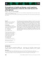
The protection of anatomical swallowing structures
was routinely perfor med by dr awing a laryngo -pharyn-
geal midline ‘shielding’ contour outside the PTVs in all
cases. This s paring structure has been defined prospec-
tively in January 2002, when we implemented IMRT
clinically, and was provided to be used in all midline
areas where no PTV was required. This structure may
include esophageal, laryngeal, and pharyngeal structures.
Aimed dose constraint for this midline shielding was a
mean dose (Dmean) below 45Gy (Figure 1).
In oropharyngeal cancer patients, this structure was
usually contoured from the level of the hyoid (below the
lateral retropharyng eal lymph nodes, corresponding ~to
the cervical vertebra 2/3, Figure 1) to the lowest level at
which PTVs were drawn. In hypopharyngeal cancer
patients, midline protection is often limited to some
aspects of the larynx to just prevent laryngeal structures
from full tumor dose.
Clinical factors
The clinical variables examined for correlation with
grade 3-4 late toxicity included age, gender, primary
site, tumor stage, tumor volume, therapy sequence, addi-
tion of systemic therapy and IMRT treatment schedules.
Dosimetric factors
The dose distribution to the swallowing structures was
calculated on the original IMRT treatment plans. Based
Figure 1 Example: midline shielding as used according to our internal IMRT guidelines (pink contour, below hyoid/C3).
Table 2 Volumetric staging in patients treated with primary radiotherapy (n = 63)
total gross tumor volume (tGTV)
Primary site mean (range) 1-15cc 16-70cc >70cc
Oropharynx
base of tongue 34cc (9-127cc) 2 13 3
tonsil/lateral mesopharynx 51cc (4-171cc) 1 11 7
Larynx
Glottis 12cc (1-29cc) 1 1 0
Supraglottis 21cc (6-54cc) 2 5 0
Hypopharynx 46cc (5-173cc) 3 11 3
Total 44cc (1-173cc) 9 (14%) 41 (65%) 13 (21%)
Peponi et al. Radiation Oncology 2011, 6:1
/>Page 3 of 8
on studies published so far [6-9], and with regard to the
swallowing appar atus, the following anatomic structures
were retrospectively identified and delineated on the
axial CT-slices of each plan: the pharyngeal c onstrictor
muscles (PCs) - superior, middle and inferior- , the glot-
tic and supraglottic larynx (GSL) and the muscular com-
partment of the esophagus inlet (eim). In brief, the
superior constrictor muscl e (scm) was defined from the
caudal tips of the pterygoid plates through the upper
edge of the hyoid bone, the middle constrictor muscle
(mcm) was defined from the upper through the lower
edge of the hyoid and the inferior constrictor musc le
(icm) was defined from below the hyoid through the
inferior edge of the cri coid. A structure named PCs was
outlined to involve the constrictors as a single structure.
The larynx (GSL) was contoured from the tip of the epi-
glottis superiorly to the bottom of the cricoid inferiorly.
Caudal to the inferi or border of the cricoid, the esopha-
gus(eim)wascontoured,withitscaudal-mostextent
corresponding to the caudal-most extent of the low
neck target volumes. Dose-volume histograms to the
swallowing structures were assessed and mean dose,
maximum point dose (Dmax), minimum point dose
(Dmin), V
30
(volume of a structure receiving >30Gy),
V
50
(volume of a structure receiving >50Gy), V
60
(volume of a structure receiving >60Gy), V
65
(volume of
a structure receiving >65Gy), V
70
(volume of a structure
receiving >70Gy), D
50
(dose received by 50% volume of
a structure/median dose) and D
80
(dose received by 80%
volume of a structure) were calculated.
Statistical analysis
Statistical calculations of Kaplan Meier curves were per-
formed using StatView
®
program (Abacus Concepts
Inc., Berkeley, CA). A p value of ≤0.05 was considered
statistically significant.
Results
Between January 2002 and November 2005, a total of 82
out o f 96 eligible patients successfully treated with SIB-
IMRT agreed to participate in our study. 80 patients
responded to all given questionnaires; 2/82 pa tients
declined to return the questionnaires, however, agreed
to allow using their objective data as assessable from
regular follow up visits. One previously irradiated
patient was excluded from the dysphagia analysis.
Late toxicity
At the first post-treatment follow-up (mean 20 month,
range 4-48), any subjective grade 3/4 toxicity (G3/4) wa s
reported by 14/80 patients (18%), while 66/80 patients
(82%) experienced grade 0-2 toxicity. At the second follow
up (objective assessment), grade 3/4 toxicity rate was 10%
(8/78) (two patients excluded because of tumor recur-
rence, two patients lost to follow-up); Table 3 shows sub-
jective and objective late toxicity. The mean dose and dose
range in definitively irradiated versus postoperatively irra-
diated patients is indicated in Table 1.
Prevalence of long term dysphagia (re-irradiated patient
excluded)
At the patient reported first assessment (mean 20
mont hs, range 4-40), 77/79 patients experienced dyspha-
gia grade 0-2; five patients (8%) experienced dysphagia
grade 2, symptom that continued to persist only in one
patient by reevaluation (objective assessment, mean 32
months, range 16-60). Persistent dysphagia grade 3/4 was
found in one patient (1%). There were no cases of
Table 3 Frequency of grade 3/4 (G3/4) late toxicity at the subjective (mean 20 months; range 4-40) and objective
(mean 32 months; range 16-60) assessment
Grade 3/4 late term toxicity
Parameter S u b j e c t i v e O b j e c t i v e
Swallow pain 1na
Dysphagia 2 (1 definitive/1postoperative) 2 (same pts. as subjective)
Taste alteration 9na
Xerostomia 3 6
(same 3 pts as subjective + 3 others)
Weight loss ≥10% na 0
Hoarseness 0 0
Total No of pts 14/80 (18%) 8/78 (10%)
Abbreviations: na: not assessable, pts: patients, No: number
Peponi et al. Radiation Oncology 2011, 6:1
/>Page 4 of 8
clinically symptomatic pneumonia as a potential conse-
quence of aspiration reported by patients or stated in the
patient charts (no radiological swallowing tests
performed).
At the second evaluation (mean 32 month post treat-
ment; n loco-reg ional ly controlled patients with no pre-
vious radiation = 77) as well as at the most recent follow
up (“patient last seen”, mean 50 months, range 16-85,
Figure 2), persisting swallowing dysfuncti on grade (2-) 3
was subjectively and objectively assessed by 1 patient.
Weight loss/PEGs
Percutaneous endoscopic gastrostomy feeding tubes
(PEGs) were placed before or during treatment in 21 of
82 patients (26%). The mean time to PEG tube removal
was 8 months (range 5-25). At the time of the first ana-
lysis (20-month follow up), 6/21 patients (7% of all, ~1/3
of the PEG patients) were still using PEG for some or all
of their nutrition. Patients sustained median weight loss
of 5.1 kg (range 0-20 kg) during treatment, while one
year post treatment there was no p atient who had lost
>10% of body weight. Only two of those 6 patients
remained PEG-dependent (10% of all PEG patients, 2% of
the entire cohort); the other 4 patients regained indepen-
denc e of PEG 14, 16, 33 and 36 months after completio n
of IMRT, respectively. In none of the patients who
remained loco-regionally disease free, a PEG had to be
placed during the monitored follow up period/replaced
once the PEG has been removed.
Swallowing structure doses
The median doses (median of the median dose) to the
swallowing structures, the partial volumes receiving spe-
cified doses (V
D
) in all patients and comparable results
of reported series are detailed in Tables 4 and 5.
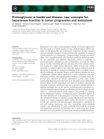
Long term local control and overall survival
The 3 and 5 year local control rates of the assessed
cohort were 78 and 75% (Figure 2) , the correspo nding
overall survival rates were 80 an d 77%, respectively
(Kaplan Meier survival curves, December 2010). None
of the local failures were found related to the midline
protection structure (all failures analysed: no medial
marginal failures).
Discussion
Recent gains in the management of head and neck can-
cer have been achieved due to concurrent chemo-radio-
therapy with altered fractionated three-dimensional
conformal radiotherapy (3D-CRT) or IMRT technique
[10-12]. The use of these high intensity treatments has
resulted in considerable rates of swallowing dysfunction,
both acute (15-63%) and long term (3-21%) [13-20].
Comprehensivedataonlatetoxicityfromrandomized
and nonrandomized trials, however, are sparse.
In our cohort of patients treated with SIB-IMRT
either alone or in combination with chemotherapy or
surgery, the rate of grade 3/4 long term dysphagia was
1%, comparable to that seen in other IMRT studies, and
considerably better than that observed in 3D-CRT stu-
dies (Table 6). These findings of a low rate of severe
dysphagia in a patient coho rt at risk motivate efforts to
reduce the doses to the swallowing structures, fact
which could reduce the severity and prevalence of dys-
phagia. This may be reached by a simple protection
structure along the midline were no PTV is needed (Fig-
ure 1). Analysis of the relationship between the swallow-
ing structure doses and the development of late
dysphagia were limited due to the single even t, pre-
cluded statistical significance. Median doses of the swal-
lowing structures in the own cohort were comparable to
reported series [7,21] (Table 4, 5). The limit ation of our
study was the retrospective nature of the analysis,
whereas midline protection contouring (Figure 1) was
prospectively performed as part of our internal IMRT
guidelines. Similar to the parotid gland protection, no
oncological compromises are acceptable in contouring
the midline sparing structure. The group of Eisbruch et
al [2] suggested that the high loco-regional control rates
have not been compromised by the efforts to spare the
parts of the swallowing structures not involved by
tumor and not at risk of subclinical disease. In addition,
in a previous evaluation of our hypopharynx-larynx
patient cohort [22] treated with IMRT using midline
sparing as far as feasible, local failures were not found
related to the midline sparing structures.
The low percentage of PEG tube dependence (7%) at
the mean 20-month follow-up may be interpreted as a
surrogate of limited swallowing problems.
Published analyses focused on predicting the probabil-
ity of s evere acute or late dysphagia during or after RT
[23,24] between patient-rated and objective assessment
of dysphagia are conflicting.
0
.2
.4
.6
.8
1
Cum. Survival
0 10 20 30 40 50 60 70 80 90
m
o
nth
s
81 63 51 45 41 30 23 6 patients at risk
loc al control rate
late grade 3 dysphagia
Figure 2 5-year local control rate (75%) and rate of freedom of
grade 3/4 late dysphagia (re-irradiated patient excluded, 99%).
Peponi et al. Radiation Oncology 2011, 6:1
/>Page 5 of 8
Table 5 Partial volumes receiving specified doses (VD) to the swallowing structures in all patients and comparison to
the data as reported by Feng et al 2007 21
PCs, median (range) GSL, median (range) eim, median (range)
V50 (%) V60 (%) V65 (%) V70 (%) V50 (%) V60 (%) V65 (%) V70 (%) V50 (%) V60 (%) V65 (%)
Current study 88.5 (10-100) 43.9 (0-94) 29 (0-60) 7.3 (0-40) 3.9 (1-100) 21.1 (0-98) 8.9 (0-94) 0 (0-67) 5.1 (0-100) 0 (0-87) 0 (0-84)
Feng et al [21] 90 (58-100) 73 (36-100) 57 (20-99) NA 69 (1-100) 37 (0-100) 20 (0-100) NA 14 (0-100) 0 (0-100) 0 (0-78)
Abbreviations: PCs: pharyngeal constrictor muscles, GSL: glottic and supraglottic larynx, eim: muscular compartment of the esophagus inlet, NA: not assessed.
Table 6 Results from selected series regarding late toxicity in head and neck cancer patients treated with RT ±
chemotherapy
Technique Authors
[reference]
year No of
patients
median follow up
(months)
stage lll/lV
(%)
Chemotherapy
(%)
grade 3/4 late
toxicity
Chao et al [25] 2003 74 30 93 23 0
I
de Arruda et al [14] 2006 50 18 92 86 3 (6%)
M
Lee et al [17] 2006 41 31 100 100 5 (12%)
R
Studer et al [1] 2006 115 18 52 78 18 (15%)
T
present study 2010 81* 55 100 85 7 (9%)
Denis et al [26] 2003 44 60 100 61 30 (68%)
3D-CRT
Huguenin et al [19] 2004 224 39 97 50 G3: 92 (41%)
G4: 5 ("%)
Abbreviations: IMRT: Intensity modulated radiation therapy; 3D-CRT: three-dimensional conformal radiotherapy (*patient with previous Cobalt radiation therapy
excluded).
Table 4 Median doses (median of the median dose) to the swallowing structures in all patients and comparison to
reported series
Swallowing structures Current study Feng et al 2007 [21] Levendag et al* 2007 [7]
Median Range Median Range Median Range
PCs Volume (cc) 20 11-32 NA NA NA NA
Dose (Gy) 59 39-66 64 51-72 48 6.0-73.0
scm Volume (cc) 12 6-21 NA NA NA NA
Dose (Gy) 59.4 24-69 68 57-74 51 22.0-73.0
mcm Volume (cc) 3.6 1.5-9 NA NA NA NA
Dose (Gy) 59 37.0-71.5 64 53-75 48 11.0-72.0
icm Volume (cc) 3.7 0.9-7 NA NA NA NA
Dose (Gy) 53 29-73 51 30-70 32 6.0-73.0
GSL Volume (cc) 15.3 6.4-23 NA NA NA NA
Dose (Gy) 53 28-70 55 22-72 NA NA
eim Volume (cc) 5.4 0.7-44 NA NA NA NA
Dose (Gy) 39 16.0-67.8 44 15-66 18 3.0-64.0
*The results are not entirely comparable with the study of Levendag et al [7], as there is a difference in the delineation of muscular structures. In Levendag et al.
the anterior part of scm and mcm were not delineated and eim was defined as the proximal 1 cm of the esophageal inlet, regardless of the caudal-most extent
of the low neck target volumes.
Abbreviations: PCs: pharyngeal constrictor muscles, scm: superior constrictor muscle, mcm: middle constrictor muscle, icm: inferior constrictor muscle, GSL: glottic
and supraglottic larynx, eim: muscular compartment of the esophagus inlet, NA: not assessed
Peponi et al. Radiation Oncology 2011, 6:1
/>Page 6 of 8
Patients’ satisfaction with their swallowing function,
in addition with the objective parameters ‘body weight’
and ‘dependency of a long term PEG’ , are reliable
answer and were found congruent with the objective
grading. No specific tests were performed to detect
potentially aspiration-related, clinically not obvious
pneumonia.
Conclusions
In conclusion, IMRT using a midline contour to spare
swallowing structures outside PTVs is relatively safe and
effective in terms of local disease control and avoidance
of persistent late dysphagia. The subjective patients’ esti-
mation of late dysphagia was compatible with the objec-
tive assessment of swallowing dysfunction.
Author details
1
Department of Radiation Oncology, University Hospital Zurich, Zurich,
Switzerland.
2
Department of Pediatrics, Civic Hospital of Lugano, Lugano,
Switzerland.
3
Department of Otorhinolaryngology, Head and Neck Surgery,
University Hospital Zurich, Zurich, Switzerland.
Authors’ contributions
GS and CG conceived of the study, carried out its design and supervised the
coordination. BW performed all phone call interviews with patients, sent out
and analysed the QoL questionnaire forms. EP carried out the specific
contouring work, analysed the related DVHs, and drafted the manuscript. GH
was mainly involved/in charge with the clinical post treatment follow up
visits of all patients. All authors read and approved the final manuscript.
Competing interests
The authors declare that they have no competing interests.
Received: 19 October 2010 Accepted: 5 January 2011
Published: 5 January 2011
References
1. Studer G, Huguenin PU, Davis JB, Kunz G, Lutolf UM, Glanzmann C: IMRT
using simultaneously integrated boost (SIB) in head and neck cancer
patients. Radiat Oncol 2006, 1:7.
2. Feng Y, Kim MHyungjin, et al: Intensity-Modulated
Chemoradiotherapy Aiming to Reduce Dysphagia in Patients With
Oropharyngeal Cancer: Clinical and Functional Results. JClinOncol
2010, 28:2732-2738.
3. Caudell JJ, Schaner PE, Meredith RF, et al: Factors Associated With Long-
Term Dysphagia After Definitive Radiotherapy for Locally Advanced
Head-and-Neck Cancer. International Journal of Radiation
Oncology*Biology*Physics 2009, 73:410-415.
4. Cooper J, Fleming ID, editors, HD: Head and Neck Cancer. AJCC Manual
for Staging of Cancer. 6 edition. Philadelphia: JB Lippincott; 2002.
5. James DC, JoAnn S, Thomas FP: Toxicity criteria of the Radiation Therapy
Oncology Group (RTOG) and the European organization for research
and treatment of cancer (EORTC). International journal of radiation
oncology, biology, physics 1995, 31:1341-1346.
6. Eisbruch A, Schwartz M, Rasch C, et al: Dysphagia and aspiration after
chemoradiotherapy for head-and-neck cancer: Which anatomic
structures are affected and can they be spared by IMRT? International
Journal of Radiation Oncology Biology Physics 2004, 60:1425-1439.
7. Levendag PC, Teguh DN, Voet P, et al: Dysphagia disorders in patients
with cancer of the oropharynx are significantly affected by the radiation
therapy dose to the superior and middle constrictor muscle: A dose-
effect relationship. Radiotherapy and Oncology 2007, 85:64-73.
8. Sanguineti G, Adapala P, Endres EJ, et al: Dosimetric Predictors of
Laryngeal Edema. International Journal of Radiation Oncology Biology
Physics 2007, 68:741-749.
9. Caglar HB, Tishler RB, Othus M, et al: Dose to Larynx Predicts for
Swallowing Complications After Intensity-Modulated Radiotherapy.
International Journal of Radiation Oncology Biology Physics 2008,
72:1110-1118.
10. Fang F-M, Chien C-Y, Tsai W-L, et al: Quality of Life and Survival Outcome
for Patients With Nasopharyngeal Carcinoma Receiving Three-
Dimensional Conformal Radiotherapy vs. Intensity-Modulated
Radiotherapy–A Longitudinal Study. International Journal of Radiation
Oncology Biology Physics 2008, 72:356-364.
11. Fu KK, Pajak TF, Trotti A, et al: A Radiation Therapy Oncology Group
(RTOG) phase III randomized study to compare hyperfractionation and
two variants of accelerated fractionation to standard fractionation
radiotherapy for head and neck squamous cell carcinomas: first report
of RTOG 9003. Int J Radiat Oncol Biol Phys 2000, 48
:7-16.
12.
Forastiere AA, Goepfert H, Maor M, et al: Concurrent Chemotherapy and
Radiotherapy for Organ Preservation in Advanced Laryngeal Cancer. N
Engl J Med 2003, 349:2091-2098.
13. Eisbruch A, Lyden T, Bradford CR, et al: Objective assessment of
swallowing dysfunction and aspiration after radiation concurrent with
chemotherapy for head-and-neck cancer. International Journal of
Radiation Oncology Biology Physics 2002, 53:23-28.
14. de Arruda FF, Puri DR, Zhung J, et al: Intensity-modulated radiation
therapy for the treatment of oropharyngeal carcinoma: the Memorial
Sloan-Kettering Cancer Center experience. Int J Radiat Oncol Biol Phys
2006, 64:363-373.
15. Teguh DN, Levendag PC, Noever I, et al: Treatment techniques and site
considerations regarding dysphagia-related quality of life in cancer of
the oropharynx and nasopharynx. Int J Radiat Oncol Biol Phys 2008,
72:1119-1127.
16. Dirix P, Nuyts S: Value of Intensity-Modulated Radiotherapy in Stage IV
Head-and-neck Squamous Cell Carcinoma. International Journal of
Radiation Oncology*Biology*Physics 2010, 78:1373-1380.
17. Lee NY, de Arruda FF, Puri DR, et al: A comparison of intensity-modulated
radiation therapy and concomitant boost radiotherapy in the setting of
concurrent chemotherapy for locally advanced oropharyngeal
carcinoma. Int J Radiat Oncol Biol Phys 2006, 66:966-974.
18. Fua TF, Corry J, Milner AD, Cramb J, Walsham SF, Peters LJ: Intensity-
modulated radiotherapy for nasopharyngeal carcinoma: Clinical
correlation of dose to the pharyngo-esophageal axis and dysphagia.
International Journal of Radiation Oncology*Biology*Physics 2007, 67:976-981.
19. Huguenin P, Beer KT, Allal A, et al: Concomitant Cisplatin Significantly
Improves Locoregional Control in Advanced Head and Neck Cancers
Treated With Hyperfractionated Radiotherapy. J Clin Oncol 2004,
22:4665-4673.
20. Caudell JJ, Schaner PE, Desmond RA, Meredith RF, Spencer SA, Bonner JA:
Dosimetric Factors Associated With Long-Term Dysphagia After
Definitive Radiotherapy for Squamous Cell Carcinoma of the Head and
Neck. International Journal of Radiation Oncology*Biology*Physics 2010,
76:403-409.
21. Feng FY, Kim HM, Lyden TH, et al: Intensity-Modulated Radiotherapy of
Head and Neck Cancer Aiming to Reduce Dysphagia: Early Dose-Effect
Relationships for the Swallowing Structures. International Journal of
Radiation Oncology Biology Physics 2007, 68:1289-1298.
22. Studer G, Peponi E, Kloeck S, Dossenbach T, Huber G, Glanzmann C:
Surviving Hypopharynx-Larynx Carcinoma in the Era of IMRT.
International Journal of Radiation Oncology*Biology*Physics 2010,
77:1391-1396.
23. Langendijk JA, Doornaert P, Rietveld DHF, Verdonck-de Leeuw IM, René
Leemans C, Slotman BJ: A predictive model for swallowing dysfunction
after curative radiotherapy in head and neck cancer. Radiotherapy and
Oncology 2009, 90:189-195.
24.
Joke W, Kim De R, Duprez F, et al: Acute Normal Tissue Reactions in
Head-and-Neck Cancer Patients Treated With IMRT: Influence of Dose
and Association With Genetic Polymorphisms in DNA DSB Repair Genes.
International journal of radiation oncology, biology, physics 2009,
73:1187-1195.
Peponi et al. Radiation Oncology 2011, 6:1
/>Page 7 of 8
25. Chao KSC, Gokhan O, Wade LT: Toxicity profile of intensity-modulated
radiation therapy for head and neck carcinoma and potential role of
amifostine. Seminars in oncology 2003, 30:101-108.
26. Denis F, Garaud P, Bardet E, et al: Late toxicity results of the GORTEC 94-
01 randomized trial comparing radiotherapy with concomitant
radiochemotherapy for advanced-stage oropharynx carcinoma:
comparison of LENT/SOMA, RTOG/EORTC, and NCI-CTC scoring systems.
International Journal of Radiation Oncology Biology Physics 2003, 55:93-98.
doi:10.1186/1748-717X-6-1
Cite this article as: Peponi et al.: Dysphagia in head and neck cancer
patients following intensity modulated radiotherapy (IMRT). Radiation
Oncology 2011 6:1.
Submit your next manuscript to BioMed Central
and take full advantage of:
• Convenient online submission
• Thorough peer review
• No space constraints or color figure charges
• Immediate publication on acceptance
• Inclusion in PubMed, CAS, Scopus and Google Scholar
• Research which is freely available for redistribution
Submit your manuscript at
www.biomedcentral.com/submit
Peponi et al. Radiation Oncology 2011, 6:1
/>Page 8 of 8