Báo cáo khoa học: "The in vivo study on the radiobiologic effect of prolonged delivery time to tumor control in C57BL mice implanted with Lewis lung cancer" pdf
Bạn đang xem bản rút gọn của tài liệu. Xem và tải ngay bản đầy đủ của tài liệu tại đây (257.72 KB, 6 trang )
RESEARCH Open Access
The in vivo study on the radiobiologic effect of
prolonged delivery time to tumor control in
C57BL mice implanted with Lewis lung cancer
Xin Wang
1,2†
, Xiao-Peng Xiong
1,3†
, Jiade Lu
1,4
, Guo-Pei Zhu
1
, Shao-Qin He
1
, Chao-Su Hu
1
, Hong-Mei Ying
1*
Abstract
Background: High-precision radiation therapy techniques such as IMRT or sterotactic radiosurgery, delivers more
complex treatment fields than conventional techniques. The increased complexity causes longer dose delivery
times for each fraction. The purpose of this work is to explore the radiobiologic effect of prolonged fraction
delivery time on tumor response and survival in vivo.
Methods: 1-cm-diameter Lewis lung cancer tumors growing in the legs of C57BL mice were used. To evaluate
effect of dose delivery prolongation, 18 Gy was divided into different subfractions. 48 mice were rando mized into 6
groups: the normal control group, the single fraction with 18 Gy group, the two subfractions with 30 min interval
group, the seven subfractions with 5 min interval group, the two subfractions with 60 min interval group and the
seven subfractions with 10 min interval group. The tumor growth tendency, the tumor growth delay and the mice
survival time were analyzed.
Results: The tumor growth delay of groups with prolonged delivery time was shorter than the group with single
fraction of 18 Gy (P < 0.05). The tumor grow delay of groups with prolonged delivery time 30 min was longer than
that of groups with prolonged delivery time 60 min P < 0.05). There was no significant difference between groups
with same delivery time (P > 0.05). Compared to the group with single fraction of 18 Gy, the groups with
prolonged delivery time shorten the mice survival time while there was no significant difference between the
groups with prolonged delivery time 30 min and the groups with prolonged delivery time 60 min.
Conclusions: The prolonged delivery time with same radiation dose shorten the tumor growth delay and survival
time in the mice implanted with Lewis lung cancer. The anti-tumor effect decreased with elongation of the total
interfractional time.
Introduction
New radiation therapy techniques such as sterotactic
radiosurgery and IMRT are featured with improving tar-
get dose conformity while minimizing radiation expo-
sure to surrounding normal tissues [1-5]. However,
these technologies require complex planning and deliv-
ery procedure thus a substantially prolongerd delivery
time for each fraction.
According to radiobiological theory, the sublethal
damage repair (SLDR) takes place not only between the
frations but also during the irradiation. Cell killing tends
to decrease with fraction delivery time increasing
because of ongoing sublethal damage repair processes
during dose delivery [6,7]. Therefore, it is reasonable to
question whether the radiobiological effectiveness of
intermittently delivered radiation over a prlonged time
has the same biological effectiveness as those delivered
continuously through conventional external beam radia-
tion therapy (EBRT).
A number of studies have been published to investi-
gate the impact of prolonged delivery time (such as
used in IMRT) on biological effects at the cellular level
and demonstrated that the total time to deliver a single
fraction may have a significant impact on treatment out-
come [8-15]. Howev er, in-vivo study about the effects of
* Correspondence:
† Contributed equally
1
Department of Radiation and Oncology, Cancer Center and Department of
Oncology, Shanghai Medical College, Fudan University, Shanghai, PR China
Full list of author information is available at the end of the article
Wang et al. Radiation Oncology 2011, 6:4
/>© 2011 Wang et al; licensee BioMed Central Ltd. This is an Open Access article distributed under the terms of the Creative Commons
Attribution License (http://crea tivecommons.org/licens es/by/2.0), which permits unrestricted use, distribution, and re prod uction in
any medium, provide d the origin al work is properly cited.
treatment time and fractionation on tumor response and
growth is lmited. To our knowledge, there were only
two studies to evaluate the effect of proloned delivery
time in SCCVII tumors using an in vivo-in vitro assay
and they found that cell survival from clamped tumors
tended to increase with elongation of the intervals, but
not significantly [16,17]. They contributed the confilic-
tion of the results in vitro and in vivo to reoxygenation.
In this study, we attempt to evaluate the impact of pro-
longed fraction delivery time on tumor control and sur-
vival in C57BL mice implanted with Lewis lung cancer
using growth delay assay and survival analysis.
Methods
Cell line and mice
The Lewis lung carcinoma (LLC) cell line purchased
from the Division of Animals of FUDAN University was
growninRPMI1640(GibcoBRL,USA)supplemented
with 10% fetal bovine serum, 100 μg/ml streptomycin
and 100 U/ml penicillin. The cell line was incubated at
37°C in 5% CO
2
until near-confluency, harvested,
washed, and counted using trypan blue exclusion. Female
C57BL/6 mice weighing 16-18 g were used. For i n vivo
implantation, LLC cells were washed in Hanks’ balanced
salt solution (HBSS) and injected subcutaneously at 1 ×
10
6
cells in 0.1 ml HBSS in the right hind limb of C57BL/
6 mice. At 10 days after the injection, 48 mice with
tumor diameter reached 0.8~1.0 cm were retained and
randomly divided into 6 groups and radiated according
to the predetermi ned schedule as described below. These
animals were fed sterilized chow and tap water in accor-
dance with Fudan University of Animal Resource Depart-
ment protocols in a laminar flow room.
Irradiation
Unanesth etized tumor-bearing mice were immobilized in
a jig with customed modules with their legs fixed using
adhesive tapes to receive a focal irradiation. Irradiation
was delivered using
60
Cotherapyunitatadoserateof
157.1cGy/min at room temperature. The dose was cali-
brated using a RAMTEC 1000 dosimeter. All mice were
shielded with a specia lly designed lead apparatus to allow
irradiation to the right hind limb. Mice were kept under
these conditions until all irradiation finished.
Radiation Schedule
To learn the radiobiological characteristics, 48 mice
were randomly divided into 6 groups with 8 mice per
group. The total dose was 18 Gy for all irradiated sub-
jects except for the control. In addition to the control, 5
radiation schedules were used: (1) 1 fraction of 18 Gy
without interruption, (2) 2 fractions of 9 Gy with inter-
fraction intervals of 60 min, (3) 7 fractions of 2.57 Gy
with interfraction intervals of 10 min, (4) 2 fractions
of 9 Gy with interfraction intervals of 30 min, (5) 7
fractions of 2.57 Gy with interfraction intervals of 5 min.
Assay
Tumor dimensions were measured with Vernier calipers
once every two days. Tumor volumes were calculated as
follows: Volume (mm
3
) = (H*W * L)/2 (H = height, W =
width, and L = length of the tumor).
The tumor growth time (TGT) was defined as the
time required for the initial tumor size to quadruple
after the first day of treatment. The tumor growth delay
time (TGDT) was defined as the TGT in each treated
animal minus the mean TGT in the contr ol group. The
curves of tumor growth and calculated tumor growth
delayed time among the 6 groups were compared.
The survival time of mice was also documented.
Statistical analysis
Statistical differences among groups were determined
using one-way analysis of variance (ANOVA). Kaplan-
Meier survival curves were analyzed. The logrank test
was used to assess if there were differences among the
six groups in overall survival. Res ults are expressed as
means ± SEM. The level of significance used for all
comparisons was P < 0.05.
Results
The effect of the total interfraction interval time on the
growth of tumor tissues
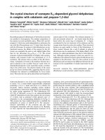
Figure 1 shows the tumor growth curves of all groups
treated with various radiation schedules and the control.
Tumors in the control group demonstrated a rapid
exponential growth. Tumors irradiated with a single
fraction of 18 Gy produced the most significant growth
delay. The other groups irradiated to 18 Gy with various
fractions and intermittent time also demonstrated delay
in tumor growth, which was significantly associated with
interfractional intermittent time. The anti-tumor effect
decreased with elongation of the total interfractional
time. However, the fractionation dose was not associated
with the tumor growth rate. (Table 1)
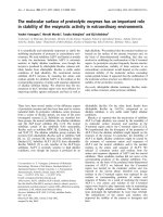
Figure2showsthetumorgrowthdelaytime(TGDT)
of each group. The tumor growth delay of the groups
with total interfraction interval time 30 min was higher
than that of the gro ups with total interfraction interval
time 60 min (P < 0.01). When the tumor growth delay
of groups with t he same total interfraction interval
times was compared, there is no statistical significance
(P > 0.05).
The effect of the total interfraction interval time on
survival time of the mice
As shown in table 2, the survival time of every irradiated
group was longer than the control group; though the
Wang et al. Radiation Oncology 2011, 6:4
/>Page 2 of 6
survival time of the four prolonged delivery time group
was similar, the survival time of 18 Gy single fractions
was much longer than the other four irradiated group.
Discussion
Our current knowledge of the effect of radiation on
tumor growth were largely based on linear-quadratic
(LQ) model, which was initially derived to fit experi-
mental observation s of the effects of dose and fracti ona-
tion on cell survival, chromosomal damage and acute
radiation effects. However, it was derived largely from in
vitro rather than in vivo observations, thus does not
consider that tumor response in vivo are affected by
other effects such as the impact of ionizing radiation on
the supporting tissues and the impact of the subpopula-
tion of radioresistant clonogens. Therefore, our under-
standing of tumor response to d ifferent radiation
fractionation or treatment time may be questionable for
in vivo irradiation.
In the current study, the potential impact of pro-
longed fraction delivery time for a fixed total dose on
the control of Lewis lung cancer implanted in C57BL
mice was studied in order to investigate the effect of
intermittent radiation exposure compared with that of
continuous radiation exposure in vivo. The results of
this in vivo study confirmed a rapid growth of tumor
after prolonged intermittent time betw een fractions thus
the total treatment time. However, the fraction size of
radiation may not be a significant factor for tumor
control.
The results of our in vivo study consisted with those
previously reported studies which had focuse d to in
vitro cell survival rates. Both in vitro radiobiological
Da
y
s after irradiation
312927252321191715131197531
Mean tumor volume(cm3)
10.00
8.00
6.00
4.00
2.00
0.00
2.57 Gy×7 at 5min intervals
9 Gy× 2 at 30min intervals
2.57 Gy×7 at 10min intervals
9 Gy× 2 at 60min intervals
18 Gy as a single dose
control
2.57 Gy×7 at 5min intervals
9 Gy× 2 at 30min intervals
2.57 Gy×7 at 10min intervals
9 Gy× 2 at 60min intervals
18 Gy as a single dose
control
group
Figure 1 The tumor growth curves of all groups treated with various radiation schedule and the control. Tumor growth delay was
significantly prolonged with the elongation of the total interfraction interval time.
Table 1 Tumor growth time* compared to 18 Gy in a
sigle fraction
Radiation schedule TGT (days) ± SE p value
Control 8.1 ± 0.6 <0.001
18 Gy as a single dose 19.9 ± 2.3
9 Gy × 2 fractions at 60 min intervals 14.0 ± 1.8 <0.001
2.57 Gy × 7 at 10 min intervals 14.3 ± 1.8 <0.001
9 Gy × 2 fractions at 30 min intervals 17.7 ± 2.5 0.034
2.57 Gy × 7 at 5 min intervals 17.7 ± 2.6 0.037
* The tumor growth time (TGT) was defined as the time required for the initial
tumor size to quadruple after treatment. Each date represents mean ± SE.
Wang et al. Radiation Oncology 2011, 6:4
/>Page 3 of 6
experiments and calculations based on the linear-quad-
ratic model have shown greater cell survival rates for
long 15-60 min compared to short 2-5 min fractional
delivery times. Benedict et al irradiated several human
GBM cell lines by the 6 MV g rays of linear accelerator
simulating intensity-modulated stereotactic radiosurgery.
They divided the total doses into several f ractions and
the intervals ranged form 16 min to 3 hours. The results
showed that the prolonged interval time will increase
the survival fraction of the cells. A 40% increase in
malignant glioma cell survival when the dose delivery
schedule for a singlefraction 12 Gy irradiation was
altered from 5 min of continuous irradiation to 60 min
of intermittent irradiation were observed. Survival rates
increased three-fold when the intermittent irradiation
was stretched over 110 min. [8] Morgan and his collea-
gue irradiated the tumor cancer cells simulating the
IMRT plans. They delivered a total dose of 2 Gy to the
cell lines over 2 min, 6 min and 20 min, and found that
compared with the 2 min and 6 min group, the survival
fraction of 20 min group increased significantly [9].
Wang et al reported total time to deliver a single frac-
tion may have a s ignificant impact on IMRT tr eatment
Figure 2 The effects of different interfraction interval time on the TGDT. The mean ± SE of tumor growth time in the group of the control
is 8.09 ± 0.61 days. P < 0.05 as compared with the group irradiated with 18 Gy single fraction.
Table 2 The survival time of each group (days)
Radiation schedule survival time
(days) ± SE
p value
Control 13.8 ± 2.4 <0.001
18 Gy as a single dose 28.8 ± 2.3
9 Gy × 2 fractions at 60 min intervals 23.5 ± 3.7 0.011
2.57 Gy × 7 at 10 min intervals 23.5 ± 3.7 0.004
9 Gy × 2 fractions at 30 min intervals 25.0 ± 2.9 0.026
2.57 Gy × 7 at 5 min intervals 24.8 ± 2.8 0.027
The survival time of each group was compared with the group irradiated with
18 Gy single fraction. Each date represents mean ± SE. We also compared the
last four groups by chi-square test, the F value is 0.428 and p value is 0.735.
(data not shown in this table)
Wang et al. Radiation Oncology 2011, 6:4
/>Page 4 of 6
outcome for tumors. They irradiated the human pros-
tate cancer cells (the repair half-time is 16 min anda/b =
3.1 Gy) with different fraction delivery times in the range
of 15-45 min. This study showed that for a prescription
dose of 81 Gy in 1.8 Gy fractions, the EUD for prostate
cancer decreased from 78 Gy for a conventional EBRT to
69 Gy for an IMRT with a fraction delivery time of 30
min; the TCP decreased almost 30% as well [10].
All the above-mentioned studies based on cell lines
and were conducted under simplified in vitro conditions.
The influence of other factors, such as proliferation,
oxygen, and nutritional states in vitro is smaller than in
tumors in vivo and repopulation, reoxygenation and
bystander effects are obviously not considered.
In order to explore the biological effect of prolonged
deliverytimeinvivo,Sugieetalconductedastudy.In
this study they used EMT6 and SCCVII tumors approxi-
mately 1 cm in diameter growing in the hind legs of
syngeneic mice. Mice received whole body irradiation
without anesthesia or physical restraint. Tumors were
excised twenty hours after radiation and cell survival
was determined by an in vivo-in vitro assay. They
reported that no statistically significant decreases were
observed by posing intervals between fractions in vivo.
It was suggested that SLDR in vivo might be counterba-
lanced by other phenomena such as reoxygenation that
sensitizes tumor cells to subsequent irradiation [16]. To
explain the discrepancy between the in vitro and in vivo
results, Tomita N et al conducted another in vivo study
to evaluate the effect of intermittent radiation by using
local irradiation to tumor-bearing legs and a tumor
growth delay assay. They found that the fractionated
groups had faster tumor regrowth than the continu-
ously-irradiated control group, and the effect of radia-
tion tended to decrease with elongation of interfraction
intervals. In the present study, we studied the influence
of the different fraction intervals to the mouse lewis
cancer model. Our results are in consistent with those
reported by Tomita although different experimental
methods and anaimal models were used.
The discrepancy be tween the results in Sugie’s study
and ours may contribute to the technical problems asso-
ciated with leg clamping and the magnitude and velocity
of reoxygenation in tumors [17]. In Sugie’s study, Mice
received whole body irradiation without anesthesia or
physical restraint. However, in Tomita’s and our studies,
unanesthetized tumor-bearing mice were immobilized in
a jig with customed modules with t heir legs fixed using
adhesive tapes to receive a focal irradiation. According
to the study conducted by Shibamoto et al, when
tumor-bearing mice were irradiated without anesthesia
or physical restraint, th e tumor had a hypoxic fraction
of 5.4% [18]. Both anesthesia and immobilization of
thetumor-bearinglegwithadhesivetapeproduced
significant increases in the hypoxic fraction (23 and
28%, respectively). Tomita’ s study showed that reoxy-
genation occurring within 5-15 min appeared to com-
pensate for SLDR in SCCVII tumors. When tumor-
bearing mice were immobilized, reoxygenation was
limited and the magnitude of reoxygenation of hypoxic
tumor cells might not be great enough to counterba-
lance SLDR, then the decrease of radiation effect
occurred due to SLDR.
Although t his study evaluated the radiation treatment
time on the response a nd growth rate of tumor in vivo
using tumor growth delay and survival analysis, a
number of issues remain to be discussed. First, the het-
erogeneity of different tumor tissues have different cap-
abilities of recovery from radiation [19], therefore the
influence due to the prolonged delivery time may be dif-
ferent according to different tumor type. It is well
known that the radiation sensitivity to low LET radia-
toin is largely determined by sublethal damage repair,
and dose-fractionation is an important factor for tumor
killing and control [20], so the results obtained from the
current series may not be applicable to all tumors. Sec-
ond, the underlying mechanism of the differences in
tumor response and delay of tumor growth due to treat-
ment break time remains unknown. Third, our data pro-
vide a simplified estimate on the significance of
prolonged delivery as a result of IMRT or radiosurgery.
However, in reality the situation includin g the effects of
instantaneous dose rate, beam-on time, and number,
size and distribution of segments may be more complex.
Moreover, the present study focused on tumor response
only, and response and recovery of various normal tis-
sues or organs from fractionated radiation o ver various
irradiation time is complex and not addressed.
In general, our study demonstrated that prolonged deliv-
ery time significantly reduce the biological effect of radia-
tion therapy in Lewis lung tumor. Treatment time may
impact clinical outcome and should be recorded along
with other established dosimetric parameters. These
effects need to be confirmed in clinical trials and consid-
ered in treatment planning. Biologically, more reliabe
experimental investigations using animal models based on
human tumors are desirable. In addition, the underlying
mechanism o f tumor response and sublethal damage
repair after radiation therapy should be inves tigated by
examine multiple endpoints including cellular motility,
metabolic activity and invasive capacities. Further studies
are needed to establish more reliable radiobiological mod-
els to evaluate the relationship between interfaction inter-
vals and the biologic effect of radiation.
Author details
1
Department of Radiation and Oncology, Cancer Center and Department of
Oncology, Shanghai Medical College, Fudan University, Shanghai, PR China.
Wang et al. Radiation Oncology 2011, 6:4
/>Page 5 of 6
2
Department of Radiation and Oncology, Huashan Hospital, Fudan
University, Shanghai, PR China.
3
Department of Nuclear Medicine, Renji
Hospital, Shanghai Jiaotong University School of Medicine,Shanghai, PR
China.
4
Department of Radiation Oncology, National University Hospital,
Singapore, Singapore.
Authors’ contributions
XW and XPX carried out the murine study, wrote the final version of the
manuscript and contributed equally on this manuscript. CSH and SQH
participated in the design of the study. HMY conceived of the study, and
participated in its design and coordination. GPZ and JL provided some
intellectual recommendation and reviewed the manuscript. All authors read
and approved the final manuscript.
Competing interests
All authors declare there were no actual or potential conflicts of interest in
this study.
Received: 7 September 2010 Accepted: 12 January 2011
Published: 12 January 2011
References
1. Mendenhall WM, Amdur RJ, Palta JR: Intensity-modulated radiotherapy in
the standard management of head and neck cancer: promises and
pitfalls. J Clin Oncol 2006, 24:2618-2623.
2. Tubiana M, Eschwege F: Conformal radiotherapy and intensity-modulated
radiotherapy-clinical data. Acta Oncol 2000, 39:555-567.
3. Gregoire V, Maingon P: Intensity modulated radiation therapy in head
and neck squamous cell carcinoma: state of the art and future
challenges. Cancer Radiother 2005, 9:42-50.
4. Benedict SH, Cardinale RM, Wu Q, Zwicker RD, Broaddus WC, Mohan R:
Intensity-modulated stereotactic radiosurgery using dynamic micro-
multileaf collimation. Int J Radiat Oncol Biol Phys 2001, 50:751-758.
5. Siochi RA: Minimizing static intensity modulation time using an intensity
solid paradigm. Int J Radiat Oncol Biol Phys 1999, 43:671-680.
6. Fowler JF, Welsh JS, Howard SP: Loss of biological effect in prolonged
fraction delivery. Int J Radiat Oncol Biol Phys 2004, 59:242-249.
7. Elkind MM, Alescio T, Swain RW, Moses WB, Sutton H: Recovery of hypoxic
mammalian cells from sub-lethal X-ray damage. Nature 1964,
202:1190-1193.
8. Benedict SH, Lin PS, Zwicker RD, Huang DT, Schmidt-Ullrich RK: The
biological effectiveness of intermittent irradiation as a function of
overall treatment time: Development of correction factors for LINAC
based stereotactic radiotherapy. Int J Radiat Oncol Biol Phys 1997,
37:765-769.
9. Morgan WF, Naqvi SA, Yu C, Smith LE, Rose M: Dose the time required to
deliver IMRT reduce its biological effectiveness. Int J Radiat Oncol Biol
Phys 2002, 54S:222.
10. Wang JZ, Li XA, D’Souza WD, Stewart RD: Impact of prolonged fraction
delivery times on tumor control: A note of caution for intensity-
modulated radiation therapy (IMRT). Int J Radiat Oncol Biol Phys 2003,
57:543-552.
11. Mu X, Lofroth PO, Karlsson M, Karlsson M, Zackrisson B: The effect of
fraction time in intensity modulated radiotherapy: Theoretical and
experimental evaluation of an optimization problem. Radiother Oncol
2003, 68:181-187.
12. Zheng XK, Chen LH, Yan X, Wang HM: Impact of prolonged fraction dose-
delivery time modeling intensity-modulated radiation therapy on
hepatocellular carcinoma cell killin. World J Gastroenterol 2005,
11:1452-1456.
13. Shibamoto Y, Ito M, Sugie C, Ogino H, Hara M: Recovery from sublethal
damage during intermittent exposures in cultured tumor cells:
Implications for dose modification in radiosurgery and IMRT. Int J Radiat
Oncol Biol Phys 2004, 59:1484-1490.
14. Ogino H, Shibamoto Y, Sugie C, Ito M: Biological effects of intermittent
radiation in cultured tumor cells: influence of fraction number and dose
per fraction.
J Radiat Res 2005, 46:401-406.
15. Paganetti H: Changes in tumor cell response due to prolonged dose
delivery times in fractionated radiation therapy. Int J Radiat Oncol Biol
Phys 2005, 63:892-900.
16. Sugie C, Shibamoto Y, Ito M, Miyamoto A, Fukaya N, Niimi H, Hashizume T:
Radiobiologic effect of intermittent radiation exposure in murine
tumors. Int J Radiat Oncol Biol Phys 2006, 64:619-624.
17. Tomita N, Shibamoto Y, Ito M, Ogino H, Sugie C, Ayakawa S, Iwata H:
Biological effect of intermittent radiation exposure in vivo: recovery
from sublethal damage versus reoxygenation. Radiother Oncol 2008,
86(3):369-374.
18. Shibamoto Y, Sasai K, Abe M: The radiation response of SCCVII tumor
cells in C3H/He mice varies with the irradiation conditions. Radiat Res
1987, 109:352-354.
19. Elkind MM, Sutton H: Radiation response of mammalian cells grown in
culture. I. Repair of X-ray damage in surviving Chinese hamster cells.
Radiat Res 1960, 13:556-593.
20. Kampinga HH, Hiemstra YS, Konings AWT, Dikomey E: Correlation between
slowly repairable double-strand breaks and thermal radiosensitization in
the human HeLa S3 cell line. Int J Radiat Biol 1997, 72:293-301.
doi:10.1186/1748-717X-6-4
Cite this article as: Wang et al.: The in vivo study on the radiobiologic
effect of prolonged delivery time to tumor control in C57BL mice
implanted with Lewis lung cancer. Radiation Oncology 2011 6:4.
Submit your next manuscript to BioMed Central
and take full advantage of:
• Convenient online submission
• Thorough peer review
• No space constraints or color figure charges
• Immediate publication on acceptance
• Inclusion in PubMed, CAS, Scopus and Google Scholar
• Research which is freely available for redistribution
Submit your manuscript at
www.biomedcentral.com/submit
Wang et al. Radiation Oncology 2011, 6:4
/>Page 6 of 6