Báo cáo khoa học: "Cationized gelatin-HVJ envelope with sodium borocaptate improved the BNCT efficacy for liver tumors in vivo" pptx
Bạn đang xem bản rút gọn của tài liệu. Xem và tải ngay bản đầy đủ của tài liệu tại đây (763.01 KB, 12 trang )
RESEARCH Open Access
Cationized gelatin-HVJ envelope with sodium
borocaptate improved the BNCT efficacy for liver
tumors in vivo
Hitoshi Fujii
1
, Akifumi Matsuyama
2
, Hiroshi Komoda
1
, Masao Sasai
2
, Minoru Suzuki
3
, Tomoyuki Asano
4
,
Yuichiro Doki
1
, Mitsunori Kirihata
4
, Koji Ono
3
, Yasuhiko Tabata
5
, Yasufumi Kaneda
6
, Yoshiki Sawa
1
,
Chun Man Lee
1,2,7*
Abstract
Background: Boron neutron capture therapy (BNCT) is a cell-selective radiation therapy that uses the alpha
particles and lithium nuclei produced by the boron neutron capture reaction. BNCT is a relatively safe tool for
treating multiple or diffuse malignant tumors with little injury to normal tissue. The success or failure of BNCT
depends upon the
10
B compound accumulation within tumor cells and the proximity of the tumor cells to the
body surface. To extend the therapeutic use of BNCT from surface tumors to visceral tumors will require
10
B
compounds that accumulate strongly in tumor cells without significant accum ulation in normal cells, and an
appropriate delivery method for deeper tissues.
Hemagglutinating Virus of Japan Envelope (HVJ-E) is used as a vehicle for gene delivery because of its high ability
to fuse with cells. However, its strong hemagglutination activity makes HVJ-E unsuitable for systemic administration.
In this study, we developed a novel vector for
10
B (sodium borocaptate: BSH) delivery using HVJ-E and cationized
gelatin for treating multiple liver tumors with BNCT without severe adverse events.
Methods: We developed cationized gelatin conjugate HVJ-E combined with BSH (CG-HVJ-E-BSH), and evaluated its
characteristics (toxicity, affinity for tumor cells, accumulation and retention in tumor cells, boron-carrying capacity
to multiple liver tumors in vivo, and bio-distribution) and effectiveness in BNCT therapy in a murine model of
multiple liver tumors.
Results: CG-HVJ-E reduced hemagglutination activity by half and was significantly less toxic in mice than HVJ-E.
Higher
10
B concentrations in murine osteosarcoma cells (LM8G5) were achieved with CG-HVJ-E-BSH than with BSH.
When administered into mice bearing multiple LM8G5 liver tumors, the tumor/normal liver ratios of CG-HVJ-E-BS H
were significantly higher than those of BSH for the first 48 hours (p < 0.05). In suppressing the spread of tumor
cells in mice, BNCT treatment was as effective with CG-HVJ-E-BSH as with BSH containing a 35-fold higher
10
B
dose. Furthermore, CG-HVJ-E-BSH significantly increased the survival time of tumor-bearing mice compared to BSH
at a comparable dosage of
10
B.
Conclusion: CG-HVJ-E-BSH is a promising strategy for the BNCT treatment of visceral tumors without severe
adverse events to surroundin g normal tissues.
* Correspondence:
1
Department of Surgery, Osaka University Graduate School of Medicine,
Osaka, Japan
Full list of author information is available at the end of the article
Fujii et al. Radiation Oncology 2011, 6:8
/>© 2011 Fujii et a l; licensee BioMed Central Ltd. This is an Open Access article distributed under the terms of the Creative Commons
Attribution License (http://crea tivecommons.org/licenses/by/2.0), which pe rmits unrestricted use, distribution, and re prod uction in
any medium, provide d the origin al work is properly cited.
Background
Boron neutron capture therapy (BNCT) is a cell-selective
radiation therapy that uses alpha particles and l ithium
nuclei produced by the boron neutron capture reaction.
These particles cause cell destruction, bouncing out to a
maximum distance of 10 μm from the target, a distance
that corresponds to the size of a cell. These particles only
destroy the cells that take up
10
Boron (
10
B) [1]. This ther-
apy is clinically indicated for multiple and diffuse tumors,
such as glioblastoma and head and neck tumors [2].
BNCT was recently evaluated for treating liver tumors
[3-8], although the prognosis of patients treated b y
BNCT with conventional
10
B compounds, particularly
sodium borocaptate (BSH), is not good because of its low
accumulation in liver tumors and the attenuation of the
epithermal neutron b eams directed toward deep lesions
[9-11]. Therefore, treating liver tumors effectively with
BNCT will require novel ways of delivering BSH, with
the characteristics of high ac cumulation in the tumor,
low toxicity fo r normal tissue, and rapid withdrawal from
normal tissue and the bloodstream [12]. Vario us carrie rs
such as liposomes have been investigated [13-16], but
until now a vector for BSH that adequately satisfies the
above requirements has not been developed.
Liver tumors, including primary and secondary
tumors, are the fifth most common solid tumor world-
wide. The incidence is increasing rapidly in most coun-
tries, at a pace that will make liver tumors the third
most common tumor by 2030 [17,18]. The mortality
rate of liver tumors, especially multiple metastatic liver
tumors, is high. Multimodal therapies for multiple liver
tumors have advanced considerably, and include radio-
frequency ablation, radiation, surgical ext irpation and
transplantation [19]. However, t herapy for multiple and
diffuse liver tumors is still difficult, because reducing the
liver volume reduces its organ function. Therefore, a
therapy selective for tumors with minimal damage to
normal liver tissue is of great interest.
Hemagglutinating Virus of Japan Envelope (HVJ-E) is
a simple vector that is converted into an inactivated
virus containing lipid envelope for gene transfer vector
orig inally [20]. HVJ-E has been used to carry anticancer
drugs with some success [21,22]. HVJ-E is reported both
to possess high fusion ability and to elicit anti-tumor
immune responses [23,24], making it an attractive can-
didate for widespread use in cancer therapy. On the
other hand, HVJ-E has strong hemagglutination activity,
making it unsuitab le to administer systemically. There
are no reports describing the systemic administration of
HVJ-E in cancer therapy, although one study reports
improved HVJ-E stability in the bloodstream when it is
administered with a cationized gelatin [25]. The devel-
opment of a novel HVJ-E-based vector that can be
admini stered into the general circulation is hi ghly desir-
able for cancer treatment.
We therefore focused on HVJ-E because of its versati-
lity, its high fusion ability, and its ability to stimulate an
immune response. We developed a cationized gelatin
conjugate of HVJ-E with BSH that can be administered
into the general circulation, and we evaluated its safety,
bio-distribution, and effectiveness in BNCT treatment
using a murine model of multiple liver tumors.
Materials and methods
Mice
Female C3H/HeN Jcl mice at 8-12 weeks of age were
obtained from CLEA Japan (Tokyo, Japan) and kept in
standard housing. Body weight of mice was 19.6 ± 1.6
(17-23) g at each experiment. All animal experiments
were performed under a protocol approved by the Ethics
Review Committee for Animal Experimentation of
Osaka University Graduate School of Medicine.
Cell line
The cell line of murine osteosarcoma (LM8G5), w hich
was isolated from LM8 cells after five successive cycles
of in vivo selection procedures, were used because of
their high potential for metastasizing to the liver [26,27].
The cells were maintained in D-MEM (Sigma Aldrich
Japan, Tokyo, Japan) containing 10% feta l bovine serum,
1% (v/v) 100 × non-essential amino acids, 1 mM sodium
pyruvate, 2 mM L-glutamine, 50 μM 2-mercaptoethanol,
100 units/ml penicillin, and 100 μg/ml streptomycin.
Animal Model
LM8G5 cells (1 × 10
6
cells in 200 μl, with serum-free
medium) were injected into the surgically exposed ileo-
colic vein of mice under general anesthesia with Avertin
(2.5% tribro moethanol at a concentration of 1 ml/100 g
live weight). Multiple small liver tumors were observed
seven days after the injection by exploratory laparotomy,
and these tumors led to the death of the mice within 20
days after tumor inoculation.
HVJ-E
HVJ was purified from chicken egg chorioallantoic fluid
by centrifugation, and the titer calculated as previously
described [20]. The virus was inactiv ated by UV irradia-
tion exposure (99 mJ/cm
2
) just before use, eliminating
the ability of the virus to replicate while leaving its
fusion capacity intact, as previously described [20].
Cationized Gelatin (CG) and BSH
Gelatin was prepared from pig skin type I collagen
through an acid process, and was kindly supplied by
Nitta Gelatin (Osaka, Japan). Ethylenediamine (ED),
Fujii et al. Radiation Oncology 2011, 6:8
/>Page 2 of 12
glutaraldehyde, 2,4,6-trinitrobenzenesulfonic acid, b-ala-
nine, and a protein assay kit (# L8900) were purchased
from Nacalai Tesque (Kyoto, Japan). The coupling
agent, 1-ethyl-3-(3-dimethylaminopropyl) carbodiimide
hydrochloride salt (EDC), was obtained from Dojindo
Laboratories (Kumamot o, Japan). The CG w as prepared
by introducing ED to the carboxyl groups of low-mole-
cular-weight gelatin (M.W. 3,100), as previously
described [28]. Sodium borocaptate (Na
2
10
B
12
H
11
SH:
BSH), was obtained from Stella Chemifa (Osaka, Japan).
Incorporation into HVJ-E
To incorporate plasmid DNA or BSH into HVJ-E, 10 μlof
HVJ-E suspension (1.0 × 10
10
particles) was added to 15 μl
of 1% protamine sulfate, and this was mixed with plasmid
DNA (200 μg) or BSH (6,667 μgboron)and40μlof3%
Tween-80 diluted with TE solution (10 mM Tris-HCl, pH
8.0, 1 mM EDTA). Qdot 6 55 ITK Carboxyl Quantum
Dots (Qdot; Invitrogen, Carlsbad, CA, USA) were intro-
duced into HVJ-E by electroporation (250 V, 750 μF). The
mixture was centrifuged at 15,000 rpm for 15 min at 4°C.
To remove the detergent and unincorporated p lasmid
DNA, BSH, or Qdot, the pellet was washed with 1 ml of
balanced salt solution (10 mM Tris-HCl, pH 7.5, 137 mM
NaCl, and 5.4 mM KCl), and the envelope vector was sus-
pended in 1,000 μl of phosphate-buffered saline (PBS). To
determine the
10
B concentration in the HVJ-E combi ned
with BSH, the complex was digested with nitric acid solu-
tion at Bio Research (Hyogo, Japan) and assayed with
inductively coupled plasma-atomic emission spectrometry
(ICP-AES, ULTIMA2, Horiba Jobin Yvon, Kyoto, Japan).
Cationized Gelatin conjugate HVJ-E (CG-HVJ-E)
The CG-HVJ-E complex was formed by mixing the two
materials in an aqueous solution. Briefly, 750 μgofCG
was added to 150 μl of 0.1 M PBS (pH 7.4) containing
4.5 × 10
9
particles of HVJ-E. The solution was mixed by
tapping several times. The solution was then incubated
on ice for 15 min to form CG-HVJ-E. The CG-HVJ-E
vector was purified by centrifugation as described above.
Zeta potential and particle size of HVJ-E compounds
The zeta potential of each HVJ-E complex (HVJ-E, CG-
HVJ-E, HVJ-E-BSH, and CG-HVJ-E-BSH) was measured
by an electrophoretic light scattering (ELS) assay (ELS-
7000AS, Otsuka E lectric Co. Ltd., Osaka, Japan) at 37°C
with an electric field strength of 100 V/cm [29]. The par-
ticle size of each compound was measured by a dynamic
light scattering (DLS) assay (Submicron Particle Analyzer
N5, Beckman Coulter, Fullerton, CA, USA).
Transmission microscopy
Ultra-thin layers of HVJ-E, CG-HVJ-E, and CG-HVJ-E-
BSH stained with 3% uranylacetate were examined with
an electron microscope (H-7650 and S-800, Hitachi,
Tokyo, Japan) to determine the particle size.
Hemagglutination assay
The hemagglutination assay was done in a 96-well
round-bottom plate using 50 μl/well of a 0.5% suspen-
sion of chicken red blood cells (Nippon Bio-Test
Laboratories, Tokyo, Japan) and 50 μl/well of an HVJ-E
solution serially diluted with PBS [30].
Acute toxicity in normal mice
Each HVJ-E complex was administered by intra-cardiac
injection (200 μl) into 8-12-week-old female C3H/HeN
mice, which were monitored for 7 days for survival.
Blood chemistry monitoring after systemic administration
of HVJ-E complexes
Indications of systemic injury were recorded, including
serum levels of total bilirubin (T. Bil), aspartate amino-
transferase (AST), and alanine aminotransferase (ALT)
as markers of liver function, lactate dehydrogenase
(LDH) and blood ur ea nitrogen (BUN) as markers of
hemagglutination, and creatinine (Cr) as a marker of
renal function. All marker levels were measured using
an automated analyzing system (BML, Tokyo, Japan) at
24 and 48 hours and at 7 days after systemic administra-
tion of 4.5 × 10
9
HVJ-E particles.
Affinity of HVJ-E complexes to tumor cells and
localization of Qdot carried in HVJ-E complexes
HVJ-E (1.5 × 10
9
particles) and CG (250 μg) were com-
bined to produce CG-HVJ-E. LM8G5 cells (2 × 10
4
) were
seeded into each well of an 8-well Lab-tek chamber (Nalge
Nunc International, Rochester, NY, USA) and cultured
overnight. The cells were incubated with Qdot alone or
Qdot with HVJ-E or CG-HVJ-E, at a concentration of
2.5 × 10
8
Qdot particles per well for 1 hour. The cells
were washed twice with PBS and fixed with 4% parafor-
maldehyde. Hoechst 33342 (10 μM, Invitrogen) was used
to stain the nuclei, and the cells were viewed with fluores-
cence microscopy (BX61, Olympus, Tokyo, Japan). To
visualize the intracellular localization of the Qdot carried
in the HVJ-E or CG-HVJ -E, the cells were stained with
Hoechst 33342 for the nucleus and Alexa Fluor 488 phal-
loidin (Invitrogen) for the cytoplasm, and were viewed by
confocal microscopy (Fluoview FV1000, Olympus).
Transfection efficiency of HVJ-E complexes into
tumor cells
The various HVJ-E complexes were incubated with
tumor cells to evaluate their transfection efficiency.
LM8G5 cells (2 × 10
4
) were seeded into each well of a
96-well plate, culture d overnight with 200 μlofculture
medium, and washed with PBS. Each HVJ-E complex
Fujii et al. Radiation Oncology 2011, 6:8
/>Page 3 of 12
with or without luciferase-expressing plasmids (50 μl;
1.5 × 10
9
particles) was incubated with tumor cells for
30 min, and then incubated for 30 min at 37°C. A fter
washing twice with PBS, the cells were incubated with
fresh medium for 24 hours and then lysed with Lysis
Buffer (Promega, Madison, WI, USA). Luciferase act ivity
in the cells was then measured with a Luciferase Assay
kit (Promega) using a fluorescence plate reader (Mithras
LB 940, Berthold Technologies, Bad Wildbad, Germany).
The pr otein content of the samples was a ssayed by the
Bradford method [31].
Accumulation and retention of BSH or CG-HVJ-E-BSH in
tumor cells in vitro
TumorcellsoftheLM8G5cellline(1×10
6
)were
seeded in 75 cm
2
tissue culture flasks and were cultured
overnight. The cells were then washed with PBS, 1 ml
of BSH (20 μg boron/ml) or CG-HVJ-E-BSH (20 μg
boron/ml) was added to each flask, and the mixture was
incubated for 30 min at 37°C. The cells were then
washed twice with PBS, and the
10
B concentration in
the cells was immediately measured by ICP-AES (Horiba
Jobin Yvon) as the initial
10
Bvalueboundtothecells.
Other flasks were incubated an a dditional 24-48 hours
at 37°C and the cells were double-washed again before
being tested for
10
B concentration, which was measured
as the
10
B value.
Bio-distribution of BSH or CG-HVJ-E-BSH in normal or
liver tumor-bearing mice
Mice were injected with 200 μl of BSH (35 μg bor on/g )
or 200 μl of CG-HVJ-E-BSH (1.2 μg boron/g ), adminis-
tered into the general circulation. At 1, 24, or 48 hours
after the injection, mice were sacrificed and peripheral
blood samples collected. The lung, liver, kidney and
spleen were removed after whole-body perfusion with
heparinized saline, and weighted. The extracted tissues
were digested with the M-Per mammalian protein
extraction reagent (Pierce Chemical Co., Rockford, IL,
USA) and ultrasonic homogenizer (H3-350, Kawajiri
Machinery, Hyogo, Japan), and the
10
B concentration in
each sample was measured by ICP-A ES (Horiba Jobin
Yvon). The
10
B accumulation into each organ was calcu-
lated as the percentage of
10
B per weight of each organ.
Neutron capture autoradiography imaging of murine liver
sections after BSH or CG-HVJ-E-BSH administration
Mice bearing liver tumors were given either 35 μg
boron/g of BSH or 1.2 μg boron/g of CG-HVJ-E -BSH,
administered into the general circulation. The mice
were sacrificed 1 hour after BSH administration or
24 hours after CG-HVJ-E-BSH administration. The liver
was removed after whole-body perfusion with hepari-
nized saline. Frozen 16-μm-thick liver sections were
mounted on Baryotrak-P detector plates (Nagase-
Landauer, Tokyo, Japan) and air-dried for 60 min. The
samples were exposed to thermal neutrons at a rate of
2.1 × 10
13
neutrons/m
2
·s
1
for 1 hour at the Japan
Research Reactor 4 (JRR-4). For a-auto-radiographic
imaging, the detector plates were etched in 7 N NaOH
at 70°C for 2 hours to reveal the proton tracks produced
by the boron neutron capture reaction [32]. The number
of a particles per 10,000 μm
2
in each section was
counted using VH Analyzer software (Biozero, Keyence,
Osaka, Japan).
Antitumor efficacy of BNCT for murine liver tumors with
BSH or CG-HVJ-E-BSH
Mice bearing liver tumors were irradiated with a ther-
mal neutron beam at the JRR-4 8 days after tumor cell
inoculation. The mice were given 1.2 μg boron/g of CG-
HVJ-E-BSH 24 hours before irradiation treatment, or 35
μg boron/g of BSH 1 hour before irradiation t reatment,
administered into the general circulation. The mice
were then set the acrylic stand, and irradiated for 17
min at the Japan Research Reactor 4 (JRR-4). Neutron
irradiation was performed in a single fraction using an
thermal beam mode I of JRR-4. In the in-air beam char-
acteristics, thermal neutron flux and the g-ray absorbed
dose were 2.1 × 10
13
neutrons/m
2
·s
1
and 3.6 Sv/h at a
reactor power of 3.5 MW, respectively. To evaluate the
effect of BNCT treatment on the liver tumors, the mice
were sacrificed 6 days after irradiation, and the livers
remov ed, weighed, and evaluated for pathologic changes.
In a separate experiment, 1.2 μg boron/g of BSH or 1.3 μg
boron/g of CG-HVJ-E-BSH was administered, the mice
were either irradiated or not, and their survival time after
irradiation was recorded.
Statistical analyses
Student’s t-test was used to determine whether the dif-
ferences between the various groups were significant.
Differences between groups in the survival experiment
were determined using the Kaplan-Meier log-rank test.
A p-value of less than 0.05 was considered statistically
significant.
Results
CG-HVJ-E characteristics
SDS-PAGE results confirmed that when mixed and cen-
trifuge d with HVJ-E, the CG bound to HVJ-E in a dose-
dependent manner within a ce rtain range (data not
shown). The optimal ratio of CG to HVJ-E, in which the
CG-HVJ-E containing luciferase plasmid was transferred
most efficiently into LM8G5 cells (data not shown), was
identified as 1 μgto6.0×10
6
particles, and the zeta
potential and particle size of the resulting CG-HVJ-E
conjugate was measured (Table 1). CG- HVJ-E was
Fujii et al. Radiation Oncology 2011, 6:8
/>Page 4 of 12
more positive (-14.7 mV) than HVJ-E (-25.1 mV). The
form and size of these particles were estimated by using
Transmission Electron Microscopy (TEM) and Scanning
Electron Microscopy (SEM). HVJ-E, CG-HVJ-E, and
CG-HVJ-E-BSH were approxima tely 300, 300, and 5 00
nm in diameter, respectively, as measured by TEM
(Additional file 1, Figure S1). The DLS assay results
were similar (data not shown). Therefore, these data ar e
able to give an estimate that incorporating BSH into the
HVJ-E complexes made them larger and slightly more
positive than either HVJ-E or CG-HVJ-E.
CG-HVJ-E had less hemagglutination activity in vitro and
was less toxic than HVJ-E in mice
Hemagglutination is caused by hemagglutinin-neurami-
dase (HN) protein on the HVJ-E membrane [33]. The
hemagglutination of chicken blood cells by C G-HVJ-E
was approximately half that of HVJ-E (data not shown).
The acute toxicity was determined by administering var-
ious concentrations of HVJ-E or CG-HVJ-E to normal
mice and monitoring their survival over 7 days; the
100% survival dosage of CG-HVJ-E (6.0 × 10
9
particles)
was higher tha n that of HVJ-E (4.5 × 10
9
particles).
Blood tests done 24 hours after the administration of
4.5 × 10
9
particles of HVJ-E or 4.5 × 10
9
particles of
CG-HVJ-E showed that blood chemistry markers in the
CG-HVJ-E-treated mice were almost within the normal
range, while those in the HVJ-E-treated mice were sig-
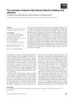
nificantly higher (Figure 1). These levels peaked
24 hours after administration in mice treated with HVJ-
E, and became normal at 7 days (data not shown).
Figure 1 Blood chemistry tests 24 hours after HVJ-E and CG-HVJ-E administration into norma l mice. Blood markers (T.Bil, AST, ALT, LDH,
BUN, and Cr) in normal mice tested 24 hours after intra-cardiac injection of PBS, HVJ-E or CG-HVJ-E. * p < 0.05. Results shown are the means ±
SD (n = 4).
Table 1 Zeta potential and particle sizes of each HVJ-E
complex
Complex Apparent molecular size (nm) Zeta potential (mV)
HVJ-E 293 ± 32 -25 ± 1
CG-HVJ-E 297 ± 21 -15 ± 3
HVJ-E-BSH 448 ± 144 -28 ± 1
CG-HVJ-E-BSH 494 ± 196 -19 ± 2
Fujii et al. Radiation Oncology 2011, 6:8
/>Page 5 of 12
High affinity and infusion ability of CG-HVJ-E
in tumor cells
CG-HVJ-E containing Qdot had a higher affinity for
tumor cells than Qdot alone or HVJ-E containing Qdot
(Figure 2A). CG-HVJ-E containing Qdot was taken into
the cytoplasm, and some Qdots were localized to the
nuclei, as seen by confocal microscopy (Figure 2B).
CG-HVJ-E transfection into tumor cells in vitro was highly
efficient
CG-HVJ-E’ s in vitro transfection efficiency into tumor
cells was 4 times greater than that of HVJ-E, as assessed
by a luciferase assay, and it was not cytotoxic (Figure 2C).
The enhanced transfection efficiency of CG-HVJ-E was
also observed in another tumor cell line (CT26: murine
colon cancer, data not shown).
CG-HVJ-E-BSH increased
10
B accumulation and retention
in tumor cells in vitro compared to BSH
The concentration of
10
B was significantly higher in cells
incubated with CG-HVJ-E-BSH than in those incubated
with BSH (p<0.05). The
10
B levels gradually decreased
in both cell groups, but the levels were significantly
higher in the cells incubated with CG-HVJ-E-BSH than
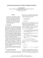
Figure 2 Affinity of CG-HVJ-E for tumor cells and the intracellular uptake of molecules incorporated into HVJ-E. A) Affinity of HVJ-E and
CG-HVJ-E for tumor cells. LM8G5 cells were incubated alone (a), or with Qdot (b), HVJ-E-Qdot (c), or CG-HVJ-E-Qdot (d) for 60 min in a Lab-tek
chamber slide and examined for Qdot (red) and Hoechst 33342 (blue) by fluorescence microscopy, Representative views are shown. B)
Intracellular localization of Qdot transported by CG-HVJ-E. Tumor cells were incubated with CG-HVJ-E-Qdot (orange) and stained with Hoechst
33342 (blue) and Alexa Fluor 488 phalloidin (green). Image shows 3-dimensional analysis with confocal microscopy. C) Luciferase activity in
tumor cells transfected with HVJ-E or CG-HVJ-E. Cells were cultured for 30 min with HVJ-E or CG-HVJ-E containing a luciferase-expressing
plasmid. Luciferase activity was measured 24 hours later to evaluate the transfection efficiency. Results are shown as means ± SD (n = 4). Similar
results were obtained in three experiments. * p < 0.05. D)
10
B accumulation and retention in tumor cells in vitro . Cells were incubated with 20
μg boron/ml of BSH or CG-HVJ-E-BSH for 30 min, then washed twice with PBS, and the
10
B concentration was measured by ICP-AES. Separately,
cells were incubated in the same manner, but after washing, were incubated in medium without BSH for 24 or 48 hours before testing for
10
B
concentration as described above. The horizontal axis shows time after co-incubation. The vertical axis shows the percent of the administered
dose (% dose) of CG-HVJ-E-BSH (open diamond) or BSH (solid square). Results shown are the means ± S.D. (n = 3). * p < 0.05.
Fujii et al. Radiation Oncology 2011, 6:8
/>Page 6 of 12
in those with BSH for at least 48 hours after incubation
(Figure 2D). These results indicate that CG-HVJ-E-BSH
binds rapidly to tumor cells and that the
10
Bcontained
in CG-HVJ- E-BSH is internalized into the cytoplasm or
the nucleus. Adding CG-HVJ-E-BSH to tumor cells in
vitro resulted in sufficient
10
B accumulation and reten-
tion in the cells to be useful for BNCT.
BSH incorporated into CG-HVJ-E accumulated in liver
tumors and rapidly disappeared from normal tissues in
tumor-bearing mice
In normal mice, the
10
B concentration in the liver 1 hour
aft er adminis tration was higher with BSH than with CG-
HVJ-E-BSH. The concentration of bo th compounds
started to d ecrease by 48 hours after administ ration. The
10
B concentration in the lung, kidney, and spleen was low
at all time points with both compounds (Figure 3A). In
the liver tumor model, BSH and CG-HVJ-E-BSH behaved
similarly in the normal liver tissue surrounding the
tumors (Figure 3B, middle panel). In the tumors, how-
ever, the concentration of
10
Bat1and24hoursafter
administration was significantly higher with CG-HVJ-E-
BSH (34.76 and 10.71% dose/g) than with BSH (2.21 and
2.29% dose/g) (Figure 3B, left panel). In the bloodstream,
the
10
B concentration at 1 hour after administration
tended to be higher with CG-HVJ-E-BSH (20.9% dose/
ml) than with BSH (7.96% dose/ml), despite the lower
quantity of
10
B administered with both boron compounds
(1.2 μg boron/g). From 24 hours after administration and
onward, the concentration of
10
Bfrombothcompounds
was the same (Figure 3B, right panel).
Tumor/Normal liver
10
B ratio in murine liver tumors was
greater with CG-HVJ-E-BSH
The Tumor/Normal (T/N) l iver
10
B ratio with CG-HVJ-
E-BSH was significantly higher than with BSH from 1 to
48 hours after administration (p < 0.05), with a peak dif-
ference at 24 hours (p < 0.05; Figure 3C). The Tumor/
Blood
10
B ratio of CG-HVJ-E-BSH also remained higher
than that of BSH from 1 to 48 hours after administra-
tion (data not shown).
CG-HVJ-E-BSH improved the T/N
10
B ratio in neutron
capture autoradiography images of murine liver tumors
Neutron capture autoradiography (NCAR) was per-
formed after BSH (35 μg boron/g) or CG-HVJ-E-BSH
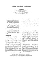
Figure 3 Bio-distribution of
10
B in mice with normal liver or with liver tumors. A) Time course of o rgan (lung, liver, kidney, and spleen)
uptake of
10
B delivered by 1.2 μg boron/g of BSH or CG-HVJ-E-BSH in normal mice. B) Time course of tumor accumulation (left panel), liver
uptake (middle panel), and blood residence (right panel) of
10
B delivered by 1.2 μg boron/g of BSH or CG-HVJ-E-BSH in tumor-bearing mice. The
horizontal axis shows the time after administration. The vertical axis shows the percent of the administered dose per gram of tissue (% dose/g).
C) Time course of the Tumor-to-Normal liver tissue (T/N)
10
B concentration ratio for CG-HVJ-E-BSH (open diamond) or BSH solution (solid square).
Data are expressed as the mean ± S.D. (n = 3). * p<0.05 compared with BSH.
Fujii et al. Radiation Oncology 2011, 6:8
/>Page 7 of 12
(1.2 μg boron/g) was injected into mice bearing liver
tumors. The
10
B particle count in the BSH- and CG-
HVJ-E-BSH-treated livers are shown in Figure 4B and
4C. The T/N ratio 1 hour after BSH administration was
0.12, and that for CG-HVJ-E-BSH at 24 hours after
administration was 2.76 (Figure 4D), which is similar to
the values obtained in the bio-distribution study. It is of
interest that the T/N
10
B ratio was higher with CG-HVJ-
E-BSH, even though the actual quantity of
10
Bwas
30 times greater in the BSH dosage. The number of a par-
ticles with CG-HVJ-E (415 ± 35) was similar to that of
BSH (451 ± 107) in the liver tumor sections (Figure 4A).
BNCT with CG-HVJ-E-BSH inhibited tumor growth,
preserved the normal surrounding liver tissue, and
prolonged survival time in the murine liver tumor model
To evaluate the use of BNCT with CG-HVJ-E-BSH for
murine liver tumors, BNCT was performed on mice
bearing LM8G5 liver tumors. To assess the T/N ratio of
CG-HVJ-E-BSH, BNCT was performed 24 hours after
CG-HVJ-E-BSH admi nistration or 1 hour after BSH
administration [2,4]. We first evaluated the anti-tumor
efficacy at 14 days after tumor cell inoculation, because
up to that time, the tumor-bearing mice were severely
damaged by the radical spread of tumors (about 50% of
the untreat ed mice were dead). Therefore, we sacrificed
the tumor-bearing mice that were alive until that time
to evaluate the efficacy of BNCT.
BNCT with CG-HVJ-E-BSH (1.2 μg boron/g) inhibited
the local growth of liver metastases as much as BNCT
with BSH (35 μgboron/g).ThisdosageofBSHwas
determined from the clinical dose for BNCT for various
malignant tumors, and effectively contained 35 times
the
10
B t hat was present in the CG-HVJ-E-BSH dosage
(Figure 5A, B). Some histological damage, which
appeared, for example as fractionated or vacuolated
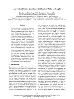
Figure 4 Neutron capture radiographic image in murine liver sections after administration of BSH or CG-HVJ-E-BSH. Liver sections from
tumor-bearing mice were prepared and frozen 1 hour after BSH (35 μg boron/g) injection or 24 hours after CG-HVJ-E-BSH (1.2 μ g boron/g)
injection. The sections were placed on CR-39 detector plates and exposed to thermal neutrons (2.1 × 10
13
neutrons/m
2
·s
1
) for 1 hour. A) The
number of a particles per 10,000 μm
2
section was counted by VH Analyzer software after NaOH etching. B) The number of a particles per
10,000 μm
2
section of BSH or C) of CG-HVJ-E-BSH (n = 3). D) Tumor-to-normal liver tissue (T/N) ratio for the number of a particles.
Fujii et al. Radiation Oncology 2011, 6:8
/>Page 8 of 12
cells, w as observed in both the tumor mass and in the
normal liver tissue after BNCT with BSH (35 μg boron/
g) (Figure 5C-b, d). In contrast, little histological damage
was detected in the normal liver tissue surrounding the
tumors after BNCT with CG-HVJ-E-BSH (Figure 5C-a,
d). We originally thought that the damage to the liver
might have been influen ced by the longer survival time
of mice treated with BSH and BNCT; however, the sur-
vival rate of these mice at 14 days after tumor cell
inoculation was 37.5% (Additional file 2, Figure S2).
This survival time was shorter than that of the untreated
tumor-bearing mice. As we were not able to be certain
if this dosage of BSH was a clinical equivalent, we used
a dose of 1.3 μg boron/g of BSH to evaluate the survival
time after BNCT, compare d to a dose of 1.2 μg boron/g
of CG-HVJ-E-BSH.
Finally, we compared the effectiveness of BNCT
against tumors when used with BSH or CG-HVJ-E-BSH,
in terms of survival after BNCT. With the assumption
thatthesurvivaltimeoftumor-bearingmiceafter
BNCT with a high dose of BSH (35 μg boron/g) was
affected by normal liver damage as well as anti-tumor
efficacy, both compounds were administered at dosages
with similar
10
B concentrations (CG-HVJ-E-BSH, 1.2 μg
boron/g or BSH,1.3 μgboron/g)intomicebearingliver
tumors at 24 hours or 1 hour before irradiation, respec-
tively. Irradiation was performed 8 days after the tumor
cell inoculation, and the survival of the mice assessed.
Figure 5 Anti-tumor efficacy of BNCT in mice with liver tumors. C3H/HeN mice were given an intra-portal injection of LM8G5 cells (1 × 10
6
cells) on day 0. Mice were given a single intra-cardiac injection of CG-HVJ-E-BSH (1.2 μg boron/g) 24 hours before irradiation, or BSH (35 μg
boron/g) 1 hour before irradiation. PBS and CG-HVJ-E-BSH solution were administered without neutron irradiation as a control. After irradiation
on day 8, mice were sacrificed on day 14 to determine the BNCT efficacy on tumor metastasis. A) Macroscopic views (a) of the liver with tumors
after administration of PBS; (b) normal liver; (c) liver with tumors after BNCT with BSH, and (d) liver with tumors after BNCT with CG-HVJ-E. B)
Liver weight after BNCT. * p < 0.05 compared with PBS or CG-HVJ-E without irradiation. (each group n = 4). C) Representative light microscopic
views of liver tumor tissue (upper panels, low magnification) or normal liver tissue (lower panels, high magnification) 6 days after BNCT with CG-
HVJ-E-BSH (1.2 μg boron/g) (a, c) or BSH (35 μg boron/g) (b, d). Sections are stained with hematoxylin-eosin. Bar: 100 μm.
Fujii et al. Radiation Oncology 2011, 6:8
/>Page 9 of 12
CG-HVJ-E-BSH was most effective in increasing the
mean survival time of mice bearing liver tumors com-
pared with the other groups (p < 0.005; Additional file
2, Figure S2). We observed little histological damage in
the normal liver tissues 6 days after B NCT with the
lower dose of BSH (1.3 μg boron/g ) besides the damage
that was already present in the tumor mass (Additional
file 3, Figure S3).
Discussion
With the goal of creating a novel BSH vector for effec-
tive BNCT, we chose HVJ-E because of its strong fusion
ability, its effectiveness as a vehicle for delivering various
drugs and genes, and its ability to stimulate an immune
response against tumors in local cancer therapy [23].
Clinical trials of locally administered HVJ-E for patients
with advanced malignant melanoma are underway in
Japan. Although HVJ-E is not suitable for systemic
administration because of its strong hemagglutinating
activity, it has been reported that combining HVJ-E with
5,000-kDa cationized gelatin greatly improves its stabi-
lity in the bloodstream [25]. In this study, we developed
CG-HVJ-E combined with BSH, which can be adminis-
tered into the general circulation, unlike HVJ-E, and
confirmed its bio-distribution.
We compared the safety and efficacy of CG-HVJ-E-
BSH in B NCT with that of BSH, using a murine model
for liver tumors. For systemic administration, we devel-
oped a smaller CG-HVJ-E with a lower molecular weight
(3,300 kDa) CG compared with the previously used CG-
HVJ-E, which had a particle diameter of 777 nm [25].
We found that this CG-HVJ-E could be safely adminis-
tered systemically in mice, with reduced toxic ity and
hemagglutination compared to HVJ-E (Figure 1). In the
bio-distribution test using normal mice, both BSH and
CG-HVJ-E-BSH accumulated in the liver immediately,
but almost all of the
10
B had disappeared from the nor-
mal liver 48 hours later (Figure 3A). In liver tumors,
however, CG-HVJ-E-BSH accumulation was greater than
that of BSH although the boron proceeding from CG-
HVJ-E-BSH was 35 times higher than that of BSH (Figure
3B); accordingly, the CG-HVJ-E-BSH T/N ratio was sig-
nificantly higher than that of BSH in tumor-bearing
mice, particularly at 24 hours after administration (Figure
3C). Neutron capture autoradiography revealed a higher
T/N
10
B ratio with CG-HVJ-E-BSH than with BSH 1
hour after administration, despite the 35 -fold-higher
quantity of
10
B contained in the BSH dosage (Figure 4).
In our experiments, BNCT was performed 1 hour
after BSH administration, because it followed the
reported procedure for the clinical use of BNCT for
liver tumors [9], and t here was little difference between
the T/N ratio an hour after admin istration and the ratio
over the next 24 hours (Figure 3C). This was due to the
protracted circulating time of CG-HVJ-E-BSH in the
bloodstream. Therefore, this complex accumulated in
the tumor by the enhanced permeability and retention
(EPR) effect [34]. In fact, the particle size of the CG-
HVJ-E-BSH was suitable for the EPR effect (Table 1)
[35]. Another reason for this finding was that CG-HVJ-
E has a high affinity and high fusion ability for tumor
cells (Figure 2A, B, C). Although
10
Bwastakenupby
the tumor cells over time, a large number of CG-HVJ-
E-BSH molecules were incorporated into the tumor cells
immediately, and high
10
B concentrations were main-
tained much longer with CG-HVJ-E-BSH than with
BSH (Figure 2D). The mechanism for the preferential
affinity of CG-HVJ-E to tumor cells as compared with
HVJ-E has not been clarified, but it has been reported
that when HVJ-E is conjugated with cationized gelatin,
the transfection efficiency improves without a loss of
cell fusion ability [25]. Therefore, the efficacy of CG-
HVJ-E-BSH was similar to the 35-fold higher dose of
10
B as BSH for s uppressing the spread of tumor cells
without normal liver injury (Figure 5A, B, C).
When used in BNCT, the CG-HVJ-E-BSH significantly
increased the survi val time o ver BSH at an equivalent
10
B dosage (Additional file 2, Figure S2). Generally, BSH
is rarely tra nsferred into the cytoplasm and, once there,
is easily removed [36]. On t he other hand, CG-HVJ-E-
BSH was highly selective for tumor cells and showed
both strong fusion ability and the ability to transfer into
the tumor cell nucleus. As a result, CG-HVJ-E-BSH
improved the effectiveness of BNCT because the
10
B
was highly concentrated and retained in the nuclei of
the tumor cells (Figure 2B, C), where its cytotoxicity
was much higher than that of
10
B bound to the tumor
cell surface [14,37,38].
Moreover, HVJ-E has the potential to induce a bystan-
der effect, so that CG-HVJ-E-BSH could be incorporated
into vicinal cells through gap junctions. It is possible
that BNCT with CG-HVJ-E-BSH induces a synergistic
effect, resulting in a greater destruction of vicinal tumor
cells than is seen with BNCT with BSH, which induces
a bystander effect that generates hereditary abnormal-
ities in vicinal cells [39].
We chose multiple liver tumors as a target for evaluat-
ing the effectiveness of BNCT with CG-HVJ-E-BSH,
because BNCT for multiple liver tumors has not gained
popularity and the T/N ratio needs to be improved for
deep-site tumors. In the absence of liver function disor-
ders, the response o f multiple liver tumors is thought to
be a good indication of BNCT effectiveness. In this
report, we treated mice bearing liver tumors with BNCT
[27] after establishing the presence o f tumors of several
millimeters in diameter. This murine model appears to
reflect the clinical stage that we targeted. BNCT with
BSH is not indicated for multiple liver tumors in clinical
Fujii et al. Radiation Oncology 2011, 6:8
/>Page 10 of 12
settings and is only at the experimental stage [9,10].
BNCT was signi ficantly more effective against liv er
tumors when used with CG-HVJ-E-BSH than with BSH,
and normal liver tissue was not injur ed. The limited
injury to normal liver tissue makes more than one
BNCT irradiation possible, which is likely to increase
the therapeutic potential. However, in these experi-
ments, only one irradiation was done. Wit h regard to
BNCT with BSH for clinical liver tumors at deep sites,
the required T/N
10
B ratio is over 15 [36,40]. Moreover,
the human trunk is much thicker than the m urine
trunk. Therefore, for BNCT with CG-HVJ-E-BSH to
become an established, effective clinical procedure,
further improvements are needed not only in the drug-
delivery system, but also in the vessel-sel ective delivery
[41] because of the attenuation of neutron beams direc-
ted toward deep lesions.
Our trial of BNCT for multiple liver tumors at deep
sites should forward its development to treat other deep-
site tumors, such as pancreatic cancer and malignant
mesothelioma [42-44], and further the investigation into
BNCT and HVJ-E. However, some problems need to be
resolved in future experiments, particularly with regard
to improving the incorporation of
10
B into the HVJ-E.
It has been reported that locally administered HVJ-E
induces immuno-responses against tumors [23. 24], and
effectively transports antitumor drugs [22,45]. Our
experiments included a single administration of HVJ-E,
which did not appear to have an anti-tumor effect
unless accompanied by irradiation (Figure 5B, Addi-
tional file 2, Figure S2). However, the fractionated
administration of HVJ-E, as is used for other vaccina-
tions, might be possible. To address the limitations of
this novel HVJ-E BSH, investigations into concurrent
chemo-radiation therapy, fractionated administration
with or without
10
B, and conjugating with ligands for
tumor-specific molecules should be performed.
In summary, we developed a form of CG-HVJ-E that
could be administered into the general circulation and
had both hi gh tumor selectivit y and high retention in
tumor cells. This vector, when comb ined with BSH,
improved the efficacy of BNCT for multiple liver tumors
in vivo. Therefore, CG-HVJ-E holds potential for a drug
delivery system with clinical applications for cancer
therapy.
Additional material
Additional file 1: Figure S1. Transmission electron microscope
photographs of HVJ-E complexes. (A) HVJ-E, (B) CG-HVJ-E, and (C) CG-
HVJ-E-BSH. Bar: 200 nm.
Additional file 2: Figure S2. Survival of mice treated with BNCT.
Mice were given a single intra-cardiac injection of CG-HVJ-E-BSH (1.2 μg
boron/g) 24 hours before irradiation, or BSH (1.3 μg boron/g) 1 hour
before irradiation. PBS and CG-HVJ-E-BSH were administered wit hout
irradiation as a control. The mean survival time of the mice that received
the BNCT treatment with CG-HVJ-E-BSH was significantly longer than that
of the other groups (n = 4). * p < 0.005 (PBS without neutron irradiation,
1.3 μg boron/g of BSH with neutron irradiation, 1.2 μg boron/g of CG-
HVJ-E-BSH without neutron irradiation vs. 1.2 μg boron/g of CG-HVJ-E-
BSH with neutron irradiation).
Additional file 3: Figure S3. Representative light microscopy views
of the liver tumor (A) and normal liver tissue (B) 6 days after BNCT
with a low dose of BSH (1.3 μg boron/g). Tissues were stained with
hematoxylin-eosin. Bar: 100 μm.
Abbreviations
BNCT: Boron Neutron Capture Therapy; BSH: sodium borocaptate; HVJ-E:
Hemagglutinating Virus of Japan Envelope.
Acknowledgements
This work was supported in part by a grant for research and development
of a Fixed Field Alternating Gradient Accelerator and DDS for BNCT from the
New Energy and Industrial Technology Development Organization (NEDO), a
Health Labour Science Research Grant from the Ministry of Health, Labour
and Welfare of Japan, and a grant-in-Aid for Exploratory Research from the
Ministry of Education, Culture, Sports, Science and Technology (MEXT).
Author details
1
Department of Surgery, Osaka University Graduate School of Medicine,
Osaka, Japan.
2
Medical Center for Translational Research, Osaka University
Hospital, Osaka, Japan.
3
Particle Radiation Oncology Research Center
Laboratory, Research Reactor Institute, Kyoto University, Osaka, Japan.
4
Department of Agriculture, Osaka Prefectural Universi ty, Osaka, Japan.
5
Department of Biomaterials, Institute for Frontier Medical Sciences, Kyoto
University, Kyoto, Japan.
6
Division of Gene Therapy Science, Osaka University
Graduate School of Medicine, Osaka, Japan.
7
Health Care Economics and
Industrial Policy, Osaka University Graduate School of Medicine, Osaka Japan.
Authors’ contributions
HF carried out the study, and contributed to the conception of the
manuscript and the interpretations of the data. AM, HK, MS, MS, AT, and YT
participated in the design of the study. YD, MK, and KO provided some
intellectual recommendation. YK and YS provided some intellectual
recommendation and reviewed the manuscript. CML conceived of the study,
and participated in its design and coordination. All authors read and
approved the final manuscript.
Competing interests
All authors declare there were no actual or potential conflicts of interest in
this study.
Received: 17 October 2010 Accepted: 20 January 2011
Published: 20 January 2011
References
1. Barth RF, Coderre JA, Vicente MG, Blue TE: Boron neutron capture therapy
of cancer: current status and future prospects. Clin Cancer Res 2005,
11:3987-4002.
2. Yamamoto T, Nakai K, Matsumura A: Boron neutron capture therapy for
glioblastoma. Cancer Lett 2008, 262:143-52.
3. Pinelli T, Zonta A: From the first idea to the application to the human
liver. Research and development in Neutron Capture Therapy; 2002.
4. Suzuki M, Sakurai Y, Hagiwara S, Masunaga S, Kinashi Y, Nagata K,
Maruhashi A, Kudo M, Ono K: First attempt of boron neutron capture
therapy (BNCT) for hepatocellular carcinoma. Jpn J Clin Oncol 2007,
37:376-81.
5. Wittig A, Malago M, Collette L, Huiskamp R, Buhrmann S, Nievaart V,
Kaiser G, Jockel KH, Sauerwein W: BNCT in liver metastases: results of the
EORTC trial 11001. Strahlentherapie Und Onkologie 2007, 183:115-115.
6. Sauerwein W, Malago M, Moss R, Altieri S, Hampel G, Wittig A, Nievaart V,
Collette L, Mauri P, Huiskamp R, Michel J, Daquino G, Gerken G, Bornfeld N,
Broelsch CE: Boron Neutron Capture Therapy (BNCT) for the treatment of
Fujii et al. Radiation Oncology 2011, 6:8
/>Page 11 of 12
diffuse, non-resectable liver metastases. Strahlentherapie Und Onkologie
2006, 182:109-109.
7. Cardose JE, Trivillin VA, Heber EM, Nigg DW, Calzetta O, Blaumann H,
Longhino J, Itoiz ME, Bumaschny E, Pozzi E, Schwint AE: Effect of Boron
Neutron Capture Therapy (BNCT) on normal liver regeneration: Towards
a novel therapy for liver metastases. International Journal of Radiation
Biology 2007, 83:699-706.
8. Chou FI, Chung HP, Liu HM, Chi CW, Lui WY: Suitability of boron carriers
for BNCT: Accumulation of boron in malignant and normal liver cells
after treatment with BPA, BSH and BA. Applied Radiation and Isotopes
2009, 67:S105-108.
9. Wittig A, Malago M, Collette L, Huiskamp R, Buhrmann S, Nievaart V,
Kaiser GM, Jockel KH, Schmid KW, Ortmann U, Sauerwein WA: Uptake of
two
10
B-compounds in liver metastases of colorectal adenocarcinoma
for extracorporeal irradiation with boron neutron capture therapy
(EORTC Trial 11001). Int J Cancer 2008, 122:1164-71.
10. Suzuki M, Masunaga SI, Kinashi Y, Takagaki M, Sakurai Y, Kobayashi T, Ono K:
The effects of boron neutron capture therapy on liver tumors and
normal hepatocytes in mice. Jpn J Cancer Res 2000, 91:1058-64.
11. Sakurai Y, Ono K, Miyatake S, Maruhashi A: Improvement effect on the
depth-dose distribution by CSF drainage and air infusion of a tumour-
removed cavity in boron neutron capture therapy for malignant brain
tumours. Phys Med Biol 2006, 51:1173-83.
12. Wu G, Barth RF, Yang W, Lee RJ, Tjarks W, Backer MV, Backer JM: Boron
containing macromolecules and nanovehicles as delivery agents for
neutron capture therapy. Anticancer Agents Med Chem 2006, 6:167-84.
13. Mehta SC, Lu DR: Targeted drug delivery for boron neutron capture
therapy. Pharm Res 1996, 13:344-51.
14. Maruyama K, Ishida O, Kasaoka S, Takizawa T, Utoguchi N, Shinohara A,
Chiba M, Kibayashi H, Eriguchi M, Yanagie H: Intracellular targeting of
sodium mercaptoundecahydrododecaborate (BSH) to solid tumors by
transferrin-PEG liposomes, for boron neutron-capture therapy (BNCT).
J Control Release 2004, 98:195-207.
15. Masunaga S, Kasaoka S, Maruyama K, NIgg D, Sakurai Y, Nagata K, Suzuki M,
Kinashi Y, Maruhashi A, Ono K: The potential of transferrin-pendant-type
polyethyleneglycol liposomes encapsulating decahydrodecaborate-(10)B
(GB-10) as (10)B-carriers for boron neutron capture therapy. Int J Radiat
Oncol Biol Phys 2006, 66:1515-22.
16. Doi A, Kawabata S, Iida K, Yokoyama K, Kajimoto Y, Kuroiwa T, Shirakawa T,
Kirihata M, Kasaoka S, Maruyama K, Kumada H, Sakurai Y, Masunaga S,
Ono K, Miyatake S: Tumor-specific targeting of sodium borocaptate (BSH)
to malignant glioma by transferrin-PEG liposomes: a modality for boron
neutron capture therapy. J Neurooncol 2008, 87:287-94.
17. Aljabiri MR, Lodato F, Burroughs AK: Surveillance and diagnosis for
hepatocellular carcinoma. Liver Transpl 2007, 13(11 Suppl 2):S2-12.
18. World Health Organization: World Health Statistics 2008/Future trends in
global mortality.[ />19. Arciero CA, Sigurdson ER: Diagnosis and treatment of metastatic disease
to the liver. Semin Oncol 2008, 35:147-59.
20. Kaneda Y, Nakajima T, Nishikawa T, Yamamoto S, Ikegami H, Suzuki N,
Nakamura H, Morishita R, Kotani H: Hemagglutinating virus of Japan (HVJ)
envelope vector as a versatile gene delivery system. Mol Ther 2002, 6:219-26.
21. Mima H, Yamamoto S, Ito M, Tomoshige R, Tabata Y, Tamai K, Kaneda Y:
Targeted chemotherapy against intraperitoneally disseminated colon
carcinoma using a cationized gelatin-conjugated HVJ envelope vector.
Mol Cancer Ther 2006, 5:1021-8.
22. Kawano H, Komaba S, Kanamori T, Kaneda Y: A new therapy for highly
effective tumor eradication using HVJ-E combined with chemotherapy.
BMC Med 2007, 5:1-7.
23. Kurooka M, Kaneda Y: Inactivated Sendai virus particles eradicate tumors
by inducing immune responses through blocking regulatory T cells.
Cancer Res 2007,
67:227-36.
24. Fujihara A, Kurooka M, Miki T, Kaneda Y: Intratumoral injection of
inactivated Sendai virus particles elicits strong antitumor activity by
enhancing local CXCL10 expression and systemic NK cell activation.
Cancer Immunol Immunother 2008, 57:73-84.
25. Mima H, Tomoshige R, Kanamori T, Tabata Y, Yamamoto S, Ito S, Tamai K,
Kaneda Y: Biocompatible polymer enhances the in vitro and in vivo
transfection efficiency of HVJ envelope vector. J Gene Med 2005, 7:888-97.
26. Asai T, Ueda T, Itoh K, Yoshioka K, Aoki Y, Mori S, Yoshikawa H:
Establishment and characterization of a murine osteosarcoma cell line
(LM8) with high metastatic potential to the lung. Int J Cancer 1998,
76:418-422.
27. Lee CM, Tanaka T, Murai T, Kondo M, Kimura J, Su W, Kitagawa T, Ito T,
Matsuda H, Miyasaka M: Novel chondroitin sulfate-binding cationic
liposomes loaded with cisplatin efficiently suppress the local growth
and liver metastasis of tumor cells in vivo. Cancer Res 2002, 62:4282-8.
28. Fukunaka Y, Iwanaga K, Morimoto K, Kakemi M, Tabata Y: Controlled
release of plasmid DNA from cationized gelatin hydrogels based on
hydrogel degradation. J Control Release 2002, 80:333-43.
29. Hosseinkhani H, Aoyama T, Ogawa O, Tabata Y: Ultrasound enhancement
of in vitro transfection of plasmid DNA by a cationized gelatin. J Drug
Target 2002, 10:193-204.
30. Nagata I, Kimura Y, Ito Y, Tanaka T: Temperature-Sensitive Phenomenon of
Viral Maturation Observed in BHK Cells Persistently Infected with HVJ.
Virology 1972, 49:453-461.
31. Guttenberger M: Protein Determination. Cell Biology A Laboratory
Handbook;295-303.
32. Ogura K, Yanagie H, Eriguchi M, Lehmann EH, Kuhne G, Bayon G,
Kobayashi H: Neutron capture autoradiographic study of the
biodistribution of
10
B in tumor-bearing mice. Appl Radiat Isot 2004,
61:585-90.
33. Saga K, Tamai K, Kawachi M, Shimbo T, Fujita H, Yamazaki T, Kaneda Y:
Functional modification of Sendai virus by siRNA. J Biotechnol 2008,
133:386-94.
34. Maeda H, Wu J, Sawa T, Matsumura Y, Hori K: Tumor vascular permeability
and the EPR effect in macromolecular therapeutics: a review. J Control
Release 2000, 65:271-84.
35. Siwak DR, Tari AM, Lopez-Berestein G: The potential of drug-carrying
immunoliposomes as anticancer agents. Commentary re: J. W. Park et
al., Anti-HER2 immunoliposomes: enhanced efficacy due to targeted
delivery. Clin Cancer Res 2002, 8:1172-1181, Clin Cancer Res 2002;8:955-6.
36. Yanagie H: Selective Enhancement of Boron Accumulation with Boron-
Entrapped Water-in-oil-in-water Emulsion in VX-2 Rabbit Hepatic Cancer
Model for BNCT. Proc of 12th International Congress of Neutron Capture
Therapy; 2006.
37. Ye SJ: Monte Carlo based protocol for cell survival and tumour control
probability in BNCT. Phys Med Biol 1999, 44:447-61.
38. Kobayashi T, Kanda K: Analytical calculation of boron- 10 dosage in cell
nucleus for neutron capture therapy. Radiat Res 1982, 91:77-94.
39. Kinashi Y, Masunaga S, Nagata K, Suzuki M, S T, Ono K: A bystander effect
observed in boron neutron capture therapy: A study of the induction of
mutations in the HPRT locus. Int J Radiat Oncol Biol Phys 2007, 68:508-14.
40. Suzuki M, Sakurai Y, Masunaga S, Kinashi Y, Nagata K, Ono K: Dosimetric
study of boron neutron capture therapy with borocaptate sodium
(BSH)/lipiodol emulsion (BSH/lipiodol-BNCT) for treatment of multiple
liver tumors. Int J Radiat Oncol Biol Phys 2004, 58:892-6.
41. Suzuki M, Nagata K, Masunaga S, Kinashi Y, Sakurai Y, Maruhashi A, Ono K:
Biodistribution of
10
B in a rat liver tumor model following intra-arterial
administration of sodium borocaptate (BSH)/degradable starch
microspheres (DSM) emulsion. Appl Radiat Isot 2004, 61:933-7.
42. Yanagie H, Tomita T, Kobayashi H, Fujii Y, Nonaka Y, Saegusa Y, Hasumi K,
Eriguchi M, Kobayashi T, Ono K: Inhibition of human pancreatic cancer
growth in nude mice by boron neutron capture therapy. Br J Cancer
1997, 75:660-5.
43. Yanagie H, Sakurai Y, Ogura K, Kobayashi T, Furuya Y, Sugiyama H,
Kobayashi H, Ono K, Nakagawa K, Takahashi H, Nakazawa M, Eriguchi M:
Evaluation of neutron dosimetry on pancreatic cancer phantom model
for application of intraoperative boron neutron-capture therapy. Biomed
Pharmacother 2007, 61:505-14.
44. Suzuki M, Sakurai Y, Masunaga S, Kinashi Y, Nagata K, Maruhashi A, Ono K:
A preliminary experimental study of boron neutron capture therapy for
malignant tumors spreading in thoracic cavity. Jpn J Clin Oncol 2007,
37:245-9.
45. Kawano H, Komaba S, Yamasaki T, Maeda M, Kimura Y, Maeda A, Kaneda Y:
New potential therapy for orthotopic bladder carcinoma by combining HVJ
envelope with doxorubicin. Cancer Chemother Pharmacol 2008, 61:973-8.
doi:10.1186/1748-717X-6-8
Cite this article as: Fujii et al.: Cationized gelatin-HVJ envelope with
sodium borocaptate improved the BNCT efficacy for liver tumors in
vivo. Radiation Oncology 2011 6:8.
Fujii et al. Radiation Oncology 2011, 6:8
/>Page 12 of 12