Báo cáo khoa học: "Radiation recall pneumonitis induced by chemotherapy after thoracic radiotherapy for lung cancer" potx
Bạn đang xem bản rút gọn của tài liệu. Xem và tải ngay bản đầy đủ của tài liệu tại đây (389.45 KB, 6 trang )
RESEARCH Open Access
Radiation recall pneumonitis induced by
chemotherapy after thoracic radiotherapy
for lung cancer
Xiao Ding
1†
, Wei Ji
1,3†
, Junling Li
2
, Xiangru Zhang
2
, Luhua Wang
1*
Abstract
Background: Radiation recall pneumonitis (RRP) describes a rare reaction in previously irradiated area of
pulmonary tissue after application of triggering agents. RRP remains loosely characterized and poorly understood
since it has so far only been depicted in 8 cases in the literature. The objective of the study is to disclose the
general characteristics of RRP induced by chemotherapy after thoracic irradiation for lung cancer, and to draw
attention to the potential toxicity even after a long time interval from the previ ous irradiation.
Methods: Medical records were reviewed. RRP induced by chemo therapy was diagnosed by the history of
chemotherapy after radiotherapy, clinical presentation and radiographic abnormalities including ground-glass
opacity, attenuation, or consolidation changes within the radiation field, plus that radiographic examination of the
thorax before showed no radiation pneumonitis. RRP was graded according to Common Terminology Criteria for
Adverse Events version 3.0. The characteristics of the 12 RRP cases were analyzed.
Results: Twelve patients were diagnosed of RRP, of who 8 received taxanes. The median time interval between
end of radiotherapy and RRP, between end of radiotherapy and beginning of chemotherapy, and between
beginning of chemotherapy and RRP was 95 days, 42 days and 47 days, respectively. Marked symptomatic and
radiographic improvement was observed in the 12 patients after withdrawal of chemotherapy and application of
systemic corticosteroids. Seven patients were rechallenged with chemotherapy, of whom four with the same kind
of agent s, and showed no recurrence with steroid cover.
Conclusions: Doctors should pay attention to RRP even after a long time from the previous radiotherapy or after
several cycles of consolidation chemotherapy. Taxanes are likely to be associated with radiation recall more
frequently. Withdrawal of causative agent and application of steroids are the treatment of choice. Patients may be
rechallenged safely with steroid cover and careful observation, which needs to be validated.
Background
Radiation recall reaction (RRR) refers to an inflamma-
tory reaction within the previously treated radiation
field in response to precipitating agents, which could
have been masked if radiotherapy is not followed by
inciting agents. It has been observed mainly with
chemotherapeutic drugs [1]. Nevertheless, antituberculo-
sis drugs, antibiotics, tamoxifen, simvastatin have also
been involved in it [2-6]. Skin is the major site of radia-
tion recall toxicity [7]. But it has been as well described
in different internal organs including lung, digestive
tract, muscle, central nervous system, and supraglottis
[8-16]. Treatment-related pneumo nitis is a major dose-
limiting toxicities resulting from thoracic radiotherapy
and chemotherapy. Radiation recall pneumonitis (RRP)
describes a rare reaction in previously irradiated area of
pulmonary tissue after application of triggering agents.
The diagnosis of RRP induced by chemotherapy is
established by a history of chemotherapy after thoracic
radiotherapy, radiographic abnormality, and clinical pre-
sentation. The typical radiologic changes of RRP include
ground-glass opacity, diffuse haziness, infiltrates or
* Correspondence:
† Contributed equally
1
Department of Radiation Oncology, Cancer Institute (Hospital), Chinese
Academy of Medical Sciences and Peking Union Medical College, Beijing, PR
China
Full list of author information is available at the end of the article
Ding et al. Radiation Oncology 2011, 6:24
/>© 2011 Ding et al; licensee BioMed Central Ltd. This is an Open Access article distributed under the terms of the Creative Commons
Attribution License ( which permits unrestricted use, distribution, and reproduction in
any medium, provided the original wor k is properly cited.
consolidation in the irradiated lung that conform to the
shape and size of the treatment porta ls [ 17]. The symp-
toms are dry cough, low-grade fever, chest pain, and
shortness of breath. The antineoplastic agents having
bee n reported to trigger RRP include taxanes, anth racy-
clines, gemcitabine and erlotinib [8,18-23].
RRP remains loosely characterized and poorly under-
stood since it has so far only been depicted in 8 cases
[8,18-23] in the literature. The objective of the present
studyistodisclosethegeneral char acteristics of RRP
induced by chemotherapy after thoracic irradiation of
lung cancer, and to draw attention to the potential toxi-
city even after a long time interval from the previous
irradiation.
Methods
We retrospectively reviewed the medical and radiation
records of lung cancer patients who were treated conse-
cutively between January 1999 and December 2007 in the
Department of Radiation Oncology at Cancer Hospital,
Chinese Academy of Medical Sciences, Peking Union
Medical College. Patientswereincludediftheyhad
newly diagnosed and pathologically confirmed lung
cancer, chemot herapy after thoracic radiotherapy, a lung
dose-volume histogram (DVH) that was recoverable from
institutional archives, and availability of both radio-
graphic images and symptom assessment for determining
the occurrence of RRP.
The total normal lung volume was defined as the total
lung volume minus the primary gross target volume
(GTV) and volume of the trachea and main bronchi.
The following dosimetric parameters were generated
from the DVH for total normal lung: mean lung dose
(MLD), and lung volumes receiving more than 5 Gy
(V5), 10 Gy (V10), 20 Gy (V20), and 30 Gy (V30).
All patients were examined by their treating radiation
oncologists weekly during radiotherapy and 4-6 weeks
after completion of radiotherapy. The patients were
then followed every 3 months for the first 3 years and
every 6 months thereafter unless they had symptoms
that required immediate examination or intervention.
Radiographic examination by c hest X-ray or CT was
performed at each follow-up visit after completion of
radiotherapy.
RRP induced by chemotherapy was diagnosed by the
history of chemotherapy after radiotherapy, clinic al pre-
sentation and radiographic abnormalities including
ground-glass opacity, attenuation, or consolidation
changes within the radiation field, plus that radiographic
examination of the thorax before showed no radiation
pneumonitis. RRP was graded according to the National
Cancer Institute’ s Common Terminology Criteria for
Adverse Events (CTC) version 3.0 (23) as follows: Grade
1 pneumonitis was asymptomatic and diagnosed by
radiographic findings only;Grade2pneumonitiswas
symptomatic but did not interfere with daily activities;
Grade 3 pneumonitis was symptomatic and interfered
with daily activities or required administration of oxygen
to the patient; Grade 4 pneumonitis required assisted
ventilation for the patient; and Grade 5 pneumonitis
was fatal. Informed consent was obtained from all the
subjects.
Results
Twelve patients were diagnosed of RRP induced by con-
solidation chemotherapy. The median age of the group
was 51 years (range, 41-66 years). 5 patients were female,
and 7 male. Three cases are limited small cell lung cancer
(SCLC), and 9 are locally-advanced non small cell
lung cancer (NSCLC). All p atients’ Karnofsky perfor-
mance status (KPS) was 80. Five patients had induction
chemotherapy, and 7 had concurrent chemotherapy. The
12 lung cancer patients’ clinical charac teristics are shown
in Table 1.
Eight patients received 3-dimentional conformal
radiotherapy (3D-CRT), and 4 received intensity-
modulated radiotherapy (IMRT). The median radiation
dose was 60.7 Gy (range, 52-66 Gy). The median MLD
was 1540.5 cGy (range, 1301-2130 cGy). The median
V5, V10, V20 and V30 was 53.3% (range, 38.0%-
65.0%), 41.0% (range, 29.0%-51.0%), 26.9% (range,
20.0%-32.0%), and 20.2% (ran ge, 15.0%-27.0%), respec-
tively. The 12 lung cancer patients’ dosimetric para-
meters are shown in Table 2.
Of the 12 intravenous consolidation chemotherapy
regimens inducing RRP, 8 included taxanes, 2 of which
included both taxanes and gemcitabine; 2 etoposide; 1
vinorelbine; and 1 epirubicin.
The median time interval between end of radiotherapy
and RRP, between end of radiother apy and beginning of
chemotherapy, and between beginning of chemot herapy
and RRP was 95 days (range, 71-202 days), 42 days
(range, 7-60 days) and 47 days (range, 22-169 days),
respectively.
Eleven patients had Grade 2 and 1 patient had G rade
3 RRP. Marked symptomatic and radiographic improve-
ment was observed in the 12 patients after withdrawal
of the chemotherapy and application of systemic corti-
costeroids. Of the 12 RRP patients, 7 were rechallenged
with chemotherapy, 3 of who were rechallenged with
the same agents and 1 with the same kind of agents,
and showed no recurrence with steroid cover. The med-
ian time interval between RRP and rechallenge was
20 days (range, 4-89 days). The characteristics of the
12 RRP cases are shown in Table 3.
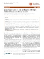
Figure 1 shows th e thoracic CT scans of Patient 10 (A)
before radiotherapy, (B) on e month after e nd of radio-
therapy, (C) 4 months after end of radiotherapy when
Ding et al. Radiation Oncology 2011, 6:24
/>Page 2 of 6
RRP took place induced by consolidation chemotherapy,
and (D) days after application of systematic steroids, sug-
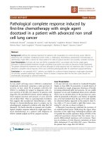
gestive o f RRP development. Figure 2 shows CT based
IMRT plan of Patient 10.
Discussion
RRR describes an inflammatory reaction in previously
irradiated area after application of certain promoting
agents. W hen it occurs in previously irradiated lung, it
is called RRP. RRP is a special subtype of radiation
induced pneumonitis, as the base of RRP is subclinical
radiation damage of pulmonary tissue. When radiation
therapy is followed by chemotherapy, subclinical damage
from irradiation can be unmasked and clinically mani-
fested as a radiation recall phenomenon.
Taxanes a nd anthracyclines have been reported to be
responsible for 20% and nearly 30% of RRR, respecti vely
[1]. The incit ing agents observed in RRP previously
reported and here included taxanes, anthracyclines,
gemcitabine, etoposide, vinorelbine and erlotinib.
Taxanes and anthracyclines are responsible for the
majority of the 20 chemotherapy-induced RRP cases
available, 50% and 25% respectively. In the present
study, of the 12 regimens, 8 (66.7%) included taxanes,
2 (16.7%) of which included both taxanes and gemcita-
bine; 2 (16.7%) etoposide; 1 vi norelbine; and 1 epirubi-
cin. Certain drugs seem to be associated with radiation
recall more frequently. On the other hand, cisplatin and
carboplatin, which are frequently used after radiother-
apy, has not been depicted in RRR. In contrast, radiation
recall induced by oxaliplatin has been reported [24].
When a combination of gemcitabine and docetaxel was
involved, we assume that RRP was induced by the com-
bination, as it could not completely be ruled out that
the pulmonary recall reaction was not caused by either,
although the time intervals from the last application of
the two agents to the RRP were different.
So far, we are the first to describe etoposide-induced
RRP with details. Moreover, we are the first to describe
RRR by Vinorelbine beyond one suspected RRR case
after a first cycle of gemcitabine and Vinorelbine with
no details [22].
Classic RRR often occurs with the initiation of the
precipitating agent but can occur after several courses of
treatment. The time delay o f cases that occurred after
several courses of treatment could be explained by a
putative drug d ose threshold for RRP or/and a time lag
effect. Clinically, these patients’ symptoms were consid-
ered to be triggered by chemotherapy. Both radiotherapy
and chemotherapy contributed to the development of
RRP, and it is difficult to tell how much each of them
contributed in each case. The reported time interval
between the end of radiation therapy and the recall
reaction ranged from 2 days [25] to 15 years [26].
Table 1 Clinical characteristics of the 12 lung cancer patients
Patient Sex Age Histology Stage
a
KPS Induction chemotherapy Concurrent chemotherapy
1 F 51 small cell lung cancer IIIa
T1N2M0
80 CE No
2 F 50 Adenocarcinoma IIIb
T2N3M0
80 No PC
3 M 54 Squamous cell carcinoma IIIa
T3N2M0
80 No EP
4 M 48 small cell lung cancer IIIa
T2N2M0
80 CE No
5 F 59 Adenocarcinoma IIIb
T3N3M0
80 NP No
6 M 49 Squamous cell carcinoma IIIa
T3N2M0
80 PC EP
7 M 58 Squamous cell carcinoma IIIa
T2N2M0
80 No EP
8 F 63 Adenocarcinoma IIIa
T2N2M0
80 No PC
9 M 44 Squamous cell carcinoma IIIb
T4N0M0
80 No PC
10 F 41 Adenocarcinoma IIIb
T4N2M0
80 No EP
11 M 46 small cell lung cancer IIIa
T2N2M0
80 EP No
12 M 66 Squamous cell carcinoma IIIa
T2N2M0
80 No No
KPS indicates Karnofsky performance status; CE, carboplatin, etoposide; NP, navelbine, cisplatin; PC, Paclitaxel, carboplatin; EP, etoposide, cisplatin.
a
Grading determined according to the American Joint Committee on Cancer 6th edition grading system.
Ding et al. Radiation Oncology 2011, 6:24
/>Page 3 of 6
The reported time interval between the first dose of
chemotherapy and the recall reaction ranged from 18
hours [27] to 15 years [26]. In the literature, the time
interval between completion of radiotherapy a nd RRP
ranged from 12 days [8] to 9 months [21], the time
interval between completion o f radiotherapy and begin-
ning of chemotherapy ranged from 12 days [8] to
8 months [21], and the time interv al between beginning
of chemotherapy and RRP ranged from several hours [8]
to 2 months [19]. In the present study, the median time
interval between end of radiotherapy and RRP, between
end of radiotherapy and beginning of chemotherapy,
and between beginning of chemotherapy and RRP was
95 days (range, 71-2 02 days), 42 days (range, 7-60 days)
and 47 days (ran ge, 22-169 days), respectively. RRP
could occur even after a long time interval from the
previous radiothe rapy or after several cycles of consoli-
dation chemotherapy. Because we generally recommend
our patients have consolidation chemotherapy 4-8 weeks
after radiotherapy in our institute if the patients are
evaluated able to take chemotherapy. The time in terval
from th e end of radiot herapy to RRP here could not be
very long.
Previous published articles have reported that recall
reactions are most severe when the time interval
between the radiotherapy and the following chemother-
apy is short. We did not find the trend in our study, the
reason for that may be there are other factors, such as
primary disease, p atient ’ s performance status, radiot her-
apy and inciting agents. Referring to all the chemother-
apy-induced RRP cases reported and here, the median
time interval from completion of radiotherapy to begin-
ning of chemotherapy was 34 days (range, 12-59 da ys)
for taxanes, 6 weeks (range, 3-8 weeks) for anthracy-
clines, 59 days (range, 56 days-8 months) f or combina-
tion of gemcitabine and docetaxel; the median time
interval from beginning of chemotherapy and RRP was
51 days (range, 36 ho urs-169 days) for taxanes, 12 hours
(range, several hours-2 months) for anthracyclines,
30 days (range, 22-38 days) for combination of gemcita-
bine and docetaxel; and the median time interval from
completion of radiotherapy and RRP was 95 days (range,
12-202 days) for taxanes, 6 weeks (range, 3 weeks-4
months) for anthracyclines, 94 days (range, 81 days-9
months) for combination of gemcitabine and docetaxel.
Probably, the time interval plays a crucial role in the
pathophysiological mechanism.
Standard treatment for radiation recall includes with-
drawal of the precipitating agent, application of corticos-
teroids and supportive care. Marked symptomatic and
radiographic improvement has been observed in all the
12 patients after withdrawal of the chemotherapy and
application of systemic corticosteroids. The most con-
fusing aspect in the treatment of RRP is to decide
whether to give up the inciting drug even chemotherapy
or not. This must be considered since it means that
an effective treatment of a patient’s malignancy stops.
Of our 12 RRP patients, 7 were rechallenged with
chemotherapy, of which 3 were rechallenged with the
same agents and 1 with the same kind of agents, and
showed no recurrence with steroid cover. In the litera-
ture 2 RRP patients rechallenged with adriamycin [19]
and paclitaxel [8] respectively showed no recurrence
with steroid cover. As for radiation recall dermatitis
(RRD) that has relatively more evidence of rechallenge
in the literature, drug rechallenge tends to produce
either only a mild recurrence or no recurrence of recall
[7]. Hence, it may work to rechallen ge RRP patient with
the same agent with steroid cover and careful observa-
tion, which needs more data to verify. However, it
should be noted that our 12 patients whose KPS was 80
received 3D-CRT or IMRT. Furthermore, with steroid
cover, we only rechallengd the patients whom we clini-
cally assessed could take it. Also, it is possible that the
rechallenged patients may have showed recurrence with-
out steroid cover or selection.
Table 2 Dosimetric parameters of the 12 lung cancer
patients
Patient Radiotherapy MLD
(cGy)
V5
(%)
V10
(%)
V20
(%)
V30
(%)
1 3D-CRT
60Gy/30F/41D
1560 46.0 41.0 26.5 22.0
2 IMRT
54Gy/24F/37D
1489 63.0 47.0 27.0 19.0
3 3D-CRT
62.6Gy/34F/
36D
1591 49.0 40.0 29.0 22.0
4 IMRT
60Gy/30F/39D
1319 55.0 38.0 24.0 15.0
5 3D-CRT
52Gy/26F/36D
1819 65.0 51.0 32.0 27.0
6 3D-CRT
63Gy/35F/56D
2130 62.0 44.0 28.0 21.0
7 3D-CRT
61.4Gy/34F/
48D
1301 42.0 30.0 20.0 17.0
8 3D-CRT
63Gy/35F/52D
1521 39.9 33.5 24.7 20.5
9 3D-CRT
64.6Gy/35F/
53D
1755 38.0 29.0 22.0 19.0
10 IMRT
66Gy/33F/45D
1667 57.2 40.9 27.8 19.9
11 IMRT
60Gy/30F/38D
1444 54.5 43.7 26.8 19.0
12 3D-CRT
56Gy/28F/38D
1445 52.0 46.0 28.0 22.0
MLD indicates mean lung dose; 3D-CRT, 3-dimensional conformal
radiotherapy; IMRT, intensity-modulated radiotherapy.
Ding et al. Radiation Oncology 2011, 6:24
/>Page 4 of 6
The etiology and pathogenesis of RRR are not comple-
tely understood. One hypothesis is that local vascular
permeability or proliferative changes induced by radio-
therapy might affect the subsequent pharmacokinetics of
the inciting drug [28]. Another is that after radiotherapy
permanent changes had been induced in stem cells’
functional features, such as capacity of prolife ration,
consequently the reaction occurs when the stem cells
are exposed to a triggering agent [29]. Nevertheless,
Abadir and Liebmann [30] suggest that the stem c ells
cycle at a fa ster rate to maintain an adequate function-
ing on the irradiated zone, thus they are more suscepti-
ble to be damaged by active drugs. However, the
absence of recurrence in cases that were rechallenged
with the same drug, and reactions caused by noncyto-
toxic d rugs do not support these hypotheses. Camidge
and Price [19] reported that the role of idiosyncratic
drug reactions should be emphasized more than the
cytotoxicity of the drug due to the rarity of reaction, the
speed of onset, and the e xtreme drug specificity. They
Table 3 Characteristics of the 12 RRP cases
Patient Consolidation
chemotherapy
Time interval
between end
of RT and
RRP (days)
Time interval
between end of
RT and
beginning of
ChT (days)
Time interval
between
beginning of
ChT and RRP
(days)
Fever Cough
Grade
Grade of
shortness
of breath
RRP
Grade
Rechallenge Time interval
between RRP
and
rechallenge
(days)
1 CEV×2 71 42 29 <38°C 2 2 2 No
2 D×1 82 31 51 <38°C 3 0 3 No
3 GD×1 81 59 22 <38°C 0 0 2 GD 14
4 CE×4 94 15 79 No 2 0 2 P 89
5 NP×2 102 60 42 <38°C 0 2 2 No
6 PC×2 86 46 40 <38.5°
C
222D 85
7 GD×2 94 56 38 <38°C 0 0 2 GD 20
8 PC×1 95 59 36 <38°C 2 0 2 PC 20
9 PC×2 105 34 71 No 2 2 2 NP 73
10 PC×2 118 41 77 <38°C 1 2 2 No
11 EP×4 171 7 164 No 2 0 2 GI 4
12 PC×3 202 31 169 <38°C 2 0 2 No
RRP indicates radiation recall pneumonitis; CEV, cyclophosphamide, epirubicin, vincristine; D, docetaxel; GD, gemcitabine, docetaxel; CE, carboplatin, etoposide;
NP, navelbine, cisplatin ; PC, paclitaxel, carboplatin; EP, etoposide, cisplatin; RT, radiotherapy; ChT, chemo therapy; P, paclitaxel; GI, gemcitabine, ifosfamide.
Figure 1 Thoracic CT scans of Patient 10 (A) before
radiotherapy, (B) one month after end of radiotherapy, (C) 4
months after end of radiotherapy when RRP took place
induced by consolidation chemotherapy, and (D) days after
application of systematic steroids. (A)(B): No pulmonary infiltrate,
(C): Pulmonary ground-glass opacity, (D): Partial resolution of the
lung infiltrate.
Figure 2 CT based IMRT plan of Patient 10.
Ding et al. Radiation Oncology 2011, 6:24
/>Page 5 of 6
also reported that radiation recall dermatitis may repre-
sent the koebner phenomenon [31]. No recurrence each
time after rechallenge with thesamedrugsupportsthe
theory of drug hypersensitivity reaction. Further studies
are needed t o elucidate the etiology and path ogenesis of
RRR.
Conclusions
Although RRP is a rarely reported phenomenon after
previous thoracic radiotherapy, doctors should pay
attention to this potential toxicity even after a long time
interval from the previous radio therapy or after several
cycles of consolidation chemotherapy. Withdrawal of
the causative agent and application of systematic ster-
oids are the treatment of choice. Patients may be rechal-
lenged safely with the same agent with steroid cover and
careful observation, which needs more data to verify.
Acknowledgements
Thank Dr. Nan Bi for revising the manuscript.
Author details
1
Department of Radiation Oncology, Cancer Institute (Hospital), Chinese
Academy of Medical Sciences and Peking Union Medical College, Beijing, PR
China.
2
Department of Medical Oncology, Cancer Institute (Hospital), Chinese
Academy of Medical Sciences and Peking Union Medical College, Beijing, PR
China.
3
Department of Radiation Oncology, Zhong Shan Hospital, Fudan
University, Shanghai, PR China.
Authors’ contributions
JL and XZ participated in the design and coordination of the study, and
helped to analyze the data. LW, XD, and WJ conceived of the study, and
participated in its design and coordination, and helped to analyze the data
and draft the manuscript. All authors read and approved the final
manuscript.
Competing interests
The authors declare that they have no competing interests.
Received: 30 November 2010 Accepted: 6 March 2011
Published: 6 March 2011
References
1. Azria D, Magné N, Zouhair A, Castadot P, Culine S, Ychou M, Stupp R, Van
Houtte P, Dubois JB, Ozsahin M: Radiation recall: a well recognized but
neglected phenomenon. Cancer Treat Rev 2005, 31:555-570.
2. Extermann M, Vogt N, Forni M, Dayer P: Radiation recall in a patient with
breast cancer treated for tuberculosis. Eur J Clin Pharmacol 1995, 48:77-78.
3. Garza LA, Yoo EK, Junkins-hopkins JM, VanVoorhees AS: Photo recall effect
in association with cefazolin. Cutis 2004, 73:79-85.
4. Parry BR: Radiation recall induced by tamoxifen. Lancet 1992, 340:49.
5. Singer EA, Warren RD, Pennanen MF, Collins BT, Hayes DF: Tamoxifen-
induced radiation recall dermatitis. Breast J 2004, 10:170-171.
6. Abadir R, Liebmann J: Radiation reaction recall following simvastatin
therapy: a new observation. Clin Oncol 1995, 7:325-326.
7. Camidge R, Price A: Characterizing the phenomenon of radiation recall
dermatitis. Radiother Oncol 2001, 59:237-245.
8. Schweitzer VG, Juillard GJ, Bajada CL, Parker RG: Radiation recall dermatitis
and pneumonitis in a patient treated with paclitaxel. Cancer 1995,
76:1069-1072.
9. Jeter MD, Pasi AJ, Brooks S, Burstein HJ, Wen P, Fuchs CS, Loeffler JS,
Devlin PM, Salgia R: Gemcitabine-induced radiation recall. Int J Radiat
Oncol Biol Phys 2002, 53:394-400.
10. Showel J, Hoover SV, Deutsch S: Radiation-recall. Int J Radiat Oncol Biol
Phys 1993, 25:929.
11. Stein RS: Radiation-recall enteritis after actinomycin-D and adriamycin
therapy. South Med J 1978, 71:960-961.
12. Kundak I, Oztop I, Soyturk M, Ozcan MA, Yilmaz U, Meydan N, Gorken IB,
Kupelioglu A, Alakavuklar M: Paclitaxel-carboplatin induced radiation
recall colitis. Tumori 2004, 90:256-258.
13. Friedlander PA, Bansal R, Schwartz L, Wagman R, Posner J, Kemeny N:
Gemcitabine-related radiation recall preferentially involves internal
tissue and organs. Cancer 2004, 100:1793-1799.
14. Ganem G, Solal-Celigny P, Joffroy A, Tassy D, Delpon A, Dupuis O:
Radiation myositis: the possible role of gemcitabine. Ann Oncol 2000,
11:1615-1616.
15. Wallenborn PA, Postma DS: Radiation recall supraglottitis. A hazard in
head and neck chemotherapy.
Arch Otolaryngol 1984, 110:614-617.
16. Wiatrak BJ, Myer CM: Radiation recall supraglottitis in a child. Am J
Otolaryngo 1991, 12:227-229.
17. Choi YW, Munden RF, Erasmus JJ, Park KJ, Chung WK, Jeon SC, Park CK:
Effects of radiation therapy on the lung: Radiologic appearances and
differential diagnosis. Radiographics 2004, 24:985-998.
18. Ma LD, Taylor GA, Wharam MD, Wiley JM: ’’Recall’’ pneumonitis:
adriamycin potentiation of radiation pneumonitis in two children.
Radiology 1993, 187:465-467.
19. McLnerney DP, Bullimore J: Reactivation of radiation pneumonitis by
adriamycin. Br J Radiol 1977, 50:224-227.
20. Hill AB, Tattersall SF: Recall of radiation pneumonitis after intrapleural
administration of doxorubicin. Med J Aust 1983, 1:39-40.
21. Schwarte S, Wagner K, Karstens JH, Bremer M: Radiation recall
pneumonitis induced by gemcitabine. Strahlenther Onkol 2007,
183:215-217.
22. Castellano D, Hitt R, Ciruelos E, Cortés-Funes H, Colomer R: Biweekly
vinorelbine and gemcitabine: a phase I dose-finding study in patients
with advanced solid tumors. Ann Oncol 2003, 14:783-787.
23. Togashi Y, Masago K, Mishima M, Fukudo M, Inui K: A case of radiation
recall pneumonitis induced by erlotinib, which can be related to high
plasma concentration. J Thorac Oncol 2010, 5:924-925.
24. Chan RT, Au GK, Ho JW, Chu KW: Radiation recall with oxaliplatin: report
of a case and a review of the literature. Clin Oncol 2001, 13:55-57.
25. Raghavan VT, Bloomer WD, Merkel DE: Taxol and radiation recall
dermatitis. Lancet 1993, 341:1354.
26. Burdon J, Bell R, Sullivan J, Henderson M: Adriamycin-induced recall
phenomenon 15 years after radiotherapy. Jama 1978, 239:931.
27. Kellie SJ, Plowman PN, Malpas JS: Radiation recall and radiosensitization
with alkylating agents. Lancet 1987, 1:1149-1150.
28. Bostrom A, Sjolin-Forsberg G, Wilking N, Bergh J: Radiation recall - another
call with tamoxifen. Acta Oncol 1999, 38:955-959.
29. Seymour CB, Mothersill C, Alper T: High yields of lethal mutations in
somatic mammalian cells that survive ionizing radiation. Int J Radiat Biol
Relat Stud Phys Chem Med
1986, 50:167-179.
30. Abadir R, Liebmann J: Radiation reaction recall following simvastatin
therapy: a new observation. Clin Oncol (R Coll Radiol) 1995, 7:325-326.
31. Camidge R, Price A: Radiation recall dermatitis may represent the
koebner phenomenon. J Clin Oncol 2002, 20:4130.
doi:10.1186/1748-717X-6-24
Cite this article as: Ding et al.: Radiation recall pneumonitis induced by
chemotherapy after thoracic radiotherapy for lung cancer. Radiation
Oncology 2011 6:24.
Ding et al. Radiation Oncology 2011, 6:24
/>Page 6 of 6