Báo cáo khoa học: "Intensity modulated radiation therapy (IMRT): differences in target volumes and improvement in clinically relevant doses to small bowel in rectal carcinoma" pps
Bạn đang xem bản rút gọn của tài liệu. Xem và tải ngay bản đầy đủ của tài liệu tại đây (1.96 MB, 9 trang )
RESEARCH Open Access
Intensity modulated radiation therapy (IMRT):
differences in target volumes and improvement
in clinically relevant doses to small bowel in
rectal carcinoma
Henry Mok
1
, Christopher H Crane
1
, Matthew B Palmer
2
, Tina M Briere
3
, Sam Beddar
3
, Marc E Delclos
1
,
Sunil Krishnan
1
and Prajnan Das
1*
Abstract
Background: A strong dose-volume relationship exists between the amount of small bowel receiving low- to
intermediate-doses of radiation and the rates of acute, severe gastrointestinal toxicity, principally diarrhea. There is
considerable interest in the application of highly conformal treatment approaches, such as intensity-modulated
radiation therapy (IMRT), to reduce dose to adjacent organs-at-risk in the treatment of carcinoma of the rectum.
Therefore, we performed a comprehensive dosimetric evaluation of IMRT compared to 3-dimensional conformal
radiation therapy (3DCRT) in standard, preoperative treatment for rectal cancer.
Methods: Using RTOG consensus anorectal contouring guidelines, treatment vol umes were generated for ten
patients treated preoperatively at our institution for rectal carcinoma, with IMRT plans compared to plans derived
from classic anatomic landmarks, as well as 3DCRT plans treating the RTOG consensus volume. The patients were
all T3, were nod e-negative (N = 1) or node-positive (N = 9), and were planned to a total dose of 45-Gy. Pairwise
comparisons were made between IMRT and 3DCRT plans with respect to dose-volume histogram parameters.
Results: IMRT plans had superior PTV coverage, dose homogeneity, and conformality in treatment of the gross
disease and at-risk nodal volume, in comparison to 3DCRT. Additionally, in comparison to the 3DCRT plans, IMRT
achieved a concomitant reduction in doses to the bowel (small bowel mean dose: 18.6-Gy IMRT versus 25.2-Gy
3DCRT; p = 0.005), bladder (V
40Gy
: 56.8% IMRT versus 75.4% 3DCRT; p = 0.005), pelvic bones (V
40Gy
: 47.0% IMRT
versus 56.9% 3DCRT; p = 0.005), and femoral heads (V
40Gy
: 3.4% IMRT versus 9.1% 3DCRT; p = 0.005), with an
improvement in absolute volumes of small bowel receiving dose levels known to induce clinically-relevant acute
toxicity (small bowel V
15Gy
: 138 -cc IMRT versus 157-cc 3DCRT; p = 0.005). We found that the IMRT treatment
volumes were typically larger than that covered by classic bony landmark-derived fields, without incurring penalty
with respect to adjacent organs-at-risk.
Conclusions: For rectal carcinoma, IMRT, compared to 3DCRT, yielded plans superior with respect to target
coverage, homogeneity, and conformality, while lowering dose to adjacent organs-at-risk. This is achieved despite
treating larger volumes, raising the possibility of a clinically-relevant improvement in the therapeutic ratio through
the use of IMRT with a belly-board apparatus.
* Correspondence:
1
Department of Radiation Oncology, The University of Texas, M.D. Anderson
Cancer Center, Houston, Texas, USA
Full list of author information is available at the end of the article
Mok et al. Radiation Oncology 2011, 6:63
/>© 2011 Mok et al; licensee BioMed Central Ltd. This is an Open Access article distributed under the terms of the Creative Commons
Attribution License ( which perm its unrestricted use, distribution, and re production in
any medium, provided the or iginal work is properly cited.
Background
Although surgery is necessary to achieve long-term cure
for locally-advanced rectal cancer, randomized data has
demonstrated the role for adjuvant therapy in this dis-
ease. The use of adjuvant radiation has been shown to
significantly reduce the rate of local f ailure [1], with
further improvement achieved with its concurrent
administration wit h chemotherapy [2,3]. Moreover,
Sauer and colleagues, demonstrated that preoperative
chemoradiation was superior with respect to the rates of
local recurrence and sphincter preservation compared to
postoperative therapy [4]. The recently published
NSABP R-03 trial demonstrated a significant improve-
ment in 5-year disease-free survival with preoperative
therapy, and a trend toward improved overall surviv al at
5-years [5].
The safe, effective, and tolerable administration of pre-
operative chemoradiation in rectal cancer is not without
challenge, owing in part to the irradiation of a large
volume at risk for microscopic disease spread, with
potential toxicity to nearby bowel, bladder, and bones.
Indeed, acute grade 3 or higher gastrointestinal toxicity
in the form of severe diarrhea was reported to be 12%
by Sauer and colleagues [4], with modern series report-
ing rates as high as 29%[6]. Additionally, a strong dose-
volume relationship between the amount small bowel
receiving intermediate- and low-doses of radiation and
the rates of severe diarrhea has been demonstrated, par-
ticularly at the 15-Gy dose level [7-10]. Higher rates of
acute severe toxicity may potentially lead to breaks in
treatment or mitigate compliance, which may confer
untoward consequences with respect to local control or
survival [11].
Tec hniques have been utilized with the aim to reduce
the volume of small bowel irradiated, such as the use of
prone positioning with a belly-board apparatus to
achieve bowel displacement away from the field [12].
Additionally, there has been interest in the application
of highly co nformal treatment approaches, such as
intensity-modulated radiation therapy (IMRT). Whole-
pelvis IMRT has been applied to gynecologic malig-
nancy, with less toxicity than traditional 3D conformal
radiation therapy (3DCRT)[13]. In anal cancer, IMRT
has been compared to 3DCRT, showing similar target
coverage with reduced dose to the genitals, femoral
heads, small bowel, and iliac crest [14,15]. In compari-
son, the data for IMRT in rectal cancer are relatively
sparse. Guerrero Urbano and colleagues compared
IMRT with 3DCRT in five patients, and found small
bowel sparing with IMRT only at the 40-Gy level and
higher [16]. Tho and colleagues selected eight patients
with the greatest volumes of small bowel irradiated from
theircohortofpatients,andobservedanoverall
reduction in s mall bowel mean dose using IMRT, wit h
evidence of sparing at high- and low- dose levels on a
case-by-case basis [8]. In one of the largest series to
date, Arbea and colleagues evaluated plans generated
from 15 patients, and found using IMRT a significant
reduction o f dose to small bowel in the range of 40-Gy
and higher; relationships at the intermediate- and low-
dose levels were not explicitly reported [17]. F urther-
more, the use o f preoperative IMRT with c oncurrent
capecitabine and oxaliplatin is currently under investiga-
tion in the recently completed phase II protocol, RTOG
0822 [18].
Therefore, the aim of our study is to further elucidate
the potential role for IMRT in the management of
locally-advanced carcinoma of the rectum with respect
to minimizing dose to relevant normal tissue structures
including the bladder, bones, and bowel, through direct
dosimetric comparisons with 3DCRT techniques.
Methods
Patients
Ten patients recently treated preoperatively for adenocar-
cinoma of the rectum at the University of Texas M.D.
Anderson Cancer Center were identified. These patients
were representative of the breadth of disease typically
encountered at this institution for preoperative chemora-
diotherapy. Six patients were male, and four were female.
All ten patients had clinical T3 disease. One patient was
clinically node-negative, while nine were clinically node-
positive. No patient had evidence of distant metastasis.
All patients received concurrent fluoropyrimidine-based
chemotherapy, typically with capecitabine.
3-field belly board plans
All patients were simulated a nd received treatment in
the prone position using a carbon-fiber belly board
apparatus (CIVCO Medical Systems, #125012) to
achieve displacement of abdominal contents, which is
the current standard practice at our institution. Com-
puted tomography (CT) simulation was used in all
patients. No specific bladder filling instructions were
given to pati ents. No bowel contrast agent was used at
the time of simulation. The plans used clinically [hence-
forth: 3-field belly board (3FBB)] consisted of a primary
treatment to a prescribed dose of 45-Gy using a 3-field
approach (PA and opposed laterals with wedges), typi-
cally without the use of any field-in-field optimization,
followed by a localized boost for an additional 5.4-Gy
using opposed lateral fields, using exclusively 18-MV
photons and 1.8-Gy daily fractions. The intended tar-
geted tissues included the gross tumor and nodal dis-
ease, which were contoured based on the CT simulation
scan, mesorectum, and the internal iliac and presacral
Mok et al. Radiation Oncology 2011, 6:63
/>Page 2 of 9
lymph nodes. Classic anatomical field borders were
employed, with the superior field border at L5/S1, and
inferior border at the level of the ischial tuberosities or
3-cm below the caudal-most extent of the tumor. For
the PA field, the lateral field borders were placed 2-cm
beyond the pelvic inlet. For the lateral fields, the ante-
rior border was 3-cm anterior to the sacral promontory,
and the pos terior border was placed sufficient to expose
a 1-cm margin on the posterior sacral bony contour.
Multileaf collimator (MLC) blocking was utilized to
block normal tissues outside o f the intended tar geted
tissues. For the purposes of this study, given a lack of
consensus with regard to delineation of boost volumes
for rectal cancer [19], only the 45-Gy primary fields
were evaluated.
Target volumes and dose prescription for 3DCRT and
IMRT planning
An IMRT plan as well as a 3DCRT plan designed to
cover the PTV (henceforth: 3DCRT) were generated for
each patient from the initial CT simulation scan data.
All cases were contoured by a single physician, and sub-
sequently reviewed b y an attending physician. Delinea-
tion of the clinical target volume (CTV) included the
gross tumor and involved lymph nodes, mesorect um,
presacral and internal iliac lymph node regions, with
appropriate margin, as described in the RTOG consen-
sus contouring atlas for anorectal cancer [19]. CTV to
planning target volume (PTV) expansions of 7-mm were
applied.
As noted above, the total prescription dose used in
this study was limited to 45-Gy in 1.8-Gy daily fractions,
without further boost.
Organs at risk (OAR)
The relevant OAR volumes for this study were the blad-
der, femoral heads/necks, pelvic bones, small bowel, sig-
moid/colon, and normal tissues. The bladder was
contoured according to the CT simulation scan. The
femoral heads/necks contours consisted of the bilateral
femoral heads and necks to the level of the lesser tro-
chanter. The pelvic bones contours were defined as t he
exterior of the bony table from top of the iliac crests to
the ischial tuberosities. Differentiation of small bowel
from sigmoid and colon was aided through correlation
with the diagnostic, contrast-enhanced CT study closest
in time to the date of simulation. The small bowel and
sigmoid/colon volumes consisted of individual loops o f
bowel, contoured up to 2-cm above the superior-most
PTV slice. The normal tissues contour s were defined by
the external contour, extending to 2-cm above and
below the superior- and inferior-most PTV slices,
respectively.
Radiotherapy planning
All plans were generated using the Pinnacle version
8.0 m treatme nt planning system (Philips Healthcare),
using MLC-equipped megavoltage linear accelerator
delivery. For the 3DCRT and IMRT plans, the original
CT simulation datasets from each patient were restored,
and contoured as delineated above. For the 3DCRT
plans, the field borders were modified from the 3FBB
plans with the goal of covering greater than 95% of the
PTV volume with the prescription dose, which was pre-
scribed to the isocenter or a calculation point, and
renormalized based on PTV coverage. Additional field-
in-fields were utilized in all cases for homogeneity con-
trol, to limit hotspots to 107% of the prescription dose,
particularly to anterior, bowel-containing regions.
18-MV photons were used for all 3DCRT plans.
IMRT treatment plans were generated with respect to
delivery using only 6-MV photons via linear accelerators
equipped with Millennium 120 MLC (Varian Medical
Systems). Several beam arrangements were tested, with
optimal results achieved using a 7-beam arrangement
with the following gantry angles: 0°, 40°, 7 0°, 95°, 265°,
290°, and 320°. The collimator was set to 90°, with a
total of 70 control points allocated t o all beams. Direct
machine parameter optimization (DMPO) was used, and
at the discretion of the optimization algorithm, fields
were split for all beam angles. In terms of general plan-
ning strategy, highest priority was given to PTV cover-
age, then to minimizing dose to small bowel. Of
intermediate priority were reducing dose to the pelvic
bones, bladder, and normal tissues outside the con-
toured regions; no specific optimization for sigmoid/
colon volume was performed, but instead a general
anterior abdominal contents avoidance structure was
used. Lowest effort was applied to minimizing dose to
the femoral head/neck. Collapsed-cone (CC) convolu-
tion methods were employed for final dose calculations.
The final IMRT plans were independently reviewed and
deemed clinically acceptable by both a gastrointestinal
clinical physicist and radiation oncologist.
Plan evaluation and statistical tools
Evaluated volumes included the PTV and relevant nor-
mal tissue volumes. The PTV, bladder, pelvic bones,
femoral heads/necks, and small bowel were reported as
whole volumes. The sigmoid/colon and normal tissue
were reported exclusive of any overlapping/encompassed
PTV.
Dosimetric parameters were calculated using tabular
cumulative dose volume histogram (DVH) data, set to a
bin size of 1-cGy, with median values reported. By con-
vention, D
X%
= dose received by X% of the volume of
interest, and V
XGy
= percent volume of interest
Mok et al. Radiation Oncology 2011, 6:63
/>Page 3 of 9
receiving at least a dose of X Gy. Maximum dose was
expressed as D
1%
, minimum dose as D
99%
, mean dose as
D
mean
, and ma ximum point dose as D
max
. The homoge-
neity index (HI) and conformality index (CI) were calcu-
lated for the 3DCRT and IMRT plans. HI was expressed
as (D
5%
-D
95%
) / prescription dose. CI was expressed as
the ratio of the absolute volume receiving the prescrip-
tion dose to the volume of the target, V
45Gy
/V
PTV
.
Plan average cumulative DVH values were calculated
by exporting tabular DVH data set to a bin size of
10-cGy, and were plotted. For the small bowel, a curve
based on th e absolute volume irradiated was also gener-
ated. Integral dose to all tissues (including PTV) was
calculated from the differen tial DVH data set to 10-cGy
bin size.
For statistical analysis, each patient’sIMRTplanwas
compared in a pairwise manner with both the 3FBB and
3DCRT plans, respectively. Non-parametric statistical
analyses were performed using the paired, two-tailed
Wilcoxon signed-rank test, with p-value < 0.05 taken to
be significant.
Results
Dose to target volumes
When comparing the 3FBB treatment volumes to the
contoured volumes based on RTOG consensus guide-
lines, it was evident that the co ntoured PTV encom-
passed a typically larger volume than that treated in the
3FBB plans. This was most pronounced superiorly, but
was also seen in the extent of the PTV anterior to the
sacral promontory, and occasionally in the inferior
extent of the field. Indeed, dosimetric comparisons
between 3FBB and IMRT plans, as shown in Table 1,
revealed that the percentage of the PTV receiving the
prescription dose was significantly lower for the 3FBB
plans than with IMRT (V
45Gy
: median 3FBB 87.2% ver-
sus IMRT 99.5%; p = 0.005). Therefore, a 3DCRT plan
was generated in each case using techniques described
in the methods to adequately cover the PTV. This was
quite effective, as the 3DCRT V
45Gy
was increased to a
median of 98.4%, though still statistically inferior com-
pared with IMRT (p = 0.02). Mean doses were similar
between the 3DCRT and IMRT plans (p = 0.46).
With respect to target coverage, the minimum dose to
the PTV, D
99%
, was higher with IMRT compared to the
3FBB (p = 0.005) and 3DCRT (p = 0.01) plans. Maximum
dose to the PTV, D
1%
, was significantly lower with IMRT
in comparison to 3FBB (p = 0.007); results were similar
between IMRT and 3DCRT (p = 0.35). Both the homoge-
neity and conformality indices were significantly better
with IMRT compared to 3DCRT (p = 0.007 and p = 0.005,
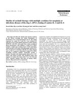
respectively). Graphically, these findings are reflected in
the averaged cumulative DVH plot (Figure 1A).
Dose to organs at risk and normal tissues
With respect to mean dose, IMRT compared to 3FBB
showed little difference for the bladder, femoral heads,
sigmoid, and small bowel. However, compared to
3DCRT, IMRT resulted in significantly lower mean dose
to the bladder (p = 0.007), sigmoid (p = 0.005), small
bowel (p = 0.005), and to the femoral hea ds (p = 0.03).
Mean dose to the pelvic bones was significantly lower
with IMRT compared with either 3FBB (p = 0.04) or
3DCRT (p = 0.005).
With respect to high dose, IMRT significantly
improved the V
40Gy
to the femoral heads (p = 0.01) and
pelvic bones (p = 0.005) compared to 3FBB, and to the
bladder (p = 0.005), femoral heads (p = 0.005), and pel-
vic bones (p = 0.005) in comparison to 3DCRT. For the
dose to sigmoid/colon, IMRT was comparable to 3FBB
at all dose levels evaluated, but was sign ificantly lower
compared to 3DCRT (p = 0.005).
Volumetric evaluation of total small bowel was per-
formed at dose levels ranging from 5- to 45-Gy. When
IMRT was compared to 3FBB, the V
15Gy
was signifi-
cantly reduced with IMRT (p = 0.03), but similar at
other doses. IMRT compared to 3DCRT showed signifi-
cant reductions in the volumes of small bowel irradiated
at levels ranging from 15- to 45-Gy (p < 0.01). With
respect to V
15Gy
, the magnitude of the difference in
median volumes was modest (138-cc IMRT versus 157-
cc 3DCRT; p = 0.005) when evaluating the ten patients
as a whole. However, the most profound bowel sparing
was evident in the subset of patients with the largest
volume of small bowel in proximity to the treatment
field. For example, in the 6 patients with the highest
volume of small bowel (range: 209 - 537-cc), the volume
of bowel receiving 15-Gy was reduced from a median of
231-cc in the 3DCRT plans to 185-cc with IMRT. Con-
versely, in the remaining four patients, only a slight
absolute reduction was evident (median V
15Gy
: 13-cc
IMRT versus 22-cc 3DCRT).
Normal tissues outside the target were evaluated, and
IMRT plans had a significantly higher mean dose (p =
0.02) and V
10Gy
(p = 0.01) to V
30Gy
(p < 0.02) in com-
parison to the 3FBB plans. However, at the highest
doses, IMRT was significantly lower (V
40Gy
, p = 0.02;
V
45Gy
, p < 0.01). IMRT, compared to 3DCRT, had a sig-
nificantly lower mean dose (p = 0.007), V
40Gy
(p =
0.005) and V
45Gy
(p = 0.005), with more modest, but
significant, differences at V
10Gy
(p = 0.005) and V
20Gy
(p = 0.01).
Averaged cumulative DVH plots for organs-at-risk
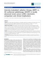
and normal tissues are depicted in Figure 1. Representa-
tive axial s lices showing isodose d istributions for an
IMRT and a 3DCRT plan for one patient are shown in
Figure 2.
Mok et al. Radiation Oncology 2011, 6:63
/>Page 4 of 9
Plan summary characteristics
Monitor units were significantly higher with IMRT com-
pared to either 3FBB (p = 0.005) or 3DCRT (p = 0.005)
(Table 2). The overall plan maximum doses were similar
between IMRT and 3FBB, but higher with IMRT com-
pared to 3DCRT (p = 0.005). Integral dose, calculated
for all tissues including the target volume, was signifi-
cantly higher for IMRT compared to 3FBB (p = 0.007),
but lower compared to 3DCRT (p = 0.007).
Discussion
In this study, we found that the application of I MRT for
rectal cancer gave excellent results in comparison to
non-IMRT based approaches. With respect to the PTV,
we found that IMRT plans achieved s uperior coverage,
homogeneity, and conformality in treating the gross dis-
ease and at-risk pelvic nodal volume, in comparison to
3DCRT plans targeting the PTV. This was not at the
expense of adjacent organs-at-risk, as some measure of
sparing was evident for all organs-at-risk evaluated: small
bowel, sigmoid, pelvic bones, bladder, and femoral he ads
(IMRT versus 3DCRT). In this comparison, IMRT actu-
ally decreased the overall integral dose to all tissues, and
achieved lower mean doses to normal tissues outside the
PTV, which was evident especially in the high dose
range. As expected, IMRT required significantly more
monitor units per fraction, compared to 3DCRT.
We found quite interesting the discrepancy between the
size of the volumes encompassed by the PTV, which were
generated according to the RTOG consensus contouring
atlas [19], and the volumes treated according to classic
anatomic landmarks (3FBB), even considering the antici-
pated patient-to-patient anatomical variation. This was
reflected in the significantly lower proportion of the PTV
Table 1 Dosimetric comparison of IMRT with 3DCRT: median value (range)
Volume Parameter IMRT 3FBB 3DCRT
PTV D
mean
(Gy) 46.6 (46.4 - 46.9) 46.0 (45.6 - 47.0)* 46.6 (46.3 - 48.3)
1547 cm
3
V
45Gy
99.5% (98.7% - 99.8%) 87.2% (80.0% - 93.4%)† 98.4% (97.7% - 99.6%)*
(1459 - D
99%
(Gy) 45.3 (44.8 - 45.8) 35.2 (10.7 - 40.3)† 44.9 (44.2 - 45.2)*
1968 cm
3
)D
1%
(Gy) 47.6 (47.2 - 47.9) 48.3 (47.7 - 50.5)† 47.5 (47.1 - 48.2)
HI 3.2% (2.5% - 3.6%) N/A 4.2% (3.0% - 5.3%)†
CI 1.16 (1.09 - 1.23) N/A 1.35 (1.27 - 1.38)†
Bladder D
mean
(Gy) 38.6 (31.1 - 42.4) 37.9 (27.5 - 44.2) 41.8 (31.0 - 45.0)†
72 cm
3
V
30Gy
74.7% (40.8% - 90.0%) 72.6% (36.7% -96.6%) 85.8% (47.2% - 100%)†
(32-652 cm
3
)V
40Gy
56.8% (26.2% - 76.6%) 58.5% (27.6% - 84.0%) 75.4% (38.0% - 100%)†
Femoral heads D
mean
(Gy) 27.1 (20.8 - 29.6) 24.9 (22.4 - 30.7) 28.5 (21.9 - 31.8)*
211 cm
3
V
30Gy
28.0% (17.8% - 44.2%) 22.6% (12.0% - 33.3%) 31.9% (13.2% - 57.4%)
(151-393 cm
3
)V
40Gy
3.4% (1.1% - 7.0%) 6.3% (1.9% - 13.2%)* 9.1% (3.5% - 14.6%)†
Pelvic bones D
mean
(Gy) 34.2 (30.5 - 36.2) 34.7 (31.9 - 36.8)* 36.7 (32.3 - 38.4)†
914 cm
3
V
30Gy
69.8% (55.6% - 76.3%) 66.7% (61.8% - 72.3%) 74.9% (63.4% - 81.0%)
(725-1338 cm
3
)V
40Gy
47.0% (35.2% - 52.8%) 53.9% (46.5% - 59.2%)† 56.9% (41.3% - 63.6%)†
Sigmoid/Colon D
mean
(Gy) 18.9 (10.4 - 27.9) 17.5 (9.8 - 23.6) 25.5 (13.7 - 31.1)†
outside PTV V
20Gy
41.6% (13.2% - 72.6%) 38.0% (11.5% - 54.0%) 60.6% (24.9% - 75.2%)†
162 cm
3
V
30Gy
17.6% (5.1% - 48.1%) 10.4% (3.0% - 36.9%) 36.9% (10.8% - 63.2%)†
(23 - 389 cm
3
)V
40Gy
4.0% (0.7% - 19.2%) 2.4% (0.4% - 30.3%) 18.3% (5.1% - 38.5%)†
Small bowel D
mean
(Gy) 18.6 (11.2 - 34.0) 21.0 (8.6 - 34.2) 25.2 (15.9 - 40.0)†
251 cm
3
V
5Gy
(cc) 224 (2.5 - 526) 225 (2.2 - 525) 234 (2.5 - 530)
(3 - 537 cm
3
)V
15Gy
(cc) 138 (0.1 - 257) 144 (0.4 - 413)* 157 (2.2 - 428)†
V
25Gy
(cc) 81 (0.0 - 142) 79 (0.0 - 149) 123 (0.5 - 183)†
V
40Gy
(cc) 45 (0.0 - 111) 50 (0.0 - 118) 76 (0.0 - 156)†
V
45Gy
(cc) 37 (0.0 - 100) 33 (0.0 - 74) 53 (0.0 - 121)†
Normal tissues D
mean
(Gy) 19.5 (12.0 - 21.6) 17.5 (12.8 - 20.6)* 20.5 (17.4 - 22.4)†
10.3*10
3
cm
3
V
10Gy
69.5% (58.0% - 76.9%) 64.9% (53.3% - 75.6%)* 73.8% (62.2% - 80.7%)†
(7.7*10
3
-V
20Gy
48.3% (39.5% - 52.7%) 42.0% (32.6% - 52.7%)* 50.5% (40.0% - 58.3%)*
18.9*10
3
cm
3
)V
30Gy
20.3% (16.5% - 27.4%) 17.3% (12.6% - 20.2%)* 23.5% (16.8% - 27.8%)
V
40Gy
6.7% (4.0% - 9.2%) 8.3% (4.2% - 10.9%)* 11.0% (6.7% - 15.7%)†
V
45Gy
2.3% (1.2% - 5.1%) 4.4% (2.1% - 6.0%)† 5.2% (2.8% - 6.4%)†
Abbreviations: PTV = planning target volume; IMRT = intensity modulated radiation therapy; 3FBB = 3 field belly board; 3DCRT = 3 dimensional conformal
radiation therapy; HI = homogeneity index; CI = conformality index; for definitions of dosimetric parameters, refer to text; denotes statistically significant
difference with IMRT as comparator, p < 0.0 5 (*) or p < 0.01 (†); otherwise, not statistically significant.
Mok et al. Radiation Oncology 2011, 6:63
/>Page 5 of 9
volume receiving the prescription dose in the 3FBB plans,
and to a certain extent the significantly lower overall inte-
gral dose, compared to IMRT. We found that despite the
significantly larger volume targeted in the IMRT plans,
IMRT achieved either similar or improved dose levels to
all organs-at-risk evaluated. For example, the small bowel
irradiated had similar mean doses, and the absolute
volumes irradiated w ere similar from the 5- to 45-Gy
levels, except at 15-Gy, where IMRT was statistically
improved, compared to the 3FBB plans.
In terms of acute, severe treatment-related toxicity,
diarrhea is the most common, and studies have
Figure 1 Averaged cumulative dose-volume histograms. Averaged cumulative dose-volume histograms for (A) PTV, (B) bl adder, (C) femoral
heads and necks, (D) pelvic bones, (E) sigmoid outside of PTV, (F) small bowel (relative), (G) small bowel (volumetric), and (H) normal tissues
outside PTV, for IMRT, 3FBB, and 3DCRT.
Mok et al. Radiation Oncology 2011, 6:63
/>Page 6 of 9
demonstrated a strong dose-volume relationship with
small bowel irradiated [7-10]. Baglan and colleagues
demonstrated a strong association between the rate of
small bowel toxicity and the V
15Gy
level; when the V
15Gy
was below 150-cc, low rates of grade 2 or higher toxicity
were observed, while the majority of patients with V
15Gy
over 300-cc had grade 3 or higher toxicity [7]. Subse-
quent studies by Robertson and colleagues have con-
firmed the significance of the V
15Gy
dose level, as well
as other intermediate dose levels, including the V
20Gy
and V
25Gy
, with respect to severe diarrhea [9,10]. In our
study, we found IMRT achieved significant sparing in
terms of the mean dose to small bowel and absolute
volumes from V
15Gy
to V
45Gy
, whereas no difference was
seen at the lowest dose level evaluated, V
5Gy
,compared
to the 3DCRT plans. This sparing at the V
15Gy
level was
most pronounced in the cases with the highest volumes
of small bowel within or nearby the PTV. Therefore, we
would predi ct a lower rate of severe, acute gastrointest-
inal toxicity in these patients treated with IMRT.
Furthermore, reduction in the small bowel V
45Gy
using
IMRT may lead to lower rates of late gastrointestinal
toxicity [20]. Again, in the comparison between IMRT
and classic bony landmark-derived 3FBB fields, despite a
moreextensivevolumetreatedwithIMRT,wewould
predict similar, or based on the V
15Gy
,possibly
improved rates of severe, acute gastrointestinal toxicity
with IMRT compared to 3FBB.
In the context of other planning studies comparing
IMRT with 3DCRT, we feel overall our results are
Figure 2 Representative axial slices. Representative axial slices showing isodose distributions for two planes for an (A), (C) IMRT and (B), (D)
3DCRT plan.
Table 2 Plan summary comparison of IMRT and 3DCRT
plans: median value (range)
Parameter IMRT 3FBB 3DCRT
MU/fraction 786 (730 - 950) 238 (224 - 272)† 242 (232 - 276)†
D
max
(Gy) 48.8 (48.4 - 49.4) 48.8 (48.1 - 51.0) 48.2 (47.8 - 49.2)†
Integral dose 2.74 (2.39 - 4.03) 2.56 (2.15 - 3.60)† 2.86 (2.49 - 4.12)†
(Gy*cm
3
*10
-5
)
Abbreviations: MU = monitor units; IMRT = intensity modulated radiation
therapy; 3FBB = 3 field belly board; 3DCRT = 3 dimensional conformal
radiation therapy;denotes statistically significant difference with IMRT as
comparator, p < 0.05 (*) or p < 0.01 (†); otherwise, not statistically significant.
Mok et al. Radiation Oncology 2011, 6:63
/>Page 7 of 9
superior and additive. Prior studies have demonstrated a
reduction in small bowel mean dose [8], or improve-
ment at the high-dose extreme [16,17], with the use of
IMRT. With respect to positioning, while all three stu-
dies employed prone positioning, one achieved immobi-
lization using a foam cushion [1 7], whereas two made
no specific reference to the use of a bowel displacement
device [8,16]. In contrast, using a rigid, carbon-fiber
belly board apparatus, we observed a significant
improvement in small bowel dose from 15-Gy through
the 45-Gy level, as well as the mean dose, with IMRT
compared to 3DCRT plans. Therefore, our study
demonstrates a f urther significant interval improvement
in small bowel dose is realized with the use of IMRT in
conjunction with the carbon-fiber belly board. An addi-
tional strength of our study is that our conto ured
volumes conformed to the RTOG consensus guidelines.
We chos e as a “ class-solution” approach to use an
asymmetric, seven-beam arrangement, biased a gainst
anterior-directed beams, thus minimizing beam entry
through anterior-lying bowel contents or through the
belly-board apparatus. This appeared to take advantage
of strengths of the 3-field beam arrangement, namely
sharp dose falloff in the intermediate- and low-dose
range anteriorly. Indeed, recently-publishe d studies of
IMRT, using 5- to 9-equispaced beams, have principally
demonstrated reduced small bowel mean dose and
V
40Gy
, compared to 3DCRT [8,16,17]. In our study, in
addition to these findings, we found IMRT capable of
reducing small bowel volumes receiving potentially toxi-
city-inducing intermediate- and low-dose irradiation, at
a statistically-significant level. Concomitantly, IMRT
achieved superior PTV target coverage, homogeneity,
and conformality, as we ll as evidence of sparing of all
other organs-at-risk evaluated in this study. Again, our
results support a clear dosimetric advantage for IMRT,
even with the use of prone-positioning on a belly-board
apparatus.
With respect to the volume of the irradiated t arget,
there are at least two different ways to consider this
issue. In our study, the PTVs, generated with a 7-mm
expansion, were typically larger than the volume treated
using classic 3FBB fields. Given the excellent historical
results obtained with the classic 3FBB fields, one inter-
pretation is that the target volumes, as delineated by the
RTOG consensus IMRT contouring atlas for anorectal
dise ase, may be more generous than necessar y. Alterna-
tively, as we found tha t the more comprehensive PTV
targ et coverage was achieved without increasing dose to
the organs-at-risk including the small bowel, it is con-
ceivable that improved efficacy is attainable without
increasing acute- and long-term t oxicities through the
use of IMRT. Long-term clinical data would be neces-
sary to provide evidence for this. As an additional point,
the use of IMRT does not automatically confer normal
tissue sparing, as an excessively voluminous target
volume may in fact lead to higher absolute volumes of
normal tissues treat ed. This reinforces the importance
of consensus target delineation to achieve standardiza-
tion from practice-to-practice.
Due to daily setup uncertainties using the rigid car-
bon-fiber belly-board apparatus, for IMRT treatment of
a CTV-to-PTV expansion of 7-mm used i n this study, it
may be worthwhile to consider daily kilovoltage imaging,
or perhaps modifications such as the inco rporation of a
vacuum-cradle device to improve setup reproducibility.
One potential criticism for intensity modulated treat-
ment approaches is with respect to integral dose,
whereby larger volumes of normal tissues are exposed
to lower r adiation doses, which may lead to increased
inciden ce of second malignancies [21]. In our study, we
found a lower integral dose with IMRT compared to
3DCRT plans targeting the PTV. However, integral dos e
was slightly higher with IMRT than in the classic 3FBB
plans.
Another potential downside of a static-field intensity
modulated therapy approach is a longer beam-delivery
time that is required as compared to 3DCRT, with
respect to intrafractional motion. This may be overcome
using volumetric-modulated arc therapy (VMAT) based
techniques.
Conclusions
For the adjuvant treatment of rectal carcinoma, IMRT,
compared to 3DCRT, yielded plans superior with
respect to target coverage, homogeneity, and conformal-
ity, while lowering dose to adjacent organs-at-risk. This
benefit was seen additive to the use of prone-positioning
on a belly-board apparatus, and with respect to small
bowel toxicity, could potentially be clinically significant.
Author details
1
Department of Radiation Oncology, The University of Texas, M.D. Anderson
Cancer Center, Houston, Texas, USA.
2
Department of Medical Dosimetry, The
University of Texas, M.D. Anderson Cancer Center, Houston, Texas, USA.
3
Department of Radiation Physics, The University of Texas, M.D. Anderson
Cancer Center, Houston, Texas, USA.
Authors’ contributions
HM carried out the study conception and design, drafted the manuscript,
and performed treatment planning. PD carried out the study conception
and design and drafted the manuscript. MBP performed treatment planning.
TMB and SB performed physics checks/plan evaluation. Patient accrual and
radiation field design were performed by CHC, MED, SK, and PD. CHC
provided mentorship for this work. All authors read and approved the final
manuscript.
Competing interests
The authors declare that they have no competing interests.
Received: 30 November 2010 Accepted: 8 June 2011
Published: 8 June 2011
Mok et al. Radiation Oncology 2011, 6:63
/>Page 8 of 9
References
1. Adjuvant radiotherapy for rectal cancer: a systematic overview of 8,507
patients from 22 randomised trials. Lancet 2001, 358:1291-1304.
2. Douglass HO Jr, Moertel CG, Mayer RJ, Thomas PR, Lindblad AS,
Mittleman A, Stablein DM, Bruckner HW: Survival after postoperative
combination treatment of rectal cancer. N Engl J Med 1986,
315:1294-1295.
3. Krook JE, Moertel CG, Gunderson LL, Wieand HS, Collins RT, Beart RW,
Kubista TP, Poon MA, Meyers WC, Mailliard JA, et al: Effective surgical
adjuvant therapy for high-risk rectal carcinoma. N Engl J Med 1991,
324:709-715.
4. Sauer R, Becker H, Hohenberger W, Rodel C, Wittekind C, Fietkau R,
Martus P, Tschmelitsch J, Hager E, Hess CF, et al: Preoperative versus
postoperative chemoradiotherapy for rectal cancer. N Engl J Med 2004,
351:1731-1740.
5. Roh MS, Colangelo LH, O’Connell MJ, Yothers G, Deutsch M, Allegra CJ,
Kahlenberg MS, Baez-Diaz L, Ursiny CS, Petrelli NJ, Wolmark N: Preoperative
multimodality therapy improves disease-free survival in patients with
carcinoma of the rectum: NSABP R-03. J Clin Oncol 2009, 27:5124-5130.
6. Braendengen M, Tveit KM, Berglund A, Birkemeyer E, Frykholm G,
Pahlman L, Wiig JN, Bystrom P, Bujko K, Glimelius B: Randomized phase III
study comparing preoperative radiotherapy with chemoradiotherapy in
nonresectable rectal cancer. J Clin Oncol 2008, 26:3687-3694.
7. Baglan KL, Frazier RC, Yan D, Huang RR, Martinez AA, Robertson JM: The
dose-volume relationship of acute small bowel toxicity from concurrent
5-FU-based chemotherapy and radiation therapy for rectal cancer. Int J
Radiat Oncol Biol Phys 2002, 52:176-183.
8. Tho LM, Glegg M, Paterson J, Yap C, MacLeod A, McCabe M, McDonald AC:
Acute small bowel toxicity and preoperative chemoradiotherapy for
rectal cancer: investigating dose-volume relationships and role for
inverse planning. Int J Radiat Oncol Biol Phys 2006, 66:505-513.
9. Robertson JM, Lockman D, Yan D, Wallace M: The dose-volume
relationship of small bowel irradiation and acute grade 3 diarrhea
during chemoradiotherapy for rectal cancer. Int J Radiat Oncol Biol Phys
2008, 70:413-418.
10. Robertson JM, Sohn M, Yan D: Predicting grade 3 acute diarrhea during
radiation therapy for rectal cancer using a cutoff-dose logistic regression
normal tissue complication probability model. Int J Radiat Oncol Biol Phys
77:66-72.
11. Fietkau R, Rodel C, Hohenberger W, Raab R, Hess C, Liersch T, Becker H,
Wittekind C, Hutter M, Hager E, et al: Rectal cancer delivery of
radiotherapy in adequate time and with adequate dose is influenced by
treatment center, treatment schedule, and gender and is prognostic
parameter for local control: results of study CAO/ARO/AIO-94. Int J Radiat
Oncol Biol Phys 2007, 67:1008-1019.
12. Gunderson LL, Russell AH, Llewellyn HJ, Doppke KP, Tepper JE: Treatment
planning for colorectal cancer: radiation and surgical techniques and
value of small-bowel films. Int J Radiat Oncol Biol Phys 1985,
11:1379-1393.
13. Mundt AJ, Lujan AE, Rotmensch J, Waggoner SE, Yamada SD, Fleming G,
Roeske JC: Intensity-modulated whole pelvic radiotherapy in women
with gynecologic malignancies. Int J Radiat Oncol Biol Phys 2002,
52:1330-1337.
14. Chen YJ, Liu A, Tsai PT, Vora NL, Pezner RD, Schultheiss TE, Wong JY: Organ
sparing by conformal avoidance intensity-modulated radiation therapy
for anal cancer: dosimetric evaluation of coverage of pelvis and
inguinal/femoral nodes. Int J Radiat Oncol Biol Phys 2005, 63:274-281.
15. Menkarios C, Azria D, Laliberte B, Moscardo CL, Gourgou S, Lemanski C,
Dubois JB, Ailleres N, Fenoglietto P: Optimal organ-sparing intensity-
modulated radiation therapy (IMRT) regimen for the treatment of locally
advanced anal canal carcinoma: a comparison of conventional and IMRT
plans. Radiat Oncol 2007, 2:41.
16. Guerrero Urbano MT, Henrys AJ, Adams EJ, Norman AR, Bedford JL,
Harrington KJ, Nutting CM, Dearnaley DP, Tait DM: Intensity-modulated
radiotherapy in patients with locally advanced rectal cancer reduces
volume of bowel treated to high dose levels. Int J Radiat Oncol Biol Phys
2006, 65:907-916.
17. Arbea L, Ramos LI, Martinez-Monge R, Moreno M, Aristu J: Intensity-
modulated radiation therapy (IMRT) vs. 3D conformal radiotherapy
(3DCRT) in locally advanced rectal cancer (LARC): dosimetric comparison
and clinical implications. Radiat Oncol 5:17.
18. A Phase II Evaluation of Preoperative Chemoradiotherapy Utilizing
Intensity Moldulated Radiation Therapy (IMRT) in Combination with
Capecitabine and Oxaliplatin for Patients with Locally Advanced Rectal
Cancer. [ />study=0822].
19. Myerson RJ, Garofalo MC, El Naqa I, Abrams RA, Apte A, Bosch WR, Das P,
Gunderson LL, Hong TS, Kim JJ, et al: Elective clinical target volumes for
conformal therapy in anorectal cancer: a radiation therapy oncology
group consensus panel contouring atlas. Int J Radiat Oncol Biol Phys 2009,
74:824-830.
20. Gallagher MJ, Brereton HD, Rostock RA, Zero JM, Zekoski DA, Poyss LF,
Richter MP, Kligerman MM: A prospective study of treatment techniques
to minimize the volume of pelvic small bowel with reduction of acute
and late effects associated with pelvic irradiation. Int J Radiat Oncol Biol
Phys 1986, 12:1565-1573.
21. Hall EJ: Intensity-modulated radiation therapy, protons, and the risk of
second cancers. Int J Radiat Oncol Biol Phys 2006, 65:1-7.
doi:10.1186/1748-717X-6-63
Cite this article as: Mok et al.: Intensity modulated radiation therapy
(IMRT): differences in target volumes and improvement in clinically
relevant doses to small bowel in rectal carcinoma. Radiation Oncology
2011 6:63.
Submit your next manuscript to BioMed Central
and take full advantage of:
• Convenient online submission
• Thorough peer review
• No space constraints or color figure charges
• Immediate publication on acceptance
• Inclusion in PubMed, CAS, Scopus and Google Scholar
• Research which is freely available for redistribution
Submit your manuscript at
www.biomedcentral.com/submit
Mok et al. Radiation Oncology 2011, 6:63
/>Page 9 of 9