Báo cáo khoa học: "Comparison of RBE values of high- LET a-particles for the induction of DNA-DSBs, chromosome aberrations and cell reproductive death" potx
Bạn đang xem bản rút gọn của tài liệu. Xem và tải ngay bản đầy đủ của tài liệu tại đây (330.04 KB, 8 trang )
RESEARCH Open Access
Comparison of RBE values of high- LET a-particles
for the induction of DNA-DSBs, chromosome
aberrations and cell reproductive death
Nicolaas AP Franken
1*
, Rosemarie ten Cate
1
, Przemek M Krawczyk
2
, Jan Stap
2
, Jaap Haveman
1
, Jacob Aten
2
and
Gerrit W Barendsen
1,2
Abstract
Background: Various types of radiation effects in mammalian cells have been studied with the aim to predict the
radiosensitivity of tumours and normal tissues, e.g. DNA double strand breaks (DSB), chromosome aberrations and
cell reproductive inactivation. However, variation in correlations with clinical results has reduced general
application. An additional type of information is required for the increasing application of high-LET radiation in
cancer therapy: the Relative Biological Effectiveness (RBE) for effects in tumours and normal tissues. Relevant
information on RBE values might be derived from studies on cells in culture.
Methods: To evaluate relationships between DNA-DSB, chromosome aberrations and the clinically most relevant
effect of cell reproductive death, for ionizing radiations of different LET, dose-effect relationships were determined
for the induction of these effects in cultured SW-1 573 cells irradiated with gamma-rays from a Cs-137 source or
with a-particles from an Am-241 source. RBE values were derived for these effects. Ionizing radiation induced foci
(IRIF) of DNA repair related proteins, indicative of DSB, were assessed by counting gamma-H2AX foci. Chromosome
aberration frequencies were determined by scoring fragments and translocations using premature chromosome
condensation. Cell survival was measured by colony formation assay. Analysis of dose-effect relations was based on
the linear-quadratic model.
Results: Our results show that, although both investigated radiation types induce similar numbers of IRIF per
absorbed dose, only a small fraction of the DSB induced by the low-LET gamma-rays result in chromosome
rearrangements and cell reproductive death, while this fraction is considerably enhanced for the high-LET alpha-
radiation. Calculated RBE values derived for the linear components of dose-effect relations for gamma-H2AX foci, cell
reproductive death, chromosome fragments and colour junctions are 1.0 ± 0.3, 14.7 ± 5.1, 15.3 ± 5.9 and 13.3 ± 6.0
respectively.
Conclusions: These results indicate that RBE values for IRIF (DNA-DSB) induction provide little valid information on
other biologically-relevant end points in cells exposed to high-LET radiations. Furthermore, the RBE values for the
induction of the two types of chromosome aberrations are similar to those established for cell reproductive death.
This suggests that assays of thes e aberrations might yield relevant information on the biological effectiveness in
high-LET radiotherapy.
* Correspondence:
1
Department of Radiation Oncology, Laboratory for Experimental Oncology
and Radiobiology (LEXOR), Centre for Experimental Molecular Medicine,
University of Amsterdam, PO Box 22700, 1100 DE Amsterdam, The
Netherlands
Full list of author information is available at the end of the article
Franken et al. Radiation Oncology 2011, 6:64
/>© 2011 Franken et al; licensee Bi oMed Central Ltd. This is an Open Access arti cle di stributed under the terms of the Creative Commons
Attribution Licens e ( nses/by/2.0 ), which permits unrestricted use, distribution, and reproduction in
any medium, provided the original work is properly cited.
Background
The individualization of cancer treatment by fractio-
nated application of ionizing radiation is expected to
benefit from a rapid assessment of the radiosensitivity of
clonogenic cells in a biopsy obtained before the treat-
ment starts, or of the effectiveness of the first fraction
dose of a schedule for damage to cells in a biopsy
obtained after this fraction [1].
The measurement of clonogenic capacity of the cells,
although it is the most relevant endpoint, requires sev-
eral weeks of culturing and is likely to depend on selec-
tion of cells in adapting to culture media.
The measurement of chromosome aberrations (CA) in
mitotic cells as a marker o f radio sensitivity may be sub -
ject to selection because damaged cells may not all pro-
ceed equally rapidly to mitosis. However, the technique
of premature chromosome condensation might provide
an applicable alternative, because the analysis can be
performed rapidly without the requirement of cells
entering into mitosis [2-5].
Another recently developed rapid technique of assess-
ment of cell damage that has been suggested to provide
information on radiosensitivity involves the measure-
ment of ionising radiation induced foci (IRIF) of DNA
repair-related proteins accumulating at DNA double-
strand breaks (DSB) [6]. Howev er, the quantitative rela-
tionship between the IRIF induction and biologically
relevant endpoints is not yet clear [7-10].
In the application of h igh-L ET radiations to the treat-
ment of cancer an additional type of quantitative infor-
mation is required: the relative biological effectiveness
value (RBE). This is especially relevant with the increas-
ing application of external radiotherapy with light ion
beams and of alpha-particle emitters in targeted radio-
nuclide therapy [11-15],
The mechanisms by which ionizing radiations produce
chromosome aberrations and reproductive death in
mammalian cells are insufficiently elucidated to derive
quantitative information applicable to the design of indi-
vidualized cancer t reatments, because this requires data
about relevant a and b values and their ratio in the bio-
physical linear-quadratic model. These parameters are
differently influenced by repair mechanisms in various
cell types and by the linear energy transfer of the radia-
tion (LET) [16-20].
Various mechanisms of damage induction by ionising
radiations have been proposed that might explain the
high RBE values of high-LET radiations.
Among the many types of DNA damage that are
induced by ionising radiation in mammalian cells, DSB
are generally recognized as the major initial lesio ns that
can result in chromosome aberrations and impairment of
the reproductive integrity which are relevant in clinical
radiotherapy. However, a simple direct causal relationship
between these effects cannot be inferred, because the
number of DNA-DSB produced by a dose of 1 Gy of low
LET ionizing ra diation is much larger than the numbers
of induced chromosome aberrations or cell reproductive
death [21]. A large majority of the induced DSB is
known to be repaired by non-homo logous end joining
(NHEJ) or homologous recombination (HR), but charac-
teristics of the minority of DSB which yield biological
damage are st ill subject of studies [22]. Furthermore the
strong dependence of frequencies of chromosome aberra-
tions and cell inactivation on the linear energy transfer
(LET) of ionizing particles, with maximum values of RBE
in the range from 5-20 compared to g-rays, is not
observed for DNA-DSBs [16-19]. In a review by Prise et
al the authors conclude that the RBE values of high LET
radiations, i.e. LET’s in the range of 50-200 keV/μm, are
between 1-2 for DSBs, although at low doses of 1-5 Gy
data were not considered sufficiently accurate [23]. New
methods of DNA-DSB detection also applied in our stu-
dies, can provide more accurate data in the range of low
doses of 1 to 5 Gy [8,9,24,25].
Two possible explanations for the discrepancy
between the RBE for DSB induction by high-LET radia-
tions compared to other biologically-relevant end points
have been advanced.
First, high-LET radiations might cause more complex
damage in DNA, which might be less easily repaired by
mechanisms in cells [26]. However, it was also suggested
that the number rather t han the molecular structure of
DSB is more important for the formation of chromoso-
mal aberrations [27]. The second explanation for the high
RBE of high-LET radiations is that interaction of two or
more DSB produced in close proximity results in a high
probability for induction of chromosome aberrations and
cell reproductive inactivation. High-LET particles produce
many DSB along their tracks in cells within distances of
less than 1-2 micrometers and this might result in an
increased probability for interaction and enhanced forma-
tion of chromosome aberrations that are known to corre-
late with cell reproductive death [16-19]. Support for this
explanation was provided by the observation of clustering
of chromosome domains containing DSB induced by
high-LET alpha particles and visualised as IRIF [28,29].
Due to the localization of the DSB along the straight
tracks of the particles the probability o f clustering and
interaction is higher than for low-LET radiations. It is
important to note that not all chrom osom e aberrations
cause cell reproductive death and therefore the RBE
values for these two effects might be different. If all chro-
mosome breaks detected by the premature chromosome
condensation technique (PCC) were to cause cell repro-
ductive death, mammalian cells would be more sensitive
Franken et al. Radiation Oncology 2011, 6:64
/>Page 2 of 8
to inactivation by at least a factor 5 [21]. Thus lethal
aberrations might be induced with a higher or lower RBE
by high-LET radiations than all aberrations but little
information on these differences has been reported. In a
recent review the observation is made that RBE values
ranging between 2 and 30 have been reported in studies
applying premature chromosome condensation (PCC)
and fluorescence in situ hybridization (FISH) [30].
Because the differences in RBE values for various
effects of high-LET r adiations in cells cannot be derived
quantitatively on the basis of the known mechanisms,
relevant data can be obtained from cell line based
experiments. This is the purpose of experiments
reported in the present communication.
Materials and methods
Cell culture
The human squamous cell lung carcinoma derived line
SW-1573 was grown at 37°C as monolayer in 75 cm
2
tissue culture flasks (Costar) in Leibowitz-15 medium
(L-15, Gibco-brl Life Technologies, Breda, The Nether-
lands) supplemented with 10% heat inactivated fetal
bovine serum and 2 mM glutamine, 100 U/ml penicillin
and 100 μg/ml streptomycin (Gibco), in an atmosphere
of 0% CO
2.
The doubling time of the cells during expo-
nential growth is 22-24 hour [31-33]. Cells were irra-
diated in plateau phase and immediately used either for
DNA-DSB detection, clonogenic assay or premature
chromosome condensation metaphase preparation. Cell
cycle distribution was monitor ed by flow cytometry and
at the time of irradiation over 90% of cells was in G
0
+G
1
phase. For irradiation with g-rays cells are grown in
6 cm diameter culture dishes.
For the a particle irradiation cells were cultured in
custom made dishes with 2 μm thick mylar bottoms
and 6 cm diameter [34,35].
Irradiation
Plateau phase cell cultures were exposed to a radiation
from an
241
Am source or g-rays from a
137
Cs source.
The 11 MBq
241
Am-sourcewaslocatedatadistance
of 50 mm underneath the dishes and the particles
passed through 50 mm of helium and the thin mylar
bottom of the culture dishes before entering the cells
through the bottom of the dishes with about 4 MeV
energy and a residual range of 25 μmintissue.The
mean LET of particles reaching the cells is 130 keV/μm
[35]. The dose rate was measured with a custom made
ionization c hamber as described in earlier publications
[35]. The length of a particle paths in cell nuclei was
about 5 μm. The average dose rate in the cell nucleus
was0.20Gy/min.Dosesofupto1.6Gywereusedfor
determining survival, 1.4 Gy for g-H2AX foci numbers
and of up to 0.8 Gy for determining chromosomal
aberrations.
Thedoserateofthe
137
Cs source used was 0.6 Gy/
min. For induction of g-H2AX foci, chromosome aberra-
tions and cell reproductive death doses of up to respec-
tively 1.4, 4.0 and 8.0 Gy were used.
Clonogenic assay
Directly after irradiation c ells were trypsini zed and
replated for clonoge nic survival assay in appropriate cell
numbers in 6-well macroplates [36]. Sub sequently, cells
were incubated for 10 days. Surviving colonies were
fixatedandstainedwithglutaraldehyde-crystal violet
solution and counted. Survival curves were analyzed
using SPSS (Chicago, IL, USA) statistical software by
means of fit of data by weighted linear regression,
according to the linear-quadratic formula: S(D)/S(0) =
exp-(aD+bD
2
) [20,36,37].
Chromosomal aberrations
Chromosomal aberrations were studied in prematurely
condensed chromosomes (PCCs). For induction of
PCCs, 80 nM of Calyculin A was added for 1 h immedi-
ately after irradiation [2,5,38]. Visualization of chromo-
somes was accomplished by fluorescent in situ
hybridization (FISH). Cells were h arvested, treated with
hypotonic KCl solution (0.075 M) for 20 min and fixed
in methanol/ acetic acid (3:1). Finally the cell suspension
was dropped on precleaned slides and air-dried. PCC
spreads w ere hybridized to whole chromosome-specif ic
FITC labeled probes for chromosome 2 (Metasystems)
using the method described earlier [4,38,39]. Slides were
counterstained with DAPI (2.5 μg/ml) and embedded
in antifade solution (Vecta shield, Vector laboratories,
Burlingame, CA. USA).
The SW-1573 cells contain between 60 and 67 chro-
mosomes. To study the relations hip between the yield
of exchanges and radiation doses, chromosome 2 was
selected. This chromosome exhibits no spontaneous
exchanges and three copies of chromosome 2 were pre-
sent in over 95% of the metaphases studied. According
to the chromosome length measurements the relative
lengths of chromosomes 2 are 7.8 ± 0.6% of the com-
plete genome [40]. Slides were examined using a fluor-
escencemicroscope(Axioskop 2 MOT Zeiss, Jena,
Germany) equipped with suitable filter block to detect
the painted chromosomes (FITC and DAPI for total
DNA) in one image. Two to four hundr ed PCCs w ere
scored for each dose. The i nduction of colour junctions
and chromosome fragments of painted chromosomes
was scored according to the method described by
Tucker et al [41]. An exchange between a fragment of a
painted chromosome and a fragment of an unp ainted
Franken et al. Radiation Oncology 2011, 6:64
/>Page 3 of 8
chromosome was scored as a colour junction . Rejoining
of two identically painted chromosome fragments with-
out a centromere was scored as fragment.
Detection of g-H2AX: Immunohistochemistry and scoring
To detect g-H2AXfociwhichareformedatsitesof
DSB, cells were grown o n plastic cover slips. The cover
slips (22 × 26 mm) were sterilized with alcohol (70%)
and were placed in 60 mm cell culture dishes. The cells
were reseeded at a density of 2.5 × 10
5
cells in cell cul-
ture dishes containing sterile cover slips and were
grown until a confluent layer was obtained. The cells
were then irradiated. For a-particle irradiation the cover
slips were placed on a dish with a mylar bottom with
the cells facing the mylar.
After a particle irradiation cells were fixated 5 min
after treatment. After g- irradiation cells were fixated 30
min after treatment. At these time intervals maximal
number of foci were counted. After irradiation, cells
were washed with PBS and fixated in PBS containing 2%
paraformaldehyde for 15 min. After three further washes
in PBS cells were treated with PBS containing 0.1% T ri-
ton X-100 & 1% FCS (TNBS) for 30 min.
The pr imary antibody used was a mouse monoclonal
anti-g-H2AX (Millipore, Ca) diluted 1:100 in TNBS. Per-
meabilized cells were incubated with 50 μl primary anti-
body under a parafilm strip for 90 min. at room
temperature. Cells were then washed with PBS for about
5 min and the parafilm strip was removed. After this,
cells were washed 2 times with TNBS. The secondary
antibody used was a Goat anti-Mouse Cy3 (Jackson,
Immunoresearch, Europe Ltd, Suffolk, UK)) also diluted
1:100 in TNBS.
Cells on cover slips were incubated with 50 μl second-
ary antibody under a para film strip for 30 min. at r oom
temperature. Cells were then washed 3 times with
TNBS for about 5 min and the parafilm strip was
removed at the first wash. Nuclei were stained with
DAPI (2.5 μg/ml) and subsequently embedded in
vectashield.
Digital image analysis was performed to determine the
number of g-H2AX IRIF (ionizing radiation induced
foci). Fluorescent photomicrographs of g-H2AX foci
were obtained using Image Pro Plus software. Stack
images of cells were obtained using a Leica DM RA HC
Upright Microscope equipped with a CCD camera.
Stack images of 100 cells per sample were taken using
Image pro plus software. One stack image consists of 40
slices with a 200 nm interval between the slices along
the z-axis. Images were then processed and the number
of foci in cells was scored using custom made software
[28,29].
All experiments were carried out in triplicates,
independently from each other. Numbers of foci in
unirradiated control cells were subtracted from numbers
in irradiated samples.
S-phase cells were excluded as an EDU staining (Invi-
trogen, Eugene, Oregon USA) was used to mark these
cells.
Results
The PCC technique was applied to measure induction of
chromosome aberrations, induction of DSB was esti-
mated by scoring g-H2AX IRIF and reproductive cell
death was measured by clonogenic assay.
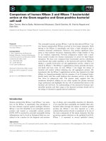
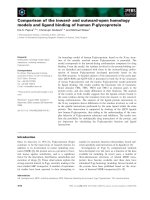
In figure 1 induction of Gamma-H2AX foci, radiation
dose survival curves, frequencies of chromosome frag-
ments and colour junctions in SW1573 cells after a-
particle and g-ray irradiation are presented. Figure 1A
shows similar induction of DSB after a- particles and
g-rays at the time i nterval s studied. Figure 1B shows the
radiation dose survival curves which demonstrate that
cell reproductive death after a- particle radiation is
much more frequently induced than af ter g-ray irradia-
tion. From figure 1C and 1D it can be observed that
after a- particle radiation the induction of chromosomal
fragments and colour junctions is much higher than
after g-ray irradiation. From these data the linear and
quadratic parameters were derived by analysis with the
formula S(D)/S(0) = exp-(aD+bD
2
) for cell survival
curves and F(D) = aD+b D
2
for the induction of DSB,
chromosome fragments end colour junctions. Except for
cell survival curves for g-irradiation, the values of the
quadratic pa rameter, b, for DSB induction, chromosom e
fragments and colour junction formation were not sig-
nificantly different from zero. Because with alpha radia-
tion only linear parameters were derived, for assessment
of RBE values the comparison with linear parameters for
gamma radiation for all endpoints is appropriate. There-
fore, in order to compare equivalent values only the
values of a are consi dered for evaluation and discussion
of RBE values. These values are summarized in table 1.
The values for the quadratic parameters for cell survival,
chromosomal fragments and colour junctions after
g-irradiation are 0.05 ± 0.01, 0.08 ± 0.08 and 0.03 ±
0.07 resp.
Discussion
For the survival curve, chromosomal fragments and col-
our junctions of cells i rradiated with g-rays linear-quad-
ratic dose response curves were obtained while for DNA
DSBs the dose-re sponse effect relation is linear. After
a- particle radiation the dose response curves f or all
endpoints studied were linear. Therefore, in order to
derive relevant RBE values, only the parameters of the
linear terms will be compared. To measure induction of
chromosome aberrations, we applied premature chro-
mosome condensation (PCC) because this method does
Franken et al. Radiation Oncology 2011, 6:64
/>Page 4 of 8
not require the treated cells to proceed to mitosis,
which may select for cells with less damage [2].
Comparison of the frequencies of induced effects
The value of a for induction of DSB (table 1) is evi-
dently much larger than the corresponding value for cell
inactivation, leading to the conclusion that only a small
fraction of the DSB (about 1% of DSB induced by g-rays
and about 1 0% by a-particles) are causing cell death.
On the other hand, the values of a for formation of
chromosome fragments and colour junctions as shown
in table 1 are about 8 and 4 resp. times larger than the
corresponding values for induction of cell reproductive
death, for a-aswellasforg-radiation. This suggests
C
0.0 0.2 0.4 0.6 0.8 1.0 1.2 1.4
0
10
20
30
40
50
Gamma-H2AX foci
Dose, Gy
# of foci/cell
A
Colour junctions
0 1 2 3 4
0
2
4
6
8
Dose, Gy
# of colour junctions/genome
Cell reproductive death
0 2 4 6 8
0.01
0.1
1
Dose, Gy
Surviving fraction
B
Chromosome fragments
0 1 2 3 4
0
3
6
9
12
15
Dose, G
y
# of fragments/genome
D
Figure 1 Number of g-H2AX foci (A), Radiation dose survival curves (B), frequency of colour junctions (C) and chromosome fragments
(D) for SW1573 cells after a particle (black squares) and gamma irradiation (black triangles). Calculated RBE values for DNA-DSBs, cell
reproductive death, chromosome fragments and colour junctions are 1.0 ± 0.3, 14.7 ± 5.1, 15.3 ± 5.9 and 13.3 ± 6.0 resp.
Table 1 Values of a of the LQ model for survival curves, chromosomal fragments, colour junctions and DNA DSBs of
SW-1573 cells after alpha particle irradiation and after g irradiation
a-particle irradiation Gy
-1
g-irradiation Gy
-1
RBE value
Survival 2.2 ± 0.38 0.15 ± 0.045 14.7 ± 5.1
Chromosomal fragments 16.8 ± 4.5 1.1 ± 0.31 15.3 ± 5.9
Colour junctions 9.2 ± 3.2 0.69 ± 0.2 13.3 ± 6.0
DSB (Gamma-H2AX foci) 25 ± 8.2 25 ± 3.0 1.0 ± 0.3
The chromosomal fragments, colour junctions were determined in chromosome 2 and the a-values are corrected for the DNA content of the complete genome.
Survival curves were analyzed using S(D)/S(0) = exp-(aD+bD
2
) [20,36,37]. The values of a for chromosomal fragments colour junctions and DSB are calculated
according to F(D) = aD+bD
2
[40]
Franken et al. Radiation Oncology 2011, 6:64
/>Page 5 of 8
that many of these aberrations are either repaired or do
not cause complete impairment of the cell reproductive
capacity. The number of fragments is higher than that
of colour junctions a s the induction of chromosomal
aberrations was studied shortly after treatment and at
that time point not all colour junctions might have be en
formed. It is generally observed that colonies arising
from cells surviving irradiation are smaller, as compare d
to colonies formed by unirradiated cells, indicating that
their genomes might be damaged, although their repro-
ductive potential is not eliminated [42]. From analyses
of cell survival curves derived for different particles in
relation to LET, it has been earlier suggested that a con-
tribution to the linear term is due to potentially lethal
damage (PLD) [16]. The present results are compatible
with this suggestion.
Comparison of RBE values
The calculated RBE value of 1.0 ± 0.3 for induction of g-
H2AX foci is much smaller than the values for induc-
tion of cell reproductive death, chromosome fragments
and colour junctions, which are not significantly differ-
ent [43].
Although there is a clear correlation between cell
reproductive death and the induction of chromosomal
aberrations, a direct causal relationship between these
effects cannot yet be inferred [44]. Further studies of
RBE values a t different time intervals post irradiation
should yield information on this problem. The RBE
value of 14.6 for cell reproductive death is similar to
values in the range of 5 to 15 pu blished for many other
lines of cultured cells [45]. The RBE of 1 derived for the
induction of DNA-DSB is consistent with published
results obtained with other methods at higher doses as
summarized by Prise et al [23]. However, data obtained
by Prise et al using the filter elution technique show a
significant contribution of the quadratic parameter b in
the dose-effect curves at large doses of X-rays [44,45].
This observation is not incompatible with our data
showing a linear dependenc e of the number of g-H2AX
focionthedoseofg-rays at low doses. The a/b ratios
that can be derived from the data obtained with the fil-
ter elution technique are equal to 27 Gy and 16 Gy for
AL-K and 250 kV X-rays, respect ively. From these large
values it is evident that at doses in the range of up to
1.4 Gy as used in our studies the quadratic term contri-
butes less then 10 percent to the total effect. This con-
tribution is not detectable as a deviation from linearity
in our results at low doses.
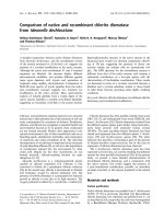
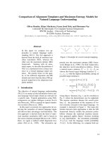
Based on the available literature, it can be suggested
that the RBE for DSB induction increases as a function
of LET between 20 and 80 keV/μm to abo ut 2 and sub-
sequently decreases to about 1 at larger LET (summar-
ized in Figure 2) [16-18,46]. The curve presented in this
figure for DNA-DSB induction was derived as an aver-
age of data published by different investigators, as sum-
marised in reference 18. This figure is included to
illustrate the small dependence of DNA-DSB on LET,
but the absolute values may vary for different cell lines.
The RB E value of 1 obtained for DS B induction by 130
keV/μm a-particles reported here is not inconsistent
with these data.
Conclusions
The final conclusion from the presented results is that
assessment of the amount of DSB induced by ionizing
radiation as measured by us shortly after radiation is
unlikely to provide information about the biological
effectiveness of high LET radiations of rel evance in the
treatment o f cancer. This is in agreement with the
report by Yoshikawa et al inferring that g-H2AX IRIF
numbers in tumour cells fail to correlate with their
radiosensitivity [7]. On the other hand, the RBE values
for induction of chromosome aberratio ns are quite
similar to the value for cell reproductive death. This
Figure 2 Relative biological effectiveness (RBE) as a function of
the linear energy transfer (LET) for different types of lethal
damage in mammalian cells and for DNA damage. ILD,
irrepairable lethal damage, derived as the contribution to the linear
parameter a of the LQ model that is not repaired after irradiation of
cells, even if maintained in conditions optimal for repair. PLD,
potentially lethal damage, derived as the contribution to the linear
parameter a that after irradiation is repaired in conditions optimal
for repair. STLD, single track lethal damage, derived as the linear
parameter a in conditions in which PLD is not repaired. SLD,
sublethal damage, derived from survival curves as the square root
of the quadratic parameter b of the LQ model. DNA-DSB, RBE for
double strand breaks in DNA., DNA-SSB, RBE for single strand breaks
in DNA. (from Barendsen et al.) [46].
Franken et al. Radiation Oncology 2011, 6:64
/>Page 6 of 8
suggests that these end-points might be more appro pri-
ate in assessment of biological effectiveness of high-LET
radiations. R ecently i t has been shown that P CC-FISH
can be applied directly to biopsy cultures and biopsies
derived from cervical cancer pati ents [2-4]. This techni-
que might, therefore, yield relevant information on the
effectiveness of high-LET radiations.
Acknowledgements
We would like to thank Maria Bozarova for technical assistance. We
acknowledge the Maurits and Anna de Kock and the Nijbakker Morra
foundations for sponsoring the fluorescence microscopes with software to
study chromosomal aberrations and g-H2AX foci. The Dutch Cancer
Foundation is acknowledged for personnel financial support (UVA 2008-
4019) and the Stichting Vanderes is ackowledged for personnel financing.
Author details
1
Department of Radiation Oncology, Laboratory for Experimental Oncology
and Radiobiology (LEXOR), Centre for Experimental Molecular Medicine,
University of Amsterdam, PO Box 22700, 1100 DE Amsterdam, The
Netherlands.
2
Department of Cell Biology and Histology, Academic Medical
Centre, University of Amsterdam, PO Box 22700, 1100 DE Amsterdam, The
Netherlands.
Authors’ contributions
NAPF performed the clonogenic survival assays, foci studies and coordinated
the study. NAPF and GWB drafted the research, performed the dosimetry of
the alpha particle irradiation and wrote the paper. RtC and JH performed
the chromosomal aberration studies. PK, JS and JA helped with the
discussion of the data. All authors read and approved the final manuscript.
Competing interests
The authors declare that they have no competing interests.
Received: 27 January 2011 Accepted: 8 June 2011
Published: 8 June 2011
References
1. Begg AC: Predicting response to radiotherapy: evolutions and
revolutions. Int J Radiat Biol 2009, 85:825-836, Review.
2. Darroudi F, Bergs JW, Bezrookove V, Buist MR, Stalpers LJ, Franken NAP:
PCC and COBRA-FISH a new tool to characterize primary cervical
carcinomas: to assess hall-marks and stage specificity. Cancer Lett 2010,
287:67-74.
3. Coco-Martin JM, Begg AC: Detection of radiation-induced chromosome
aberrations using fluorescence in situ hybridization in drug-induced
premature chromosome condensations of tumour cell lines with
different radiosensitivities. Int J Radiat Biol 1997, 71:265-273.
4. Suzuki M: The PCC assay can be used to predict radiosensitivity in
biopsy cultures irradiated with different types of radiation. Oncol Rep
2006, 16:1293-1299.
5. Gotoh E, Asakawa I, Kosaka H: Inhibition of protein serine/threonine
phosphatases directly induces premature chromosome condensation in
mammalian somatic cells. Biomed Res 1995, 16:63-68.
6. Olive PL, Banáth JP: Phosphorylation of histone H2AX as a measure of
radiosensitivity. Int J Radiat Oncol Biol Phys 2004, 58:331-335.
7. Yoshikawa T, Kashino G, Ono K, Watanabe M: Phosphorylated H2AX foci in
tumor cells have no correlation with their radiation sensitivities. J Radiat
Res 2009, 50:151-160.
8. Leatherbarrow EL, Harper JV, Cucinotta FA, O’Neill PL: Induction and
quantification of gamma-H2AX foci following low and high LET-
irradiation. Int J Radiat Biol 2006, 82:111-118.
9. Vandersickel V, Depuydt J, Van Bockstaele B, Perletti G, Philippe J,
Thierens H, Vral A: Early increase of radiation-induced γH2AX foci in a
human Ku70/80 knockdown cell line characterized by an enhanced
radiosensitivity. J Radiat Res 2010, 51:633-641.
10. Takahashi A, Yamakawa N, Kirita T, Omori K, Ishioka N, Furusawa Y, Mori E,
Ohnishi K, Ohnishi T: DNA damage recognition proteins localize along
heavy ion induced tracks in the cell nucleus. J Radiat Res 2008,
49:645-652.
11. Dale RG, Jones B, Cárabe-Fernández A: Why more needs to be known
about RBE effects in modern radiotherapy. Appl Radiat Isot 2009,
67:387-392, review.
12. Sgouros G, Roeske JC, McDevitt MR, Palm S, Allen BJ, Fisher DR, Brill AB,
Song H, Howell RW, Akabani G, SNM MIRD Committee, Bolch WE, Brill AB,
Fisher DR, Howell RW, Meredith RF, Sgouros G, Wessels BW, Zanzonico PB:
MIRD Pamphlet No. 22 (abridged): radiobiology and dosimetry of alpha-
particle emitters for targeted radionuclide therapy. J Nucl Med 2010,
51:311-328.
13. Okada T, Kamada T, Tsuji H, Mizoe JE, Baba M, Kato S, Yamada S,
Sugahara S, Yasuda S, Yamamoto N, Imai R, Hasegawa A, Imada H,
Kiyohara H, Jingu K, Shinoto M, Tsujii H: Carbon Ion Radiotherapy: Clinical
Experiences at National Institute of Radiological Science (NIRS). J Radiat
Res 2010, 51:355-364.
14. Vandersickel V, Mancini M, Slabbert J, Marras E, Thierens H, Perletti G, Vral A:
The radiosensitizing effect of Ku70/80 knockdown in MCF10A cells
irradiated with X-rays and p(66)+Be(40) neutrons.
Radiat Oncol 2010, 5:30.
15.
Jingu
K, Hasegawa A, Mizo JE, Bessho H, Morikawa T, Tsuji H, Tsujii H,
Kamada T: Carbon ion radiotherapy for basal cell adenocarcinoma of the
head and neck: preliminary report of six cases and review of the
literature. Radiat Oncol 2010, 5:89.
16. Barendsen GW: The relationships between RBE and LET for different
types of lethal damage in mammalian cells: biophysical and molecular
mechanisms. Radiat Res 1994, 139:257-270, review.
17. Barendsen GW: RBE-LET relationships for different types of lethal
radiation damage in mammalian cells: comparison with DNA dsb and
an interpretation of differences in radiosensitivity. Int J Radiat Biol 1994,
66:433-436.
18. Barendsen GW: Sublethal damage and DNA double strand breaks have
similar RBE-LET relationships: evidence and implications. Int J Radiat Biol
1993, 63:325-330.
19. Barendsen GW: Parameters of linear-quadratic radiation dose-effect
relationships: dependence on LET and mechanisms of reproductive cell
death. Int J Radiat Biol 1997, 71:649-655.
20. Barendsen GW: Dose fractionation, dose rate and iso-effect relationships
for normal tissue responses. Int J Radiat Oncol Biol Phys 1982, 8:1981-1997,
review.
21. Bedford JS: Sublethal damage, potentially lethal damage, and
chromosomal aberrations in mammalian cells exposed to ionizing
radiations. Int J Radiat Oncol Biol Phys 1991, 21:1457-1469.
22. Goodhead DT: Mechanisms for the Biological Effectiveness of High-LET
Radiations. J Radiat Res 1999, 40(Suppl):S1-S13.
23. Prise KM, Pinto M, Newman HC, Michael BD: A review of studies of
ionizing radiation-induced double-strand break clustering. Radiat Res
2001, 156:572-576, review.
24. Goodhead DT, Thacker J, Cox R: Weiss Lecture: Effects of radiations of
different qualities on cells: molecular mechanisms of damage and
repair. Int J Radiat Biol 1993, 63:543-556, review.
25. Kitajima S, Nakamura H, Adachi M, Ijichi K, Yasui Y, Saito N, Suzuki M,
Kurita K, Ishizaki K: AT Cells Show Dissimilar Hypersensitivity to Heavy-Ion
and X-rays Irradiation. J Radiat Res 2010, 51:251-255.
26. Pinto M, Prise KM, Michael BD: Evidence for complexity at the nanometer
scale of radiation-induced DNA DSBs as a determinant of rejoining
kinetics. Radiat Res 2005, 164:73-85.
27. Obe G, Johannes C, Ritter S: The number and not the molecular structure
of DNA double-strand breaks is more important for the formation of
chromosomal aberrations: a hypothesis. Mutat Res 2010, 701:3-11.
28. Aten JA, Stap J, Krawczyk PM, van Oven CH, Hoebe RA, Essers J, Kanaar R:
Dynamics of DNA double-strand breaks revealed by clustering of
damaged chromosome domains. Science 1994, 303:92-95.
29.
Stap
J, Krawczyk PM, van Oven CH, Barendsen GW, Essers J, Kanaar R,
Aten JA: Induction of linear tracks of DNA double-strand breaks by
alpha-particle irradiation of cells. Nat Methods 2008, 5:261-266.
30. Ritter S, Durante M: Heavy-ion induced chromosomal aberrations: a
review. Mutat Res 2010, 701:38-46, review.
31. Franken NAP, van Bree C, Streefkerk JO, Kuper MJA, Kipp JBA, Haveman J,
Barendsen GW: Radiosensitization by iodo-deoxyuridine in cultured SW-
1573 human lung tumor cells: Effects on alpha and beta of the linear-
quadratic model. Oncol Rep 1997, 4:1073-1076.
Franken et al. Radiation Oncology 2011, 6:64
/>Page 7 of 8
32. Franken NAP, van Bree C, Veltmaat MA, Ludwików G, Kipp JBA,
Barendsen GW: Increased chromosome exchange frequencies in iodo-
deoxyuridine-sensitized human SW-1573 cells after gamma-irradiation.
Oncol Rep 1999, 6:59-63.
33. Franken NAP, van Bree C, Veltmaat MA, Rodermond HM, Haveman J,
Barendsen GW: Radiosensitization by bromodeoxyuridine and
hyperthermia: analysis of linear and quadratic parameters of radiation
survival curves of two human tumor cell lines. J Radiat Res 2001,
42:179-190.
34. Barendsen GW: Dose-survival curves of human cells in tissue culture
irradiated with alpha-, beta-, 20-kV. x- and 200-kV. x-radiation. Nature
1962, 193:1153-1155.
35. Barendsen GW: Impairment of the proliferative capacity of human cells
in culture by alpha-particles with differing linear-energy transfer. Int J
Radiat Biol 1964, 8:453-466.
36. Franken NAP, Rodermond HM, Stap J, Haveman J, van Bree C: Clonogenic
assay of cells in vitro. Nat Protoc 2006, 1:2315-2319.
37. Franken NAP, van Bree C, Kipp JBA, Barendsen GW: Modification of
potentially lethal damage in irradiated Chinese hamster V79 cells after
incorporation of halogenated pyrimidines. Int J Radiat Biol 1997,
72:101-109.
38. Bergs JW, ten Cate R, Haveman J, Medema JP, Franken NAP, van Bree C:
Chromosome fragments have the potential to predict hyperthermia-
induced radio-sensitization in two different human tumor cell lines.
J Radiat Res 2008, 49:465-472.
39. Bergs JW, Franken NAP, ten Cate R, van Bree C, Haveman J: Effects of
cisplatin and gamma-irradiation on cell survival, the induction of
chromosomal aberrations and apoptosis in SW-1573 cells. Mutat Res
2006, 594:148-154.
40. Franken NAP, Ruurs P, Ludwików G, van Bree C, Kipp JB, Darroudi F,
Barendsen GW: Correlation between cell reproductive death and
chromosome aberrations assessed by FISH for low and high doses of
radiation and sensitization by iodo-deoxyuridine in human SW-1573
cells. Int J Radiat Biol 1999, 75:293-299.
41. Tucker JD, Morgan WF, Awa AA, Bauchinger M, Blakey D, Cornforth M,
Littlefield LG, Natarajan AT, Shasserre C: A proposed system for scoring
structural aberrations detected by chromosome painting. Cytogenet Cell
Genet 1995, 68:211-221.
42. Westra A, Barendsen GW: Proliferation characteristics of cultured
mammalian cells after irradiation with sparsely and densely ionizing
radiations. Int J Radiat Biol 1966, 11:477-485.
43. Schmid TE, Dollinger G, Beisker W, Hable V, Greubel C, Auer S, Mittag A,
Tarnok A, Friedl AA, Molls M, Röper B: Differences in the kinetics of
gamma-H2AX fluorescence decay after exposure to low and high LET
radiation. Int J Radiat Biol 2010, 86:682-691.
44. Prise KM, Davies S, Michael BD: The relationship between radiation-
induced DNA double-strand breaks and cell kill in hamster V79
fibroblasts irradiated with 250 kVp X-rays, 2.3 MeV neutrons or 238Pu
alpha-particles. Int J Radiat Biol 1987, 52:893-902.
45. Prise KM, Folkard M, Davies S, Michael BD: Measurement of DNA damage
and cell killing in Chinese hamster V79 cells irradiated with aluminum
characteristic ultrasoft X rays.
Radiat Res 1989, 117:489-499.
46. Barendsen GW, van Bree C, Franken NAP: Importance of cell proliferative
state and potentially lethal damage repair on radiation effectiveness:
implications for combined tumor treatments (review). Int J Oncol 2001,
19:257-256, review.
doi:10.1186/1748-717X-6-64
Cite this article as: Franken et al.: Comparison of RBE values of high-
LET a-particles for the induction of DNA-DSBs, chromosome aberrations
and cell reproductive death. Radiation Oncology 2011 6:64.
Submit your next manuscript to BioMed Central
and take full advantage of:
• Convenient online submission
• Thorough peer review
• No space constraints or color figure charges
• Immediate publication on acceptance
• Inclusion in PubMed, CAS, Scopus and Google Scholar
• Research which is freely available for redistribution
Submit your manuscript at
www.biomedcentral.com/submit
Franken et al. Radiation Oncology 2011, 6:64
/>Page 8 of 8