Báo cáo khoa học: " Neoadjuvant chemoradiation compared to neoadjuvant radiation alone and surgery alone for Stage II and III soft tissue sarcoma of the extremitie" pps
Bạn đang xem bản rút gọn của tài liệu. Xem và tải ngay bản đầy đủ của tài liệu tại đây (328.03 KB, 11 trang )
RESEARCH Open Access
Neoadjuvant chemoradiation compared to
neoadjuvant radiation alone and surgery alone
for Stage II and III soft tissue sarcoma of the
extremities
Kelly K Curtis
1
, Jonathan B Ashman
2*
, Christopher P Beauchamp
3
, Adam J Schwartz
3
, Matthew D Callister
2
,
Amylou C Dueck
4
, Leonard L Gunderson
2
and Tom R Fitch
1
Abstract
Background: Neoadjuvant chemoradiation (NCR) prior to resection of extremity soft tissue sarcoma (STS) has been
studied, but data are limited. We present outcomes with NCR using a variety of chemotherapy regimens compared
to neoadjuvant radiation without chemotherapy (NR) and surgery alone (SA).
Methods: We conducted a retrospective chart review of 112 case s.
Results: Treatments included SA (36 patients), NCR (39 patients), and NR (37 patients). NCR did not improve the
rate of margin-negative resections over SA or NR. Loco-regional relapse-free survival, distant metastases-free
survival, and overall survival (OS) were not different among the treatment groups. Patients with relapsed disease
(OR 11.6; p = 0.01), and tumor size greater than 5 cm (OR 9.4; p = 0.01) were more likely to have a loco-regional
recurrence on logistic regression analysis. Significantly increased OS was found among NCR-treated patients with
tumors greater than 5 cm compared to SA (3 year OS 69 vs. 40%; p = 0.03). Wound complication rates were
higher after NCR compared to SA (50 vs. 11%; p = 0.003) but not compa red to NR (p = 0.36). Wet desquamation
was the most common adverse event of NCR.
Conclusions: NCR and NR are acceptable strategies for patients with STS. NCR is well-tolerated, but not clearly
superior to NR.
Keywords: Neoadjuvant, chemotherapy, radiation, chemoradiation, soft tissue sarcoma, extremity
Background
Extremity soft tissue sarcoma (STS) treatment strategies
gradually have shifted awayfromamputationtowarda
limb preservation approach. For most patients with low-
grade extremity STS, (i.e., T1-2, N0, M0) surgical resec-
tion is the primary treatment, followed by adjuvant
radiation for margins less than or equal to 1 c entimeter
[1]. Fo r patients with high-g rade STS of the extremities
(i.e., Stages II or III), neoadjuvant radiation with or
without chemotherapy often is employed to improve
local control and functional outcome [1].
Experience with neoadjuvant chemoradiation (NCR) in
STS has been reported by several groups. Eilber and col-
leagues published a regimen of intra-arterial doxorubicin
infused o ver 24-hours for 3 days prior to radiation, fol-
lowed by surgery [2]. Other single agents that have been
studied with pre-operative radiation include ifosfamide
and gemcitabine [3,4]. Multi-agent chemotherapy regi-
mens given pre-operatively with radiation include MAID
(mesna, doxorubicin, ifosfamide and dacarbazine) or
IMAP/MAP (ifosfamide, mitomycin, doxorubicin, and
cisplatin) [5-7]. These strategies have shown promising
results, including 5-year overall survival rates up to 70%
[8-11], 5-year local control rates up to 92% [5] and limb
preservation rates up to 100% [4]. Toxicities of NCR
* Correspondence:
2
Department of Radiation Oncology, Mayo Clinic, 13400 East Shea Blvd.,
Scottsdale, AZ 85259, USA
Full list of author information is available at the end of the article
Curtis et al. Radiation Oncology 2011, 6:91
/>© 2011 Curtis et al; licensee BioMed Central Ltd. Thi s is an O pen Access article distributed under the terms of the Creative Commons
Attribution License ( which permits unrestricted use, distribution, and reproduction in
any medium, pro vided the original work is properly cited.
typically include wound complications, many of which
require re-operation, and long bone fracture [12].
At Mayo Clinic in Arizona (MCA), the decision to use
NCR, neoadjuvant radiation (NR) or surgery alone (SA)
is based on initial magnetic resonance imaging (MRI)
findings. Patients likely to have narrow resection mar-
gins, with high grade tumors, large tumor size, and an
unfavorable location relative to the neuro-vascular bun-
dles and bone a re referred to radiation oncology and
medical oncology for consideration of NR or NCR.
Despite its use, data on outcomes with NCR for Stage II
and III extremity STS are l imited. A prospecti ve, rando-
mized trial comparing NCR to NR and SA is needed to
provide mo re robust knowledge. In the absence of such
information, a retrospective analysis c an provide preli-
minary insight and be used for hypothesis generation.
Therefore, we conducted a retrospective analysis of
patients with extremity STS treated at MCA to increase
our understanding of NCR-related outcomes as com-
pared to NR- and SA-treated patients.
Methods
A retrospective chart review was conducted of 112
extremity STS cases treated between January 1, 1998
and December 31, 2009 a t MCA. We included patients
greater than 15 years of age with Stage II and III extre-
mity STS as defined by the 2010 7
th
Edition American
Joint Committee on Cancer (AJCC) Staging System of
STS. Pati ents with relapsed extremity STS being treated
with curative intent were included. Non-extremity sar-
comas, low grade (Stage I) extremity STS, and bone/car-
tilage sarcomas were excluded. Patients treated with
post-operative radiation and patients with metastatic or
recurrent disease receiving only palliative treatments
were excluded. The review was approved by the Mayo
Clinic Institutional Review Board.
The following informatio n was recorded: age a t diag-
nosis, date of first MCA evaluation, sex, primary disease
site, histology, grade, tumor size and depth (s uperficial
or deep as d efined by the 2010 AJCC Staging System of
STS), margin status, notation of periosteal or nerve
stripping in the operative summary, limb preservation or
amputation, occurrence of wound complications follow-
ing surgery, date of first local recurrence (if any), date of
appearance of distant metastases (if any), any documen-
tation of treatment-related toxicity, and date of death or
last follow-up at MCA. I t was not possible to determine
toxicity grading from medical records. Sarcoma treat-
ment was categorized as follows: SA (defined as any
curative-intent surgical procedure performed without
pre- or post-operative chemotherapy or radiation), NCR
(defined as any combination of chemotherapy with
radiation given prior to a curative-intent surgical resec-
tion), or NR (defined as radiation given without
chemotherapy prior to a cura tive-intent surgical resec-
tion). Patients treated with sequential pre-operative che-
motherapy followed by pre-operative radiation were
included in the NCR group, since historically such ther-
apy has been considered a form of NCR [2,13]. Use of
intra-operative electron radiation therapy (IOERT) or
perioperative brachytherapy was documented.
Surgical margins were recorded as negative (R0 resec-
tion) if the pathology report noted all margins to be free
of tumor microscopically. If tumor extended to the sur-
gical margin microscopically, or if the surgical margin
was less than or equal to 1 mm, th e margin was consid-
ered to be positive (R1 resection). It was not possible to
determine pathologic response rates to NCR or NR
from the records. Loco-regional recurrences were
def ined as any relapse of sarcoma at the previous sur gi-
cal site or in regional lymph nodes. A “wound complica-
tion” was defined as any post-operative wound event
requiring a return to th e operating room for an
unplanned additional procedure.
All time-to-failure endpoints were calculated from th e
date of first MCA contact. Overall survival (OS) was
defined as death as a result of any cause; time to loco-
regional recurrence was defined as time to date of a
local or regional relapse diagnosis or amputation for any
reason; time to distant metastases w as defined as ti me
to date of discovery of distant metastases, excluding new
primary cancers. Kaplan-Meier methods were used to
estimate OS, loco-regional relapse-free survival (LR-
RFS), and distant metastasis-free survival (DMFS) for
each of the treatment modality received. Contingency
analyses using the Chi-square test of independence were
conducted for different treatment modalities and surgi-
cal outcome, limb preservation, presence or absence of
local recurrence and distant metastases, and presence or
absence of wound complications. Logistic regression
analyses were performed to determine factors associated
with amputation for relapsed disease, as well as factors
associated with a greater likelihood of wound complica-
tions. Logistic regression analysis also was conducted to
determine factors associated with loco-regional recur-
rence. SA patients who were treated primarily with
amputation were excluded from the analysis of LR-RFS
and wound complications because of potential imbal-
ances among this sub-group compared to the majority
of patients treated with limb-preservation intent.
Results
Patient population
A total of 112 Stage II and III extremity STS cases were
identified. Table 1 lists patien t demographics. The med-
ian follow-up was 22.1 months (range 2.5 to 96.4
months). For SA, median follow-up was 26.6 months
(range = 2.5 to 96.4 months); for NCR, 18.4 mo nths
Curtis et al. Radiation Oncology 2011, 6:91
/>Page 2 of 11
(range = 4.5 to 95.3 months); and for NR, 29.4 months
(range = 3.0 to 90.9 months). A majo rity of patients
(79%) had lower extremity involvement, but there were
no significant differences observed be tween disease site
and treatment type. T he median tumor size for the
cohort was 7.9 cm (ra nge = 0.4 cm - 29.6 cm). The
median size of SA-treated tumors was significantly smal-
ler than NCR-treated tumors (p = 0.003), but not signif-
icantly different from NR-treated tumors (p = 0.08).
Tumors greater than 5 cm were treated typically with
either NCR or NR (59 of 72 tumors, 82%), whereas only
40%oftumorsunder5cmreceivedNCRorNR(12of
30). Patients with recurrent disease did not have a sig-
nificant difference in median tumor size compared to
patients with primary disease (p = 0.32).
Treatment
Treatment s included: SA, 36 patients; NCR, 39 patients;
and NR, 37 patients. One patient each in the NCR and
NR group did not undergo surgery, due to the discovery
of distant metastatic disease prior to surgery. NCR and
NR use increased significantly after 2004, with 87% and
57% of NCR- and NR-treated patients having received
therapy after 2004, respectively, compared t o 69% of
SA-tr eated patients who were treated prior to 2004 (p <
0.001). Patients w ith an anti cipated marginal resection
were selected for pre-operative therapy. Chemotherapy
was utilized in a subset of these patients based on a
multidisciplinary assessment of the tumor status,
planned surgical procedure, co-morbidit ies, and perfor-
mance status. When eligible, patients were enrolled on
prospective trials using NCR. NCR strategies included
sequential doxorubicin and ifosfamide followed by radia-
tion (n = 1); sequential MAID followed by radiation (n
= 1) ; sequential MAID followed by weekly cisplatin with
radiation (n = 3); ifosfamide, mitomycin, doxorubicin
and cisplatin with radiation (n = 7); gemcitabine plus
docetaxel with radiation (n = 1); mitomycin, doxorubicin
and cisplatin (without ifosfamide) with radiation (n = 1).
A regimen of cisplatin weekly with radiation (n = 20)
was typically used as the NCR regimen for patients trea-
ted off-protocol. This regimen was selected for its radio-
sensitization properties, for its limited acute toxicity,
and its relative ease of standardization. No chemother-
apy-related information was available for 5 NCR-treated
patients because they received chemotherapy elsewhere
and returned to MCA for surgery only.
The median external beam irradiation (EBRT) dose
was 50.4 Gy in 28 fractions (range 25.2 Gy in 14 frac-
tions to 54 Gy in 30 fractions). All patients were treated
on linear accelerators with photon beam energies
between 6-18MV using standard once-daily fractionation
sizes of 1.8-2.0 Gy. Most of the patients (n = 58) were
treated using three-dimensional conformal radiation
techniques, but, more recently, intensity modulated
radiatio n therapy (IMRT) was used for selected patients
(n = 10). Detai ls of radiation therapy planning were not
available for 8 patients treated at outside facilities. No
significant differences in the use of IOERT versus perio-
perative brachyt herapy were observed betw een the NCR
and NR groups; no SA patients received IO ERT or peri-
operative brachytherapy. There were no significant dif-
ferences in use of IOERT or brac hytherapy with r egard
to patient age or sex. No significant difference in
Table 1 Characteristics of 112 high-grade, Stage II and III
soft-tissue sarcoma cases
CHARACTERISTIC NCR NR SA P
All 39 37 36
Sex/
Male 19 22 23 0.39
Female 20 15 13
Grade*
2 3 5 8 0.08
317710
4162113
Age (years)
Median (range) 58 (17-88) 71 (32-93) 54.5 (18-86) 0.03
Anatomic site
Upper extremity 8 7 9 0.81
Lower extremity 31 30 27
Histology
Leiomyosarcoma 3 3 4
Liposarcoma 4 10 4
MFH 4 9 10 0.09
Myxofibrosarcoma 12 8 2
Sarcoma NOS 5 2 1
Other 11
a
5
b
15
c
Tumor size (cm)
d
Median (range) 10.6 (0.9-29.6) 8 (2.7-25) 4 (0.4-25) 0.01
<5 cm 4 9 19 0.0002
5-10 cm 14 13 6
>10 cm 20 11 6
Primary disease 37 31 23 0.002
Relapsed disease 2 6 13
NCR, neoadjuvant chemoradiation; NR, neoadjuvant radiation alone; SA,
surgery alone; MFH, malignant fibrous histiocytoma; NOS, not otherwise
specified; cm, centimeters.
*: Grade data missing on 3 NCR, 4 NR and 5 SA-treated patients.
a:
synovial sarcoma (n = 5); epithelioid sarcoma (n = 2); myxoid liposarcoma (n
= 1); malignant peripheral nerve sheath tumor (n = 1); extraskeletal myxoid
chondrosarcoma (n = 1); sclerosing epithelioid fibrosarcoma (n = 1).
b:
synovial sarcoma (n = 1); myxoid liposarcoma (n = 2); malignant peripheral
nerve sheath tumor (n = 1); clear cell sarcoma of soft tissue (n = 1).
c:
synovial sarcoma (n = 4); epithelioid hemangiosarcoma (n = 1); epithelioid
sarcoma (n = 1); myxoid liposarcoma (n = 1); malignant peripheral nerve
sheath tumor (n = 2); mixed histologies (n = 1); clear cell sarcoma of soft
tissue (n = 2); adult fibrosarcoma (n = 1); mesenchymal chondrosarcoma (n =
1); angiosarcoma (n = 1).
d:
Does not total 112 due to missing tumor size data for 10 patients.
Curtis et al. Radiation Oncology 2011, 6:91
/>Page 3 of 11
median tumor size could be detected between IOERT
and perioperative brachytherapy groups (p = 0.52).
Surgical outcome
Among patients undergoing limb preservation surgery,
R0 resections were achieved in 81 patients (88%). R1
resections occurred in 11 patients (12%). As noted, 2
patients did not undergo res ection due to discovery of
distant metastatic disease prior to surgery. In the limb
preservatio n group, R0 resections were ac hieved in 91%,
86% and 86% of NCR, NR, and SA-treated patients,
respectively. As shown in Table 2, no significant differ-
ences in R0 resection rate could be detected between
NR and SA (p = 0.95), NCR and SA (p = 0.55), or NCR
and NR (p = 0.45). Periosteal or n erve stripping was
performed in 25 patients undergoing limb preserv ation
surgery(SA,2patients;NCR,17patients;NR,6
patients). Patients treated with NCR or NR were signifi-
cantly more likely to have periosteal or nerve stripping
performed compared to SA-treated patients (p = 0.01).
Of the 112 patients analyzed, 18 patients had a limb
amputation (16%). The median tumor size among these
patients was 6.1 cm (range 0.8-18.5 cm) compared to
7.9 cm (range 0.4-29.6 cm) among patients with limb
preservation (p = 0.45). Among SA-treated patients, 14
patients (39%) had a limb amputation, 6 of whom had
tumors larger than 5 cm. Limb amputation occurred in
3 NCR-treated patients (8%), all with tumors larger than
5 cm. In the NR group, 1 patient (3%) had a limb ampu-
tation, with a tumor of 5.5 cm. There was no significant
difference in the limb amputation rate between NCR-
treated and NR-treated patients (p = 0.32). Patients pre-
senting with recurrent disease were significantly more
likely to have limb amputation than patients with pri-
mary disease (43 vs. 10%; p = 0.001). Among patients
treated for recurrent disease, all limb amputations
occurred in the SA group compared to no amputations
for patients treated with NCR or NR (p = 0.002). Logis-
tic regression analysis of patients undergoing amputa-
tion for recurrent disease showed that these patients
werenotmorelikelytohavereceivedpriorchemother-
apy or radiation than patients with recurrent disease
receiving limb preservation (p = 0.77).
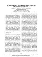
Local Recurrence
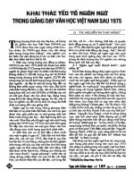
Among patients treated with limb-preservation intent,
loco-regional recurrences occurred in 12 patients, 4 in
each treatment group. At 3 years, freedom from local
recurrence was 84%, 88%, and 96% for SA, NR, and
NCR respectively (Figure 1; p = 0.88). Logistic regres-
sion analysis of factors associated with loco-regional
recurrence found no associat ion between age at diagno -
sis (p = 0.72) or tumor site (upper extremity vs. lower
extremity; p = 0.2) and recurrence risk. Patients present-
ing with recurrent disease (OR 11.6; p = 0.01) and
tumor size greater than 5 cm (OR 9.4; p = 0.01) were
more li kely to have a loco-regional recurrence on logis-
tic regression analysis. None of the ten patients treated
with IMRT have developed a local recurrence, but any
possible differences in local control based on radiation
technique did not reach statistical significance (p =
0.43).
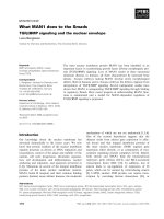
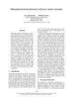
Distant Metastases
Metastatic disease developed in 30 patients. Three-year
DMFS was 83%, 68%, and 58% for patients treated with
SA, NR, and NCR, respectively, but these were n ot sta-
tistically significant differences (Figure 2; p = 0.27).
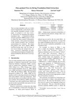
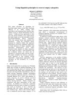
DMFS was significantly inferior at 3 years for patients
treated with SA for recurrent disease (60%) compared to
patients treated with SA for primary d isease (94%; Fig-
ure 3; p = 0.03). In contrast, no differences in DMFS for
patients with relapsed or primary disease treated with
NCR or NR could be found.
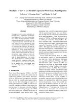
Overall survival
The median OS was 54.7 months (95% CI; range 41.6 to
96.4 months). No significant differences in OS were
observed among the treatment groups (Figure 4). Three-
year OS was 59%, 67%, and 73% for SA, NR, and NCR,
respectively (p = 0.58). For patients with tumors greater
than 5 cm, superior OS was observed for patients trea-
ted with NCR versus SA (3-year OS 69 vs. 40%; p =
0.03; Figure 5). OS also appeared improved for patients
with tumors greater than 5 cm treated with NR versus
SA (3-year OS 63 vs. 40%; p = 0.02; Figure 5). There
was no difference in OS among patients with tumors
greater than 5 cm treated with NCR compared to NR (p
=0.57).Table3summarizestheLR-RFS,DMFS,OS,
and limb preservation rates by treatment modality, w ith
an additional summary of these outcomes by primary or
recurrent disease status.
Toxicity and wound complications
Any-toxicity recorded was significan tly higher among
NCR-treated patients (21 of 39 patients, 54%) compar ed
to NR-treated patients (10 of 37 patients, 27%; p =
0.02). No toxicity was documented among SA-treated
Table 2 Outcomes of surgical resections among 92 high-
grade, Stage II and III soft-tissue sarcoma cases treated
with limb preservation
RESECTION TYPE NCR NR SA
R0 32 30 19
R1 3 5 3
p = 0.55 NCR-SA; p = 0.45 NCR-NR; p = 0.95 NR-SA
NCR, neoadjuvant chemoradiation; NR, neoadjuvant radiation alone; SA,
surgery alone; R0, surgical resection with microscopically negative margins;
R1, surgical resection with margins involved microscopically.
Curtis et al. Radiation Oncology 2011, 6:91
/>Page 4 of 11
patients, significantly less when compared to toxicity
among NCR-treated patients (p < 0.0001). The most
common toxicity among NCR-treated patients was wet
desquamation in the EBRT field and gastrointestinal
toxicity (nausea) from chemotherap y, each in 5 patients.
Wet desquamation occurred in 4 patients treated with
NR. Other toxicities observed in NCR-treated patients
included myelosuppression (n = 2), electrolyte imbal-
ance (n = 1), elevated liver biochemistries (n = 1), ifosfa-
mide-related encephalopathy (n = 1), and venous
thromboembolism (n = 1). No long term complications
were documented.
Wound complications occurred in 19 of 38 (50%)
NCR-treated patients (1 had limb amputation), 15 of 36
(42%) NR-treated patients, and 4 of 36 (11%) SA-treated
patients (1 had limb amputation). Excluding patients
treated with limb amputation, the rate of wound com-
plications was significantly higher among the NCR-trea-
tedgroupcomparedtoSA(p=0.003;Table4).Italso
was higher a mong the NR-treated group compared to
SA (p = 0.02). Wound complication rates were not sig-
nificantly differen t between NR and NCR groups for
patients treated with limb p reservation (p = 0.36). The
majority of wound complications occurred among lo wer
extremity tumors in e ach group (34 of 38 total wound
complications). Significantly more limb-preservation
patients who were treated with NCR and IOERT/perio-
perative brachytherapy had wound complications (16 of
30 patients, 53%) compared to NR-treated patients trea-
ted with IOERT/perioperative brachytherapy (11 of 25
patients, 44%, p = 0.009). However, using logistic regres-
sion analysis, no significant associations were found
between the incidence of wound complications and the
use of NCR or NR (OR 3.39; p = 0.21), use of IOERT or
perioperative brachytherapy (OR 4.61; p = 0.21), or
tumor size (OR 1.06; p = 0.37; Table 5).
Discussion
The primary treatment for Stage II and III extremity
STS is typically surgery combined with pre- or post-
operative radiation. Chemotherapy remains a co ntrover-
sial component of management. Based on the results of
this study, NCR does not appear to improve outcomes
compared to NR.
Patients at risk
Time
(mos)
10
20
30
40
50
60
70
80
90
SA
18
17
10
8
7
5
4
3
2
NCR
28
17
14
9
6
3
2
2
1
NR
29
20
18
14
12
9
9
4
1
0.0
0.1
0.2
0.3
0.4
0.5
0.6
0.7
0.8
0.9
1.0
0
10
20
30
40
50
60
70
80
90
100
Months
Proportion event-free
___
Surgery alone
___
Neoadjuvant chemoradiation
___
Neoadjuvant radiation alone
p = 0.88
Figure 1 Loco-regional relapse free survival. Kaplan-Meier plot of 92 Stage II and III extremity soft-tissue sarcoma patients treated with limb-
preservation by treatment modality (surgery alone, neoadjuvant chemoradiation, or neoadjuvant radiation alone).
Curtis et al. Radiation Oncology 2011, 6:91
/>Page 5 of 11
Neither NCR nor NR appeared to improve LR-RFS
compared to SA. Previous phase III randomized trials
have shown pre- and post-operative EBRT [14-19] and
peri-operative brachytherapy [20-22] improve LR-RFS
compared to SA. Our findings are likely impacted by
the h igh degree of pre-treatment patient selection. Fac-
tors such as tumor grade, large size, and location rela-
tive to neuro-vascular structures or bone typically
prompt referra l for multimodality pre-operative therapy.
Accordingly, given that patients in the NCR and NR
cohorts had significantly larger tumor sizes and were
more likely to undergo periosteal or nerve stripping, the
equivalent local control likely reflects the benefit of
neoadjuvant therapy to SA, but also lessens the likeli-
hood of finding a significant improvement in local con-
trol with neoadjuvant treatment. No patients treated
with IMRT experienced loco-regional recurrence, but no
definitive conclusio ns can be made with regards to
radiation technique and local failure. IMRT has pre-
viously been demonstrated to result in equivalent or
possibly superior local control compared to conventional
radiation planning [23].
NCR did not improv e the R0 resection rate compared
to NR or S A. This finding is similar to a randomized
trial of NR followed by surgery versus surgery with
post-operative radiation [15]. In that study, negative
microscopic margins were seen in 83% of patients trea-
ted with NR and 85% of patients treated with post-
operative r adiation, suggesting no difference in surgical
outcome with either strategy [15]. Therefore, as in pre-
vious studies, we are unable to demonstrate an improve-
ment in surgical outcomes with pre-operative therapy.
No improvement in DMFS or OS was detected with
NCR compared to SA or NR. Due to the heterogeneity
of chemotherapy regimens used in this study cohort, we
are unable to determine which, if any, chemotherapy
regimen added to pre-operative radiation is optimal for
impacting DMFS. Additionally, we cannot conclude
which, if any, chemotherapy regimen added to pre-
operative radiation might impact OS. The 5-year OS
Patients at Risk
Time
(mos)
10
20
30
40
50
60
70
80
90
SA
24
22
14
12
10
7
6
4
4
NCR
25
15
10
7
6
3
3
3
2
NR
26
19
18
13
11
9
9
4
2
0.0
0.1
0.2
0.3
0.4
0.5
0.6
0.7
0.8
0.9
1.0
0
10
20
30
40
50
60
70
80
90
100
Surgery alone
Neoadjuvant
chemoradiotherapy
Neoadjuvant
radiation alone
p = 0.27
Months
Surgery alone
Neoadjuvant chemoradiation
Neoadjuvant radiation alone
p = 0.27
Proportion event-free
A
Figure 2 Distant metastasis free survival. Kaplan-Meier plot of 112 Stage II and III extremity so ft-tissue sarcoma patient s treated with surgery
alone, neoadjuvant chemoradiation, or neoadjuvant radiation alone.
Curtis et al. Radiation Oncology 2011, 6:91
/>Page 6 of 11
with NCR we found initially appears inferior to other
studies o f NCR and NR, in which 5-year OS up to 90%
has been reported [8,24]. However, one analysis reported
OS of 66% at 5 years for patients with tumors measur-
ing 6 -10 cm [24]. Therefore, the apparently inferior OS
we observed with NCR compared to other studies likely
is due to selection of higher risk patients with a larger
median tumor size in our cohort. As in previous studies,
addition of radiation to surgery does not appear to
impact OS compared to SA [14,20,25].
We are unable to conclude whether pre-operative
treatment with either NCR or NR improves limb preser-
vation rate. A higher rate of limb amputations among
SA-treated patients was observe d compared to the NCR
and NR groups. How ever, most of these SA-treated
patients were deemed poor limb preservation candidates
at presentation. Therefore, conclusions cannot be made
as to whether a neoadjuvant strategy improved limb pre-
servation. Differences in limb preservation rates between
NCR and NR were not detected, making it unclear if
the addition of chemotherapy to pre-operative therapy
improves limb preservation outcomes. Logistic regres-
sion analysis showed that patients with recurrent disease
treated with limb amputation were not more likely to
have received previous chemotherapy or radiation than
patients undergoing limb preservation for recurrent dis-
ease. Thus, many relapsed patients treated with SA pos-
sibly could have received NCR or NR, but it is likely
that their disease presentation itself precluded functional
limb-preservation.
A possible advantage of pre-operative treatment is the
improvement in OS observed among patients with
extremity STS larger than 5 cm. When compared to SA,
OS was improved significantly both by NCR and NR in
this subset of patients. However, no difference in OS
was found between NCR and NR-treated patients with
extremity STS larger than 5 cm, suggesting that the OS
benefit may be derived mainly from pre-operative radia-
tion therapy rather than from chemotherapy. No r ando-
mized c ontrolled trials have compared NCR to SA,
although previous studies failed to demonstrate an OS
benefit when radiation was added t o surgery versus SA
[14,20,25]. Thus, the potential OS advantage for patien ts
with large extremity STS treated pre -operatively, as sug-
gested by our data, is intriguing, and should be con-
firmed prospectively. Caution must be used when
Patients at risk
Time
(mos)
10
20
30
40
50
60
70
80
90
Primary
19
17
11
10
9
6
5
3
3
Relapse
6
5
4
3
2
2
2
2
2
0.0
0.1
0.2
0.3
0.4
0.5
0.6
0.7
0.8
0.9
1.0
0
10
20
30
40
50
60
70
80
90
100
Months
Primary Disease
Relapsed Disease
p = 0.27
0.0
0.1
0.2
0.3
0.4
0.5
0.6
0.7
0.8
0.9
1.0
0
10
20
30
40
50
60
7
80
90
100
Primary Disease
Relapsed Disease
p = 0.03
Proportion event-free
B.
Figure 3 Distant metastasis free survival. Kaplan-Meier plot of 36 patients treated with surgery alone for primary versus relapsed disease.
Curtis et al. Radiation Oncology 2011, 6:91
/>Page 7 of 11
interpreting this finding, since only 12 patients with
extremity STS larger than 5 cm were treated with SA.
An inferior DMFS was observed among patients pre-
senting with recurrent disease treated with SA compared
to patients wit h primary disease treated with SA. This
result suggests that patients presenting with recurrent
extremity STS likely have micrometastases at the time
of relapse. Such patients might benefit from more
aggressive multi-agent chemotherapy either pre- or
post-operatively. An improvement in DMFS for recur-
rent patients given chemotherapy could not be demon-
strated in this analysis, although the exceedingly small
number of relapsed patients (n = 2) treated with NCR
greatly limits our ability to make conclusions about the
value of chemotherapy for improving DMFS in these
patients. Further analyses of outcomes among a higher
number of patients with recurrent disease should be
conducted to determi ne whether chemotherapy is bene-
ficial in this subgroup of patients.
Potential drawbacks of NCR are increased toxicity and
wound complication rates. In a phase III trial of pre-
versus post-operative radiation without chemotherapy,
wound complications occurred in 35% of patients trea-
ted with pre-operative radiation therapy [15]. While
wound complication rates of just 7.5% have been
reported with intra-arterial doxorubici n and radiation in
single institution experience [13], a multi-center trial of
intra-arteri al doxorubicin with radiation reported a 41%
wound complication rate [9]. Logistic regression analysis
did not find a significant association between use of
NCR or NR and wound complications, nor with use of
IOERT/perioperative brachytherapy. Additionally, we
found n o significant difference in the wound complica-
tion rate between NCR and NR. We cannot conclude
that NCR worsens the wound complications rate based
on these results. The apparent higher rate of wound
complications we observed may be attributable to differ-
ent definitions of wound complications among studies.
Due to small patient numbers, it is not entirely clear
that the observed rate of wound complications in our
study is significantly different than rates reported in
other studies. Working closely with our plastic surgery
Patients at risk
Time
(mos)
10
20
30
40
50
60
70
80
90
SA
27
22
15
12
9
6
5
4
4
NCR
30
18
15
10
8
4
4
4
2
NR
32
21
19
16
13
10
9
5
2
0.0
0.1
0.2
0.3
0.4
0.5
0.6
0.7
0.8
0.9
1.0
0
10
20
30
40
50
60
70
80
90
100
Months
Surgery alone
Neoadjuvant
chemoradiotherapy
Neoadjuvant
radiation alone
p = 0.58
Surgery alone
Neoadjuvant chemoradiation
Neoadjuvant radiation alone
p = 0.58
Proportion event-free
A.
Figure 4 Overall survival. Kaplan-Meier plot of 112 Stage II and III extremity soft-tissue sarcoma patients treated with surgery alone,
neoadjuvant chemoradiation, or neoadjuvant radiation alone.
Curtis et al. Radiation Oncology 2011, 6:91
/>Page 8 of 11
colleagues, we have not appreciated long-term negative
impacts on function or quality of life in patients who
experience wound complication s. Beyond w ound com-
plications, the overall degree of toxicity associated with
NCR appeared higher compared to NR. However, we
were unable to grade toxicities from medical records,
and due to inconsistencies in documentation, the
increased rate of any-toxicity with NCR reported here
must be viewed with caution. Our group is actively pur-
suing further analyses of wound complications in order
to better understand these findings and improve
practice.
There are several limitations to this study. Foremost is
its retrospective nature, which may lead to biased results
Patients at risk
Time
(mos)
10
20
30
40
50
60
70
80
SA
7
7
5
4
0
0
0
0
NCR
26
15
12
8
6
3
3
3
NR
21
14
13
12
10
7
6
3
0.0
0.1
0.2
0.3
0.4
0.5
0.6
0.7
0.8
0.9
1.0
0
10
20
30
40
50
60
70
80
90
Months
p = 0.03
Surgery alone
Neoadjuvant chemoradiation
Neoadjuvant radiation alone
Proportion event-free
B.
Figure 5 Overall survival. K aplan-Meier plot of 70 patients with tumors greater than 5 cm treated with surgery alone, neoadjuvant
chemoradiation, or neoadjuvant radiation alone.
Table 3 Treatment outcomes with regard to overall survival, disease relapse (local, distant) and limb preservation by
treatment method and disease presentation among 112 Stage II/III extremity soft-tissue sarcoma cases
Treatment/Disease
Presentation
No.
Pts
Survival
Median (mos)
Overall Survival (%) Local recurrence (%) Distant Metastases (%) Limb Preserved
3-yr 5-yr P No (%) 3-yr p No. (%) 3-yr P No. (%) P
SA 36 41.9 59 34 4 (11) 84 6 (17) 83 22 (61)
Primary 23 51.9 68 35 0.23 1 (4) 95 0.02 2 (9) 94 0.03 18 (78) 0.005
Recurrent 13 24.3 39 39 3 (23) 47 4 (31) 60 4 (31)
NR 37 74.4 67 57 4 (11) 88 11 (30) 68 1 (3)
Primary 31 74.3 67 61 0.75 2 (6) 94 0.006 9 (29) 71 0.63 29 (94) 0.54
Recurrent 6 37.4 67 * 2 (33) 56 2 (33) 50 6 (100)
NCR
39 * 73 59 4 (10) 85 13 (34) 58 34 (87)
SA, surgery alone; NR, neoadjuvant radiation alone; NCR - neoadjuvant chemoradiation; *, not reached; ¶, only 2 patients in NCR group had recurrent disease and
have not developed local recurrence, distant metastases and were living at time of analysis.
Curtis et al. Radiation Oncology 2011, 6:91
/>Page 9 of 11
because of potential imbalances in the treatment groups
being compared. Secondly, we studied a diverse mixture
of patients, with differing primary disease sites, limiting
conclusions as to which primary disease location might
benefit most from neoadjuvant therapy. Furthermore,
any conclusion as to which chemotherapy regi men may
be optimal is limited by the relatively small numbers o f
patients were treated over the 11-year period with var-
ious chemotherapy agents and schedules.
Conclusions
Despite the limitations of the methodology, the results
of this study have merit. We conclude that both NCR
and NR result in a low rate of loco-regional relapse,
high rates of limb preservation, and acceptable toxicity.
The improved OS of patients with tumors greater than
5 cm treated with pre-operative therapy (both with NCR
and NR) compared to patients with tumors greater than
5 cm receiving SA is compelling. We continue to track
outcomes of patients treated with weekly cisplatin given
with radiation, but cannot make conclusions about its
effectiveness from the available data at this time.
Wound complications remain an important manage-
ment issue for patients treated with a pre-operative
strategy, but NCR did not significantly increase the risk
of wound complications compared to NR.
In addition to cure, goals of extremity STS therapy
include limb preservation, minimizing treatment-related
toxicity, and maximizing quality of life both during and
after treatment. The results of this analysis suggest that
NCR and NR appear to be effective strategies for Stage
II and III STS, perhaps with improved outcomes com-
pared to SA, but NCR is not clearly superior to NR.
List of Abbreviations
STS: soft tissue sarcoma; NCR: neoadjuvant chemoradiation; MAID: mesna,
doxorubicin, ifosfamide and dacarbazine; IMAP/MAP: ifosfamide, mitomycin,
doxorubicin and cisplatin; MCA: Mayo Clinic in Arizona; NR: neoadjuvant
radiation; SA: surgery alone; MRI: magnetic resonance imaging; AJCC:
American Joint Committee on Cancer; IOERT: intra-operative electron
radiation therapy; OS: overall survival; LR-RFS: loco-regional recurrence-free
survival; DMFS: distant metastases-free survival; EBRT: external beam
irradiation; IMRT: intensity modulated radiation therapy.
Acknowledgements
The authors thank Jorge Rakela, MD and James A. Wilkens, MD, Department
of Internal Medicine, and Steven E. Schild, MD, Department of Radiation
Oncology.
Author details
1
Department of Internal Medicine, Division of Hematology/Oncology, Mayo
Clinic, 13400 East Shea Blvd., Scottsdale, AZ 85259, USA.
2
Department of
Radiation Oncology, Mayo Clinic, 13400 East Shea Blvd., Scottsdale, AZ
85259, USA.
3
Department of Surgery, Division of Orthopedic Surgery, Mayo
Clinic, 5779 East Mayo Blvd., Phoenix, AZ 85054, USA.
4
Division of Biomedical
Statistics and Informatics, Mayo Clinic, 13400 East Shea Blvd., Scottsdale, AZ
85259, USA.
Authors’ contributions
All authors have read and approved the final manuscript.
KKC was involved in clinical care of patients included in the data set,
conceived of the study, collected and analyzed data, and helped draft the
manuscript.
JBA was involved in clinical care of patients included in the data set,
conceived of the study, collected and analyzed data, and helped draft the
manuscript.
CPB was involved in clinical care of patients included in the data set and
reviewed the manuscript.
AJS was involved in clinical care of patients included in the data set and
helped draft the manuscript.
MDC was involved in clinical care of patients included in the data set and
helped draft the manuscript.
ACD assisted with statistical analysis of the data.
LLG was involved in clinical care of patients included in the data set and
helped draft the manuscript.
TRF was involved in clinical care of patients included in the data set and
reviewed the manuscript.
Competing interests
The authors declare that they have no competing interests.
Received: 22 March 2011 Accepted: 9 August 2011
Published: 9 August 2011
References
1. National Comprehensive Cancer Network. NCCN clinical practice
guidelines in oncology soft tissue sarcoma. [ />professionals/physician_gls/PDF/sarcoma.pdf].
2. Eilber FR, Morton DL, Eckardt J, Grant T, Weisenber T: Limb salvage for
skeletal and soft tissue sarcomas multidisciplinary preoperative therapy.
Cancer 1984, 53:2579-2584.
3. Cormier JN, Patel SR, Herzog CE, Ballo MT, Burgess MA, Feig BW, Hunt KK,
Raney RB, Zagars GK, Benjamin RS, Pisters PW: Concurrent ifosfamide-
based chemotherapy and irradiation analysis of treatment-related
toxicity in 43 patients with sarcoma. Cancer 2001, 92:1550-1555.
4. Pisters PW, Ballo MT, Bekele N, Thall PF, Feig BW, Lin P, Cormier JN,
Benjamin RS, Patel SR: Phase I trial using toxicity severity weights for
dose finding of gemcitabine combined with radiation therapy and
Table 4 Wound complications among 92 high-grade,
Stage II and III soft-tissue sarcoma cases treated with
limb preservation
TREATMENT TYPE NO. OF WOUND COMPLICATIONS SITE
NCR 18 16 LE, 2 UE
NR 15 13 LE, 2 UE
SA 3 3 LE, 0 UE
p = 0.003 NCR-SA wound complications; p = 0.02 NR-SA wound
complications; p = 0.36 NCR-NR wound complications;
NCR, neoadjuvant chemoradiation; NR, neoadjuvant radiation alone; SA,
surgery alone; LE, lower extremity; UE, upper extremity.
Table 5 Logistic regression analysis of factors associated
with wound complications
Variable OR p value
Age at diagnosis 1.00 0.8
Tumor size (cm) 1.06 0.31
Sex (male versus female) 1.12 0.82
Use of IOERT/Brachytherapy 4.61 0.21
Use of NCR or NR versus SA 3.39 0.21
Upper extremity versus lower extremity 0.47 0.33
IOERT, intra-operative electron radiation therapy; NCR, neoadjuvant
chemoradiation; NR, neoadjuvant radiation alone; SA, surgery alone.
Curtis et al. Radiation Oncology 2011, 6:91
/>Page 10 of 11
subsequent surgery for patients with extremity and trunk soft tissue
sarcomas [abstract]. J Clin Oncol 2004, 22:s820.
5. DeLaney TF, Spiro IJ, Suit HD, Gebhardt MC, Hornicek FJ, Mankin HJ,
Rosenberg AL, Rosenthal DI, Miryousefi F, Ancukiewicz M, Harmon DC:
Neoadjuvant chemotherapy and radiotherapy for large extremity soft-
tissue sarcomas. Int J Radiat Oncol Biol Phys 2003, 56:1117-1127.
6. Kraybill WG, Harris J, Spiro IJ, Ettinger DS, DeLaney TF, Blum RH, Lucas DR,
Harmon DC, Letson GD, Eisenberg B: Phase II study of neoadjuvant
chemotherapy and radiation therapy in the management of high-risk,
high-grade, soft tissue sarcomas of the extremities and body wall:
Radiation Therapy Oncology Group Trial 9514. J Clin Oncol 2006,
24:619-625.
7. Edmonson JH, Petersen IA, Shives TC, Mahoney MR, Rock MG, Haddock MG,
Sim FH, Maples WJ, O’Connor MI, Gunderson LL, Pritchard DJ, Bucnker JC,
Stafford SL: Chemotherapy, irradiation, and surgery for function-
preserving therapy of primary extremity soft-tissue sarcoma: initial
treatment with ifosfamide, mitomycin, doxorubicin, and cisplatin plus
granulocyte-colony-stimulating factor. Cancer 2002, 94:786-792.
8. Soulen MC, Weissman JR, Sullivan KL, Lackman RD, Shapiro MJ, Bonn J,
Weiss AJ, Gardiner GA Jr: Intraarterial chemotherapy with limb-sparing
resection of large soft-tissue sarcomas of the extremities. J Vasc Interv
Radiol 1992, 3:659-663.
9. Wanbeo HJ, Temple WJ, Popp MB, Constable W, Aron B, Cunningham SL:
Preoperative regional therapy for extremity sarcoma. A tricenter update.
Cancer 1995, 75:2299-2306.
10. Levine EA, Trippon M, Das Gupta TK: Preoperative multimodality
treatment for soft tissue sarcomas. Cancer 1993, 71:3685-3689.
11. Rossi CR, Vecchiato A, Foletto M, Nitti D, Ninfo V, Fornasiero A, Sotti G,
Tregnaghi A, Melanotte P, Lise M: Phase II study on neoadjuvant
hyperthermic-antiblastic perfusion with doxorubicin in patients with
intermediate or high grade limb sarcomas. Cancer 1994, 73:2140-2146.
12. Pisters PW, Ballo MT, Patel SR: Preoperative chemoradiation treatment
strategies for localized sarcoma. Ann Surg Onc 2002, 9:535-542.
13. Hegazy MA, Kotb SZ, Sakr H, El Dosoky E, Amer T, Hegazi RA, Farouk O:
Preoperative isolated limb infusion of Doxorubicin and external
irradiation for limb-threatening soft tissue sarcomas. Ann Surg Oncol
2007, 14:568-576.
14. Yang JC, Chang AE, Baker AR, Sindelar WF, Danforth DN, Topalian SL,
DeLaney T, Glatstein E, Steinberg SM, Merino MJ, Rosenberg SA:
Randomized prospective study of the benefit of adjuvant radiation
therapy in the treatment of soft tissue sarcomas of the extremity. J Clin
Oncol 1998, 16:197-203.
15. O’Sullivan B, Davis AM, Turcotte R, Bell R, Catton C, Chabot P, Wunder J,
Kandel R, Goddard K, Sadura A, Pater J, Zee B: Preoperative versus
postoperative radiotherapy in soft-tissue sarcoma of the limbs: a
randomized trial. Lancet 2002, 359:2235-2241.
16. Zagars GK, Ballo MT: Sequencing radiotherapy for soft tissue sarcoma
when re-resection is planned. Int J Radiat Oncol Biol Phys 2003, 56:21-27.
17. Zagars GK, Ballo MT, Pisters PW, Pollock RE, Patel SR, Benjamin RS:
Preoperative vs. postoperative radiation therapy for soft tissue sarcoma:
a retrospective comparative evaluation of disease outcome. Int J Radiat
Oncol Biol Phys 2003, 56:482-488.
18. Suit HD: The George Edelstyn memorial lecture: radiation in the
management of malignant soft tissue tumors. Clin Oncol (R Coll Radiol)
1989, 1:5-10.
19. Cheng EY, Dusenbery KE, Winters MR, Thompson RC: Soft tissue sarcomas:
preoperative versus postoperative radiotherapy. J Surg Oncol 1996,
61:90-99.
20. Pisters PW, Harrison LB, Leung DH, Woodruff JM, Casper ES, Brennan MF:
Long-term results of a prospective randomized trial of adjuvant
brachytherapy in soft tissue sarcoma. J Clin Oncol 1996, 14:859-69.
21. Brennan MF, Hilaris B, Shiu MH, Lane J, Magill GC, Friedrich C, Hajdu SI:
Local recurrence in adult soft-tissue sarcoma. A randomized trial of
brachytherapy. Arch Surg 1987, 122:1289-1293.
22. Harrison LB, Franzese F, Gaynor JJ, Brennan MF: Long-term results of a
prospective randomized trial of adjuvant brachytherapy in the
management of completely resected soft tissue sarcomas of the
extremity and superficial trunk. Int J Radiat Oncol Biol Phys 1993,
27:259-265.
23. Alektiar KM, Brennan MF, Healey JH, Singer S: Impact of intensity-
modulated radiation therapy on local control in primary soft tissue
sarcoma of the extremity. J Clin Oncol 2008, 26:3440-3444.
24. Parsons JT, Zlotecki RA, Reddy KA, Mitchell TP, Marcus RB Jr,
Scarborough MT: The role of radiotherapy and limb-conserving surgery
in the management of soft-tissue sarcomas in adults. Hematol Oncol Clin
North Am 2001, 15:377-388.
25. Rosenberg SA, Tepper J, Glastein E, Costa J, Baker A, Brennan M,
DeMoss EV, Seipp C, Sindelar WF, Sugarbaker P, Wesley R: The treatment of
soft-tissue sarcomas of the extremities: prospective randomized
evaluations of (1) limb-sparing surgery plus radiation therapy compared
with amputation and (2) the role of adjuvant chemotherapy. Ann Surg
1982, 196:305-315.
doi:10.1186/1748-717X-6-91
Cite this article as: Curtis et al.: Neoadjuvant chemoradiation compared
to neoadjuvant radiation alone and surgery alone for Stage II and III
soft tissue sarcoma of the extremities. Radiation Oncology 2011 6:91.
Submit your next manuscript to BioMed Central
and take full advantage of:
• Convenient online submission
• Thorough peer review
• No space constraints or color figure charges
• Immediate publication on acceptance
• Inclusion in PubMed, CAS, Scopus and Google Scholar
• Research which is freely available for redistribution
Submit your manuscript at
www.biomedcentral.com/submit
Curtis et al. Radiation Oncology 2011, 6:91
/>Page 11 of 11