Báo cáo khoa học: "Increased betulinic acid induced cytotoxicity and radiosensitivity in glioma cells under hypoxic conditions" pptx
Bạn đang xem bản rút gọn của tài liệu. Xem và tải ngay bản đầy đủ của tài liệu tại đây (490.68 KB, 9 trang )
RESEARC H Open Access
Increased betulinic acid induced cytotoxicity and
radiosensitivity in glioma cells under hypoxic
conditions
Matthias Bache
1*
, Martin P Zschornak
1†
, Sarina Passin
1†
, Jacqueline Keßler
1
, Henri Wichmann
1
, Matthias Kappler
1,2
,
Reinhard Paschke
3
, Goran N Kaluđerović
3
, Harish Kommera
3
, Helge Taubert
2,4
and Dirk Vordermark
1
Abstract
Background: Betulinic acid (BA) is a novel antineoplastic agent under evaluation for tumor ther apy. Because of the
selective cytotoxic effects of BA in tumor cells (including gliomas), the combination of this agent with conservative
therapies (such as radiotherapy and chemotherapy) may be useful. Previously, the combination of BA with
irradiation under hypoxic conditions had never been studied.
Methods: In this study, the effects of 3 to 30 μM BA on cytotoxicity, migration, the protein expression of PARP,
survivin and HIF-1a, as well as radiosensitivity under normoxic and hypoxic conditions were analyzed in the
human malignant glioma cell lines U251MG and U343MG. Cytotoxicity and radiosensitivity were analyzed with
clonogenic survival assays, migration was analyzed with Boyden cham ber assays (or scratch assays) and protein
expression was examined with Western blot analyses.
Results: Under normoxic conditions, a half maximal inhibitory concentration (IC
50
)of23μMwasobservedin
U251MG cells and 24 μM was observed in U3 43MG cells. U nder hypoxic conditions, 10 μMor15μMofBA
showed a significantly increased cytotoxicity in U251MG cells (p = 0.00 4 and p = 0.01, respectively) and
U343MG cells
(p < 0.05 and p = 0.01, respective ly). The combination of BA with radiotherapy resulted in an additive effect in
the U343MG cell line under normoxic and hypoxic conditions. Weak radiation enhancement was observed in
U251MG cell line after treatment with BA under normoxic conditions. Furthermore, under hypoxic conditions,
the incubation w ith BA resulted in increased radiation enhancement. The enhancement fac tor, at an irradiation
dose of 15 Gy after treatment with 10 or 15 μM BA, was 2.20 (p = 0.02) and 4.50 (p = 0.03), respectively.
Incubation with BA led to decrease d cell migration, cleavage of PARP and decre ased expression levels of
survivin in both cell line s. Additionally, BA treatment resulted in a reduction of HIF-1a protein under hypoxic
conditions.
Conclusion: Our results suggest that BA is capable of improving the effects of tumor therapy in human malignant
glioma cells, particularly under hypoxic conditions. Further investigations are necessary to characterize its potential
as a radiosensitizer.
Keywords: betulinc acid, glioma cells, hypo xia, irradiation
* Correspondence:
† Contributed equally
1
Department of Radiotherapy, Martin-Luther-University Halle-Wittenberg,
Dryanderstr. 4, 06110 Halle, Germany
Full list of author information is available at the end of the article
Bache et al. Radiation Oncology 2011, 6:111
/>© 2011 Bache et al; licensee BioMed Central Ltd. This is an Open Access article distributed under the terms of the Creative Commons
Attribution Licens e ( which permits unrestricted use, distribution, and reproduction in
any medium, provided the original work is properly cited.
Background
Glioblastoma is the most frequent primary brain tumor
and is characterized by a poor patient prognosis.
Although radiotherapy is widely used for the treatment
of patients with glioblastoma, the intrinsic radioresis-
tance of these tumors remains a critical problem in the
management of such patients [1]. Betulinic acid (BA)
represents a new therapeutic agent with possible use in
the treatment of glioblastoma. BA, a pentacyclic triter-
pene, can be synthesized by the oxidation of betulin, a
substance found in the bark of birch trees. Additionally,
it can also be directly isolated from certain plants. BA
has several therapeutic uses. It has been used to treat
inflammation, malaria, HIV and as an antimicrobial
drug. In addition, BA seems capable of improving tumor
therapies. For example, BA is cytotoxi c in different
tumor cell lines, including neuroectodermal tumors,
melanoma, colon, lung and ovarian carcinoma, head and
neck cancers and sarcoma [2-4]. Experiments in animals
revealed that BA also has an antitumor effect in vivo
[5-7]. Interestingly, the cytotoxicity of BA in tumor cells
occ urs regardless of w hether there is a genetic defect in
p53 [6,8]. Remarkably, untransformed, normal cells (in
comparison to tumor cells) seem to t olerate relatively
high concentrations of BA. Thus, BA is not toxic up to
a concentration of 200-400 mg/kg of body weight in
rats or 500 mg/kg of body weight in mice [5,9].
Different studies have shown that BA induces apopto-
sis [8,10-13]. In addition, BA’s effects on cell migration,
cell invasion and angiogenesis inhibition have been
demonstrated [1 4-16]. Furthermore, reactive oxygen
radicals generated by BA have been shown to cause sig-
nificant DNA damage [17-19]. The finding that BA can
both induce the formation of reactive oxygen radicals
and induce apoptosis could make it attractive for the
treatment of hypoxic tumors. The role of hypoxia in
developing a more aggressive t umor phenotype in
glioma has been previously discussed [20-22]. Because
of the selective and wide-range cytotoxic effects of BA
in tumor cells, the combination of BA with cons ervative
therapies (such as radiotherapy and chemotherapy)
seemed like a promising therapeutic strategy to investi-
gate. Indeed, investigations have shown that BA
enhances the cytotoxic effects of vincristine in the
B16F10 melanoma cell line [7]. Additionally, sublines of
SNU-C5 colon cancer cells that are resistant to che-
motherapy showed a significantly increased cytotoxicity
when either 5-fluorouracil, irinotecan, or oxaliplatin
were combined with BA treatment [23]. Two studies
examining the combinati on of BA and radi otherapy (in
melanoma or head and neck cancer cell lines) detected
an additive effect on clonogenic survival [24,25]. How-
ever, there have been no studies examining BA treat-
ment in combination with irradiation under hypoxic
conditions. In thi s study, we analyzed t he effects of BA
on the cytotoxicity, migration, protein expression of
PARP, survivin and HIF-1a and rad iosensitiv ity under
normoxic and hypoxic conditions in the radioresistant
glioma U251MG and U343MG cell lines.
Methods
Cell culture conditions, treatments with BA and
irradiation
The human malignant glioma cell lines U251MG and
U343MG (American Type Culture Collection) were
grown in RPMI 1640 medium (Lonza, Walkersville, MD,
USA) containing 10% fetal bovine serum (PAA, Cölbe,
Germany), 1% sodium pyruvate (Invitrogen, Karlsruhe,
Germany), 185 U/ml penicillin (Invitrogen) and 185 μg/
ml streptomycin (Invitrogen) at 37°C in a humidified
atmosphere containing 3% CO
2
. Hypoxia (< 1% O
2
) was
achieved using a gas generator system as previously
described [26]. All experiments were performed with
cells in their logarithmic growth phase. BA (Biosolution
GmbH, Halle, Germany) was dissolved in dimethyl sulf-
oxide (DMSO) to achieve a 20 mM stock solution. Cells
(3 × 10
5
) were seeded in 25 cm
2
flasks 24 h before treat-
ment with 3 to 30 μM BA. Cells were treated with BA
or DMSO for 24 h at 37°C under normoxic or hypoxic
conditions. Additionally, cells were irradiated in tissue
culture flasks (Greiner, Frickenhausen, Germany) with 2,
6or15Gy24hafterincubationwithBA.Irradiation
was accomplished with 6 MV photons and adequate
bolus material on a SIEMENS ONCOR (Erlangen, Ger-
many) linear accelerator at a dose rate of 2 Gy/min. At
1, 24 or 48 h after irradiation, cells were harvested for
clonogenic assays, protein extraction and migration
assays.
Clonogenic survival assays and radiosensitivity
The cytotoxicity of BA was ev aluate d using the clono-
genic survival assay. The cells were trypsinized 1 h after
irradiation. Based on the optimal plating efficacy
(depending on the BA treatment and irradiation dose),
500-5,000 cells were seeded in 25 cm
2
flasks. The cells
were cultured in RPMI supplemented with 10% FCS in
a humidified atmosphere of 3% CO
2
at 37°C. The med-
ium was changed after 5 days. Between 10 and 14 days
after irradi ation, the cells were fixed with paraformalde-
hyde (Sigma, Deisenhofen, Germany), and colony forma-
tion was visuali zed by staining with 10% Giemsa
solution (Sigma, Deisenhofen, Germany). Only colonies
with > 50 cells were scored to determine the surviving
fraction (SF). The cytotoxicity of BA was defined as the
ratio of colonies formed after treatment with different
concentrations of BA to DMSO-treated control cells.
The SF was defined as the ratio of colonies formed after
irradiation with 0, 2, 6 or 15 Gy to the number of
Bache et al. Radiation Oncology 2011, 6:111
/>Page 2 of 9
colonies formed in the unirradiated controls. The
enhancement factor (EF) was define d as the ratio of the
SF of BA-treated cells to DMSO-treated control cells
dependent on the dose of irradiation. The data represent
at least three independent experiments.
Migration assays and cell cycle analysis
Cell migration was assessed using modified Boyden
chambers as previously described [27]. Cells (2 × 10
4
)
were suspended in 300 μl of RPMI without FCS and
were then added to the upper chamber (membrane filter
with 8 μm pore size), while the bottom chamber was
filled with 1 ml of RPMI supplemented with 20% FCS
(as a chemoattractant). T he assay was performed at 37 °
C in a humidified atmosp here containing 3% CO
2
for at
least 16 h. Non-migrating cells on the upper side of the
transwell inserts were removed. The cells that had
migrated to the bottom side of the membrane were
trypsinized and counted with CASY DT (Schärfe System
GmbH, Reutlingen, Germany). The data represent at
least three independent experiments.
Furthermore, we used a wound scratch assay to deter-
mine the migration of cells after treatment with BA.
Cells were grown in 6-well cell culture plates in RPMI
medium containing 10% FCS and were cultured to 100%
confluence. A uniform cell-free area was created by
scratching the confluent monolayer with a 200 μl pipette
tip. To determine the migration of glioma cells, the
wound closure was observed at different time points.
The wound scratch assay was performed three times in
independent experiments.
Cells were analyzed for cell cycle distribution. About 5
×10
5
cells were harvested and washed in PBS. Subse-
quently, 95% ethanol was added slowly until a final con-
centration of 80% was reached. The DNA content,
which was indicated by the extent of staining of propi-
dium iodide, was measured by flow cytometry in an
FACSscan (Becton D ickinson, Heidelberg, Germany),
using the CellFit software (Version 2.0).
Western blotting
Cells were washed, trypsinized and centrifuged. The
supernatant of cells was washed with PBS and r esus-
pended in 100 μloflysisbuffer(50mMTrisatpH8.0,
0.3 M NaCl, 1 mM EDTA, 0.5 mM dithiothreitol, 0.1%
NP40 and protease inhibitors), followed by ultrasonic
homogenization. After centrifugation at 14,000 g for 15
min, the supernatant was collected and the protein con-
centration was determined using the Bradford assay
(BioRad, Munich, Germany). About 30 μg of t otal pro-
tein from each cell lysate was separated on a 10%
NuPAGE Bis-Tris (Invitrogen) gel that was placed in an
X-C ell SureLock Mini-Cell (Invitrogen). The memb rane
was blocked with 10% non-fat milk in TBST (50 mM
NaCl,30mMTris-HClatpH8.0and0.1%Tween)for
1 h and incubated with rabbit anti-human survivin anti-
body (1:1, 000 dilution, clone AF 886, R&D Systems,
Wiesbaden, Germany), rabbit anti-human cleaved PARP
(1:2,000, Cell Signaling, Danvers, MA, USA), mouse
anti-human HIF1a antibody (1:1,000, BD Transd uctio n
Laboratories, Lexington, KY) and mouse anti-b-actin
(1:5,000, Sigma, Deisenhofen, Germany) at 4°C over-
night. After washing, the membranes were incubated
with a horseradish peroxidase-labeled goat anti-rabbit or
anti-mouse IgG (1:2,000, DAKO, Glostrup, Denmark)
for 1 h at room temperature. For protein detection,
membranes were incubated with ECL substrate or ECL
Plus Blotting Detect ion System for 1 min (Amersham
Pharmacia Biotech, F reiburg, Germany) and exposed to
X-ray film (Biomax, Kodak, Braunschweig, Germany).
Statistical analyses
The experimental results were analyzed by paired Stu-
dent’ s t-tests. A p-value of 0.05 was considered to be
significant.
Results
Effects of BA on clonogenic survival
The effects of BA on the cl onogenic survival, cell migra-
tion, cell cycle, protein expression and radiosensitivity in
U251MG and U343MG glioma cell lines under nor-
moxic and hypoxic conditions were determined. With
higher concentrations (from 3 - 30 μM), a decline in
clonogenic survival was observed, with an IC
50
of 23
μM in U251MG cells and 24 μM in U343MG cells
under normoxic conditions after an incubation time of
24 h (Figure 1). In addition, longer incubation with BA
led to increased cytotoxicity in both cell lines (data not
shown). Additionally, incubation of BA caused a n
increase of subG1-cells in both cell lines. However, we
did not find effects of BA on cell distribution in other
cell cycle phase (data not shown). Under hypoxic condi-
tions, BA had significantly increased cytotoxicity in both
glioma cell lines (Figure 2). After a 24 h incubation with
10 μMor15μM BA under normoxic conditions, the
clonogenic survival was reduced to 79% (p = 0.07) or
57% (p = 0.03) in U251MG cells and 87% (p = 0 .15) or
82% (p = 0.07) in U343MG cells, respectively. Under
hypoxic conditions, an increased reduction in survival to
30% (p = 0.01) or 9% (p = 0.03) and 46% (p = 0.10) or
0.8% (p = 0.03), respectively, was detected (Figure 2).
Effects of BA on cell migration and protein expression
The effects of BA on the migration rates of both glioma
cell lines were determined with the Boyden chamber
assay a nd the scratch assay. Decreased migration rates
were detected after incubation with a higher concentra-
tion of BA in both cell lines. Compared to DMSO-
Bache et al. Radiation Oncology 2011, 6:111
/>Page 3 of 9
0
20
40
60
80
100
120
0102030
betulinic acid [
μ
M]
c
l
onogen
i
c surv
i
va
l
[%
]
U251MG
U343MG
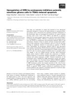
Figure 1 Cytotoxicity in U251MG and U343MG cell lines after treatment with BA. Both glioma cell lines were treated with increasing doses
of BA from 3 - 30 μM. The half maximal inhibitory concentration (IC
50
) with an incubation time of 24 h was 24 μM in U343MG cells and 23 μM
in U251MG cells. Data represent mean values (± SD) of three independent experiments.
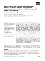
Figure 2 Effects of BA on clonogenic survival of glioma cells under normoxic or hypoxic conditions. Clonogenic survival in U251MG (A)
and U343MG (B) cells after treatment with 10 or 15 μM BA under normoxic or hypoxic conditions. Under hypoxia, when compared to normoxic
conditions, BA showed increased cytotoxicity in both glioma cell lines. Data represent mean values (± SD) of three independent experiments (*
p < 0.05).
Bache et al. Radiation Oncology 2011, 6:111
/>Page 4 of 9
treated control cells, incubation with 5, 10 and 20 μM
BA led to decreased cell migration rates in U251MG
cells to 92% (p = 0.21), 87% (p = 0.12) and 67% (p =
0.09), or in U343MG cells to 93% (p = 0.10), 70% (p =
0.20) and 53% (p = 0.08), respectively, under normoxic
conditions (Figure 3A). Similarly, reduced migration
rates were found after cells were incubated with BA in
the scratch assay (Figure 3B).
Using Western blot analysis, we examined the clea-
vage of PARP (as an indicator for the induction of apop-
tosis) and the expression of survivin (as an inhibitor of
apoptosis) (Figure 4). Incubation with 20 or 25 μMBA
led to PARP’ s cleavage, and to a decrease in survivin
levels under normoxic conditions. Additionally,
increased PARP’ s cleavage and a decrease in survivin
protein levels were observed after treatment with 10 or
15 μM BA i n the U251MG cells under hypoxic condi-
tions. BA also decreased hypoxia-induced levels of the
HIF-1a protein in both cell lines (Figure 4). However,
combination of BA with radiotherapy showed no addi-
tional effects on PARP cleavage or the expression of sur-
vivin under normoxic or hypoxic conditions.
Effects of BA on radiosensitivity
Irradiation at 2 Gy reduced clonogenic survival to 70%
(SF2 = 0.70) in U251MG cells and 71% (SF2 = 0.71) in
0
20
40
60
80
100
U251MG U343MG
relative rate of
migration [%]
untreated
DMSO
5 μM BA
10 μM BA
20 μM BA
U
251MG
U
343MG
DMSO 5 μM BA 10μM BA
A
B
0
20
40
60
80
100
U251MG U343MG
relative rate of
migration [%]
untreated
DMSO
5 μM BA
10 μM BA
20 μM BA
U
251MG
U
343MG
DMSO 5 μM BA 10μM BA
Figure 3 Effects of BA on cell migration of glioma cells. Migration rates of U251MG and U343MG cells treated with BA analyzed by Boyden
chamber assays (A) and scratch assays (B) under normoxic conditions. Compared to DMSO-treated control cells, incubation with 5, 10 and 20
μM BA led to a decrease in cell migration rates in both glioma cell lines. Similarly, cells had a reduced migration rate after BA treatment as
measured by the scratch assay. Data represent the average values (± SD) of three independent experiments.
Bache et al. Radiation Oncology 2011, 6:111
/>Page 5 of 9
U343MG cells under normoxic conditions. Irradiation-
induced clonogenic survival of U251MG and U343MG
cells was increased under hypoxic conditions when com-
pared to normoxic conditions (Figure 5). The combina-
tion of BA with radiotherapy resulted in an additive
effect for U343MG cells under normoxic and hypoxic
conditions. However, a weak not significant radioprotec-
tive effect was observed at 10 μMBAunderhypoxic
conditions. In addition, a weak radiation enhancement
was observed for U251 MG cells under normoxic condi-
tions. The enhancement factor at a radiation dose of 6
Gy after treatment with 20 μMand25μM BA was 1.22
(p = 0.02) and 1.34 (p = 0.15), respectively (Figure 5).
However, under hypoxic conditions, the effects of BA
on radiosensitivity were strongly enhanced in U251MG
cells. The enhancement factor at an irradiation dose of
15 Gy after 10 μMor15μM BA treatment was 2.20 (p
= 0.02) and 4.50 (p = 0.03), respectively (Figure 5).
Discussion
Betulinic acid (BA) represents a new therapeutic agent
with possible uses in the treatment of glioblastoma [10].
Because of the selective cytotoxic effects of BA in tumor
cells, combining BA treatment with conservative tumor
therapies (such as radiot herapy and chemotherapy) is
attractive. Here, we report that BA triggers cytotoxicity
in human mal ignant glioma cel ls in a dose-dependent
manner (Figure 1). In addition, the cytotoxic effects of
BA were increased in both cell lines under hypox ic con-
ditions (Figure 2). In accordance with our investigations,
BA was found to be a highly potent cell-death promot-
ing agent in primary glioblastoma cells and cell lines
[10,28]. However, 17% (4 of 24) primary glioblastoma
cells did not respond to treatment with BA [10]. An
activated EGFR/AKT pathway and the expression of sur-
vivin contributed to a lower sensitivity in response to
BA treatment in human melanoma cells [29].
In the present study, the increased cytotoxicity in both
glioma cell lines was dependent on BA concentration.
Additionally, it was coupled with an inhibition of cell
migration, the cleavage of the apoptotic protein PARP
and a decrease in the protein level of the apoptosis inhi-
bitor survivin (Figure 3 and 4). In agreement with our
current findings, BA was also found to inhibit the migra-
tion of glioma (C6), lung carcinoma (A549) and medullo-
blastoma (TE671) cells [15]. In addition, BA-induced
inhibition of migration was associated with the suppres-
sion of mRNA and protein levels of MMP-2 and MMP-9
in vascular smooth muscle cells [30]. It is well known
that the activation of these two matrix metalloproteinases
is involved in cellular invasion and migration. Recent stu-
dies also detected BA as an inhibitor of migration, inva-
sion and angiogenesis [14,16]. Furthermore, different
analyses have shown that BA induces apoptosis in tumor
cell lines [8,11,12,31]. BA-induced apoptosis can be asso-
ciated with cytochrome c release, the activation of cas-
pases, the cleavage of PARP and modulation of Bcl2
fam ily protein levels in glioma cells [10,17, 32]. However,
overexpression of the anti-apoptotic protein Bcl-2 o nly
partially delayed the induction of apoptosis in Jurkat cells
Figure 4 Effects of BA and irradiation on protein expression levels of glioma cells. BA treatment affects the clea vage of PARP, the
expression of survivin and hypoxia-induced HIF-1a protein levels in U251MG (left) and U343MG (right) cells. Cell lines were untreated (UT),
treated with DMSO or with increasing doses of BA from 10, 20 or 25 μM under normoxic conditions (A, B) and untreated (UT), treated with
DMSO or with doses of 10 or 15 μM BA plus irradiation at 15 Gy under hypoxic conditions (C, D). Actin served as an internal loading control.
The Western blot shows one representative result out of three independent experiments.
Bache et al. Radiation Oncology 2011, 6:111
/>Page 6 of 9
[13]. Somewhat controversial is the finding that in head
and neck cancer cells, BA-induced cytotoxic effect s were
linked to a decreased level of Bax, an inducer of apoptosis
[33]. In prostate cancer cells, the combination of doce-
taxel and BA increased NF- B activity and stimulated
apoptosis [34]. Altogether, the exact mechanisms by
which BA might act as an effective and wide-range anti-
cancer agent remain unclear.
First investigations studying the effect of BA treatment
in combination with other chemotherapeutic drugs
showed that BA improved the cytotoxic effects of differ-
ent agents. In the mouse melanoma cell line B16F10,
BA improved vincristine-induced cytotoxic effects in
vitro, in addition to reducing the number of metastases
in vivo [7]. Sublines of the colon cancer cell line SNU-
C5 that are resistant to chemotherapy sho wed
Figure 5 Effects of BA on radiosensitivity of glioma cells. U251MG (left) and U343MG (right) ce lls were either treated with 10, 20 or 25 μM
BA and irradiated with a dose of 2 and 6 Gy under normoxic conditions (A, B), or treated with 5, 10 or 15 μM BA and irradiated with a dose of
6 and 15 Gy under hypoxic conditions (C, D) and compared to DMSO-treated control cells. Data represent the mean values (± SD) of three
independent experiments.
Bache et al. Radiation Oncology 2011, 6:111
/>Page 7 of 9
significantly increased cytotoxicity when 5-fluorouracil,
irinotecan or oxali platin were combined with BA treat-
ment [23]. In addition, BA augmented doxorubicin- or
cisplatin-induced apoptosis in several different tumor
cell lines, while no apoptosis was induced by BA treat-
ment in untr ansformed fibroblasts [31]. However, the
addition of BA in SCC9 and SCC25 head and neck
tumor cell lines had no effects on cisplatin -induced
apoptosis [35]. In recent studies, glioma cell lines were
characterized as radioresistant, with a low rate of irra-
diation-induced apoptosis [36-38]. Our analyses show
that BA, in combination with radiotherapy, resulted in
an additive effect for the U343MG cells and a weak
radiation enhancement for U251MG cells under nor-
moxic conditions (Figure 5). This is in agreement with
two studies that dealt with testing a combination of BA
treatment and radiotherapy for its effects on two mela-
noma [24] and two head and neck cancer cell lines [25].
These studies showed that these two treatments were
more effective in combination. The present data a lso
demonstrate that BA strongly enhances the radiosensi-
tivity of U251MG cells under hypoxic conditions (Figure
5). To ou r knowledge, this is the first study demo nstrat-
ing that BA can increase cytotoxicity and radiosensitivity
under hypoxic conditions. These effects are coupled
with the inhibition of the hypoxia-induced increase of
HIF-1a protein level (Figure 4). In accordance with
results presented here, a decrease of HIF-1a after BA
treatment has been described in adenocarcinoma cells
[39]. Recently, our group showed that the silencing of
HIF-1a by siRNA or chetomin resulted in a significantly
enhanced cytotoxicity and radiosensitivity in both
human glioma cell lines [38], in addition to HT1080
human fibrosarcoma cells [40,41]. The downregulation
of HIF-1a consistent ly increased the sensitivity of
human glioma cells to doxorubicin and etoposide [42].
Conclusions
In summary, BA affects the clonogenic survival, migra-
tion and apoptosis in human malignant glioma cells.
Remarkably, additional effects on cytotoxicity and radia-
tion sensitivity were obse rved under hypoxic conditions.
TheseresultssuggestthatBAmaybesuitablefor
improving the treatment of malignant gliomas. However,
more investigations are necessary to characte rize its role
as chemotherapeutic drug and potential radiosensitizer.
List of abbreviations
BA: betulinic acid, IC
50
: half maximal inhibitory concentration, SF: survival
fraction, EF: Enhancement factor, UT: untreated, DMSO: dimethyl sulfoxide
Acknowledgements
We would like to thank our colleagues from the Department of
Radiotherapy for contributing to this study and for their continuous support.
We would also like to thank Gabriele Thomas and Kathrin Spröte for their
excellent technical assistance. Betulinic acid was obtained as a kind gift from
BioSolutions Halle GmbH (Halle, Germany). This work was supported by the
Wilhelm Roux program of BMBF/NBL3 (grant number: FKZ: 21/30).
Author details
1
Department of Radiotherapy, Martin-Luther-University Halle-Wittenberg,
Dryanderstr. 4, 06110 Halle, Germany.
2
Department of Oral and Maxillofacial
Plastic Surgery, Martin-Luther-University Halle-Wittenberg, Ernst-Grube-Str. 40,
06120 Halle, Germany.
3
Biozentrum, Martin-Luther-Universität Halle-
Wittenberg, Weinbergweg 22, 06120 Halle, Germany.
4
Div. Molecular
Urology, Clinic of Urology, University Hospital Erlangen, Erlangen, Germany
and Nikolaus-Fiebiger-Center for Molecular Medicine , Friedrich Alexander
University Erlangen-Nürnberg, Germany.
Authors’ contributions
MB and DV designed the study, analyzed the data and drafted the
manuscript.
MPZ, SP performed experimental procedures, analyzed the data and drafted
the manuscript.
JK, HW, MK, RP, GNK, HK and HT aided in study design, analyzed the data
and reviewed the manuscript. All authors read and approved the final
manuscript.
Competing interests
The authors declare that they have no competing interests.
Received: 25 May 2011 Accepted: 9 September 2011
Published: 9 September 2011
References
1. Sheline GE: Radiation therapy of brain tumors. Cancer 1977, 39:873-881.
2. Eiznhamer DA, Xu ZQ: Betulinic acid: a promising anticancer candidate.
IDrugs 2004, 7:(4): 359-373.
3. Alakurtti S, Mäkelä T, Koskimies S, Yli-Kauhaluoma J: Pharmacological
properties of the ubiquitous natural product betulin. Eur J Pharm Sci
2006, 29(1):1-13.
4. Kessler JH, Mullauer FB, de Roo GM, Medema JP: Broad in vitro efficacy of
plant-derived betulinic acid against cell lines derived from the most
prevalent human cancer types. Cancer Lett 2007, 251(1):132-145.
5. Pisha E, Chai H, Lee IS, Chagwedera TE, arnsworth NR, Cordell GA,
Beecher CW, Fong HH, Kinghorn AD, Brown DM, et al: Discovery of
betulinic acid as a selective inhibitor of human melanoma that
functions by induction of apoptosis. Nat Med 1995, 1(10):1046-1051.
6. Zuco V, Supino R, Righetti SC, Cleris L, Marchesi E, Gambacorti-Passerini C,
Formelli F: Selective cytotoxicity of betulinic acid on tumor cell lines, but
not on normal cells. Cancer Lett 2002, 175(1):17-25.
7. Sawada N, Kataoka K, Kondo K, Arimochi H, Fujino H, Takahashi Y,
Miyoshi T, Kuwahara T, Monden Y, Ohnishi Y: Betulinic acid augments the
inhibitory effects of vincristine on growth and lung metastasis of
B16F10 melanoma cells in mice. Br J Cancer 2004, 90(8):1672-1678.
8. Fulda S, Friesen C, Los M, Scaffidi C, Mier W, Benedict M, Nuñez G,
Krammer PH, Peter ME, Debatin KM: Betulinic acid triggers CD95 (APO-1/
Fas)- and p53-independent apoptosis via activation of caspases in
neuroectodermal tumors. Cancer Res 1997, 57(21):4956-4964.
9. Sandberg F, Dutschewska H, Christov V, Spassov S: Spondianthus preussii
var. glaber Engler: Pharmacological screening and occurrence of
triterpenes. Acta Pharm Suec 1987, 24:253-256.
10. Fulda S, Jeremias I, Steiner HH, Pietsch T, Debatin KM: Betulinic acid: a new
cytotoxic agent against malignant brain-tumor cells. Int J Cancer 1999,
82(3):435-441.
11. Kasperczyk H, La Ferla-Brühl K, Westhoff MA, Behrend L, Zwacka RM,
Debatin KM, Fulda S: Betulinic acid as new activator of NF-kappaB:
molecular mechanisms and implications for cancer therapy. Oncogene
2005, 24(46):6945-6956.
12. Rabi T, Shukla S, Gupta S: Betulinic acid suppresses constitutive and
TNFalpha-induced NF-kappaB activation and induces a poptosis in
human prostate carcinoma PC-3 cells. Mol Carcinog 2008,
47(12):964-973.
13. Mullauer FB, Kessler JH, Medema JP: Betulinic acid induces cytochrome c
release and apoptosis in a Bax/Bak-independent, permeability transition
pore dependent fashion. Apoptosis 2009, 14(2):191-202.
Bache et al. Radiation Oncology 2011, 6:111
/>Page 8 of 9
14. Kwon HJ, Shim JS, Kim JH, Cho HY, Yum YN, Kim SH, Yu J: Betulinic acid
inhibits growth factor-induced in vitro angiogenesis via the modulation
of mitochondrial function in endothelial cells. Jpn J Cancer Res 2002,
93(4):417-425.
15. Rzeski W, Stepulak A, Szymański M, Sifringer M, Kaczor J, Wejksza K,
Zdzisińska B, Kandefer-Szerszeń M: Betulinic acid decreases expression of
bcl-2 and cyclin D1, inhibits proliferation, migration and induces
apoptosis in cancer cells. Naunyn Schmiedebergs Arch Pharmacol 2006,
374(1):11-20.
16. Chintharlapalli S, Papineni S, Ramaiah SK, Safe S: Betulinic acid inhibits
prostate cancer growth through inhibition of specificity protein
transcription factors. Cancer Res 2007, 67(6):2816-2823.
17. Wick W, Grimmel C, Wagenknecht B, Dichgans J, Weller M: Betulinic acid-
induced apoptosis in glioma cells: A sequential requirement for new
protein synthesis, formation of reactive oxygen species, and caspase
processing. J Pharmacol Exp Ther 1999, 289(3):1306-1312.
18. Tan Y, Yu R, Pezzuto JM: Betulinic acid-induced programmed cell death
in human melanoma cells involves mitogen-activated protein kinase
activation. Clin Cancer Res 2003, 9(7):2866-2875.
19. Ganguly A, Das B, Roy A, Sen N, Dasgupta SB, Mukhopadhayay S,
Majumder HK: Betulinic acid, a catalytic inhibitor of topoisomerase I,
inhibits reactive oxygen species-mediated apoptotic topoisomerase I-
DNA cleavable complex formation in prostate cancer cells but does not
affect the process of cell death. Cancer Res 2007, 67(24):11848-11858.
20. Collingridge DR, Piepmeier JM, Rockwell S, Knisely JP: Polarographic
measurements of oxygen tension in human glioma and surrounding
peritumoral brain tissue. Radiother Oncol 1999, 53(2):127-131.
21. Jensen RL: Hypoxia in the tumorigenesis of gliomas and as a potential
target for therapeutic measures. Neurosurg Focus 2006, 20(4):E24.
22. Flynn JR, Wang L, Gillespie DL, Stoddard GJ, Reid JK, Owens J, Ellsworth GB,
Salzman KL, Kinney AY, Jensen RL: Hypoxia-regulated protein expression,
patient characteristics, and preoperative imaging as predictors of
survival in adults with glioblastoma multiforme. Cancer 2008,
113(5):1032-1042.
23. Jung GR, Kim KJ, Choi CH, Lee TB, Han SI, Han HK, Lim SC: Effect of
betulinic acid on anticancer drug-resistant colon cancer cells. Basic Clin
Pharmacol Toxicol 2007, 101(4):277-285.
24. Selzer E, Pimentel E, Wacheck V, Schlegel W, Pehamberger H, Jansen B,
Kodym R: Effects of betulinic acid alone and in combination with
irradiation in human melanoma cells. J Invest Dermatol 2000,
114(5):935-940.
25. Eder-Czembirek C, Erovic BM, Czembirek C, Brunner M, Selzer E, Pötter R,
Thurnher D: Betulinic Acid a Radiosensitizer in Head and Neck Squamous
Cell Carcinoma Cell Lines. Strahlenther Onkol 2010, 186:143-148.
26. Kappler M, Rot S, Taubert H, Greither T, Bartel F, Dellas K, Hänsgen G,
Trott KR, Bache M: The effects of knockdown of wild-type survivin,
survivin-2B or survivin-delta3 on the radiosensitization in a soft tissue
sarcoma cells in vitro under different oxygen conditions. Cancer Gene
Ther 2007, 14(12):994-1001.
27. Hahnel A, Wichmann H, Kappler M, Kotzsch M, Vordermark D, Taubert H,
Bache M: Effects of osteopontin inhibition on radiosensitivity of MDA-
MB-231 breast cancer cells. Radiat Oncol 2010, 5:82.
28. Jeremias I, Steiner HH, Benner A, Debatin KM, Herold-Mende C: Cell death
induction by betulinic acid, ceramide and TRAIL in primary glioblastoma
multiforme cells. Acta Neurochir 2004, 146(7):721-729.
29. Qiu L, Wang Q, Di W, Jiang Q, Schefeller E, Derby S, Wanebo H, Yan B,
Wan Y: Transient activation of EGFR/AKT cell survival pathway and
expression of survivin contribute to reduced sensitivity of human
melanoma cells to betulinic acid. Int J Oncol 2005, 27(3):823-830.
30. Yoon JJ, Lee YJ, Kim JS, Kang DG, Lee HS: Betulinic acid inhibits high
glucose-induced vascular smooth muscle cells proliferation and
migration. J Cell Biochem 2010, 111(6):1501-1511.
31. Fulda S, Debatin KM: Sensitization for anticancer drug-induced apoptosis
by betulinic Acid. Neoplasia 2005, 7(2):162-170.
32. Li Y, He K, Huang Y, Zheng D, Gao C, Cui L, Jin YH: Betulin induces
mitochondrial cytochrome c release associated apoptosis in human
cancer cells. Mol Carcinog 2010, 49(7):630-640.
33. Thurnher D, Turhani D, Pelzmann M, Wannemacher B, Knerer B,
Formanek M, Wacheck V, Selzer E: Betulinic acid: a new cytotoxic
compound against malignant head and neck cancer cells. Head Neck
2003, 25(9):732-740.
34. Parrondo R, de las Pozas A, Reiner T, Rai P, Perez-Stable C: NF-kappaB
activation enhances cell death by antimitotic drugs in human prostate
cancer cells. Mol Cancer 2010, 9:182.
35. Eder-Czembirek C, Czembirek C, Erovic BM, Selzer E, Turhani D, Vormittag L,
Thurnher D: Combination of betulinic acid with cisplatin–different
cytotoxic effects in two head and neck cancer cell lines. Oncol Rep 2005,
14(3):667-671.
36. Shu HK, Kim MM, Chen P, Furman F, Julin CM, Israel MA: The intrinsic
radioresistance of glioblastoma-derived cell lines is associated with a
failure of p53 to induce p21(BAX) expression. Proc Natl Acad Sci USA
1998, 95:14453-14458.
37. Bassi C, Mello SS, Cardoso RS, Godoy PD, Fachin AL, Junta CM, Sandrin-
Garcia P, Carlotti CG, Falcão RP, Donadi EA, Passos GA, Sakamoto-Hojo ET:
Transcriptional changes in U343 MG-a glioblastoma cell line exposed to
ionizing radiation. Hum Exp Toxicol 2008, 27(12):919-929.
38. Kessler J, Hahnel A, Wichmann H, Rot S, Kappler M, Bache M, Vordermark D:
HIF-1α inhibition by siRNA or chetomin in human malignant glioma
cells: effects on hypoxic radioresistance and monitoring via CA9
expression. BMC Cancer 2010, 10:605.
39. Karna E, Szoka L, Palka JA: Betulinic acid inhibits the expression of
hypoxia-inducible factor 1alpha and vascular endothelial growth factor
in human endometrial adenocarcinoma cells. Mol Cell Biochem 2010, 340:
(1-2):15-20.
40. Staab A, Loeffler J, Said HM, Diehlmann D, Katzer A, Beyer M, Fleischer M,
Schwab F, Baier K, Einsele H, Flentje M, Vordermark D: Effects of HIF-1
inhibition by chetomin on hypoxia-related transcription and
radiosensitivity in HT 1080 human fibrosarcoma cells. BMC Cancer 2007,
7:213.
41. Staab A, Fleischer M, Loeffler J, Said HM, Katzer A, Plathow C, Einsele H,
Flentje M, Vordermark D: Small interfering RNA targeting HIF-1α reduces
hypoxia-dependent transcription and radiosensitizes hypoxic HT 1080
human fibrosarcoma cells in vitro. Strahlenther Onkol 2011, 187(4):252-259.
42. Chen L, Feng P, Li S, Long D, Cheng J, Lu Y, Zhou D: Effect of hypoxia-
inducible factor-1alpha silencing on the sensitivity of human brain
glioma cells to doxorubicin and etoposide. Neurochem Res 2009,
34(5):984-990.
doi:10.1186/1748-717X-6-111
Cite this article as: Bache et al.: Increased betulinic acid induced
cytotoxicity and radiosensitivity in glioma cells under hypoxic
conditions. Radiation Oncology 2011 6:111.
Submit your next manuscript to BioMed Central
and take full advantage of:
• Convenient online submission
• Thorough peer review
• No space constraints or color figure charges
• Immediate publication on acceptance
• Inclusion in PubMed, CAS, Scopus and Google Scholar
• Research which is freely available for redistribution
Submit your manuscript at
www.biomedcentral.com/submit
Bache et al. Radiation Oncology 2011, 6:111
/>Page 9 of 9