Báo cáo khoa học: "Parotid gland-recovery after radiotherapy in the head and neck region: 36 months follow-up of a prospective clinical study" pdf
Bạn đang xem bản rút gọn của tài liệu. Xem và tải ngay bản đầy đủ của tài liệu tại đây (1.9 MB, 25 trang )
Radiation Oncology
This Provisional PDF corresponds to the article as it appeared upon acceptance. Fully formatted
PDF and full text (HTML) versions will be made available soon.
Parotid gland-recovery after radiotherapy in the head and neck region: 36
months follow-up of a prospective clinical study
Radiation Oncology 2011, 6:125
doi:10.1186/1748-717X-6-125
Jeremias Hey ()
Juergen Setz ()
Reinhard Gerlach ()
Martin Janich ()
Guido Hildebrandt ()
Dirk Vordermark ()
Christian R Gernhardt ()
Thomas Kuhnt ()
ISSN
Article type
1748-717X
Research
Submission date
15 June 2011
Acceptance date
27 September 2011
Publication date
27 September 2011
Article URL
/>
This peer-reviewed article was published immediately upon acceptance. It can be downloaded,
printed and distributed freely for any purposes (see copyright notice below).
Articles in Radiation Oncology are listed in PubMed and archived at PubMed Central.
For information about publishing your research in Radiation Oncology or any BioMed Central journal,
go to
/>For information about other BioMed Central publications go to
/>© 2011 Hey et al. ; licensee BioMed Central Ltd.
This is an open access article distributed under the terms of the Creative Commons Attribution License ( />which permits unrestricted use, distribution, and reproduction in any medium, provided the original work is properly cited.
Parotid gland-recovery after radiotherapy in the head and
neck region – 36 months follow-up of a prospective clinical
study
Jeremias Hey 1, Juergen Setz 1, Reinhard Gerlach 2, Martin Janich 2, Guido
Hildebrandt 4, Dirk Vordermark 2, Christian R Gernhardt 3, Thomas Kuhnt 4
1. Department of Prosthetic Dentistry, University School of Dental Medicine,
Martin- Luther- University Halle- Wittenberg, Halle, Germany
2. Department of Radiotherapy, University Clinic, Martin- Luther- University
Halle- Wittenberg, Halle, Germany
3. Department of Operative Dentistry and Periodontology, University School
of Dental Medicine, Martin- Luther- University Halle- Wittenberg, Halle,
Germany
4. Department of Radiotherapy, University Clinic, University Rostock,
Rostock, Germany
Author email addresses:
JH:
JS:
RG:
MJ:
GH:
DV:
CRG:
TK:
Corresponding Author:
Thomas Kuhnt M.D.
Department of Radiotherapy
University Clinic
University Rostock
Suedring 75
18059 Rostock
Germany
Tel.: ++49 381 4949030
Fax: ++49 381 4949002
Email:
Abstract
Background
The aim of the present study was to evaluate the recovery potential of the
parotid glands after using either 3D-conformal-radiotherapy (3D-CRT) or
intensity-modulated radiotherapy (IMRT) by sparing one single parotid gland.
Methods
Between 06/2002 and 10/2008, 117 patients with head and neck cancer were
included in this prospective, non-randomised clinical study. All patients were
treated with curative intent. Salivary gland function was assessed by
measuring stimulated salivary flow at the beginning, during and at the end of
radiotherapy as well as 1, 6, 12, 24, and 36 months after treatment.
Measurements were converted to flow rates and normalized relative to rates
before treatment. Mean doses (Dmean) were calculated from dose-volume
histograms based on computed tomographies of the parotid glands.
Results
Patients were grouped according to the Dmean of the spared parotid gland
having the lowest radiation exposure: Group I - Dmean<26Gy (n=36), group II Dmean 26-40Gy (n=45), and group III - Dmean >40Gy (n=36). 15/117 (13%)
patients received IMRT. By using IMRT as compared to 3D-CRT the Dmean of
the spared parotid gland could be significantly reduced (Dmean IMRT vs. 3DCRT: 21.7 vs. 34.4Gy, p <0.001). The relative salivary flow rates (RFSR) as a
function of the mean parotid dose after 24 and 36 months was in group I 66%
and 74%, in group II 56% and 49%, and in group III 31% and 24%,
respectively. Multiple linear regression analyses revealed that the parotid
2
gland dose and the tumor site were the independent determinants 12 and 36
months after the end of RT. Patients of group I and II parotid gland function
did recover at 12, 24, and 36 months after the end of RT.
Conclusions
If a Dmean <26Gy for at least one parotid gland can be achieved then this is
sufficient to reach complete recovery of pre-RT salivary flow rates. The
radiation volume which depends on tumor site did significantly impact on the
Dmean of the parotids, and thus on the saliva flow and recovery of parotid
gland.
Keywords
head and neck cancer; irradiation, saliva; hyposalivation; parotid gland
sparing; recovery
Background
Sparing salivary glands during radiotherapy (RT) is an important research
field in the treatment of head and neck tumors because avoiding xerostomia
or reduction of hyposalivation improves oral health and quality of life of the
patients [1-3].
The functional changes of the parotid glands as well as the impact on oral
structures depend on radiation dose and the irradiated volume [4]. Eisbruch et
al. suggested that xerostomia could be avoided until a dose lower than 26 Gy
[5]. Recently, a multicenter randomized study (PARSPORT trial) investigated
the advantage of the parotid sparing of intensity-modulated radiotherapy
3
(IMRT) technique as compared to conventional 3D-conformal-radiotherapytechnique (3D-CRT) in terms of clinical outcome [6]. The authors described
that after 12 months, 39% of IMRT patients suffered from dry mouth as
compared to 74% of conventional 3D-CRT patients. However, Dmean values
<26 Gy of both parotid glands cannot be achieved in all patients even by
using more advanced 3D-CRT or IMRT during uni- or bilateral radiotherapy in
the head neck region. In addition, a functional recovery could be expected [7].
Moreover, most studies focusing on the recovery of the salivary glands after
curative radiotherapy had only a follow-up period of 12 months. Just one
single study by Braam et al. examined the quality of life and salivary flow
rates after irradiation of head and neck cancer over a period of 5 years [8].
At the University Hospital of Halle, Germany, in the year 2002 an
individualized 3D-CRT technique has been implemented in clinical practice to
spare parotid glands [9]. Since 2007 the IMRT technique was implemented.
The aim of this investigation was to measure of the whole salivary flow rate
12, 24 and 36 months after the end of radiotherapy under the circumstances
that the protection of at least one single parotid is achieved either with 3DCRT and IMRT. Depending on the radiation dose to the salivary glands the
time of recovery of the parotid glands should be examined.
4
Methods
Patients selection
Between 06/2002 and 10/2008, 117 patients (90 male, 27 female, average
age: 57 years) with squamous cell carcinoma of the head and neck were
included in a prospective, non-randomised clinical study. These patients
represent a cross-section of all patients receiving bilateral irradiation during
tumor treatment of head and neck cancer at Martin-Luther-University HalleWittenberg (MLU-Halle), Germany. The tumors were classified in accordance
with UICC TNM classification. All described schemes corresponded to the
criteria of the official guideline. Patients’ characteristics are described in table
1. The protocol was accepted by the ethics committee of the Martin-LutherUniversity Halle-Wittenberg. Study was supported by the German Cancer Aid
e.V. The data in study Grant No. 106386 were prolonged in follow-up, and
added to the data of the IMRT patients enrolled in study Grant No. 108429.
.
Treatment planning, definition of target volumes and radiation dose
All patients received 3D-CRT or IMRT, the treatment of the bilateral neck was
indicated, thus they were irradiated generally at primary tumor region and
additionally regional lymph nodes.
Patients were immobilized with individual thermoplastic head-neck-shoulder
masks. A computed tomography (CT) scan (General Electric Lightspeed, US)
5
with slice thickness 5 mm of the head and neck region was performed for 3DCRT or IMRT treatment planning.
The Helax TMS (Version 6.1) and Oncentra Masterplan (V1.5/3.0 Nucletron
B.V., Veenendaal, NL) was used as 3D treatment planning system. The 3DCRT was performed by standardized six to seven portals arrangements [10].
6 - 10 MV photons of a linear accelerator were used (Primus or Oncor,
Siemens Medical Solutions, Germany). IMRT was based on the step-andshoot approach with seven or 9 equidistant 6 MV beams. The treatment
technique was similar to the previously described one by Georg et al. [11].
The treatment planning system used was Oncentra Masterplan (V1.5/3.0,
Nucletron B.V., Veenendaal, NL). The planning strategy was to cover 95% of
the PTVs with 95% of the prescribed dose. The mean dose of at least one
parotid gland was limited to 26 Gy without compromising the PTV, and the
maximum dose to the spinal cord was 45 Gy, Figure 1.
Two different clinical target volumes (CTVs) were delineated: the CTV 1
harbouring the region of the primary tumor or postoperative tumor bed,
including pathologically lymph nodes. The low dose volume was named CTV
2 and included the adjuvant treated regions of the neck without a histological
or clinical proof of pathological changed lymph nodes. The primary planning
target volume (PTV 1) was defined as CTV 1 with adequate safety margin of
5 mm. The secondary PTV (PTV 2) included PTV 1 and different lymph node
chains of the neck (CTV 2) with a safety margin of 5 - 8 mm. The safety
margin could be reduced close to the organs at risk. PTV 2 was irradiated five
6
days a week, each fraction with a single dose of 2 Gy, until a cumulative dose
of 50 Gy was reached. Afterwards PTV1 was continued to be irradiated in the
same way until a total dose of 64 - 70 Gy. Dose specifications are related to a
reference point in the target volume as described in ICRU reports 50, 62 and
83.
Determination of the parotid gland doses
The planning target volumes and both parotid glands, the mandible, and the
larynx were outlined on the transversal slices of the planning CT-scans. The
planning goal was – while maintaining a homogeneous dose distribution in
the target volumes – to minimize mean dose in the contra-lateral parotid
gland. No effort was undertaken to spare the submandibular, the sublingual or
minor salivary glands.
The mean dose and the partial volumes receiving specified doses were
determined for each gland from dose-volume histogram (DVH). Based on an
algorithm initially proposed by Lyman the DVHs, which represent non-uniform
irradiation of the glands, were transformed to single step DVHs [12].
Afterwards, mean doses of the ipsilateral and contralateral parotid glands
were calculated for every patient in Gy (Dmean). The patients were grouped
according to the Dmean of the lowest irradiated parotid gland: Group I - Dmean <
26 Gy (n = 36), group II - Dmean 26 - 40 Gy (n = 45), and group III - Dmean > 40
Gy (n = 36).
7
Determination of the saliva flow rate
All patients underwent saliva collection at different stages: within one week
before radiation treatment, 1, 6, 12, 24, and at least 36 months after the end
of RT. All salivary samples were collected at least one hour after a meal at a
standardized time of the day (9:00 am to 11:00 pm). Patients were asked to
rinse the mouth and swallow any residual saliva. Then, the patients were
instructed to chew on a paraffin pellet (Ivoclar Vivadent®, Liechtenstein) for 5
min. After 5 min samples were collected with the patients expectorating all
saliva into cups. Saliva was drawn up into one way syringes and salivary flow
rates were expressed in millilitre (ml) per 5 min [13,14]. Saliva measurement
was normalized in relation to pre-treatment results in relative salivary flow
rates (RSFRs). In some cases patients produced a larger amount of saliva
after radiotherapy than in the beginning. These measurements were regarded
as free of complication and as 100 per cent of post therapeutic salivary flow
rate.
Statistics
The statistical analysis was performed using SPSS 17.0 for Windows. Direct
comparisons (paired t tests) were used for the evaluation of differences in the
lowest Dmean parotid dose and RSFRs. Comparison of salivary flow rates
(RSFRs) and Dmean of the lowest parotid gland on months 12, 24 and 36
was accomplished by one-way ANOVA followed by post-hoc Bonferroni
multiple-comparison test. Linear regressions were carried out on the results,
assuming a normal distribution of the parameters Dmean lowest parotid gland,
tumor size, T and N stage and the correlation coefficients were determined.
Level of significance was set to 5% (p < 0.05).
8
Results
Mean parotid gland dose of 3D-CRT and IMRT
15/117 (13%) patients received IMRT. In group I the number of patients with
IMRT was 12/36 (33%), in group II 3/45 (13%) and in group III 0/36 (0%). By
use of IMRT, the mean dose value of the spared parotid gland was
significantly reduced compared to 3D-CRT (Table 2).
Relative salivary flow rates
During the whole treatment course time the RSFRs decreased continuously
and followed an exponential curve till 6 months after irradiation. The decline of
RSFRs began directly after initiation of the radiation treatment. The reduction
was already less pronounced in group I as compared to group II and
particularly to group III (Table 3). Six months after radiotherapy the RFSR as
compared to the initial flow rate was decreased to 50% in group I, 33% in
group II, and 13% in group III. The comparison between group I and II did not
demonstrate significant differences after 6, 12, 24 and 36 months. However,
the comparison between group I and III did reveal significant differences at all
re-examination time points (p < 0.05).
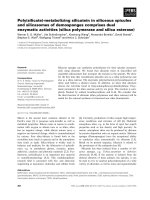
Recovery of parotid glands
After 12, 24, and 36 months in group I and II a recovery effect could be
measured. After 36 months, patients in group I had reached again about 74%
of the initial value of saliva flow. The recovery during a follow up period of 24
9
months or 36 months was significant for group I and group II, whereas in
group III no recovery potentials were measured neither at 12, 24, or 36
months (Figure 1).
Impact of parotid dose, tumor site, tumor- and lymph node stage
Analysis of the RSFR as a function of the mean parotid dose between the
different tumor sites (oral cavity, oropharynx, and larynx/ hypopharynx), T
stage and N stage was performed. A significantly greater parotid flow ratio
after 36 months after RT in favour of the tumor sites larynx/ hypopharynx
(62%) and oropharynx (56%) as compared to oral cavity (31%) was shown
(Table 4). Multiple linear regression analyses revealed that the parotid gland
dose and the tumor site were the independent determinants 12 and 36
months after the end of RT (Table 5).
Discussion
Bilateral irradiation in patients with head and neck cancer leads to a dosedependent change of salivary output and altered salivary composition
[9,15,16]. Small salivary glands in oral cavity are a part of mucosal target
volume. The submandibular glands just as sublingual glands reside in the
midst of level I. Recently Wang et al. have shown that with modern IMRT a
partial sparing of single submandibular gland is probably feasible [17]. By
consequently performed radiation treatment of carcinomas of the oral cavity,
the oropharynx, and the larynx/ hypopharynx, the sparing of submandibular
salivary glands can only be taken into consideration in special cases. Sparing
of parotid glands as well as submandibular glands with dose reduction of
10
mucous membranes seems to be the most effective way to prevent
hyposalivation after treatment. 3D-CRT as well as IMRT do allow the
generation of high dose gradients around target volumes, and thus to spare
organs at risk inclusive mucous membranes. In previous investigations we
have proven that sparing the parotid gland alone by using 3D-CRT produces
less hyposalivation than a conventional radiation technique (2D-RT) [9].
Recent investigations have shown that more and more patients can take the
advantage from more advanced RT techniques such as IMRT. In a
multicenter randomized study (PARSPORT trial) the advantage of the parotid
sparing by using IMRT technique as compared to conventional 3D-CRT in
terms of clinical outcome was investigated [6]. The authors found that 12
months after treatment, 39% of IMRT patients suffered from dry mouth as
compared to 74% of conventional RT. We also have found that by using
IMRT, the mean dose value of the spared parotid gland was significantly
reduced as compared to 3D-CRT. By using IMRT a mean parotid gland dose
<26 Gy was reached in 12/15 patients (80%), and a dose range >26 to 40 Gy
in 3/15 patients (20%). No patient with IMRT has had a mean dose of >40 Gy
within the spared parotid gland.
Currently, in the literature only limited data is available providing long-term
salivary flow measurements over several years. Solely Braam et al. have
demon-strated a recovery concerning a time period of 5 years [8]. Most of the
other analysis did not cover more than 12 months [18,19]. In the present
prospective analysis, we have shown results for a time period of 36 months
recovery of the salivary glands. We assume that most of the recovery
11
processes have been completed within this period. To demonstrate doserelated differences in the recovery potential of the parotid gland, we have
divided our patients into 3 groups. Our separation with Dmean < 26 Gy, 26 – 40
Gy and > 40Gy was based on common reports from the literature and was
done by reasons of comparison with previous investigations and particularly
to complement our own objective measurements with the investigations of the
quality of life after salivary gland protection [20,21].
As described in other studies hyposalivation can be prevented by restricting
mean parotid gland doses to 26 - 30 Gy [15,22-24]. In our study, nearly one
quarter of the patients did benefit from sparing the parotid gland by using 3DCRT. With IMRT this was possible for 75% of the patients. Three years after
irradiation 76% of the pre-treatment salivary flow can be preserved in this
group. These results are excellent and highlight the significant advantage of
IMRT as compared to the conventional 3D-CRT-technique [25,26]. With a
mean parotid gland dose lower than 26 Gy, the recovery of salivary gland
function reaches about 74% of the initial value at 3 years. Otherwise, patients
with a mean parotid gland dose above 40 Gy did not show significant
recovery values.
Our analysis of the flow ratio as a function of the mean parotid dose between
the tumor sites oral cavity and larynx/hypopharynx did demonstrate a
significantly higher parotid flow ratio in favour of the lower sites in the neck
(larynx/hypopharynx) after radiotherapy. Significant differences over a time
period of 12 and 36 months after end of RT were observed. The independent
12
influence of T and N stage could not be demonstrated clearly also due to the
limited number of patients 24 and 36 months after end of RT.
Taking into consideration the tumor localization, still one third of the patients
received despite the use of 3D-CRT more than 40 Gy to the spared parotid
gland. These patients suffered a total damage of salivary gland function after
irradiation. In a further study, we have already shown that the remaining
stimulated saliva in these patients is not able to maintain oral health due to its
pH and its buffer capacity [20]. In fact it promotes dental caries [27].
Considering the low pH of 6.4, remineralisation is not possible any more,
instead dentine and root areas are demineralised. Accordingly, dental
prearrangements have to accommodate these circumstances.
We know of some weaknesses in our analysis. At 3 years after irradiation, of
117 initially included patients only a limited number of patients have been
available for follow-up measurements, respective 14-17-11 patients in group I,
II and III. This number of patients shows the reality concerning investigations
of recovery effects of the salivary glands over a long time period. We also
know that the method of the whole stimulated salivary flow rate measure
resulting in a higher salivary flow rate compared to the more detailed
examination techniques of parotid gland alone with Lashley cups. But the
method is robust, easy to use and non-invasive, simulating a physiological
situation and showed the smallest variability for measuring the salivary flow
rate [13,22,28].
13
Also, we mention the expected anatomical changes of the parotid glands
during the head and neck irradiation [29]. This is known from studies in
centers with the use of helical tomotherapy. Due to weight loss and tumor
shrinkage especially in head and neck patients the parotid gland is expected
to get higher doses than predicted. Studies, whether these changes have a
significant influence on the salivary flow rates are not available.
Hence, it has to be accepted that approximately three quarter of IMRT and
only one third of 3D-CRT patient’s benefit from salivary gland sparing by an
increasing of the salivary flow rates do to 12 and 24 months after radiation.
However, the aim of the radiation protocol used in this study was to preserve
salivary flow rate as high as possible. Lack of saliva predisposes the
development of atypical, unusual and rapidly progressive and aggressive
dental decay [4,30,31].
Conclusions
IMRT provides remarkable success rates as compared to conventional 3DCRT in terms of parotid gland sparing. The IMRT technique should therefore
represent the standard of care for the treatment of head and neck tumors.
Parotid-gland-sparing up to mean doses of 26 Gy proved to be a reliable
method to avoid distinct long lasting xerostomia.
List of Abbreviations
14
3D-CRT = Three- dimensional conformal radiotherapy, IMRT = intensity
modulated radiotherapy, RT = Radiation therapy, RSFR = Relative Salivary
Flow Rate, Dmean = Dose mean value, SD = Standard deviation, SE =
Standard error, T = Tumor, N = Lymph node
Competing interests
The authors declare that they have no competing interests.
Authors' contributions
JH gathered data and was the main author of the manuscript. JS performed
statistical analysis. RG and MJ gathered treatment planning data. GH revised
the manuscript and aided in the analysis. DV participated in the coordination.
CG and TK conceived of the study, and participated in its design and
coordination. All authors have approved the final manuscript.
Acknowledgements
The authors acknowledge funding received from the German Cancer Aid e.V..
References
[1] Lin A, Kim HM, Terrell JE, Dawson LA, Ship JA, Eisbruch A: Quality of life
after parotid-sparing IMRT for head-and-neck cancer: a prospective
longitudinal study. Int J Radiat Oncol Biol Phys 2003, 57: 61-70.
15
[2] Nishimura Y, Nakamatsu K, Shibata T, Kanamori S, Koike R, Okumura M,
Suzuki M: Importance of the initial volume of parotid glands in xerostomia for
patients with head and neck cancers treated with IMRT. Jpn J Clin Oncol
2005, 35: 375-379.
[3] Porter SR, Fedele S, Habbab KM: Xerostomia in head and neck
malignancy. Oral Oncol 2010, 46: 460-463.
[4] Bekes K, Francke U, Schaller HG, Kuhnt T, Gerlach R, Vordermark D,
Gernhard CR: The influence of different irradiation doses and desensitizer
application on demineralization of human dentin. Oral Oncol 2009, 45: e80e84.
[5] Eisbruch A, Ship JA, Martel MK, Ten Haken RK, Marsh LH, Wolf GT,
Esclamado RM, Bradford CR, Terrell JE, Gebarski SS, Lichter AS: Parotid
gland sparing in patients undergoing bilateral head and neck irradiation:
techniques and early results. Int J Radiat Oncol Biol Phys 1996, 36: 469-480.
[6] Nutting CM, Morden JP, Harrington KJ, Urbano TG, Bhide SA, Clark
C,Miles EA, Miah AB, Newbold K, Tanay M, Abad F, Jefferies SJ, Scrase C,
Yap BK, A'hern RP, Sydenham MA, Emson M, Hall E: Parotid-sparing
intensity modulated versus conventional radiotherapy in head and neck
cancer (PARSPORT): a phase 3 multicentre randomised controlled trial.
Lancet Oncol 2011,12:127-136.
[7] Eisbruch A, Dawson LA, Kim HM, Bradford CR, Terrell JE, Chepeha DB,
Teknos TN, Anzai Y, Marsh LH, Martel MK, Ten Haken RK, Wolf GT, Ship
JA: Conformal and intensity modulated irradiation of head and neck cancer:
16
the potential for improved target irradiation, salivary gland function, and
quality of life. Acta Otorhinolaryngol Belg 1999, 53: 271-275.
[8] Braam PM, Roesink JM, Moerland MA, Raaijmakers CP, Schipper M,
Terhaard CH: Long-term parotid gland function after radiotherapy. Int J Radiat
Oncol Biol Phys 2005, 62: 659-664.
[9] Kuhnt T, Jirsak N, Muller AC, Pelz T, Gernhardt C, Schaller HG, Janich M,
Gerlach R, Dunst J: [Quantitative and qualitative investigations of salivary
gland function in dependence on irradiation dose and volume for reduction of
xerostomia in patients with head-and-neck cancer]. Strahlenther Onkol 2005,
181: 520-528.
[10] Kuhnt T, Janich M, Gotz U, Gerlach R, Chiricuta IC, Hansgen G:
[Presentation of a 3D conformal radiotherapy technique for head-and-neck
tumors resulting in substantial protection of the parotid glands]. Strahlenther
Onkol 2006, 182: 325-330.
[11] Georg D, Kroupa B, Georg P, Winkler P, Bogner J, Dieckmann K, Potter
R: Inverse planning--a comparative intersystem and interpatient constraint
study. Strahlenther Onkol 2006, 182: 473-480.
[12] Lyman JT, Wolbarst AB: Optimization of radiation therapy, IV: A dosevolume histogram reduction algorithm. Int J Radiat Oncol Biol Phys 1989, 17:
433-436.
[13] Chao KS, Deasy JO, Markman J, Haynie J, Perez CA, Purdy JA, Low
DA: A prospective study of salivary function sparing in patients with headand-neck
cancers
receiving
intensity-modulated
or
three-dimensional
17
radiation therapy: initial results. Int J Radiat Oncol Biol Phys 2001, 49: 907916.
[14] Burlage FR, Coppes RP, Meertens H, Stokman MA, Vissink A: Parotid
and submandibular/sublingual salivary flow during high dose radiotherapy.
Radiother Oncol 2001, 61: 271-274.
[15] Beer KT, Zehnder D, Lussi A, Greiner RH: Sparing of contralateral major
salivary glands has a significant effect on oral health in patients treated with
radical radiotherapy of head and neck tumors. Strahlenther Onkol 2002, 178:
722-726.
[16] Konings AW, Coppes RP, Vissink A: On the mechanism of salivary gland
radiosensitivity. Int J Radiat Oncol Biol Phys 2005, 62: 1187-1194.
[17] Wang ZH, Yan C, Zhang ZY, Zhang CP, Hu HS, Tu WY, Kirwan J,
Mendenhall WM: Impact of Salivary Gland Dosimetry on Post-IMRT Recovery
of Saliva Output and Xerostomia Grade for Head-and-Neck Cancer Patients
Treated with or without Contralateral Submandibular Gland Sparing: A
Longitudinal Study. Int J Radiat Oncol Biol Phys 2010, Oct 7. [Epub ahead of
print].
[18] Stock M, Dorr W, Stromberger C, Mock U, Koizar S, Potter R, Georg D:
Investigations on Parotid Gland Recovery after IMRT in Head and Neck
Tumor Patients. Strahlenther Onkol 2010, 186: 665-671.
[19] Hey J, Setz J, Gerlach R, Janich M, Sehlleier S, Schaller HG, Gernhardt
CR, Kuhnt T: Parotid-gland-sparing 3D conformal radiotherapy in patients
18
with bilateral radiotherapy of the head and neck region--results in clinical
practice. Oral Oncol 2009, 45: e11-e17.
[20] O'Neill M, Heron DE, Flickinger JC, Smith R, Ferris RL, Gibson M:
Posttreatment Quality-of-Life Assessment in Patients With Head and Neck
Cancer Treated With Intensity-modulated Radiation Therapy. Am J Clin Oncol
2011, Apr 29. [Epub ahead of print].
[21] van Rij CM, Oughlane-Heemsbergen WD, Ackerstaff AH, Lamers EA,
Balm AJ, Rasch CR: Parotid gland sparing IMRT for head and neck cancer
improves xerostomia related quality of life. Radiat Oncol 2008, 3: 41.
[22] Maes A, Weltens C, Flamen P, Lambin P, Bogaerts R, Liu X, Baetens J,
Hermans R, Van den, Bogaert W: Preservation of parotid function with
uncomplicated conformal radiotherapy. Radiother Oncol 2002, 63: 203-211.
[23] Munter MW, Karger CP, Hoffner SG, Hof H, Thilmann C, Rudat V, Nill S,
Wannenmacher M, Debus J: Evaluation of salivary gland function after
treatment of head-and-neck tumors with intensity-modulated radiotherapy by
quantitative pertechnetate scintigraphy. Int J Radiat Oncol Biol Phys 2004,
58: 175-184.
[24] Blanco AI, Chao KS, El N, I, Franklin GE, Zakarian K, Vicic M, Deasy JO:
Dose-volume modeling of salivary function in patients with head-and-neck
cancer receiving radiotherapy. Int J Radiat Oncol Biol Phys 2005, 62: 10551069.
[25] Rades D, Fehlauer F, Wroblesky J, Albers D, Schild SE, Schmidt R:
Prognostic factors in head-and-neck cancer patients treated with surgery
19
followed
by
intensity-modulated
radiotherapy
(IMRT),
3D-conformal
radiotherapy, or conventional radiotherapy. Oral Oncol 2007, 43: 535-543.
[26] Chen WC, Hwang TZ, Wang WH, Lu CH, Chen CC, Chen CM,Wneg HH,
Lai CH, Chen MF: Comparison between conventional and intensitymodulated post-operative radiotherapy for stage III and IV oral cavity cancer
in terms of treatment results and toxicity. Oral Oncol 2009, 45: 505-510.
[27] Lacatusu S, Francu L, Francu D: Clinical and therapeutical aspects of
rampant caries in cervico-facial irradiated patients. Rev Med Chir Soc Med
Nat Iasi 1996, 100: 198-202.
[28] Bardow A, Moe D, Nyvad B, Nauntofte B: The buffer capacity and buffer
systems of human whole saliva measured without loss of CO2. Arch Oral Biol
2000, 45: 1-12.
[29] Duma MN, Kampfer S, Wilkens JJ, Schuster T, Molls M, Geinitz H:
Comparative analysis of an image-guided versus a non-image-guided setup
approach in terms of delivered dose to the parotid glands in head-and-neck
cancer IMRT. Int J Radiat Oncol Biol Phys 2010, 77: 1266-1273.
[30] Gernhardt CR, Kielbassa AM, Hahn P, Schaller HG: Tensile bond
strengths of four different dentin adhesives on irradiated and non-irradiated
human dentin in vitro. J Oral Rehabil 2001, 28: 814-820.
[31] Brown LR, Dreizen S, Handler S, Johnston DA: Effect of radiationinduced xerostomia on human oral microflora. J Dent Res 1975, 54: 740-750.
Legends
20
Figure 1 Recovery potentials 1, 6, 12, 24, and 36 months after end of
radiation therapy (RT). The initial flow-rate was 100%. Saliva measurement
was normalized in relation to pre-treatment results in relative salivary flow
rates (RSFR’s in %).
Table 1 Patient and tumor characteristics.
Study population
Patient number
117
Male/female
90/27
Median age in years (range)
57 (27 – 88)
Unilaterale/ bilaterale radiotherapy
117
3D-CRT/IMRT
102/15
Tumour sites
Oral cavity/ Oropharynx
81
Larynx/ Hypopharynx
29
Unknown primary (CUP)
2
Other (Myeloma, Lymphoma, Nasal
Cavity, Paranasal Sinus)
5
Staging
UICC-I
7
UICC-II
11
UICC-III
34
UICC-IVA
61
UICC-IVB
2
Myeloma and Lymphoma IA/IIA
2
Table 2 Mean parotid gland doses with 3D-CRT and IMRT.
IMRT
Spared (lowest)
parotid
gland
Spared (lowest)
parotid
gland
No
Yes
Mean
Patients
n
Dose (Gy)
102
34.4
15
21.7
Standard
Deviation
13.6
p
0.001
6.2
21
Table 3 Mean and standard deviation of relative salivary flow rate at 1, 6, 12,
24, and 36 months after radiotherapy.
Patients (n)
mean RSFR
(%)
SD (%)
34
55.6
32.71
6 months after RT
35
50.2
36.44
12 months after RT
27
59.7
36.46
24 months after RT
19
65.8
34.10
36 months after RT
14
74.3
27.85
1 month after RT
40
30.8
26.97
6 months after RT
40
33.4
31.03
12 months after RT
37
46.7
33.05
24 months after RT
26
56.4
31.16
36 months after RT
17
48.7
33.19
1 month after RT
37
17.6
16.84
6 months after RT
31
12.8
15.05
12 months after RT
35
19.2
23.21
24 months after RT
18
30.6
26.68
36 months after RT
11
24.2
28.55
Group
I Dmean < 26 Gy 1 month after RT
II Dmean 26-40
Gy
III Dmean > 40
Gy
Table 4 Tumor site with mean and standard deviation of relative salivary flow
rate at 1, 6, 12, 24, and 36 months after radiotherapy.
Patients (n)
mean RSFR
(%)
SD (%)
1month after RT
29
34.3
32.42
6 months after RT
29
32.7
35.63
12 months after RT
28
35.9
37.54
24 months after RT
14
43.4
37.84
36months after RT
13
31,0
28.20
1month after RT
51
30.2
27.67
6 months after RT
47
27.4
29.87
12 months after RT
41
34.1
33.56
24 months after RT
30
51.3
34.75
36months after RT
16
55.9
39.70
Tumor site
Oral cavitiy
Oropharynx
22
Hypopharynx/
Larynx
1month after RT
28
42.7
32.32
6 months after RT
27
42.6
34.50
12 months after RT
29
53.1
31.90
24 months after RT
17
59.6
28.94
36months after RT
11
61.6
29.99
Table 5 Multiple linear regression analyses for relative salivary flow rates
(RSFRs) in the observation periods 12, 24 and 36 months.
Variables
RSFRs (%)
of 12
months
after RT
(R² = 0.299)
ß (SE)
RSFRs (%)
of 24
months
after RT
(R² = 0.199)
ß (SE)
RSFRs (%)
of 36
months
after RT
(R² = 0.416)
Dmean lowest
parotid gland
-1.187 (0.244)
pvalue
0.0001
-0.736 (0.338)
pvalue
0.034
Tumor site
8.886 (2.815)
0.002
7.796 (4.178)
0.068
T stage
-2.429 (3.173)
0.446
-5.880 (4.671)
0.214
N stage
-2.870 (2.143)
0.184
-2.499 (3.232)
0.443
ß (SE)
-1.160 (0.395)
11.310
(4.640)
-10.047
(5.230)
0.866 (4.153)
23
pvalue
0.006
0.021
0.064
0.836