Báo cáo khoa học: "The prognostic value of the nodal ratio in N1 breast cancer" pps
Bạn đang xem bản rút gọn của tài liệu. Xem và tải ngay bản đầy đủ của tài liệu tại đây (253.79 KB, 30 trang )
This Provisional PDF corresponds to the article as it appeared upon acceptance. Fully formatted
PDF and full text (HTML) versions will be made available soon.
The prognostic value of the nodal ratio in N1 breast cancer
Radiation Oncology 2011, 6:131 doi:10.1186/1748-717X-6-131
Tae Jin Han ()
Eun Young Kang ()
Wan Jeon ()
Sung Won Kim ()
Jee Hyun Kim ()
So Yeon Park ()
Jae Sung Kim ()
In Ah Kim ()
ISSN 1748-717X
Article type Research
Submission date 8 June 2011
Acceptance date 6 October 2011
Publication date 6 October 2011
Article URL />This peer-reviewed article was published immediately upon acceptance. It can be downloaded,
printed and distributed freely for any purposes (see copyright notice below).
Articles in Radiation Oncology are listed in PubMed and archived at PubMed Central.
For information about publishing your research in Radiation Oncology or any BioMed Central journal,
go to
/>For information about other BioMed Central publications go to
/>Radiation Oncology
© 2011 Han et al. ; licensee BioMed Central Ltd.
This is an open access article distributed under the terms of the Creative Commons Attribution License ( />which permits unrestricted use, distribution, and reproduction in any medium, provided the original work is properly cited.
- 1 -
The prognostic value of the nodal ratio in N1 breast cancer
Tae Jin Han
1
, Eun Young Kang
1
, Wan Jeon
1
, Sung-Won Kim
2
, Jee Hyun Kim
2
, Yu
Jung Kim
2
, So Yeon Park
2
, Jae Sung Kim
1
, In Ah Kim
1,2
*
1
Department of Radiation Oncology, Seoul National University, Bundang Hospital,
166 Gumiro Seongnamsi Kyeonggido, Korea, 463-707.
2
Breast Care Center, Seoul National University, Bundang Hospital, Korea
*Corresponding author: In Ah Kim, M.D., Ph.D.
Department of Radiation Oncology, Seoul National University Bundang Hospital,
166 Gumiro Seongnamsi Kyeonggido, Korea, 463-707
E-mail:
, Phone: +82(31) 787-7651, Fax +82(31) 787-4019
Email addresses:
TJH:
EYK:
WJ:
SWK:
JHK:
YJK:
SWP:
JSK:
IAK:
- 2 -
Abstract
Background: Although the nodal ratio (NR) has been recognized as a prognostic
factor in breast cancer, its clinical implication in patients with 1-3 positive nodes (N1)
remains unclear. Here, we evaluated the prognostic value of the NR and identified
other clinico-pathologic variables associated with poor prognosis in these patients.
Methods: We analyzed 130 patients with N1 invasive breast cancer who were treated
at Seoul National University Bundang Hospital from March 2003 to December 2007.
Disease-free survival (DFS), locoregional recurrence-free survival (LRRFS), and
distant metastasis-free survival (DMFS) were compared according to the NR with a
cut-off value of 0.15.
Results: We followed patients’ recovery for a median duration of 59 months. An NR
>0.15 was found in 23.1% of patients, and a median of 18 nodes were dissected per
patient (range 1-59). The NR was statistically independent from other prognostic
variables, such as patient age, T stage, extent of surgery, pathologic factors in the chi
square test. On univariate analysis, patients with a NR >0.15 had significantly lower
5-year LRRFS (88.7% vs. 97.9%, p=0.033) and 5-year DMFS (81.3% vs. 96.4%,
p=0.029) and marginally lower 5-year DFS (81.3% vs. 94.0%, p=0.069) than those
with a NR ≤0.15, respectively. Since the predictive power of the NR was found to
differ with diverse clinical and pathologic variables, we performed adjusted analysis
stratified by age, pathologic characteristics, and adjuvant treatments. Only young
patients with a NR >0.15 showed significantly lower DFS (p=0.027) as well as those
presenting an unfavorable pathologic profile such as advanced T stage (p=0.034),
histologic grade 3 (p=0.034), positive lymphovascular invasion (p=0.037), involved
resection margin (p=0.007), and no chemotherapy (p=0.014) or regional radiotherapy
- 3 -
treatment (p=0.039). On multivariate analysis, a NR >0.15 was significantly
associated with lower DFS (p=0.043) and DMFS (p=0.012), but not LRRFS
(p=0.064).
Conclusions: A NR >0.15 was associated with an increased risk of recurrence,
especially in young patients with unfavorable pathologic profiles.
Key words: breast cancer, N1, nodal ratio, prognostic factor
- 4 -
Background
The presence of axillary lymph node metastasis is one of the most important factors
affecting prognosis in patients with breast cancer [1]. According to the current 7th
edition of the American Joint Committee on Cancer staging system, N stage in breast
cancer is solely determined by the number of positive nodes [2]. In patients with
inappropriately dissected axillary nodes, however, a discrepancy may exist between
the absolute number of positive nodes and the substantive extent of axillary node
metastasis [3]. Therefore, the nodal ratio (NR), defined as the absolute number of
involved nodes/number of excised nodes, has been suggested to address this
discrepancy [4]. Recent studies have shown the prognostic value of the NR and even
proposed the possibility of NR as an alternative or a complement to N staging in
node-positive breast cancer [5-13]. However, no consensus has been reached for the
appropriate criteria to discriminate between low- and high-risk groups of NR for
breast cancer with 1-3 positive nodes.
In the current study, we evaluated the prognostic value of the NR and identified
other clinico-pathologic variables associated with poor prognosis in N1 breast cancer
patients.
- 5 -
Methods
Patients
We retrospectively analyzed 130 patients with N1 invasive breast cancer who were
treated at Seoul National University Bundang Hospital (SNUBH) from March 2003 to
December 2007. Patients who had received neoadjuvant chemotherapy prior to
surgery were excluded. We collected not only treatment modality information such as
type of surgery, type of systemic treatment, and radiation field, but also detailed
clinico-pathologic prognostic factors such as age, pathologic stage, histologic type
and grade, number of excised and positive nodes, estrogen/progesterone receptor
(ER/PR) status, human epithelial growth factor receptor family 2 (HER2) status,
presence of extracapsular extension (ECE), presence of lymphovascular invasion
(LVI), and resection margin status. A close margin was defined as the presence of
invasive carcinoma within 2 mm of the surgical margin of resection.
Patient grouping according to the nodal ratio
We categorized the patients into two NR groups: low NR (LNR; ≤0.15) and high NR
(HNR; >0.15). Disease-free survival (DFS), locoregional recurrence-free survival
(LRRFS), and distant metastasis-free survival (DMFS) were compared between
groups. We defined locoregional recurrence as the first site of recurrence involving
residual breast or chest wall (local) tissue and/or axillary, supra- or infraclavicular,
and internal mammary nodes (regional). For cases in which locoregional recurrence
and distant metastasis simultaneously occurred, we counted both failure patterns.
Statistical analysis
- 6 -
To make comparisons between the two groups, we used the chi-square test or
Fisher’s exact test for categorical data and independent sample t-test for continuous
data. The Kaplan-Meier method was used for DFS, LRRFS, and DMFS probability,
and survival according to different variables was compared by the log-rank test. The
Cox proportional hazard method was used to perform multivariate analysis for
predictors of survival. We included variables that showed significance in the
univariate analysis or were otherwise were considered to be confounders in the
multivariate analysis. All statistical analyses were performed with Statistical Package
for the Social Sciences (version 17.0; SPSS, Chicago, IL). We considered p values
equal to or less than 0.05 to be statistically significant.
- 7 -
Results
Patient and tumor characteristics
Of the 130 patients, the LNR group included 100 patients and the HNR group
included 30 patients. Patient characteristics for these two groups are summarized in
Table 1. The median number of excised nodes per patient was 18 (range, 1-59) for
both groups combined, and was significantly higher in the LNR group than in the
HNR group (20 vs. 7, p<0.001). RT was used to treat 46 (46%) LNR patients and 20
(66.7%) HNR patients; among these, regional RT was more frequently used in the
HNR group (50.0% vs. 10.9%, p=0.001). The local RT field consisted of the whole
breast or chest wall only. In contrast, supraclavicular lymph nodes and/or internal
mammary lymph nodes were included in the locoregional RT field. Chemotherapy
was used to treat 92 (92%) LNR patients and 25 (83.3%) HNR patients. Taxane-
containing regimens such as AC (adriamycin and cyclophosphamide) were most
frequently prescribed. The tumor characteristics in the two groups are summarized in
Table 2. Infiltrating ductal carcinoma was the most frequent tumor histology in both
groups, but was more dominant in the LNR group (92.0% vs. 73.3%, p=0.011). The
NR was a statistically independent variable from other prognostic variables including
patient age, extent of surgery, and pathologic factors such as ECE, LVI, tumor grade,
margin status, ER/PR, and HER2 status.
Follow-up and patterns of failure
We followed patients’ recovery for a median duration of 59 months (range, 10-89
months) for both groups although the LNR group had a longer duration of follow-up
(p=0.013). Both groups showed distant metastasis as the dominant failure pattern in
- 8 -
eight of nine patients who experienced any failures. These details are summarized in
Table 3.
Univariate analysis of different prognostic factors
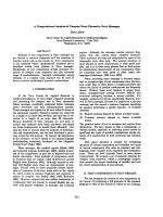
The univariate analysis results for prognostic factors are summarized in Table 4.
According to the univariate analysis, patients with a NR >0.15 had significantly lower
5-year LRRFS (88.7% vs. 97.9%, p=0.033) and 5-year DMFS (81.3% vs. 96.4%,
p=0.029) and marginally lower 5-year DFS (81.3% vs. 94.0%, p=0.069) than those
with a NR ≤0.15 (Figure 1).
The effect of NR on DFS stratified by other prognostic factors
Since the prognostic power of the NR was found to differ according to diverse
clinical and pathologic variables, we performed adjusted analysis stratified by age,
pathologic characteristics, and adjuvant treatments. The HNR group showed
significantly lower 5-year DFS exclusively in those presenting an unfavorable clinico-
pathologic profile: young age (p=0.027), advanced T stage (p=0.034), high grade
(p=0.034), the presence of LVI (p=0.037), involved resection margin (p=0.007) and
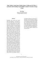
the lack of chemotherapy (p=0.014) or regional RT (p=0.039; Table 5). The DFS
curves according to NR with and without regional RT are presented in Figure 2.
Multivariate analysis of different prognostic factors
According to the multivariate analysis, a NR >0.15 was significantly associated with
lower DFS (p=0.043) and DMFS (p=0.012) but not LRRFS (p=0.064; Table 6).
Patients not treated with chemotherapy showed a tendency of increased distant
metastasis.
- 9 -
Discussion
The prognostic value of NR is supported by several studies [4-13]. Vinh-Hung et al.
reported the superiority of NR over pN stage in predicting disease-specific survival,
and Danko et al. revealed that the prognostic value of NR for disease-free survival
remained significant even when stratified by pN stage [8, 10]. Recently, Ahn et al.
showed that NR is a better predictor of disease-free survival than pN stage, especially
in patients with high-risk features such as young age, HER2-enriched or triple-
negative tumor, and recommended that NR should be preferentially considered in
decision making for adjuvant treatment [13].
Although most studies used a value between 0.20 and 0.25 as a minimal cut-off
threshold to distinguish risk groups, there is no consensus on which value is the most
reliable [5-14]. We used 0.15 as a cut-off value, which may be considered somewhat
low. Because the number of positive nodes is inevitably limited in the N1 category,
however, the distribution of the NR is strongly affected by the number of nodes
sampled. While other studies have focused on patients with between 10 and 16
excised nodes, the present study investigated patients with a median of 18 excised
nodes.
Extensive data suggest that other clinico-pathologic findings also can predict an
increased risk of locoregional recurrence and even distant metastasis, such as young
age, higher histologic grade, negative hormone receptors, presence of ECE, presence
of LVI, and inadequate resection margins [15-19]. Recently, Truong et al. reported
that T1-T2 breast cancer patients with one to three positive nodes, young age (<50
years), histologic grade 3, or ER-negative disease had high 10-year locoregional
- 10 -
recurrence risks (up to 20%), even after breast-conserving surgery was followed by
whole breast radiotherapy [15]. In the current study, those findings were not
significant factors for locoregional recurrence or distant metastasis independently but
showed selective significance in adjusted analysis combined with the NR.
Regardless of the extent of surgery, substantially high locoregional recurrence rates
have been reported in patients with 1-3 positive nodes [15, 20-24]. Locoregional
recurrence also has been linked to distant metastasis and long-term breast cancer
mortality [25-28]. In a meta-analysis of five National Surgical Adjuvant Breast and
Bowel Project (NSABP) trials, patients who experienced locoregional recurrence had
a considerably lower 5-year DMFS: 51.4% after ipsilateral breast tumor recurrence,
31.5% after axillary recurrence, and 12.1% after supraclavicular metastasis,
respectively [27]. Data from the Breast Cancer Trialists’ Collaborative Group
(BCTCG) showed the overall absolute reduction of 5-year locoregional recurrence by
19%, resulting in a 5% overall absolute reduction of 15-year breast cancer mortality
risk in patients who underwent either breast-conserving surgery or mastectomy [28].
In the current study, the HNR group showed lower LRRFS, DMFS, and DFS.
However, it is inconclusive whether decreased risk of distant metastasis resulted from
decreased locoregional recurrence because only a small number of patients
experienced locoregional recurrence.
The National Cancer Institute of Canada Clinical Trials Group (NCIC-CTG) has
suggested that adding regional RT may improve survival compared with whole breast
RT only when administered after breast-conserving surgery in patients who have T1-
T2 breast cancer with N1 or moderate to high risk N0 [29]. The current study revealed
that regional RT reduced the risk of distant metastasis in the HNR group only;
however, this finding could also support the interpretation that regional RT is
- 11 -
unnecessary for LNR patients who have undergone adequate axillary dissection and
had no poor prognostic factors. For optimization of the locoregional modality, it is
necessary to better define the selection criteria for adjuvant RT. The NR may be a
useful indicator for deciding whether to use adjuvant regional RT to treat patients
with N1 disease.
Inadequate nodal sampling (less than 10), histology grade 3, estrogen receptor-
negative breast carcinomas, or presence of LVI are all considered to be related to the
risk of regional recurrence. Previous studies have shown that sampling fewer than 10
axillary nodes is associated with an increased risk of subsequent locoregional
recurrence [15, 23-24, 30]. Tai et al. included in their study only patients with 10 or
more excised nodes in order to avoid the possibility of an increased regional relapse
rate resulting from understaging or undertreatment [6]. The adjuvant regional RT
could compensate for the compromised regional control resulting from inadequate
axillary dissection; however, this result does not directly apply to patients in the HNR
group who have undergone adequate axillary dissection and remain at substantial risk
for locoregional recurrence [31].
- 12 -
Conclusions
The results of this study associate a NR >0.15 with increased risk of disease
recurrence, especially in young patients with unfavorable pathologic profiles.
- 13 -
Abbreviations
LN, lymph node; BCS, breast-conserving surgery; MRM, modified radical
mastectomy; SLNBx, sentinel lymph node biopsy; ALND, axillary lymph node
dissection; CMF, cyclophosphamide/methotrexate/5-fluorouracil; FEC, 5-
FU/epirubicin/cyclophosphamide; FAC, 5-FU/adriamycin/cyclophosphamide; ACT,
adriamycin/cyclophosphamide/paclitaxel; NR, nodal ratio; IDC, infiltrating ductal
carcinoma; ER, estrogen receptor; PR, progesterone receptor; ECE, extracapsular
extension; LVI, lymphovascular invasion; RM, resection margin NED, no evidence of
disease; LRR, locoregional recurrence; DM, distant metastasis; LRRFS, locoregional
recurrence-free survival; DMFS, distant metastasis-free survival; DFS, disease-free
survival
- 14 -
Competing interests
Authors declare that they have no conflict of interests
Authors' contributions
IAK designed this study and is responsible for the preparation of manuscript with TJH.
TJH, EYK and WJ contributed to the management of clinical data. SWK, JHK, YJK,
JSK, and IAK provided clinical expertise in clinical breast oncology. SYP contributed
to the pathologic work. All authors read and approved the content of manuscript.
Acknowledgements
This work was supported by Nuclear R&D Program (BAERI#2011-0006312) from
National Research Foundation, Korean Ministry of Education, Science & Technology
and Cancer Control Program (#0820010) from Korean Ministry of Health & Welfare
to Kim IA.
- 15 -
References
1. Vinh-Hung V, Burzykowski T, Cserni G, Voordeckers M, Van De Steene J,
Storme G: Functional form of the effect of the numbers of axillary nodes
on survival in early breast cancer. Int J Oncol 2003, 22:697-704.
2. American Joint Committee on Cancer. Chapter 32. Breast. In AJCC cancer
staging manual. 7th edition. New York: Springer; 2010:347-369.
3. Recht A, Edge SB, Solin LJ, Robinson DS, Estabrook A, Fine RE, Fleming GF,
Formenti S, Hudis C, Kirshner JJ, Krause DA, Kuske RR, Langer AS, Sledge
GW Jr, Whelan TJ, Pfister DG; American Society of Clinical Oncology:
Postmastectomy radiotherapy: Clinical practice guidelines of the
American Society of Clinical Oncology. J Clin Oncol 2001, 19:1539-1569.
4. Woodward WA, Vinh-Hung V, Ueno NT, Cheng YC, Royce M, Tai P,
Vlastos G, Wallace AM, Hortobagyi GN, Nieto Y: Prognostic value of nodal
ratios in node-positive breast cancer. J Clin Oncol 2006, 24:2910-2916.
5. Truong PT, Berthelet E, Lee J, Kader HA, Olivotto IA: The prognostic
significance of the percentage of positive/dissected axillary lymph nodes in
breast cancer recurrence and survival in patients with one to three
positive axillary lymph nodes. Cancer 2005, 103:2006-2014.
6. Tai P, Joseph K, Sadikov E, Mahmood S, Lien F, Yu E: Nodal ratios in
node-positive breast cancer long-term study to clarify discrepancy of role
of supraclavicular and axillary regional radiotherapy. Int J Radiat Oncol
Biol Phys 2007, 68:662-666.
- 16 -
7. Truong PT, Woodward WA, Thames HD, Ragaz J, Olivotto IA, Buchholz TA:
The ratio of positive to excised nodes identifies high-risk subsets and
reduces inter-institutional differences in locoregional recurrence risk
estimates in breast cancer patients with 1-3 positive nodes: an analysis of
prospective data from British Columbia and the M. D. Anderson Cancer
Center. Int J Radiat Oncol Biol Phys 2007, 68:59-65.
8. Vinh-Hung V, Verkooijen HM, Fioretta G, Neyroud-Caspar I, Rapiti E, Vlastos
G, Deglise C, Usel M, Lutz JM, Bouchardy C: Lymph node ratio as an
alternative to pN staging in node-positive breast cancer. J Clin Oncol
2009, 27:1062-1068.
9. Hatoum HA, Jamali FR, El-Saghir NS, Musallam KM, Seoud M, Dimassi H,
Abbas J, Khalife M, Boulos FI, Tawil AN, Geara FB, Salem Z, Shamseddine
AA, Al-Feghali K, Shamseddine AI: Ratio between positive lymph nodes
and total excised axillary lymph nodes as an independent prognostic
factor for overall survival in patients with nonmetastatic lymph node-
positive breast cancer. Ann Surg Oncol 2009, 16:3388-3395.
10. Danko ME, Bennett KM, Zhai J, Marks JR, Olson JA Jr: Improved staging in
node-positive breast cancer patients using lymph node ratio: results in
1,788 patients with long-term follow-up. J Am Coll Surg 2010, 210:797-805.
11. Schiffman SC, McMasters KM, Scoggins CR, Martin RC, Chagpar AB:
Lymph node ratio: a proposed refinement of current axillary staging in
breast cancer patients. J Am Coll Surg 2011, 213:45-52.
12. Chagpar AB, Camp RL, Rimm DL: Lymph Node Ratio Should Be
Considered for Incorporation into Staging for Breast Cancer. Ann Surg
Oncol, in press.
- 17 -
13. Ahn SH, Kim HJ, Lee JW, Gong GY, Noh DY, Yang JH, Jung SS, Park HY:
Lymph node ratio and pN staging in patients with node-positive breast
cancer: a report from the Korean breast cancer society. Breast Cancer Res
Treat, in press.
14. Fortin A, Dagnault A, Blondeau L, Vu TT, Larochelle M: The impact of the
number of excised axillary nodes and of the percentage of involved nodes
on regional nodal failure in patients treated by breast-conserving surgery
with or without regional irradiation. Int J Radiat Oncol Biol Phys 2006,
65:33-39.
15. Truong PT, Jones SO, Kader HA, Wai ES, Speers CH, Alexander AS,
Olivotto IA: Patients with T1 to T2 breast cancer with one to three
positive nodes have higher local and regional recurrence risks compared
with node-negative patients after breast-conserving surgery and whole-
breast radiotherapy. Int J Radiat Oncol Biol Phys 2009, 73:357-364.
16. Wallgren A, Bonetti M, Gelber RD, Goldhirsch A, Castiglione-Gertsch M,
Holmberg SB, Lindtner J, Thürlimann B, Fey M, Werner ID, Forbes JF, Price
K, Coates AS, Collins J: Risk factors for locoregional recurrence among
breast cancer patients: results from International Breast Cancer Study
Group Trials I through VII. J Clin Oncol 2006, 24:2028-2037.
17. Katz A, Strom EA, Buchholz TA, Theriault R, Singletary SE, McNeese MD:
The influence of pathologic tumor characteristics on locoregional
recurrence rates following mastectomy. Int J Radiat Oncol Biol Phys 2001,
50:735-742.
18. Veronesi U, Marubini E, Del Vecchio M, Manzari A, Andreola S, Greco M,
Luini A, Merson M, Saccozzi R, Rilke F: Local recurrences and distant
- 18 -
metastases after conservative breast cancer treatments: partly
independent events. Int J Radiat Oncol Biol Phys 2001, 50:735-742.
19. Yang PS, Chen CM, Liu MC, Jian JM, Horng CF, Liu MJ, Yu BL, Lee MY,
Chi CW: Radiotherapy Can Decrease Locoregional Recurrence and
Increase Survival in Mastectomy Patients with T1 to T2 Breast Cancer
and One to Three Positive Nodes with Negative Estrogen Receptor and
Positive Lymphovascular Invasion Status. Int J Radiat Oncol Biol Phys
2010, 77:516-522.
20. Recht A, Gray R, Davidson NE, : Locoregional failure 10 years after
mastectomy and adjuvant chemotherapy with or without tamoxifen
without irradiation: Experience of the Eastern Cooperative Oncology
Group. J Clin Oncol 1999, 17:1689-1700.
21. Katz A, Strom EA, Buchholz TA, Thames HD, Smith CD, Jhingran A,
Hortobagyi G, Buzdar AU, Theriault R, Singletary SE, McNeese MD:
Locoregional recurrence patterns after mastectomy and doxorubicin-
based chemotherapy: Implications for postoperative irradiation. J Clin
Oncol 2000, 18:2817-2827.
22. Woodward WA, Strom EA, Tucker SL, Katz A, McNeese MD, Perkins GH,
Buzdar AU, Hortobagyi GN, Hunt KK, Sahin A, Meric F, Sneige N, Buchholz
TA: Locoregional recurrence after doxorubicinbased chemotherapy and
postmastectomy: Implications for breast cancer patients with early-stage
disease and predictors for recurrence after postmastectomy radiation. Int
J Radiat Oncol Biol Phys 2003, 57:336-344.
23. Truong PT, Olivotto IA, Kader HA, Panades M, Speers CH, Berthelet E:
Selecting breast cancer patients with T1-T2 tumors and one to three
- 19 -
positive axillary nodes at high postmastectomy locoregional recurrence
risk for adjuvant radiotherapy. Int J Radiat Oncol Biol Phys 2005, 61:1337-
1347.
24. Lukens JN, Vapiwala N, Hwang WT, Solin LJ: Regional nodal recurrence
after breast conservation treatment with radiotherapy for women with
early-stage breast carcinoma. Int J Radiat Oncol Biol Phys 2009, 73:1475-
1481.
25. Buchholz TA, Woodward WA, Duan Z, Fang S, Oh JL, Tereffe W, Strom EA,
Perkins GH, Yu TK, Hunt KK, Meric-Bernstam F, Hortobagyi GN, Giordano
SH: Radiation use and long-term survival in breast cancer patients with
T1, T2 primary tumors and one to three positive axillary lymph nodes. Int
J Radiat Oncol Biol Phys 2008, 71:1022-1027.
26. Whelan TJ, Julian J, Wright J, Jadad AR, Levine ML: Does locoregional
radiation therapy improve survival in breast cancer? A meta-analysis. J
Clin Oncol 2000, 18:1220-1229.
27. Wapnir IL, Anderson SJ, Mamounas EP, Geyer CE Jr, Jeong JH, Tan-Chiu E,
Fisher B, Wolmark N: Prognosis after ipsilateral breast tumor recurrence
and locoregional recurrences in five National Surgical Adjuvant Breast
and Bowel Project node-positive adjuvant breast cancer trials. J Clin
Oncol 2006, 24:2028-2037.
28. Clarke M, Collins R, Darby S, Davies C, Elphinstone P, Evans E, Godwin J,
Gray R, Hicks C, James S, MacKinnon E, McGale P, McHugh T, Peto R,
Taylor C, Wang Y; Early Breast Cancer Trialists' Collaborative Group
(EBCTCG): Effects of radiotherapy and of differences in the extent of
- 20 -
surgery for early breast cancer on local recurrence and 15-year survival:
an overview of the randomised trials. Lancet 2005, 366:2087-2106.
29. Olivotto IA, Chua B, Elliott EA, Parda DS, Pierce LJ, Shepherd L, Vallow
LA, White JR, Whelan TJ: A clinical trial of breast radiation therapy
versus breast plus regional radiation therapy in early-stage breast cancer:
the MA20 trial. Clin Breast Cancer 2003, 4:361-363.
30. Joslyn SA, Konety BR. Effect of axillary lymphadenectomy on breast
carcinoma survival. Breast Cancer Res Treatment 2005, 91:11-18.
31. Fowble B. Postmastectomy radiation in patients with one to three positive
axillary nodes receiving adjuvant chemotherapy: An unresolved issue.
Semin Radiat Oncol 1999, 9:230-240.
- 21 -
Figure Legends
Figure 1: LRRFS, DMFS, and DFS according to NR
(a) LRRFS according to NR
(b) DMFS according to NR
(c) DFS according to NR
Abbreviations: LRRFS, locoregional recurrence-free survival; DMFS, distant
metastasis-free survival; DFS, disease-free survival; NR, nodal ratio
Figure 2: Adjusted analysis for DFS with or without regional RT
(a) DFS according to nodal ratio with regional RT
(b) DFS according to nodal ratio without regional RT
Abbreviations: DFS, disease-free survival; NR, nodal ratio
- 22 -
Table 1. Patient characteristics
No. of patients
(%)
NR <0.15
(n=100)
NR >0.15
(n=30)
p value Total
(n=130)
Age (years)
median (range)
46 (25-79)
50 (32-82)
0.225
47 (25-82)
Excised LN (No.)
median (range)
20 (7-59)
7 (1-18)
<0.001
18 (1-59)
Breast resection
BCS
MRM
49 (49.0)
51 (51.0)
18 (60.0)
12 (40.0)
0.290
67 (51.5)
63 (48.5)
LN resection
SLNB only
SLNB + ALND
ALND
3 ( 3.0)
68 (68.0)
29 (29.0)
4 (13.3)
20 (66.7)
6 (20.0)
0.071
7 ( 5.4)
88 (67.7)
35 (26.9)
Radiotherapy
no
yes
54 (54.0)
46 (46.0)
10 (33.3)
20 (66.7)
0.047
64 (49.2)
66 (50.8
)
Extent of radiotherapy
local
locoregional
41 (89.1)
5 (10.9)
10 (50.0)
10 (50.0)
0.001
51 (77.2
)
15 (22.8
)
Chemotherapy
no
yes
8 ( 8.0)
92 (92.0)
5 (16.7)
25 (83.3)
0.176
13 (10.0)
117 (90.0)
Regimen
CMF
FEC/FAC
ACT
12 (13.0)
31 (33.7)
49 (53.3)
7 (28.0)
9 (36.0)
9 (36.0)
0.141
19 (16.2)
40 (34.2)
58 (49.6)
Abbreviations: LN, lymph node; BCS, breast-conserving surgery; MRM, modified
radical mastectomy; SLNBx, sentinel lymph node biopsy; ALND, axillary lymph
node dissection; CMF, cyclophosphamide/methotrexate/5-fluorouracil; FEC, 5-
FU/epirubicin/cyclophosphamide; FAC, 5-FU/adriamycin/cyclophosphamide; ACT,
adriamycin/cyclophosphamide/paclitaxel
- 23 -
Table 2: Tumor characteristics
No. of patients
(%)
NR ≤0.15
(n=100)
NR >0.15
(n=30)
p value
Total
(n=130)
Histology
IDC
others
92 (92.0)
8 ( 8.0)
22 (73.3)
8 (26.7)
0.011
114 (87.7)
16 (12.3)
T stage
T1a/T1b
T1c
T2
T3
8 ( 8.0)
42 (42.0)
49 (49.0)
1 ( 1.0)
6 (20.0)
12 (40.0)
10 (33.3)
2 ( 6.7)
0.166
14 (10.8)
54 (41.5)
59 (45.4)
3 ( 2.3)
ER
(-)
(+)
23 (23.0)
77 (77.0)
5 (16.7)
25 (83.3)
0.459
28 (21.5)
102 (78.5)
PR
(-)
(+)
41 (41.0)
59 (59.0)
8 (26.7)
22 (73.3)
0.155
49 (37.7)
81 (62.3)
HER2
(-)
(+)
unknown
72 (72.0)
18 (18.0)
10 (10.0)
25 (83.3)
5 (16.7)
0 ( 0.0)
0.866
107 (82.3)
23 (17.7)
ECE
(-)
(+)
unknown
74 (74.0)
26 (26.0)
0 ( 0.0)
22 (73.4)
7 (23.3)
1 ( 3.3)
0.183
97 (74.6)
33 (25.4)
LVI
(-)
(+)
unknown
52 (52.0)
48 (48.0)
0 ( 0.0)
14 (47.7)
15 (50.0)
1 ( 3.3)
0.175
67 (51.5)
63 (48.5)
Resection margin
(-)
close or (+)
91 (91.0)
9 ( 9.0)
27 (90.0)
3 (10.0)
0.868
118 (90.8)
12 ( 9.2)
Tumor grade
G1/G2
G3
63 (63.0)
37 (37.0)
22 (73.3)
8 (26.7)
0.297
85 (65.4)
45 (34.6)
Abbreviations: NR, nodal ratio; IDC, infiltrating ductal carcinoma; ER, estrogen
receptor; PR, progesterone receptor; ECE, extracapsular extension; LVI,
lymphovascular invasion; RM, resection margin
- 24 -
Table 3: Clinical status and patterns of failure
No. of patients (%)
NR ≤0.15
(n=100)
NR> 0.15
(n=30)
Total
(n=130)
Follow-up (months)
median (range)
61 (10-89)
48 (26-78)
59 (10-89)
Clinical Status
NED
alive with disease
cause-specific death
intercurrent death
96 (96.0)
4 ( 4.0)
0 ( 0.0)
0 ( 0.0)
26 (86.6)
2 ( 6.7)
2 ( 6.7)
0 ( 0.0)
122 (93.9
)
6 ( 4.6)
2 ( 1.5)
0 ( 0.0)
Patterns of failure
LRR only
DM only
LRR+DM
1 (20.0)
3 (60.0)
1 (20.0)
0 ( 0.0)
1 (25.0)
3 (75.0)
1 (11.0)
4 (44.5)
4 (44.5)
Abbreviations: NR, nodal ratio; NED, no evidence of disease; LRR, locoregional
recurrence; DM, distant metastasis