Báo cáo khoa học: "Mitochondrial DNA alterations of peripheral lymphocytes in acute lymphoblastic leukemia patients undergoing total body irradiation therapy" ppt
Bạn đang xem bản rút gọn của tài liệu. Xem và tải ngay bản đầy đủ của tài liệu tại đây (1.75 MB, 25 trang )
This Provisional PDF corresponds to the article as it appeared upon acceptance. Fully formatted
PDF and full text (HTML) versions will be made available soon.
Mitochondrial DNA alterations of peripheral lymphocytes in acute lymphoblastic
leukemia patients undergoing total body irradiation therapy
Radiation Oncology 2011, 6:133 doi:10.1186/1748-717X-6-133
Quan Wen ()
Yide Hu ()
Fuyun Ji ()
Guisheng Qian ()
ISSN 1748-717X
Article type Research
Submission date 26 May 2011
Acceptance date 6 October 2011
Publication date 6 October 2011
Article URL />This peer-reviewed article was published immediately upon acceptance. It can be downloaded,
printed and distributed freely for any purposes (see copyright notice below).
Articles in Radiation Oncology are listed in PubMed and archived at PubMed Central.
For information about publishing your research in Radiation Oncology or any BioMed Central journal,
go to
/>For information about other BioMed Central publications go to
/>Radiation Oncology
© 2011 Wen et al. ; licensee BioMed Central Ltd.
This is an open access article distributed under the terms of the Creative Commons Attribution License ( />which permits unrestricted use, distribution, and reproduction in any medium, provided the original work is properly cited.
- 1 -
Title page
Title: Mitochondrial DNA alterations of peripheral lymphocytes in acute
lymphoblastic leukemia patients undergoing total body irradiation therapy
Quan Wen
1
, Yide Hu
1§
, Fuyun Ji
2
, Guisheng Qian
2
1
Third Department of Oncology, The second affiliated hospital, Third Military
Medical University, Chongqing 400037, China
2
Institute of Human Respiratory Disease, The second affiliated hospital, Third
Military Medical University, Chongqing 400037, China
§
Corresponding author
Email addresses:
YH:
QW:
F J:
GQ:
- 2 -
Abstract
Background: Mitochondrial DNA (mtDNA) alterations, including mtDNA copy
number and mtDNA 4977bp common deletion (CD), are key indicators of
irradiation-induced damage. The relationship between total body irradiation (TBI)
treatment and mtDNA alterations in vivo, however, has not been postulated yet. The
aim of this study is to analyze mtDNA alterations in irradiated human peripheral
lymphocytes from acute lymphoblastic leukemia (ALL) patients as well as to take
them as predictors for radiation toxicity.
Methods: Peripheral blood lymphocytes were isolated from 26 ALL patients 24 hours
after TBI preconditioning (4.5 and 9 Gy, respectively). Extracted DNA was analyzed
by real-time PCR method.
Results: Average 2.31 times mtDNA and 0.53 fold CD levels were observed after 4.5
Gy exposure compared to their basal levels. 9 Gy TBI produced a greater response of
both mtDNA and CD levels than 4.5 Gy. Significant inverse correlation was found
between mtDNA content and CD level at 4.5 and 9 Gy (P = 0.037 and 0.048).
Moreover, mtDNA content of lymphocytes without irradiation was found to be
correlated to age.
Conclusions: mtDNA and CD content may be considered as predictive factors to
radiation toxicity.
Keywords: mtDNA; 4977-bp Common deletion; Total body irradiation;
Real-time-PCR; Acute lymphoblastic leukemia
- 3 -
Background
Breakage of cellular DNA following radiation is a dose dependent phenomenon and
occurs in both the nuclear and extra-nuclear DNA. Thus, besides nuclear nDNA,
mitochondrial DNA (mtDNA) is equally affected as an only extra-nuclear genome
[1-2]. Numerous investigations showed that mtDNA can be an easily available target
for endogenous reactive oxygen species (ROS) and free radicals caused by ionizing
radiation (IR), which resulted in mtDNA copy number alteration and mtDNA damage
(such as mutation and depletion) [3-4].
The mechanisms of cellular response to radiation with regard to mtDNA alterations
were mainly involved in the following two ways. On one hand, mtDNA has few repair
mechanisms and continued mitochondrial function is preserved primarily due to its
high copy number. One of possible radio-protective mechanism is that enhanced
replication of mtDNA reduces the mutation frequency of total mtDNA and delays the
onset of lethal radiation damage to the mitochondria [5-6]. This hypothesis has been
recently supported by Zhang et al with exhibiting increased mtDNA copy number in
gut and bone marrow of total body irradiated rats [7]. On the other hand, IR usually
prompts cell apoptosis by displaying an accumulation of large scale mtDNA deletions,
especially the specific 4977 bp deletion, referred to as the “common deletion
(CD)”[8]. The site of CD is flanked by two13 bp direct repeats (ACCTCCCTCACCA)
at mtDNA nucleotide site 8470 and 13447 respectively, and easy to make deletion for
its unique formation mechanism[9]. Studies have shown that CD can be as a sensitive
marker of oxidative damage to mtDNA[10-12]. Unfortunately, only few experiments
- 4 -
have evaluated the association between CD and IR till now. For example,
accumulation of CD has been identified by qualitative PCR method on several
irradiated cell lines (such as human skin fibroblasts, glioblastoma and colon
carcinoma lines) and primary lymphocytes [13-15]. Furthermore, CD was induced by
IR in human hepatoblastoma cell line performing on real-time PCR with nonspecific
dsDNA-binding dye SYBR Green. However, their conclusions were largely
controversial. The inconsistency may be due, in part, to the use of non-quantitative
PCR strategies. Additionally, none of these studies have assayed mtDNA or CD level
in peripheral blood lymphocytes (PBLs) after in vivo irradiation exposure for lack of
appropriate human beings radiation model.
In this study, we performed real-time PCR technique with a specific fluorogenic
TaqMan probe conjugated with minor groove binder (MGB) groups, which is more
sensitive and appropriate than nonspecific dsDNA-binding dye PCR methods
previously used [16]. Besides, we taken the acute lymphoblastic leukemia (ALL)
patients undergoging total body irradiation (TBI) precondionting as human beings in
vivo irradiation model. The advantage of using this model lies in full view of in vivo
microenvironment, and without need for irradiating healthy individuals. We attempted
to address the mtDNA status in irradiated human peripheral blood lymphocytes in
vivo to elucidate whether alterations in mtDNA can be linked to exposure to total
body irradiation.
Materials and methods
Study participants
- 5 -
This study comprised peripheral blood (PB) samples from 26 high risked ALL
patients undergoing TBI as pre-transplantation treatment in their first complete
remission (CR1) at hematology department of our institution. The diagnoses
were .according to world healthy organization (WHO) classification and high risk
factors were measured on Ribeca’s report [17]. The patients age from 19 to 56 years
with a mean of 39.4 ± 10.5. Of these, 10 are females and 16 males. Besides, a total of
39 healthy volunteer individuals without IR were included in this study for comparing
the difference of basal mtDNA and CD levels between ALL patients and normal
donors before IR. The donors age from 18 to 55 years with a mean of 37.2 ± 9.4. 19
are females and 20 males. All tested subjects signed an informed consent to the use of
blood samples in accordance with the Declaration of Helsinki and with the approval
from our Institutional Review Board. The amount of CD in skeletal muscle under
physiological conditions is relatively high (up to 1-2% from total mtDNA
content)[18]. Therefore, DNA isolated from skeletal muscle of a 75-year-old male at
autopsy was used as positive control in the present study.
In vivo irradiation and peripheral blood lymphocyte isolation
All patients were treated with two 4.5 Gy TBI sessions daily using an Elekta SLi
8MV linear accelerator (Elekta Co., Stockholm, Sweden) set to deliver a dose rate of
4.5-4.9 cGy/min over two successive days. None of the patients had prior exposure to
any cytotoxic treatment for at least 2 weeks before the start of radiotherapy. All
patients had 7 ml of PB collected prior to and 24 h following exposure for each
radiation treatment. Besides, 39 healthy donors had the same volume of PB collected
- 6 -
without ionizing radiation. Preparation of PBLs followed standard methods, using
human lymphocyte isolation reagent (TBD Biological Technology Co., Tianjin, China)
for separation of mononuclear cells.
DNA extraction
DNA from lymphocytes in vivo and the skeletal muscle was obtained with the
TIANamp Genomic DNA Kit (Tiangen Ltd, Beijing, China), and stored at -70
o
C until
further study.
Analysis of amount of mtDNA and CD by real-time PCR
TaqMan probes with conjugated MGB groups were performed to ensure maximal
specificity in real-time PCR reaction. Nuclear DNA content was estimated by
measuring the human ß-actin gene. The hypervariable region 2 (HVR2) in the
mitochondrial D-Loop was used to represent the total amount of mtDNA since this
region is relatively conserved in Han Chinese [19]. The forward primer (ß-actin:
5'–AGGACCCTGGATGTGACAGC–3'; HVR2:
5'–GCTTTCCACACAGACATCATAACAA–3'; CD:
5'–CTTACACTATTCCTCATCACCCAACTAAAAA–3'), reverse primer (ß-actin:
5'–TGGCATTGCCGACAGGAT–3'; HVR2:
5'–GTTTAAGTGCTGTGGCCAGAAG–3'; CD:
5'–GGAGTAGAAACCTGTGAGGAAAGG–3') and TaqMan MGB hybridization
probes (ß-actin: 5'–AAAGACACCCACCTTGAT–3'; HVR2:
5'–AATTTCCACCAAACCCC–3; CD: 5'–CATTGGCAGCCTAGCATT–3') were
synthesized by GeneCore Bio Technologies Co. Ltd., Shanghai, China.
- 7 -
Dose-dependent plasmid-constructed ß-actin, HVR2 and CD standards were used in
each run of real-time PCR. Of these, both plasmids containing the CD breakpoint and
the HVR2 region were kindly provided by Professor E. Kirches [20]. All TaqMan
reactions were carried out in 96-well plates on an ABI 7500 Real-Time PCR
instrument (Applied Biosystems, Foster City, CA, USA) using the Real-Time PCR
Master Mix kit from Toyobo Co. (Osaka, Japan). Each reaction was carried out in
total volume of 25 µl with 50 ng total DNA template, 300 nM each primer, and 100
nM TaqMan-MGB probe. After an initial denaturation step at 94
o
C for three minutes,
40-45 PCR cycles of 15 s at 94
o
C, 20 s at 60
o
C, and 30 s at 72
o
C were performed.
Real-time PCR of all samples and standards were carried out in quadruplicate. The
data from a PCR run were rejected if the correlation coefficient was less than 0.98.
Statistical analysis
All statistical computations were done using the SPSS v15.0 (SPSS, Chicago, IL).
Logarithmic transformation of data was essential for further parameter statistical
analysis since the original values of the mtDNA and CD copy number in lymphocytes
showed a nonnormal distribution. Univariate analysis of variance and
Student-Newman-Keuls post hoc tests were used to analyze the difference in mtDNA
and CD level with IR exposure. The relative change of mtDNA and CD levels after
different dosage exposure were tested by nonparametric Friedman test. The Pearson’s
correlation test was used to explore association between mtDNA and CD levels. The
correlation between mtDNA, CD level and gender, age was analyzed by the
nonparametric Spearman’s rho correlation test and the Pearson’s correlation test
- 8 -
individually. P values <0.05 are considered statistically significant. All reported P
values are two sided.
Results
Reliability and reproducibility of the TaqMan-MGB PCR assay
The level of mtDNA and CD from lymphocytes was determined in a set of
independent experiments. First, a TaqMan reaction targeting the house keeping gene
ß-actin was used to measure the amount of genomic DNA present in cells. A second
TaqMan assay was designed to the HVR2 region to quantitate the total amount of
mitochondrial DNA. The mtDNA content was normalized to the amount of genomic
DNA in a lymphocyte and expressed as a ratio of mtDNA molecules relative to total
genomic DNA molecules per cell. A third TaqMan assay targeted the CD breakpoint
and measured the abundance of the CD in the samples. The level of CD was
normalized using mtDNA amount and was expressed as a ratio to the mitochondrial
DNA amounts. In other words, the CD ratio was expressed as a percentage of deleted
mtDNA molecules relative to total mtDNA molecules in per genomic DNA molecules.
These primer sets have been used extensively for measuring the CD and mtDNA in
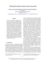
tissues containing low CD and give reliable results [20-21]. Figure 1 showed the
standard curve for the mtDNA common deletion and the CD amplification plots for
the samples examined. It demonstrated that employed TaqMan assay was sensitive
enough to detect single molecule of CD and high linearity was found (y = 3E
-12e-0.6358x
)
in the range of standard samples. CD levels in most of the samples were detected
between Ct 35 and 39. In all samples examined, PCR products were amplified within
- 9 -
the linear range of assays (r
2
> 0.98). Positive control DNA from a 75 year old male
skeletal muscle contained about 0.729% CD ratio and most of the lymphocytes
samples contained from 0.003% to 0.04% CD ratio, consistent with other
measurements [18, 22]. These results suggest that the TaqMan-MGB PCR approach
produces high sensitivity, and could give reliable and corroborating data in our study.
Basal level of mtDNA content and CD ratio from healthy donors and ALL
patients
We first quantified the mtDNA content (median = 197, minimum = 65, maximum =
1124 in ALL; median = 398, minimum = 39, maximum = 1283 in healthy donors) and
CD ratio (median = 0.0116%, minimum = 0.0019%, maximum = 0.085% in ALL;
median = 0.0193%, minimum = 0.0027%, maximum = 0.121%) per cell in PBLs from
ALL patients and healthy donors before irradiation to determine the distribution
pattern. Since both variables did not show normal distribution (P < 0.01,
Kolmogorov–Smirnov test), a logarithm of the mtDNA content and CD ratio was
made for normal distributions (see details in additional file 1, Figure S1). Data of
mtDNA content and CD ratio after logarithm in the three study groups (0, 4.5 and 9
Gy TBI respectively) were given in Table 1 as mean ± SD, median and range. Mean ±
SD values of initial mtDNA and CD level in healthy donors cohort were at 2.507 ±
0.281 and -3.683 ± 0.414. No statistically significant difference was found for
logarithm of basal mtDNA and CD level between healthy donors and patients with
ALL.
Changes of mtDNA content and CD ratio after TBI in patients
- 10 -
Next, we investigated whether the irradiation dose has an effect on mtDNA and CD
level with lymphocytes. Significant differences were found between IR status and
mtDNA alteration among lymphocytes 24h after the irradiation (P = 0.038 for mtDNA
content, 0.027 for CD ratio, Univariate analysis of variance). Furthermore, Student
Newman–Keuls post-hoc tests were used to compare the difference among the three
groups. mtDNA content was significantly increased in 4.5 and 9 Gy irradiation groups
compared with 0 Gy group (mean value of mtDNA content 2.526 and 2.711 compared
with 2.360 ), as well as CD ratio reduced in 4.5 and 9 Gy irradiation groups compared
with 0 Gy group (mean value of CD ratio -4.148 and -4.233 compared with -3.935 ).
Relative change of mtDNA and CD in lymphocytes from each patient after TBI
The results above obtained from in vivo lymphocytes isolated from patients suggest a
correlation of increased mtDNA and decreased CD level with dosage (4.5, 9 Gy)
irradiation in cohort study. To better examine the association between mtDNA
alterations and IR in individuals, relative changes of mtDNA and CD levels after
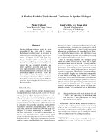
different dose TBI were compared for each patient. As shown in Figure 2, the increase
in mtNDA content was average 1.87 and 2.13 times individually after 4.5 and 9 Gy
TBI (P < 0.001, Friedman test). Meanwhile, decrease in CD was 0.78 and 0.61 when
4.5, 9 vs. 0 Gy cohorts respectively (P < 0.001, Friedman test). Moreover, significant
difference was observed in mtDNA copy (P = 0.041) and CD ratio (P < 0.001) in each
patient when comparing 9 Gy vs. 4.5 Gy exposure. Besides, proportions of increased
mtDNA content in lymphocytes was found to be 80.8% (21/26) and decreased CD
ratio to be 84.6% (22/26) after 4.5 Gy of TBI. Similar trends occurred after 9 Gy
- 11 -
exposure, where 84.6% of increased mtDNA content (22/26) and 88.5% of decreased
CD ratio (23/26) observed.
Relation between mtDNA and CD level after irradiation
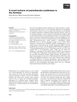
No relation was found between the level of mtDNA and CD at 0, 4.5 and 9 Gy, when
they were analyzed as continuous variables (Pearson test used in all correlations).
However, when CD values were segregated in two populations (the lower third
against the two upper thirds of the distribution), a modest inverse correlation was
found reaching significant level for mtDNA content at different dosage (P = 0.037 for
4.5 Gy, 0.048 for 9 Gy, shown in Figure 3). Besides, significant elevated mtDNA
content was observed not in high but in low CD population (P = 0.021) after 4.5 Gy
TBI exposure.
Effect of age and gender
Finally, the correlations between age, gender, mtDNA and CD level were analyzed
individually. No relationship was found between mtDNA, CD level and gender.
However, a significant positive effect of age was found for basal logarithm mtDNA
content in PBLs. A regression analysis allowed quantification of the effect of age on
basal mtDNA content (regression coefficient = 0.0085 y−1; r2 = 0.251; P = 0.011).
The corresponding graphs are presented in Fig. 4. These results suggest that older
people contained higher mtDNA content in general in the age range of 19-56.
Discussion
In this paper, we described a sensitive and reliable real-time PCR assay of identifying
the mtDNA and common deletion levels. As expected, employed TaqMan-MGB
- 12 -
probe was sensitive enough to detect single molecule of CD in our experiment. The
sensitivity increased at least 5 fold compared with non specific SYBR Green dye
real-time PCR experiment [23]. Besides the improvement of PCR method, we used
human tissues and in vivo irradiation model, whereas the cell strains and ex vivo
irradiation model was exclusively used in other studies. As we known, the ex vivo
cultured cells is unlikely to reflect full view of in vivo microenvironment. What is
more, a lots of apoptotic cell occurs after IR, which is hardly to isolate from the whole
cell population of strain, and will extremely affect the accurate quantification of
mtDNA and CD level for cell heterogeneity [23]. In contract, lymphocytes in vivo
mostly consist of survival cells (> 95%) and could avert the effect of apoptosis [24].
No doubt, it had integrity advantage and is a big step up compared to ex vivo model.
Based on these evidences above mentioned, we can declare that direct analysis of
lymphocytes isolated from human bodies who received TBI would greatly improve
specificity and reliability. These technique refinements take us closer to a
methodology that is likely to produce reliable and quantitative results.
The role of mtDNA content has been investigated in relation to TBI therapy for the
first time in ALL patients. The number of mtDNA copies was elevated in lymphocytes
from above 80% of cases after TBI. Besides, mtDNA content of irradiated PBLs
elevated consisting with a dose response. This phenomenon has been explained as a
compensatory replication of mtDNA to replace damaged mtDNA
7
.
Our statistical analysis showed induced levels of CD after TBI in PBLs, compared to
some other reports that IR-induced oxidative stress may cause increase of CD ratio
- 13 -
[13-14]. Considering that high deletion level of mtDNA increases the susceptibility of
human cell to apoptosis[25], the difference is most likely due to the fact that IR
exposure causes lymphocytes differentiate into two major populations immediately:
apoptotic population usually containing relative high CD level and thus being
sensitive to apoptosis, while surviving population containing relatively low CD level
and more resistant to IR. The cell source in other studies is likely mixed with many
apoptotic cells, which may resulted in relatively high CD level detection.
Here, we report a statistical inversely association between these two predictive values
(mtDNA content and CD ratio) for radiation toxicity. That is to say, lowest values of
CD ratio were related to higher values of mtDNA content, at the same radiation dose
in our experiment. Cell response to IR is individual, and the amount of initial mtDNA
and CD levels depend on each patient. The mechanism behind the relationship
remains unclear. One possible reason is that lymphocytes containing lower level of
deleted mtDNA have stronger ability to replicate wild mtDNA than cells with high
CD level, in order to resist the irradiation induced mitochondrial damage [26].
Besides, abundant mtDNA replication only occurred in low CD population after
moderate dosage treatment, which suggests the stronger replication ability of low CD
population and a mass of mtDNA copy number production. However, the strong
replication ability was not shown in high CD population after modest dosage
treatment.
Cellular oxidative stress is thought to play a role in the aging process and may affect
mtDNA replication. In the present study, we found that mtDNA copy number is
- 14 -
increased with age by lineal regression in our limited cohorts. Similar results has been
described that individuals after middle age may be attributed to the enhanced
oxidative stress than young adults [27], suggesting age factor should be considered
when measuring mtDNA content from both nonirradiated and irradiated lymphocytes.
Conclusion
This study describes the development of a rapid, sensitive, and practical real-time
PCR method to quantify the mtDNA copy number and common deletion in PBL
samples. Our results suggest that radiation increased mtDNA content and declined
common deletion ratio in peripheral lymphocytes of ALL patients, and an inverse
association was observed between both parameters after irradiation, which may be
considered as predictive factors to radiation toxicity.
List of abbreviations: mtDNA, mitochondrial DNA; CD, common deletion; PB,
peripheral blood; PBLs, peripheral blood lymphocytes; TBI, total body irradiation; IR,
ionizing radiation; HVR2, hypervariable region 2; nDNA, nuclear DNA; ALL, acute
lymphoblastic leukemia; MGB, minor groove binder; ROS, reactive oxygen species;
Competing interest: The authors report no conflicts of interest.
Authors’ contributions
QW and YH designed the study, FJ provided real-time PCR assay, QW analyzed the
- 15 -
data and written the paper,GQ contributed to revising the paper. All authors read and
approved the final manuscript.
Acknowledgments
We would like to acknowledge Professor E. Kirches for his assistance with plasmids
donation. We are indebted to associate Professor Jieqiong Lei (Mathematics
department, College of biotechnology, TMMU University) for statistical assistance, to
the patients and donors who donated blood for this study. This study was supported by
grants from the Keystone Project of the “Eleventh Five-year Plan” for Medical
Science Development of PLA (No.06G068) and the National Natural Science
Foundation of China (No.30772144).
Reference
1. Iliakis G, Wang H, Perrault AR, Boecker W, Rosidi B, Windhofer F, Wu W, Guan J, Terzoudi
G, Pantelias G: Mechanisms of DNA double strand break repair and chromosome
aberration formation. Cytogenet Genome Res 2004, 104:14-20.
2. Purkayastha S, Milligan JR, Bernhard WA: On the chemical yield of base lesions, strand
breaks, and clustered damage generated in plasmid DNA by the direct effect of X rays.
Radiat Res 2007, 168:357-366.
3. Yakes FM, Van Houten B: Mitochondrial DNA damage is more extensive and persists
longer than nuclear DNA damage in human cells following oxidative stress. Proc Natl
Acad Sci U S A 1997, 94:514-519.
4. Rodemann HP, Blaese MA: Responses of normal cells to ionizing radiation. In. Elsevier;
2007: 81-88.
5. Larsen NB, Rasmussen M, Rasmussen LJ: Nuclear and mitochondrial DNA repair: similar
pathways? Mitochondrion 2005, 5:89-108.
6. Yang JL, Weissman L, Bohr VA, Mattson MP: Mitochondrial DNA damage and repair in
neurodegenerative disorders. DNA repair 2008, 7:1110-1120.
7. Zhang H, Maguire D, Swarts S, Sun W, Yang S, Wang W, Liu C, Zhang M, Zhang D, Zhang L,
et al: Replication of murine mitochondrial DNA following irradiation. Adv Exp Med Biol
2009, 645:43-48.
8. Cortopassi GA, Arnheim N: Detection of a specific mitochondrial DNA deletion in tissues
of older humans. Nucleic Acids Res 1990, 18:6927-6933.
- 16 -
9. Krishnan KJ, Reeve AK, Samuels DC, Chinnery PF, Blackwood JK, Taylor RW, Wanrooij S,
Spelbrink JN, Lightowlers RN, Turnbull DM: What causes mitochondrial DNA deletions in
human cells? Nat Genet 2008, 40:275-279.
10. Gerhard GS, Benko FA, Allen RG, Tresini M, Kalbach A, Cristofalo VJ, Gocke CD:
Mitochondrial DNA mutation analysis in human skin fibroblasts from fetal, young, and
old donors. Mech Ageing Dev 2002, 123:155-166.
11. Chabi B, Mousson de Camaret B, Chevrollier A, Boisgard S, Stepien G: Random mtDNA
deletions and functional consequence in aged human skeletal muscle. Biochem Biophys
Res Commun 2005, 332:542-549.
12. Pavicic WH, Richard SM: Correlation analysis between mtDNA 4977-bp deletion and
ageing. Mutat Res 2009, 670:99-102.
13. Prithivirajsingh S, Story MD, Bergh SA, Geara FB, Ang KK, Ismail SM, Stevens CW,
Buchholz TA, Brock WA: Accumulation of the common mitochondrial DNA deletion
induced by ionizing radiation. Febs Letters 2004, 571:227-232.
14. Kubota N, Hayashi J, Inada T, Iwamura Y: Induction of a particular deletion in
mitochondrial DNA by X rays depends on the inherent radiosensitivity of the cells.
Radiat Res 1997, 148:395-398.
15. Wardell TM, Ferguson E, Chinnery PF, Borthwick GM, Taylor RW, Jackson G, Craft A,
Lightowlers RN, Howell N, Turnbull DM: Changes in the human mitochondrial genome
after treatment of malignant disease. Mutat Res 2003, 525:19-27.
16. Willoughby K, Valdazo-Gonzalez B, Maley M, Gilray J, Nettleton PF: Development of a real
time RT-PCR to detect and type ovine pestiviruses. J Virol Methods 2006, 132:187-194.
17. Ribera JM, Oriol A, Bethencourt C, Parody R, Hernandez-Rivas JM, Besalduch J, Sanz MA,
Arias J, Fernandez-Calvo J, Moraleda JM, et al: Comparison of intensive chemotherapy,
allogeneic or autologous stem cell transplantation as post-remission treatment for adult
patients with high-risk acute lymphoblastic leukemia. Final results of the PETHEMA
ALL-93 Trial. Bone Marrow Transplantation 2005, 35:S14-S14.
18. Moraes CT, Sciacco M, Ricci E, Tengan CH, Hao H, Bonilla E, Schon EA, DiMauro S:
Phenotype-genotype correlations in skeletal muscle of patients with mtDNA deletions.
Muscle Nerve 1995, 3:S150-153.
19. Yao Y, Kong Q, Bandelt H, Kivisild T, Zhang Y: Phylogeographic differentiation of
mitochondrial DNA in Han Chinese. The American Journal of Human Genetics 2002,
70:635-651.
20. Sabunciyan S, Kirches E, Krause G, Bogerts B, Mawrin C, Llenos IC, Weis S: Quantification
of total mitochondrial DNA and mitochondrial common deletion in the frontal cortex of
patients with schizophrenia and bipolar disorder. Journal of Neural Transmission 2007,
114:665-674.
21. Pogozelski WK, Hamel CJ, Woeller CF, Jackson WE, Zullo SJ, Fischel-Ghodsian N, Blakely
WF: Quantification of total mitochondrial DNA and the 4977-bp common deletion in
Pearson's syndrome lymphoblasts using a fluorogenic 5'-nuclease (TaqMan) real-time
polymerase chain reaction assay and plasmid external calibration standards.
Mitochondrion 2003, 2:415-427.
22. Mohamed SA, Wesch D, Blumenthal A, Bruse P, Windler K, Ernst M, Kabelitz D, Oehmichen
M, Meissner C: Detection of the 4977 bp deletion of mitochondrial DNA in different
- 17 -
human blood cells. Experimental Gerontology 2004, 39:181-188.
23. Wang L, Kuwahara Y, Li L, Baba T, Shin RW, Ohkubo Y, Ono K, Fukumoto M: Analysis of
Common Deletion (CD) and a novel deletion of mitochondrial DNA induced by ionizing
radiation. International Journal of Radiation Biology 2007, 83:433-442.
24. Schmitz A, Bayer J, Dechamps N, Thomas G: Intrinsic susceptibility to radiation-induced
apoptosis of human lymphocyte subpopulations. International Journal of Radiation
Oncology Biology Physics 2003, 57:769-778.
25. Liu CY, Lee CF, Hong CH, Wei YH: Mitochondrial DNA mutation and depletion increase
the susceptibility of human cells to apoptosis. Ann N Y Acad Sci 2004, 1011:133-145.
26. Wai T, Teoli D, Shoubridge EA: The mitochondrial DNA genetic bottleneck results from
replication of a subpopulation of genomes. Nature Genetics 2008, 40:1484-1488.
27. Liu CS, Tsai CS, Kuo CL, Chen HW, Lii CK, Ma YS, Wei YH: Oxidative stress-related
alteration of the copy number of mitochondrial DNA in human leukocytes. Free Radic
Res 2003, 37:1307-1317.
Figure Legends
Figure 1. TaqMan PCR assay for measuring the common mitochondrial deletion
in DNA extracted from lymphocytes. The top panel shows the amplification plot for
the standard curve whereas the bottom panel shows the amplification plot for the
lymphocyte samples. The level of the common mitochondrial deletion in the
lymphocyte samples is within the linear range of the standard curve.
Figure 2. Relative change of mtDNA content (A) and CD ratio (B) from patients’
PBLs (n = 26) after different dose of total body irradiation therapy. Significant
difference was observed in relative mtDNA (*P = 0.041) and CD (*P < 0.001) change
of every patient when comparing 9 Gy vs. 4.5 Gy exposure. A circle represents mean
value of relative change level from each patient undergoing irradiation compared to
their basal levels. The lines connect the mean values of relative change level from all
cases.
- 18 -
Figure 3. Box plot shows an association between CD ratio and mtDNA content.
The lines connect the medians, the boxes cover the 25
th
to 75
th
percentiles, and the
minimal and maximal values are shown by the ends of the bars. Patients with lower
amount of CD ratio suffered higher levels of mtDNA.
Figure 4. Regression analysis of the relationship between age and basal mtDNA
content from patients’ lymphocytes (n = 26).
Tables
Table 1. Logarithm of mtDNA and CD levels in peripheral blood lymphocytes from
patients before and after irradiation
Log (mtDNA content) Log (CD ratio)
Group
Median (range) Mean ± SD
Median (range) Mean ± SD
0 Gy 2.294 (1.811~
3.051)
2.360 ± 0.
320
-3.934 (-4.730 ~
-3.071)
-3.935 ±
0.459
4.5 Gy
2.566 (1.950 ~
3.069)
2.526 ± 0.
384
-4.069 (-4.857 ~
-3.063)
-4.148 ± 0.
531
9 Gy 2.715 (1.956 ~
3.186)
2.711 ± 0.
363
-4.437 (-4.952 ~
-3.255)
-4.233 ±
0.527
P
a
0.038 0.027
Abbreviations: SD, standard devitation; CD, common deletion;
P value was demonstrated by univariate analysis of variance.
- 19 -
Additional files
Additional file 1: Figure S1
The histograms show the frequency distribution of logarithm of both mtDNA content
(A) and CD ratio (B) from patients (n = 26) after different dose of irradiation. Both
population showed normal distributions (P = 0.488 and P = 0.753 respectively,
Kolmogorov–Smirnov test).
Figure 1
Figure 2
Figure 3
Figure 4
Additional files provided with this submission:
Additional file 1: Additional Figure S1.doc, 874K
/>