Báo cáo khoa học: "In vitro studies on the modification of low-dose hyper-radiosensitivity in prostate cancer cells by incubation with genistein and estradiol" pot
Bạn đang xem bản rút gọn của tài liệu. Xem và tải ngay bản đầy đủ của tài liệu tại đây (1.13 MB, 12 trang )
BioMed Central
Page 1 of 12
(page number not for citation purposes)
Radiation Oncology
Open Access
Research
In vitro studies on the modification of low-dose
hyper-radiosensitivity in prostate cancer cells by incubation with
genistein and estradiol
Robert Michael Hermann*
†1,5
, Hendrik Andreas Wolff
†1,5
, Hubertus Jarry
2,5
,
Paul Thelen
3,5
, Carsten Gruendker
4,5
, Margret Rave-Fraenk
1,5
,
Heinz Schmidberger
5
and Hans Christiansen
1,5
Address:
1
Department of Radiotherapy and Radiooncology, University hospital Goettingen, Robert-Koch-Str. 40, 37075, Goettingen, Germany,
2
Department of Experimental Endocrinology, University hospital Goettingen, Robert-Koch-Str. 40, 37075, Goettingen, Germany,
3
Department of
Urology, University hospital Goettingen, Robert-Koch-Str. 40, 37075, Goettingen, Germany,
4
Department of Gynecology, University hospital
Göttingen, Robert-Koch-Str. 40, 37075, Göttingen, Germany and
5
Department of Radiotherapy, University of Mainz, Langenbeckstr, 1, 55131,
Mainz, Germany
Email: Robert Michael Hermann* - ; Hendrik Andreas Wolff - ;
Hubertus Jarry - ; Paul Thelen - ; Carsten Gruendker - ;
Margret Rave-Fraenk - ; Heinz Schmidberger - ;
Hans Christiansen -
* Corresponding author †Equal contributors
Abstract
Background: As the majority of prostate cancers (PC) express estrogen receptors, we evaluated the combination of
radiation and estrogenic stimulation (estrogen and genistein) on the radiosensitivity of PC cells in vitro.
Methods: PC cells LNCaP (androgen-sensitive) and PC-3 (androgen-independent) were evaluated. Estrogen receptor
(ER) expression was analyzed by means of immunostaining. Cells were incubated in FCS-free media with genistein 10 μM
and estradiol 10 μM 24 h before irradiation and up to 24 h after irradiation. Clonogenic survival, cell cycle changes, and
expression of p21 were assessed.
Results: LNCaP expressed both ER-α and ER-β, PC-3 did not. Incubation of LNCaP and PC-3 with genistein resulted
in a significant reduction of clonogenic survival. Incubation with estradiol exhibited in low concentrations (0.01 μM)
stimulatory effects, while higher concentrations did not influence survival. Both genistein 10 μM and estradiol 10 μM
increased low-dose hyper-radiosensitivity [HRS] in LNCaP, while hormonal incubation abolished HRS in PC-3. In LNCaP
cells hormonal stimulation inhibited p21 induction after irradiation with 4 Gy. In PC-3 cells, the proportion of cells in
G2/M was increased after irradiation with 4 Gy.
Conclusion: We found an increased HRS to low irradiation doses after incubation with estradiol or genistein in ER-α
and ER-β positive LNCaP cells. This is of high clinical interest, as this tumor model reflects a locally advanced, androgen
dependent PC. In contrast, in ER-α and ER-β negative PC-3 cells we observed an abolishing of the HRS to low irradiation
doses by hormonal stimulation. The effects of both tested compounds on survival were ER and p53 independent. Since
genistein and estradiol effects in both cell lines were comparable, neither ER- nor p53-expression seemed to play a role
in the linked signalling. Nevertheless both compounds targeted the same molecular switch. To identify the underlying
molecular mechanisms, further studies are needed.
Published: 14 July 2008
Radiation Oncology 2008, 3:19 doi:10.1186/1748-717X-3-19
Received: 28 April 2008
Accepted: 14 July 2008
This article is available from: />© 2008 Hermann et al; licensee BioMed Central Ltd.
This is an Open Access article distributed under the terms of the Creative Commons Attribution License ( />),
which permits unrestricted use, distribution, and reproduction in any medium, provided the original work is properly cited.
Radiation Oncology 2008, 3:19 />Page 2 of 12
(page number not for citation purposes)
Background
Curative therapy of prostate carcinoma (PC) is of major
concern, as PC is the leading cancer diagnosis in the male
population [1]. In locally advanced tumor stages the rec-
ommended treatment is radiotherapy combined with
simultaneous application of LHRH-agonists. [2].
Several studies reported that the majority of PC express
estrogen receptors (ER-α and/or ER-β) [3-5]. The soy iso-
flavone genistein is a well-known ER-agonist. In contrast
to estradiol it activates especially ER-β [6]. Therefore both
substances exhibit distinct effects, and genistein contain-
ing soy products seem to have fewer side effects than estra-
diol in patients [7]. As estradiol (e.g. diethylstilbestrol) is
associated with a high risk of cardiovascular side effects in
patients, we compared the effects of estradiol with the bet-
ter tolerable genistein in irradiated PC cell lines in vitro.
Other mechanisms for genistein action besides the ER
mediated effects have been reported. It has been shown
that genistein acts as an inhibitor of steroidogenesis,
blocks several protein tyrosine- and histidine kinases [8-
10], and inhibits topoisomerase I and II [11]. These effects
result in alterations of several intracellular and extracellu-
lar pathways including cell cycle control, apoptosis, and
angiogenesis [12-14].
In PC cell lines genistein incubation proved to have many
effects in vitro. Among others, it inhibits proliferation
[15], reduces PSA secretion [16] and induces dose-
dependent apoptosis [17]. In vivo, soy extracts let to signif-
icant reduction in tumour progression on mice after sub-
cutaneous implantation of PC cell lines [18].
The combination of radiotherapy and estrogenic stimula-
tion can increase cytotoxicity [19]. This has been shown in
particular for breast cancer cells [20]. Recently several
studies have reported an enhancement of radiosensitivity
by genistein in different tumor cell lines in vitro: in human
esophageal squamous cell cancer cell lines (TP 53 mutant
and wild-type) [21], hepatoma cells [22], leukemia cells
[23], and PC cell lines [24,25]. Furthermore, increased
radiosensitivity in the androgen independent PC cell line
PC-3 has been demonstrated in vitro and in vivo [26,27].
Our study analyzes the interactions of irradiation and gen-
istein or estradiol incubation in androgen sensitive
LNCaP and androgen independant PC-3 cells in vitro.
Clinically relevant irradiation doses between 0 and 4 Gy
were tested.
Methods
Cell lines and cultures
PC cell lines LNCaP and PC-3 were purchased from DSMZ
(Braunschweig, Germany). All cells were cultured in Dul-
becco's minimal essential medium (phenol red free, high
glucose [4,5 g/l]) supplemented with 2% glutamine, 1%
sodium pyruvate (Sigma, Taufkirchen., Germany), 1%
penicillin and streptomycin (Biochrom, Berlin, Germany)
and 10% fetal bovine serum (PAA, Cölbe, Germany) in
10% CO2 atmosphere. The cells were grown as a monol-
ayer culture, harvested and replated twice per week (PC-3)
or once per week (LNCaP). To avoid genetic alterations in
late cell passages, early passages were regularly taken from
frozen stocks.
Hormonal treatment and irradiation
Genistein and estradiol were purchased from Sigma. Both
were dissolved in ethanol stock solution. To exclude any
other than the studied hormonal effects, 24 h before gen-
istein or estradiol were added the cell cultures were
washed with PBS and supplemented with medium with-
out FCS ("serum withdrawal").
LNCaP cells showed a long doubling time (about 5 days).
Defined cell numbers were plated in 25 cm
2
tissue flasks.
After attachment of the cells (about 48 h later) serum
withdrawal was done, the next day genistein or estradiol
in different concentrations and ethanol in the highest
used concentration for the controls were added to the
medium to incubate for another 24 h. Radiation was
given with a linear accelerator (Varian, Palo Alto, USA)
with 6 MeV and a dose rate of 2.4 Gy/min. 24 h later the
medium was changed and the cells were incubated in
medium supplemented with FCS.
As PC-3 cells had a short doubling time (about 1 day),
irradiation experiments were performed as „immediate
plating“. PC-3 cells were seeded in 25 cm
2
tissue culture
flasks in 5 ml medium. After growing to 80% confluence,
serum was withdrawn. 24 h later genistein or estradiol in
different concentrations and ethanol in the highest used
concentration for the controls were added to the medium
to incubate for another 24 h. Immediately after irradia-
tion, cells were trypsinized and counted. Serial dilution
allowed to plate between 300 – 1000 cells in four new cul-
ture flasks in FCS supplemented medium.
Colony forming assay
The cell survival was evaluated using a standard colony-
forming assay. A total of 300 – 1000 cells were plated per
25 cm
2
flask for low to high doses of radiation. After more
than 5 doublings the experiments were stopped. The cell
layer was fixed with 70% ethanol and stained with crystal
violet. Scoring was done under a microscope. Colonies
with more than 50 cells were counted as survivors.
Each experiment was performed at least 3 times; each sur-
vival point was calculated from at least 12 single results.
Cell survival was calculated as follows:
Radiation Oncology 2008, 3:19 />Page 3 of 12
(page number not for citation purposes)
Staining of ER-
α
and ER-
β
Antibodies were purchased from Novocastra (Newcastle,
UK). The protocols for immunostaining have been pub-
lished previously [28]. In short, 10.000 cells of the cell
lines were seeded in each well of an 8-chamber slide. 24 h
later the cells were fixed with methanol and H
2
O
2
. After
incubation with blocking solution, primary monoclonal
mouse antibodies were given for 1 h (to stain for ER-α:
NCL-ER-6F11 [Novocastra, Newcastle, UK] 1:80; for ER-β:
NCL-ER- β [Novocastra] 1:200). After washing, the sec-
ondary anti-mouse antibody was incubated for 30 min.
The plates were washed and stained with DAB (Sigma). To
serve as positive and negative controls EFO-21 and BG-1
ovarian cancer cell lines were used.
Protein extraction and Western Blot analysis of p21
Cells were grown to 80% confluence in 25 cm
2
culture
flasks. After serum withdrawal for 24 h the cells were incu-
bated with genistein and estradiol in different concentra-
tions. 24 h later the culture flasks were irradiated with 0
Gy, 0.5 Gy and 4 Gy (linear accelerator, Varian). Protein
extraction and Western Blots have been published else-
where [28]. In short, 6 h later the cells were trypsinized
washed and incubated with 200 μl 1 mM PMSF in PBS on
ice. The probes were frozen three times in liquid nitrogen,
and then centrifuged at 10.000 × g for 30 min. The protein
concentration was measured in the supernatant using the
DC protein assay kit (Bio-Rad, Hercules, USA) following
the recommendations of the manufacturer. Protein aliq-
uots (50 μg) were separated by size on a 10% SDS resolv-
ing gel and transferred to a nitrocellulose membrane. For
protein detection the Western Breeze Chromogenic
Immunodetection system (Invitrogen, Carlsbad, USA)
was used following the instructions of the manufacturer.
Primary antibodies were (all mice) for WAF-1 (Ab-1):
monoclonal mouse IgG (Oncogene); and for actin IgG1
(Santa Cruz, Santa Cruz, USA). Incubation time of these
antibodies was 90 min in a dilution of 1:1000.
FACS analysis of cell cycle distribution
500.000 cells were seeded in 25 cm
2
flasks. After attaching
and growing to 80% confluence, FCS was withdrawn. The
next day hormones in different concentrations were
added, after 24 h of incubation they were irradiated. Dur-
ing the whole process and at different time intervals after
irradiation samples were washed twice with PBS,
trypsinized, washed again and fixed with cold ethanol and
stored at -18°C. After washing off ethanol, the cells were
stained in 1 ml DAPI – solution (Partec, Muenster, Ger-
many) and analyzed for cell cycle distribution in a flow
cytometer (Partec).
Statistical analysis
All experiments were repeated three times. For descriptive
statistics, the software package KaleidaGraph 3.5 (Synergy
Software, Reading, USA) was used. Means and standard
deviations were calculated for each of the data points; sta-
tistical comparison of the survival data was done using the
t-test and one-way ANOVA (Tukey HSD for post hoc test-
ing). P < 0.05 was considered statistically significant. Sur-
vival curves, each referring to its specific control, were
fitted to the data using the linear-quadratic model if pos-
sible (S = exp(-aD-βD
2
), S = surviving cells, D = radiation
Dose, a,β = cell specific constants) [29].
Results
Receptor expression
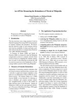
Immunocytological staining for ER-α and ER-β revealed
that LNCaP expressed both receptors (figure 1). In con-
trast, in our passages of PC-3 cells we could not stain any
of these receptors.
Genistein inhibits clonogenic cell survival in LNCaP and
PC-3
In PC-3 cells we tested genistein concentrations between
0.1 μM and 25 μM, and estradiol concentrations between
0.01 μM and 10 μM. In LNCaP both hormones were used
in concentrations between 0.01 μM and 10 μM.
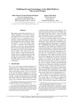
Incubation of LNCaP and PC-3 with genistein without
irradiation resulted in a significant reduction in clono-
genic survival in both cell lines (figure 2). In PC-3 cells,
this effect appeared to be dose-dependent. In contrast,
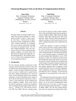
incubation with estradiol exhibited in low concentrations
(0.01 μM) stimulatory effects on the clonogenic survival
of both cell lines, while higher concentrations did not
alter colony formation ability as compared to controls
(figure 3).
Genistein or estradiol sensitize LNCaP to low radiation
doses
Clonogenic survival of irradiated LNCaP cells without
hormonal incubation did not follow the linear-quadratic
model. Instead, a marked hypersensitivity of the cells to
low irradiation doses (<0.1 Gy – 0.3 Gy) was revealed (fig-
ure 4). Radiation with 0.2 Gy decreased colony formation
to 60% compared to unirradiated controls. Higher radia-
tion doses led to a sharp increase in radioresistance: 0.4
Gy reduced clonogenic survival to 95%. This effect has
been described before as ”low-dose hyper-radiosensitiv-
ity“ [HRS] [30].
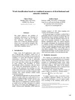
However, when cells were incubated with estradiol 10 μM
or genistein 10 μM before irradiation, the sensitivity to
radiation doses between 0.4 and 2 Gy was significantly
increased compared to irradiation alone controls (figure
4). Combination of estradiol incubation and irradiation
S
no. of colonies counted at a given dose
no. of cells plat
=
eed at a given dose
control no. of cells plated
control no.
×
of colonies counted
.
Radiation Oncology 2008, 3:19 />Page 4 of 12
(page number not for citation purposes)
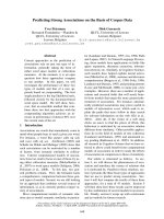
Immuncytological staining of ER-α and -β in EFO-21 (positive control), BG-1 (negative control), LNCaP and PC-3Figure 1
Immuncytological staining of ER-α and -β in EFO-21 (positive control), BG-1 (negative control), LNCaP and
PC-3. In the first row ER-α has been stained, in the second ER-β. EFO-21 and BG-1 cells served as controls: EFO-21 is an
ovarian carcinoma cell line that expresses both ER-α and ER-β, whereas BG-1 is an ovarian cell that does not express these
receptors. Expression of the receptors reflects a brown staining. For easier analysis, staining of the nuclei with DAB was not
performed in the presented samples of LNCaP and PC-3. While LNCaP cells showed expression of ER-α and ER-β, PC-3 cells
did not.
Clonogenic survival LNCaP (left side) and PC-3 (right side) after incubation with genistein (LNCaP 48 h incubation, PC-3 24 h)Figure 2
Clonogenic survival LNCaP (left side) and PC-3 (right side) after incubation with genistein (LNCaP 48 h incu-
bation, PC-3 24 h). Survival was expressed relative to untreated controls. Error bars represent standard errors. In both cell
lines a significant reduction in colony forming is observed after incubation with genistein. Colony formation was reduced to
50% of the controls in LNCaP afer incubation with genistein 0.01 μM (p = 0.004). Higher genistein concentrations (0.1 μM and
10 μM) did not further suppress clonogenic survival. In PC-3 incubation with genistein 0.1 μM decreased colony formation to
75% of the controls (p = 0.027), higher concentrations reduced clonogenic survival further (10 μM: p < 0.001; 25 μM: p <
0.001).
0
0.2
0.4
0.6
0.8
1
1.2
1.4
Surviving fraction [S/S0]
control 0.01μM 0.1μM 10 μM
LNCaP and Genistein
0
0.2
0.4
0.6
0.8
1
1.2
1.4
Surviving fraction [S/S0]
control 0.1μM 10μM 25 μM
PC3 and Genistein
Radiation Oncology 2008, 3:19 />Page 5 of 12
(page number not for citation purposes)
with 1 Gy resulted in a reduction of clonogenic survival to
45%, after incubation with genistein to 50%. With higher
radiation doses (2 Gy or higher) hormonal incubation did
not alter clonogenic survival of LNCaP cells significantly
compared to controls.
Genistein and estradiol enhance radioresistance of PC-3 to
low radiation doses
Irradiation of PC-3 cells without hormonal incubation
revealed a marked HRS to low radiation doses (figure 5).
Radiation with 0.3 Gy decreased colony formation to 57%
Clonogenic survival LNCaP (left side) and PC-3 (right side) after incubation with estradiol (LNCaP 48 h incubation, PC-3 24 h)Figure 3
Clonogenic survival LNCaP (left side) and PC-3 (right side) after incubation with estradiol (LNCaP 48 h incu-
bation, PC-3 24 h). Survival was expressed relative to untreated controls. Error bars represent standard errors. In both cell
lines estradiol 0.01 μM increased colony formation significantly (LNCaP: p < 0.0001; PC-3: p < 0.0001), while higher concentra-
tions of estradiol did not influence colony formation in comparison to untreated controls.
0
0.5
1
1.5
2
Surviving fraction [S/S0]
control 0.01μM
0.1μM 10 μM
LNCaP and Estradiol
0
0.5
1
1.5
2
Surviving fraction [S/S0]
control 0.01μM
0.1μM 10 μM
PC-3 and Estradiol
Survival of LNCaP cells after 24 h pretreatment with genistein 10 μM (left) and with estradiol 10 μM (right), followed by irradi-ation with single doses between 0.5 and 4 Gy, and by further 24 h of hormonal incubationFigure 4
Survival of LNCaP cells after 24 h pretreatment with genistein 10 μM (left) and with estradiol 10 μM (right),
followed by irradiation with single doses between 0.5 and 4 Gy, and by further 24 h of hormonal incubation.
Survival was expressed relative to sham-irradiated controls. Error bars represent standard errors. A polynominal equation was
used to fit the low-dose hyper-radiosensitivity region of all curves. Incubation with genistein 10 μM and estradiol 10 μM
enlarged the area of radiohypersensitivity to doses of up to 1 Gy when compared to untreated controls. p-values for LNCaP
control vs. genistein 10 μM were p < 0.05 at the following dose points: 0.4 Gy, 0.5 Gy, 0.6 Gy, 0.8 Gy, 1 Gy. No significant dif-
ferences were found between the clonogenic survival curves at 0 Gy, 0.2 Gy, 2 Gy, 3 Gy and 4 Gy. p-values for LNCaP con-
trols vs. estradiol 10 μM were p < 0.05 at the following dose points: 0.4 Gy, 0.6 Gy, 0.8 Gy, 1 Gy and 3 Gy. No significant
differences were found at 0 Gy, 0.5 Gy, 2 Gy and 4 Gy.
0,1
1
01234
LNCaP controls (drawn line)
vs genistein 10 μM (dashed line)
surviving fraction [S/S0]
dose [Gy]
0,1
1
01234
dose [Gy]
surviving fraction [S/S0]
LNCaP controls (drawn line)
vs estradiol 10 μM (dashed line)
Radiation Oncology 2008, 3:19 />Page 6 of 12
(page number not for citation purposes)
compared to unirradiated controls. With higher radiation
doses (0.5 Gy – 1 Gy) radiosensitivity did not increase fur-
ther, while at 4 Gy clonogenic survival was about 28%.
In contrast to the results obtained with LNCaP, incuba-
tion with estradiol 10 μM or genistein 10 μM increased
resistance to low irradiation doses in PC-3. Clonogenic
survival was significantly higher after hormonal incuba-
tion when compared to radiation alone at 1 Gy. The sur-
vival curves after hormonal incubation followed the
linear-quadratic model. At higher irradiation doses, we
did not find a significant difference between hormonal
incubation and control.
Estrogenic stimulation inhibits p21-induction after
irradiation in LNCaP
On protein level the expression of p21 was analyzed, as
these proteins are involved in cell cycle control. To control
for effects of serum withdrawal, controls without serum-
withdrawal were investigated, too. Because of mutation of
p53 in PC-3 cells, we could not detect any expression of
p21 in this cell line [31] (plots not shown).
p21 expression was increased in LNCaP 6 h after irradia-
tion in a radiation-dose dependent manner in controls
and after incubation with low concentration of genistein
or estradiol (0.01 μM) (figure 6). In contrast, incubation
with high hormone concentrations (10 μM) abolished the
increase in p21 expression.
Irradiation increases fraction of cells in G2/M in PC-3
Analysis of cell cycle distribution did not show significant
differences in LNCaP cells incubated with estradiol (10
μM) or genistein (10 μM) before irradiation (0.5 Gy, 4
Gy) when compared to controls (serum withdrawal). A
high proportion of these cells rested in G0/G1 (about
70%), this proportion was not significantly reduced by
hormonal stimulation (data not shown).
In PC-3 cells unirradiated controls exhibited a nearly con-
stant G2/M fraction during the whole time course (about
20%, figure 7). However, the S-phase fraction decreased
from 15% at the beginning of the observation (24 h after
serum withdrawal) to 5% 66 h after serum withdrawal.
Comparable cell cycle distribution characteristics were
seen after incubation with 10 μM genistein. Only 66 h
after serum withdrawal a higher proportion of the cells in
S-phase were detected.
Survival of PC-3 cells after 24 h pretreatment with genistein 10 μM (left) and with estradiol 10 μM (right), followed by irradia-tion with single doses between 0.5 and 4 Gy, followed by immediate-platingFigure 5
Survival of PC-3 cells after 24 h pretreatment with genistein 10 μM (left) and with estradiol 10 μM (right), fol-
lowed by irradiation with single doses between 0.5 and 4 Gy, followed by immediate-plating. Survival was
expressed relative to sham-irradiated controls. Error bars represent standard errors. A polynominal equation was used to fit
the low-dose hyper-radiosensitivity region of the control curves while the genistein and estradiol curves followed a linear-
quadratic equation. Incubation with genistein and estradiol abolished the HRS to low irradiation doses seen in the controls
(genistein 10 μM vs. controls: 0.4 Gy: p < 0.01; 0.6 Gy: p = 0.027; estradiol 10 μM vs. controls: 0.4 Gy: p < 0.001; 0.6 Gy: p <
0.001). At and above 2 Gy there were no significant differences between the surviving clones after hormonal incubation and
controls.
0,1
1
01234
PC-3 control (drawn line)
vs genistein 10 μM (dashed line)
Surviving fraction [S/S0]
dose [Gy]
0,1
1
01234
surviving fraction [S/S0]
dose [Gy]
PC-3 control (drawn line)
vs estradiol 10 μM (dashed line)
Radiation Oncology 2008, 3:19 />Page 7 of 12
(page number not for citation purposes)
In contrast, irradiation (4 Gy) of PC-3 cells after serum
withdrawal resulted in a significant increase in the frac-
tion of cells in G2/M (from 21% before irradiation to 34%
12 h after irradiation [timepoint 60 h, figure 7]). At the
same time-course a reduction of cells in G1 from 73%
before irradiation to 62% 12 h after irradiation was
noticed.
Interestingly, incubation with genistein 10 μM reduced
the amount of cells in G2/M after 4 Gy irradiation when
compared to irradiation alone (figure 7). Comparable
results were achieved with estradiol incubation and irradi-
ation (data not shown).
Irradiation with 0.5 Gy had no significant influence on
cell cycle distribution neither after serum withdrawal nor
after hormone incubation (data not shown).
Discussion
For easier reading the results of the study are summarized
in table 1.
Our passages of LNCaP cells stained positive for ER-α and
ER-β, but no expression of ER-α or ER-β was seen in the
investigated PC-3 passages. RNA expression of these
receptors in LNCaP has been shown before [3,4]. How-
ever, other groups reported contradictory results [32,33].
In PC-3 cells RNA expression of ER-β has been reported
[3]. Others found PC-3 cells to be positive for both ER
types [4,32]. These differing results are explained by dif-
ferences in the passages of the studied cell lines, previous
and present growth conditions, and in the applied meth-
odologies.
In the interpretation of our data on clonogenic survival we
have to take different incubation times for both cell lines
into account. Due to methodological problems (slow
growing LNCaP cells – see materials and methods),
LNCaP were incubated for 48 h and PC-3 for 24 h with
genistein or estradiol. We do not feel that these protocol
variations may sufficiently explain the differences
observed in clonogenic survival between both cell lines.
Effects of hormone incubation
Incubation with genistein for 24 h – 48 h reduced clono-
genic survival in both studied cell lines. Similar results
have been reported by other groups [16,18,34,35]. Taken
Western-Blot analysis of p21 and actin in LNCaP after 24 h of hormonal incubation, irradiation with 0.5 Gy and 4 GyFigure 6
Western-Blot analysis of p21 and actin in LNCaP after 24 h of hormonal incubation, irradiation with 0.5 Gy
and 4 Gy. 6 h after irradiation the cells were harvested and subjected to protein extraction. The shown results represent one
of three assays. In controls incubated in FCS containing media and in controls incubated in media with FCS-withdrawal there
was a induction of p21 expression. The same was seen in cells after incubation with low doses of estradiol (0.01 μM) and gen-
istein (0.01 μM). After incubation with high concentrations of estradiol (10 μM) and genistein (10 μM) the induction of p21 was
completely abolished.
Radiation Oncology 2008, 3:19 />Page 8 of 12
(page number not for citation purposes)
together, the effects of genistein on survival in these cell
lines seem to be independent of ER- and p53-expression.
In contrast, low concentrations of estradiol (0.01 μM)
stimulated clonogenic growth in both cell lines, while
higher concentrations did not exhibit a significant effect
in comparison to controls. In LNCaP cells, these results
are supported by proliferation studies where cells were
incubated up to 5 days with concentrations between
0.0001 – 10 μM [36,37]. In these studies even high estra-
diol concentrations induced cell proliferation. In contrast
to our results, other studies (using serum-containing
media and other endpoints than clonogenic survival)
reported reduction of cell proliferation in PC-3 after incu-
bation with 0.1 μM estradiol [38].
Effects of irradiation alone
HRS of LNCaP and PC-3 cells to low irradiation doses
(<0.1 Gy – 0.3 Gy) has been described before [30]. The
cells are very sensitive to small single radiation doses but
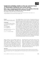
Cell cycle distribution of PC-3 cellsFigure 7
Cell cycle distribution of PC-3 cells. At point "0 h" serum was withdrawn. At point "24 h" incubation genistein 10 μM was
added to designated probes. At point "48 h" designated probes were irradiated with 4 Gy. In the following time, every 6 h
probes were stained with DAPI and analyzed in a flow cytometer up to point "72 h". Every result reflects 3 independent assays.
Proportion of cells in G0/G1 is symbolised by a bar, G2/M by a white bar and S-phase by a black bar. In the controls 15% of the
cells were in S-phase, 65% in G0/G1 and 20% in G2/M. In the following, the S-phase was reduced to 5%, while G0/G1 increased
to 73%. The proportion of cells in G2/M showed minimal changes. After incubation with genistein 10 μM no significant differ-
ences when compared to untreated controls were observed. After irradiation with 4 Gy the proportion of cells in G0/G1
decreased from 73% to 62%, in S-phase decreased from 8% to 3.6% and in G2/M phase increased from 21% to 34.3%. Similar
results were observed after incubation with genistein 10 μM and irradiation with 4 Gy. In the controls 15% of the cells were in
S-phase, 65% in G0/G1 and 20% in G2/M. In the following, the S-phase was reduced to 5%, while G0/G1 increased to 73%. The
proportion of cells in G2/M was only minimal changes. After incubation with genistein 10 μM no significant differences when
compared to untreated controls were observed. After irradiation with 4 Gy the proportion of cells in G0/G1 decreased from
73% to 62%, in S-phase decreased from 8% to 3.6% and in G2/M phase increased from 21% to 34.3%. Similar results were
observed after incubation with genistein 10 μM and irradiation with 4 Gy.
4 Gy
0 Gy
0%
20%
40%
60%
80%
100%
24 48 54 60 66 72
time [h]
cells [%]
G0/G1-phase S-phase G2/M-phase
0%
20%
40%
60%
80%
100%
24 48 54 60 66 72
time [h]
cells [%]
G0/G1-phase S-phase G2/M-phase
Control Genistein 10 μM
0%
20%
40%
60%
80%
100%
24 48 54 60 66 72
time [h]
cells [%]
G0/G1-phase S-phase G2/M-phase
0%
50%
100%
24 48 54 60 66 72
time [h]
cells [%]
G0/G1-phase S-phase G2/M-phase
Radiation Oncology 2008, 3:19 />Page 9 of 12
(page number not for citation purposes)
become more resistant to larger single doses (at about 1
Gy). Explanations for this phenomen have been proposed
by Marples et al. in regard to damage recognition, signal
transduction and damage repair [39]. Amongst others
they postulate a rapidly occurring dose-dependent pre-
mitotic cell cycle checkpoint that is specific to cells irradi-
ated in the G2-phase. The activation of this checkpoint
seems to be dependent on a threshold dose. However, the
clinical relevance of HRS is debatable. To our knowledge,
up to now HRS effects were only described in vitro. An in
vivo proof has not been published yet.
Our data support an independence of the HRS in regard
to ER- or p53/p21-expression.
Effects of the combination of irradiation and hormone
incubation
In combination with irradiation both tested hormones
exhibited similar effects on clonogenic survival dependent
on the investigated cell line. In LNCaP, incubation with
genistein as well as estradiol increased the area of HRS
(including the 1 Gy dose point). To our knowledge, such
effect has not been reported before.
In PC-3 we found a completely different effect as hormo-
nal incubation abolished the HRS observed in the irradi-
ated controls by increasing radioresistance. Clonogenic
survival was best described with the linear-quadratic
model also at low irradiation dose points.
Hillman et al. investigated the combination between irra-
diation and genistein incubation (5–30 μM, 24 h before
irradiation – 10 d after irradiation) on clonogenic survival
of PC-3 cells, too [24]. Only a concentration of 15 μM
genistein reduced clonogenic survival at all measured irra-
diation doses, lower genistein doses had no effect. Sur-
vival curves followed the linear-quadratic model, too.
They did not describe HRS to low irradiation doses, as
only doses of 2 Gy and higher (photon beam) were eval-
uated. Further methodological differences to our study
were the use of FCS-containing media during genistein
incubation and the incubation with higher genistein
doses.
Mechanisms of interaction
To search for mechanisms of interaction we investigated
protein expression of cell cycle controlling proteins. As
PC-3 cells expressed not functional p53 we could not
detect p21 expression in these cells. In LNCaP p21 expres-
sion was increased as a downstream signal transduction
protein of p53 after irradiation when compared to con-
trols. Incubation with high concentrations of genistein or
estradiol abolished this p21 expression after irradiation.
In unirradiated controls no p21 expression was detecta-
ble.
Table 1: Summary of the results
cell line treatment results
receptor expression LNCaP ER-α + ER-β +
PC-3 ER-α 0 ER-β 0
clonogenic survival LNCaP genistein survival reduced
estradiol survival increased (0.01 μM)/no effect (higher concentrations)
genistein + RT area of HRS enlarged
estradiol + RT area of HRS enlarged
PC-3 genistein survival reduced
estradiol survival increased (0.01 μM)/no effect (higher concentrations)
genistein + RT HRS abolished
estradiol + RT HRS abolished
p21 expression LNCaP RT increased expression
RT + genistein 10 μM reduced expression
RT + estradiol 10 μM reduced expression
PC-3 no expression
FACS (cell cycle) LNCaP controls G0/G1 arrest
genistein or estradiol G0/G1 arrest
RT G0/G1 arrest
PC-3 genistein or estradiol no influence
RT G2/M arrest
RT + genistein or estradiol G2/M arrest reduced
Radiation Oncology 2008, 3:19 />Page 10 of 12
(page number not for citation purposes)
These data are in contrast to the literature. Shen et al.
showed a dose-dependent increase in p21 expression after
incubation with genistein (without irradiation, 0 – 80 μM
for 24 h) [17]. Similar results were obtained by another
study after incubation with 5 μM for 6 h – 12 h [40].
With FACS-analysis we tried to verify our Western Blot
results in terms of cell cycle regulation. However, as our
passages of LNCaP cells proliferated very slowly in FCS-
free medium (time for cell doubling 5 days), the majority
of cells was in G0/G1. Incubation with hormones did not
dissolve this accumulation. With such a high level of cells
in G0/G1 in control cells, the increase after irradiation in
this proportion of cells did not reach significance. There-
fore, short term effects as seen in western blotting did not
result in significant changes in cell cycle distribution.
Effects described in clonogenic survival were not
explained by the results of cell cycle analysis.
In PC-3 cells, incubation with estradiol 10 μM or genistein
10 μM did not alter cell cycle distribution significantly
when compared to controls. However, irradiation with 4
Gy induced a G2/M cell cycle arrest, but not irradiation
with 0.5 Gy. This result is explained by the missing of
functional p53, thus lacking of any G0/G1 arrest. The G2/
M arrest after irradiation with high doses was not abol-
ished by estrogenic stimulation. These results are sup-
ported by another study that reported a G2/M cell cycle
arrest 72 h and 96 h after irradiation with 3 Gy or after
incubation with genistein concentrations of 15 – 30 μM in
FCS-containing media [41]. In this study the NF-κB activ-
ity was investigated, too. An inhibition of radiation-
induced activation of NF-κB activity by genistein pretreat-
ment could be shown. Furthermore, a significant increase
in cleaved PARP protein was measured following com-
bined genistein and radiation treatment, indicating
increased apoptosis. The authors proposed a mechanism
of increased cell death by genistein and radiation via inhi-
bition of NF-κB, leading to altered expression of regula-
tory cell cycle proteins, thus promoting G2/M arrest and
increased radiosensitivity [41]. However, as it is doubtful
whether apoptosis is clinical relevant in irradiated solid
tumor cells, we did not measure this endpoint in our
study [42].
One potential interaction between estrogenic stimulation
and irradiation could be identified in ER-α and ER-β pos-
itive LNCaP cells. Irradiation induces double strand
breaks, these are recognized and via phosphorylation of
ATM, p53 and p21 a G0/G1 arrest is induced. Activated
ERs interfere with this cascade by inducing degradation of
p21, thus abolishing G0/G1 arrest [43]. However, these
cascades do not explain the increased area of HRS seen in
clonogenic survival analysis after incubation with genis-
tein or estradiol. Furthermore, we could not identify the
molecular mechanism of the results observed in ER-α and
ER-β negative PC-3 cells.
Taken together, since we showed comparable effects of
genistein and estradiol in combination with irradiation in
both studied cell lines neither ER- nor p53-expression
seemed to play a role in the linked signalling. Neverthe-
less, both compounds targeted the same molecular
switch, that we were not able to identify.
Conclusion
We observed an increased HRS to low irradiation doses
after incubation with estradiol 10 μM and genistein 10
μM in ER-α and ER-β positive LNCaP cells. In contrast, in
ER-α and ER-β negative PC-3 cells, we observed an abol-
ishing of the HRS to low irradiation doses by hormonal
stimulation. In conclusion, HRS was independent from
ER- or p53/p21-expression. It was modulated by genistein
and estradiol dependent from the genetic background of
the investigated cell line. Furthermore, the effects of both
tested compounds on survival were ER and p53 independ-
ent. Since genistein and estradiol effects in both cell lines
were comparable, neither ER- nor p53-expression seemed
to play a role in the linked signalling. Nevertheless both
compounds targeted the same molecular switch. To iden-
tify the underlying molecular mechanisms, further studies
are needed.
The observation of an extended HRS of PC cells after incu-
bation with genistein or estradiol would be of high clini-
cal interest, especially as LNCaP reflects a locally
advanced, androgen dependent PC. This would mean,
that PC could be irradiated with decreased irradiation
doses, resulting in reduced normal tissue toxicity. How-
ever, as our data are based on in vitro observations only,
these results have to be interpreted with caution. To our
knowledge, no in vivo proof for HRS to low irradiation
doses has been published up to day.
Competing interests
The authors declare that they have no competing interests.
Authors' contributions
RMH outlined the study, helped HAW to perform the
majority of the experimental work and drafted the manu-
script. MRF supervised the radiobiological experiments
and molecularbiological work. PT carried out the cell
cycle analyses. HJ participated in the planning of the
experiments and the Western Blot analyses. HS conceived
the study and helped with coordination. CG performed
immunostaining. HC participated in its design and coor-
dination and helped to draft the manuscript.
All authors read and approved the final manuscript.
Radiation Oncology 2008, 3:19 />Page 11 of 12
(page number not for citation purposes)
Acknowledgements
This research was supported by the medical faculty of the University of
Goettingen with financial funding.
References
1. Gesellschaft der epidemiologischen Krebsregister in Deutschland e.V:
Krebs in Deutschland: Häufigkeiten und trends. Saarbrücken
2006 [
].
2. Bolla M, Collette L, Blank L, Warde P, Dubois JB, Mirimanoff RO,
Storme G, Bernier J, Kuten A, Sternberg C, Mattelaer J, Lopez Tore-
cilla J, Pfeffer JR, Lino Cutajar C, Zurlo A, Pierart M: Long-term
results with immediate androgen suppression and external
irradiation in patients with locally advanced prostate cancer
(an EORTC study): a phase 3 randomised trial. Lancet 2002,
360:103-106.
3. Ito T, Tachibana M, Yamamoto S, Nakashima J, Murai M: Expression
of estrogen receptor (ER-alpha and ER-beta) mRNA in
human prostate cancer. Eur Urol 2001, 40:557-563.
4. Maruyama S, Fujimoto N, Asano K, Ito A, Usui T: Expression of
estrogen receptor alpha and beta mRNAs in prostate can-
cers treated with leuprorelin acetate. Eur Urol 2000,
38:635-639.
5. Sasaki M, Tanaka Y, Perinchery G, Dharia A, Kotcherguina I, Fujimoto
S, Dahiya R: Methylation and inactivation of estrogen, proges-
terone, and androgen receptors in prostate cancer. J Natl
Cancer Inst 2002, 94:384-390.
6. Kuiper GGJM, Lemmen JG, Barlsson B, Corton JC, Safe SH, Saag PT
van der: Interaction of estrogenic chemicals and phytoestro-
gens with estrogen receptor β. Endocrinology 1998,
139:4252-4263.
7. Fischer L, Mahoney C, Jeffcoat AR, Koch MA, Thomas BE, Valentine
JL, Stinchcombe T, Boan J, Crowell JA, Zeisel SH: Clinical charac-
teristics and pharmacokinetics of purified soy isoflavones:
multiple-dose administration to men with prostate neopla-
sia. Nutr Cancer 2004, 48:160-170.
8. Akiyama T, Ishida J, Nakagawa S, Ogawara H, Watanabe SI, Itoh N,
Shibuya M, Fukami Y: Genistein, a specific inhibitor of tyrosine-
specific protein kinase. J Biol Chem 1987, 262:5592-5595.
9. Huang J, Nasr M, Kim Y, Matthews HR: Genistein inhibits protein
histidine kinase. J Biol Chem 1992, 267:15511-15515.
10. Bektic J, Guggenberger R, Eder IE, Pelzer AE, Berger AP, Bartsch G,
Klocker H: Molecular effects of the isoflavonoid genistein in
prostate cancer. Clin Prostate Cancer 2005,
4:124-129.
11. Kaufmann WK: Human topoisomerase II function, tyrosine
phosphorylation and cell cycle checkpoints. Proc Soc Exp Biol
Med 1998, 217:327-334.
12. Molteni A, Brizio-Molteni L, Persky V: In vitro hormonal effects of
soybean isoflavones. J Nutr 1995, 125(3 Suppl):751-756.
13. Kim H, Peterson TG, Barnes S: Mechanisms of action of the soy
isoflavone genistein: emerging role for ist effects via trans-
forming growth factor β signalling pathways. Am J Clin Nutr
1998, 68:1418-1425.
14. Mitchell JH, Duthie SJ, Collins AR: Effects of phytoestrogens on
growth and DNA integrity in human prostate cell lines: PC-
3 and LNCaP. Nutr Cancer 2000, 38:223-228.
15. Davis JN, Singh B, Bhuiyan M, Sarkar FH: Genistein-induced upreg-
ulation of p21WAF1, downregulation of cyclin B, and induc-
tion of apoptosis in prostate cancer cells. Nutr Cancer 1998,
32:123-131.
16. Onozawa M, Fukuda K, Ohtani M, Akaza H, Sugimura T, Wakabayashi
K: Effects of soybean isoflavones on cell growth and apoptosis
of the human prostatic cancer cell line LNCaP. Jpn J Clin Oncol
1998, 28:360-363.
17. Shen JC, Klein RD, Wei Q, Guan Y, Contois JH, Wang TT, Chang S,
Hursting SD: Low-dose genistein induces cyclin-dependent
kinase inhibitors and G(1) cell-cycle arrest in human pros-
tate cancer cells. Mol Carc 2000, 29:92-102.
18. Zhou JR, Gugger ET, Tanaka T, Guo Y, Blackburn GL, Clinton SK:
Soybean phytochemicals inhibit the growth of transplanta-
ble human prostate carcinoma and tumor angiogenesis in
mice. J Nutr 1999, 129:1628-1635.
19. Schmidberger H, Hermann RM, Hess CF, Emons G: Interactions
between radiation and endocrine therapy in breast cancer.
Endocr Relat Cancer 2003, 10:375-388.
20. Villalobos M, Becerra D, Nunez MI, Valenzuela MT, Shiles E, Olea N,
Pedraza V, Ruiz de Almodovar JM: Radiosensitivity of human
breast cancer cell lines of different hormonal responsiveness.
Modulatory effects of oestradiol. Int J Radiat Biology 1996,
70:161-169.
21. Akimoto T, Nonaka T, Ishikawa H, Sakurai H, Saitoh JI, Takahashi T,
Mitsuhashi N: Genistein, a tyrosine kinase inhibitor, enhanced
radiosensitivity in human esophageal cancer cell lines in
vitro: possible involvement of inhibition of survival signal
transduction pathways. Int J Radiat Oncol Biol Phys 2001,
50:195-201.
22. van Rijn J, Berg J van den: Flavonoids as enhancers of x-ray-
induced cell damage in hepatoma cells. Clin Cancer Res 1997,
3:1775-1779.
23. Papazisis KT, Zambouli D, Kimoundri OT, Papadakis ES, Vala V, Gero-
michalos GD, Voyatzi S, Markala D, Destouni E, Boutis L, Kortsaris
AH: Protein tyrosine kinase inhibitor, genistein, enhances
apoptosis and cell cycle arrest in K564 cells treated with γ-
irradiation. Cancer Letters 2000, 160:107-113.
24. Hillman GG, Forman JD, Kucuk O, Yudelev M, Maughan RL, Rubio J,
Layer A, Tekyi-Mensah S, Abrams J, Sarkar FH: Genistein potenti-
ates the radiation effect on prostate carcinoma cells. Clin Can-
cer Res 2001, 7:382-390.
25. Yan SX, Ejima Y, Sasaki R, Zheng SS, Demizu Y, Soejima T, Sugimura
K: Combination of genistein with ionizing radiation on
androgen-independent prostate carcinoma cells. Asian J Androl
2004, 6:280-290.
26. Hillman GG, Wang Y, Kucuk O, Che M, Doerge DR, Yudelev M,
Joiner MC, Marples B, Forman JD, Sarkar FH: Genistein potenti-
ates inhibition of tumor growth by radiation in a prostate
cancer orthotopic model. Mol Cancer Ther 2004, 3:1271-1279.
27. Julian J, Raffoul JJ, Banerjee S, Che M, Knoll ZE, Doerge DR, Abrams
J, Kucuk O, Sarkar FH, Hillman GG: Soy isoflavones enhance radi-
otherapy in a metastatic prostate cancer model. Int J Cancer
2007, 120:2491-2498.
28. Hermann RM, Fest J, Christiansen H, Hille A, Rave-Fränk M, Nitsche
M, Gründker C, Viereck V, Jarry H, Schmidberger H: Radiosensiti-
zation dependent on p53 function in bronchial carcinoma
cells by the isoflavone genistein and estradiol in vitro. Strahl-
enther Onkol 2007, 183:195-202.
29. Hall EJ: Cell-survival curves.
In Radiobiology for the Radiologist Phil-
adelphia: Lippincott; 1998.
30. Joiner MC, Marples B, Lambin P, Short SC, Turesson I: Low-dose
hypersensitivity: current status and possible mechanisms. Int
J Radiat Oncol Biol Phys 2001, 49:379-389.
31. An J, Chervin AS, Nie A, Ducoff HS, Huang Z: Overcoming the
radioresistance of prostate cancer cells with a novel Bcl-2
inhibitor. Oncogene 2007, 26:652-661.
32. Lau KM, LaSpina M, Long J, Ho SM: Expression of estrogen recep-
tor (ER)-alpha and ER-beta in normal and malignant pros-
tatic epithelial cells: regulation by methylation and
involvement in growth regulation. Cancer Res 2000,
60:3175-3182.
33. Hobisch A, Hittmair A, Daxenbichler G, Wille S, Radmayr C,
Hobisch-Hagen P, Bartsch G, Klocker H, Culig Z: Metastatic
lesions from prostate cancer do not express oestrogen and
progesterone receptors. J Pathol 1997, 182:356-361.
34. Bemis DL, Capodice JL, Desai M, Buttyan R, Katz AE: A concen-
trated aglycone isoflavone preparation (GCP) that demon-
strates potent anti-prostate cancer activity in vitro and in
vivo. Clin Cancer Res 2004, 10:5282-5292.
35. Ouchi H, Ishiguro H, Ikeda N, Hori M, Kubota Y, Uemura H: Genis-
tein induces cell growth inhibition in prostate cancer
through the suppression of telomerase activity. Int J Urol 2005,
12:73-80.
36. Arnold JT, Le H, McFann KK, Blackman MR: Comparative effects
of DHEA vs. testosterone, dihydrotestosterone, and estra-
diol on proliferation and gene expression in human LNCaP
prostate cancer cells. Am J Physol Endocrinol Metab 2005,
288:573-584.
37. Castagnetta LA, Miceli MD, Sorci CM, Pfeffer U, Farruggio R, Oliveri
G, Calabro M, Carruba G: Growth of LNCaP human prostate
cancer cells is stimulated by estradiol via its own receptor.
Endocrinology 1995, 136:2309-2319.
Publish with Bio Med Central and every
scientist can read your work free of charge
"BioMed Central will be the most significant development for
disseminating the results of biomedical research in our lifetime."
Sir Paul Nurse, Cancer Research UK
Your research papers will be:
available free of charge to the entire biomedical community
peer reviewed and published immediately upon acceptance
cited in PubMed and archived on PubMed Central
yours — you keep the copyright
Submit your manuscript here:
/>BioMedcentral
Radiation Oncology 2008, 3:19 />Page 12 of 12
(page number not for citation purposes)
38. Tinley TL, Leal RM, Randall-Hlubek DA, Cessac JW, Wilkens LR, Rao
PN, Mooberry SL: Novel 2-methoxyestradiol analogues with
antitumor activity. Cancer Res 2003, 63:1538-1549.
39. Marples B, Wouters BG, Collis SJ, Chalmers AJ, Joiner MC: Low-
dose hyper-radiosensitivity: A consequence of ineffective cell
cycle arrest of radiation-damaged G2-phase cells. Radiation
Res 2004, 161:247-255.
40. Rao A, Coan A, Welsh JE, Barclay WW, Koumenis C, Cramer SD:
Vitamin D receptor and p21/WAF1 are targets of genistein
and 1,25-dihydroxyvitamin D3 in human prostate cancer
cells. Cancer Res 2004, 64:2143-2147.
41. Raffoul JJ, Wang Y, Kucuk O, Forman JD, Sarkar FH, Hillman GG:
Genistein inhibits radiation-induced activation of NF-kappaB
in prostate cancer cells promoting apoptosis and G2/M cell
cycle arrest. BMC Cancer 2006, 6:107-117.
42. Dewey WC, Ling CC, Meyn RE: Radiation-induced apoptosis:
relevance to radiotherapy. Int J Radiat Oncol Biol Phys 1995,
33:781-796.
43. Foster JS, Henley DC, Bukovsky A, Seth P, Wimalasena J: Multifac-
eted regulation of cell cycle progression by estrogen: regula-
tion of Cdk inhibitors and Cdc25A independent of cyclin D1-
Cdk4 function. Mol Cell Biol 2001, 21:794-810.