Báo cáo khoa học: " The impact of functional imaging on radiation medicine" doc
Bạn đang xem bản rút gọn của tài liệu. Xem và tải ngay bản đầy đủ của tài liệu tại đây (674.8 KB, 13 trang )
BioMed Central
Page 1 of 13
(page number not for citation purposes)
Radiation Oncology
Open Access
Review
The impact of functional imaging on radiation medicine
Nidhi Sharma
1
, Donald Neumann
2
and Roger Macklis*
3
Address:
1
Research fellow, Department of Radiation Oncology, Cleveland Clinic Foundation, 9500 Euclid Avenue, Cleveland, OH 44195, USA,
2
Staff physician, Department of Nuclear Medicine, Cleveland Clinic Foundation, 9500 Euclid Avenue, Cleveland, OH 44195, USA and
3
Professor
of Medicine (Radiation Oncology), Cleveland Clinic Lerner College of Medicine and Department of Radiation Oncology, 9500 Euclid Avenue,
Cleveland, OH 44195, USA
Email: Nidhi Sharma - ; Donald Neumann - ; Roger Macklis* -
* Corresponding author
Abstract
Radiation medicine has previously utilized planning methods based primarily on anatomic and
volumetric imaging technologies such as CT (Computerized Tomography), ultrasound, and MRI
(Magnetic Resonance Imaging). In recent years, it has become apparent that a new dimension of
non-invasive imaging studies may hold great promise for expanding the utility and effectiveness of
the treatment planning process. Functional imaging such as PET (Positron Emission Tomography)
studies and other nuclear medicine based assays are beginning to occupy a larger place in the
oncology imaging world. Unlike the previously mentioned anatomic imaging methodologies,
functional imaging allows differentiation between metabolically dead and dying cells and those
which are actively metabolizing. The ability of functional imaging to reproducibly select viable and
active cell populations in a non-invasive manner is now undergoing validation for many types of
tumor cells. Many histologic subtypes appear amenable to this approach, with impressive sensitivity
and selectivity reported.
For clinical radiation medicine, the ability to differentiate between different levels and types of
metabolic activity allows the possibility of risk based focal treatments in which the radiation doses
and fields are more tightly connected to the perceived risk of recurrence or progression at each
location.
This review will summarize many of the basic principles involved in the field of functional PET
imaging for radiation oncology planning and describe some of the major relevant published data
behind this expanding trend.
Review
Introduction and background
Recent advances in high precision radiation treatment
methodologies have focused on developing a tighter cor-
respondence between the visualized location of neoplas-
tic target structures and the radiation dose deposition
patterns chosen in an attempt to control the target tissue
proliferation. The ability to map the real time or near-real-
time positional information has been facilitated by the
rapid growth over the last few decades in high speed com-
puting and algorithms for shape recognition and manipu-
lation. These processing algorithms are gleaned from
diverse fields including industrial manufacturing, military
applications, and the entertainment industry. These
advances have now essentially made it possible to "paint"
recognizable target structures with modulated pulses of
Published: 15 September 2008
Radiation Oncology 2008, 3:25 doi:10.1186/1748-717X-3-25
Received: 2 August 2007
Accepted: 15 September 2008
This article is available from: />© 2008 Sharma et al; licensee BioMed Central Ltd.
This is an Open Access article distributed under the terms of the Creative Commons Attribution License ( />),
which permits unrestricted use, distribution, and reproduction in any medium, provided the original work is properly cited.
Radiation Oncology 2008, 3:25 />Page 2 of 13
(page number not for citation purposes)
ionizing radiation using the complex beam-shaping rou-
tines developed for intensity modulated radiotherapy
(IMRT). The validity of such dose painting is, however,
currently the source of intense debate. In order to deter-
mine the optimal dose deposition patterns, methods are
required to correlate three dimensional anatomic struc-
tures with function, physiology, and change over time.
The use of PET (positron emission tomography) provides
one important medical methodology being optimized for
this purpose. This review will summarize the current sta-
tus of the incorporation of physiologic "functional" med-
ical imaging into radiation medicine and radiotherapy
treatment plan design.
Though PET is not really a new field, it has recently under-
gone a dramatic revitalization as new clinical indicators
are validated for this type of functional imaging. The prin-
ciples behind PET involve the non-invasive analysis and
positional correlation of biochemical processes, typically
with a level of quantization not easily achieved using
other nuclear medicine methodologies. This superiority is
based on the fact that PET uses the positron-emitting
annihilation event that occurs when an electron and pos-
itron collide and vanish with the creation of two opposed
photons of a precise characteristic energy 511 keV. This
sort of annihilation reaction can be demonstrated in nat-
ural radioisotopes such oxygen-15, fluorine-18, and car-
bon-11. The invention of complex detectors capable of
sensing the emitted energy stream allowed PET to be vali-
dated as a reproducible physiologic biomarker, originally
for cardiac and neuroanatomic studies and more recently
for many physiologic processes found in oncology. The
high sensitivity of PET for cancer processes relates to the
partially planned and partially fortuitous discovery that
the glucose analog fluorodeoxyglucose (FDG) accumu-
lates in most human cancers and is physiologically
"trapped" within the cell by phosphorylation. Positron
radio-labeled
18
FDG provides some of the highest signal-
to-noise ratios observed in the sometimes murky domain
of oncology imaging due to factors such as neoplastic
over-expression of glucose transport proteins, increased
glycolysis (the "Warburg Effect") and modified cellular
hexokinase activity. The kinetics of this trapping process
produces a gradual rise in the signal and the resolution
limit of the image (typically several millimeters) produces
an imaging envelope representing the total region in
which abnormal glycolysis patterns may be differentiated
from baseline metabolism. There is a delayed physiologic
signal (typically becoming maximal after several hours or
more) and reasonable quantitation may be achieved by
calculating the "standardized uptake value" (SUV) which
normalizes signal size to infused isotope dose and patient
mass. While the typical PET signal produced by FDG
uptake cannot be considered specific for neoplasia, the
PET process has the tremendous advantage over other
oncologic imaging methods of producing rapid whole-
body images capable of delineating and differentiating
between normal structures and many different sites of pri-
mary cancers and metastatic disease. Though the half-lives
of PET radiopharmaceuticals are typically very short (< 0.5
hr) the test may be repeated in a serial fashion in order to
define a valid time course for the observed physiologic
processes. Thus, for the investigator interested in signature
cancer biomarkers, PET provides an entirely new dimen-
sion of physiologic information that may be highly com-
plementary to the routine 3-D anatomic information
obtained through volume-based methods such as CT,
ultrasound, and MRI. Table 1 shows some of the primary
Medicare-accepted indications for the use of this test. For
the radiation oncologist, functional information such as
18
FDG-PET thus provides much useful data on oncologic
process in addition to tumor location. PET has been used
as an adjunct to traditional anatomic modalities to more
accurately assess local and regional disease extent and to
detect early sites of metastasis. Preoperative evaluation of
regional metastases has been tested in a number of disease
sites, including the axilla [1,2] in breast cancer, the neck in
squamous cell carcinomas of head and neck, [3,4] and the
liver in colorectal carcinoma [5,6]. FDG-PET has been
most extensively studied in non-small cell lung cancer
(NSCLC), where surgical assessment of the mediastinal
lymph nodes is typically performed before definite resec-
tion. Using appropriately designed and informative
reporter molecules, PET can be used to trace the evolution
Table 1: Medicare-accepted indications (2007) for positron emission tomography (PET) for Cancers
INDICATION PURPOSE
Breast cancer Staging, restaging, evaluating treatment response
Colorectal cancer Diagnosis, staging, restaging
Esophageal cancer Diagnosis, staging, restaging
Head and neck cancer Diagnosis, staging, restaging
Lung cancer Diagnosis, staging, restaging
Lymphoma Diagnosis, staging, restaging
Melanoma Diagnosis, staging, restaging
Solitary pulmonary nodules Characterization
Thyroid cancer Restaging(with negative iodine-131 scan and positive thyroglobulin)
Radiation Oncology 2008, 3:25 />Page 3 of 13
(page number not for citation purposes)
of the sorts of abnormal physiologic signals which are
often considered the metabolic hallmark of the transfor-
mation event.
Basis of PET scan technology
With the push for new of technology in the fields of
nuclear medicine and radiation oncology, the PET scan
has become a valuable modality in the hands of the phy-
sicians. It has proved to be of immense importance in
modifying the radiation treatment therapy for patients
with malignancies. The basic principle of oncologic PET
scan is based on the characteristic of the malignant cells
which may divide continuously in an uncontrollable
manner, thus altering their metabolic profile compared to
the normal cells. In the past, numerous radiological trac-
ers have been put to practice, but presently 2-[18 F]-
fluoro-2-deoxy-D-glucose (FDG) is the most popular one.
Its role in functional imaging is unique, as it helps differ-
entiate groups of active cancer cells, allowing further
imaging and intervention in the specific diseased site.
Across oncological applications, the sensitivity and specif-
icity of FDG-PET ranged from 84 to 87% and 88 to 93%
respectively [7].
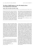
Upon its intravenous administration, the membrane
bound glucose transporter takes up FDG into the cells,
where it gets phosphorylated to
18
FDG-6-phosphate by
the enzyme hexokinase. This product cannot enter the gly-
colytic pathway and thus keeps accumulating inside the
cells (See Figure 1). The uncontrolled proliferation and
metabolic activity of the tumor cells is picked up by PET
scan as it detects the photons emitted by radiotracers like
18
FDG (or C-11, N-13 etc.). These photons are emitted at
a specific energy (511 keV) in opposite directions. There-
fore, PET scanners have detectors placed on the opposite
sides of the region from where the photons are emitted
(within the patient) and the detectors register an event
FDG Mechanism in Functional ImagingFigure 1
FDG Mechanism in Functional Imaging. Abbreviations: 18-FDG: 2-[18 F]-fluoro-2-deoxy-D-glucose; Gl: Glucose; Fru:
fructose.
Radiation Oncology 2008, 3:25 />Page 4 of 13
(page number not for citation purposes)
only if both the detectors record the photon emission at
the same time [8].
There are a few limitations of the PET-only images like
lack of anatomic details required for therapy, physiologic
update of FDG by normal tissues, fat, muscle and lym-
phoid tissue, increasing confounding and also lack of an
easy method to incorporate this information into treat-
ment planning.
Roles for PET imaging in radiotherapy
Malignant lymphoma
The role of PET and PET-CT in oncology is currently most
fully embodied in the relevant work on malignant Hodg-
kin's and non-Hodgkin's Lymphoma. For Hodgkin's Lym-
phoma staging,
18
FDG-PET was shown to be somewhat
more useful than other more traditional anatomic imag-
ing technologies such as CT and MRI and has been
claimed recently to be the "most accurate imaging tech-
nology for staging malignant lymphoma." It is now fairly
routine to obtain a pretreatment baseline
18
FDG-PET
study for Hodgkin's and aggressive non-Hodgkin's Lym-
phoma prior to the initiation of chemotherapy and
18
FDG-PET studies have largely replaced gallium scans as
a pretreatment and post-treatment whole-body radionu-
clide studies for lymphoma. While some of the earliest
studies evaluating
18
FDG-PET for malignant lymphoma
date from the 1980s, investigation in this area has
expanded dramatically in the last decade as evidence
mounted for the sensitivity and cost effectiveness of the
technology. For malignant lymphoma, both tumor grade
and proliferative activity appeared to be somewhat corre-
lated with the uptake intensity of the FDG signal. How-
ever, these findings have not always been reproducible
and at present it appears that the correlation of high SUV
levels to tumor grade are still insufficient to be used in
clinical treatment decision making.
In addition to providing a sensitive and noninvasive tool
for oncologic staging, FDG-PET has also shown utility in
assessing response to treatment. This is particularly help-
ful in-lymphoma, where post-treatment fibrosis can
obscure detection of residual disease [9,10]. In a study of
44 patients with abdominal presentations of Hodgkin's
disease (HD) and non-Hodgkin's lymphoma (NHL) [11],
FDG-PET proved superior to anatomic imaging in deter-
mining post-treatment tumor viability. Thirty seven of the
44 patients had residual CT abnormality following chem-
otherapy with or without radiation therapy. Thirteen
patients were also shown to be positive by FDG-PET, and
all of these patients eventually relapsed. Only 1 patient,
negative by FDG-PET but positive by CT, relapsed. The
relapse-free survival rate was 0% for those patients posi-
tive by FDG-PET, and 95% for those negative by FDG-PET
at 2 years. Clearly, patients shown to have residual disease
by FDG-PET should be considered for additional treat-
ment.
The role of FDG-PET in Hodgkin's Lymphoma workups
and management has been the subject of several recent
reviews. Castellucci et al evaluated 967 consecutive PET
studies in 706 individual patients treated previously for
malignant lymphoma. They found that over 20 percent
showed focal FDG uptake unrelated to the presence of
known tumor deposits (e.g., a "false positive"). This "false
positive" uptake appeared to result from a number of
potential causes including either "brown fat" (mean SUV:
11.7) thymic hyperplasia (mean SUV: 4.1) muscle con-
traction (mean SUV: 7.4) or various types of inflamma-
tion or infection (mean SUV levels 4–7) [12]. These
authors suggest that the use of correlated single-platform
PET-CT should minimize the number of spurious "false
positives" produced by non-tumor FDG signals. At a min-
imum, it suggests that FDG hot-spots should not be eval-
uated in the absence of additional anatomic information.
FDG-PET can also serve as a sensitive means to monitor
therapy in progress, with an eye to changing ineffectual
treatments in midcourse. A provocative study from Ger-
many used early response to FDG-PET to predict outcome.
The treatment course of 11 patients with NHL was moni-
tored by Romer et al [13]. All patients underwent FDG-
PET imaging before treatment, at 1 week, and again at 6
weeks. The mean decrease in SUV at day 42 was 79%.
Interestingly, the tumor SUV levels at week 1 were signifi-
cantly lower in the group of 6 patients remaining in remis-
sion after 16 months follow-up, than in the group of
patients eventually relapsing. Patients showing no
response by FDG-PET at 1 week might be candidates for
more aggressive/altered treatment regimens. Others have
used FDG-PET in a similar fashion to monitor response to
neoadjuvant chemotherapy in patients with locally
advanced breast cancer [14,15].
For evaluation of response, the PET or PET-CT appears to
be gaining ground with respect to accepted clinical utility.
The "International Workshop Criteria for Response in
NHL" recently adopted PET as the "gold standard in
response evaluation." For NHL patients treated with
CHOPR chemotherapy, response after just 2–3 cycles was
shown to predict eventual clinical outcomes. This "early
look" at response is of extreme importance in choosing
therapies likely to produce long-term control without the
necessity of a protracted and potentially dangerous course
of treatment. Other investigators are evaluating F-18
fluorothymidine (
18
FLT) rather than
18
FDG due to the
more specific uptake of this analog into DNA [16]. While
FDG mirrors glycolysis,
18
FLT is thought to mirror DNA
synthesis. Patients with positive PET studies after chemo-
therapy had a significantly higher risk of relapse than
Radiation Oncology 2008, 3:25 />Page 5 of 13
(page number not for citation purposes)
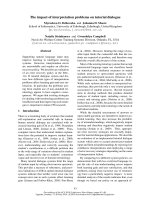
those with negative scans (P < 0.0001) though not all
patients with persistently positive scans ultimately
showed evidence of clinical progression and a negative
post-treatment PET was not an accurate predictor that
local progression was contained (See Figure 2).
For radiotherapy, one interesting question is whether the
PET studies can be used to pick out those patients who
might benefit from post-chemotherapy involved-field
radiation, and whether the location and intensity of the
PET signal can be used to guide radiotherapy treatment
planning. Kahn et al [17] showed that FDG-PET was use-
ful in identifying the patients likely to recur and the sites
at which they were most at risk for recurrence. However,
patients with positive post-chemotherapy PET studies
were not fully protected by local field radiotherapy as
administered in this trial. The authors note that the fields
were designed to include only the persistent PET-positive
regions of assumed disease, and that dose and fractiona-
tion schemes were "highly individualized" with median
doses of 30.6 Gy and dose ranges of 9–46 Gy. Over half of
the relapses observed in this study occurred infield. Thus,
either the treated region or the dose was insufficient to
control disease sites showing post-chemotherapy positive
PET signals.
With respect to radiotherapy field design, some have dis-
cussed the use of PET and other similar functional studies
in what they call "Theragnostic imaging" appropriate for
use as a guide for radiation "dose painting." The term
Theragnostic is meant to refer to the use of medical images
to guide treatment decisions and intensity. For radiother-
apy, the suggestion is that tumor burden and clonogen
density may be indicated by FDG or FLT PET SUV required
levels and that these levels may be used as a proxy for
recurrence risk and therefore required dose of radiation
necessary to achieve local control [18]. If this conjecture
proves true, the deliberately inhomogeneous dose deliv-
ery algorithms currently used in IMRT technology may be
fitted using "inverse planning" to estimated risk maps
incorporating indices of proliferation, hypoxia, and other
known local recurrence risk factors. In a sense, this is a
more dosimetrically rigorous version of the now-accepted
risk-adjustment methodology commonly used in current
clinical radiotherapy approaches in which NHL complete
responders (CR) to chemotherapy are given lower doses
than patients showing only partial responses. Whether
this general principle, clinically validated for aggressive
lymphomas, can be applied to small sub-portions of non
localized tumors will require additional study. One could
construct reasonable arguments to support the hypothesis
Assessment of treatment response of lymphoma with PETFigure 2
Assessment of treatment response of lymphoma with PET. Images of pre- and post-therapy PET scans in a lymphoma
patient treated with chemotherapy. The pre therapy image (left) shows increased FDG uptake in the left supraclavicular region
(red arrow), mesentery, retroperitoneum (yellow arrow), and spleen (olive arrow). The post-therapy image (right) shows no
residual disease, with a bone marrow activation commonly seen after chemotherapy and which can be seen with other treat-
ments such as granulocyte colony-stimulating factor.
Radiation Oncology 2008, 3:25 />Page 6 of 13
(page number not for citation purposes)
that either the FDG-intense areas or the FDG-cold areas
would require higher doses, depending on whether one
proposes to dose-intensify regions of higher proliferation
or lower oxygenation. While specific PET markers of
hypoxia such as
18f
Misonidazole are currently being stud-
ied in both pre-clinical and clinical trials, some investiga-
tors claim that images obtained on untreated patients may
show significant changes over a few hours or days ("inter-
mittent hypoxia") and hence are not reproducible mark-
ers of a fixed biology [19]. If the hypoxia markers show us
only temporary biologic indications of intermittent vascu-
lar status then dose adjustments based on these images
would be invalid. The idea of dose-painting based on
"theragnostic imaging" though intellectually appealing, is
thus still in the hypothesis stage and will require substan-
tial clinical validation before it can be incorporated into
clinical practice. Several recent sets of authoritative guide-
lines have now appeared emphasizing the importance of
PET imaging in the interpretation of lymphoma responses
[20-22].
Specific tumor types
Head and neck tumors
FDG-PET has an expanding role in head and neck cancer
management as it provides improved staging, treatment
response delineation and recurrence detection for a wide
range of solid cancers [23] including head and neck dis-
ease [24]. It has excellent sensitivity and specificity rates
(96% and 98.5%) for cervical nodal staging [25]. In com-
parison to FDG-PET, the sensitivity and specificity of CT
and MRI were lower in many studies, ranging from 64%
[26] to 95% [27] and from 41% [28] to 97% [27], respec-
tively. Post treatment FDG-PET is often of great value in
predicting residual viable tumor [29]. Early work from a
number of groups suggests that FDG-PET/CT disease tar-
geting can help assist conformal radiotherapy and IMRT
planning in several diseases including head and neck dis-
ease [30]. Lowe et al. investigated 44 patients with head-
and-neck tumors after primary radio chemotherapy. A
year after treatment, FDG-PET showed viable tumor tissue
in 16 cases and histological data confirmed the diagnosis
made by PET. The sensitivity was 100% for FDG-PET and
38% for CT plus MRI. The specificity of FDG-PET was 93%
and of CT and MRI 85% [31]. Kunkel et al. found a signif-
icant correlation between FDG uptake after neoadjuvant
radiation treatment and histological response of mouth
carcinoma [32]. Also, Nishioka et al. showed that the inte-
gration of FDG-PET in radiation treatment planning for
oropharyngeal (twelve patients) and nasopharyngeal
(nine patients) carcinomas may also cause a reduction in
the radiation fields. The GTV for primary tumor was not
changed by image fusion in 19/21 patients (90%). Of the
nine patients with nasopharyngeal cancer, the GTV was
enlarged by 49% in only one patient and decreased by
45% in one patient. In 15/21 patients (71%) the tumor-
free FDG-PET detection allowed normal tissue to be
spared. Particularly, parotid glands were spared and, thus,
xerostomia could be avoided. The authors concluded that
the image fusion between FDG-PET and MRI/CT was use-
ful for encompassing the whole tumor area in the irradia-
tion field and for sparing of normal tissue in GTV, CTV
and PTV determination [33]. FDG-PET/CT provides more
accurate assessment than CT imaging of treatment
response and in high index suspicion patients, PET-CT
performed within four weeks after radiotherapy treatment
were highly predictive for residual disease [34]. FDG-PET
can also aid in determining response to organ preserva-
tion treatment in head and neck cancer, where true disease
status after radiation is often obscured by fibrosis. Greven
et a1 [35] reviewed the utility of FDG-PET in 31 patients
suspected of persistent disease after definitive radiation
therapy for carcinoma of the larynx. The overall sensitivity
of FDG-PET was 80% and the specificity was 81%. The
authors concluded that potentially morbid post-treatment
biopsy can be postponed in FDG-PET-negative patients,
despite clinical evidence of persistent disease. Similarly,
Farber et a1 [36] reviewed their experience with 28
patients with head and neck cancers treated with defini-
tive radiation therapy, all suspected of harboring recur-
rent/persistent disease. Twelve of 13 patients with FDG-
positive scans had biopsy-proven active disease; 2 of 15
patients with negative PET imaging did have residual dis-
ease, yielding an overall accuracy of 89%. Others have
also observed high sensitivity and specificity values for
FDG-PET in a similar setting of suspected residual/recur-
rent disease after definitive treatment [37,38]. Thus the
results of FDG-PET imaging can guide early intervention
following treatment, potentially at a stage when surgical
salvage is still possible.
Breast tumors
Breast cancer is the most common cause of cancer death
in women in the western world and imaging is essential
for its diagnosis and staging. Also, most of the patients
need adjuvant chemo-radiation therapy as a standard of
care. The increasing experience with PET scanning in
breast cancer patients is revealing a significant role for this
imaging modality. PET plays an important role in investi-
gation of metastatic disease and evaluation of pathologi-
cal response to various chemotherapeutic regimens.
According to Wolfort et al, for patients with stages II and
III breast cancer who present with a suspicion for recur-
rent disease, a whole-body FDG-PET scan may act as a use-
ful adjunct in the evaluation of recurrence. However, its
added benefit over conventional imaging can be ques-
tioned [39]. PET has proved superior to conventional
imaging modalities and has a high positive predictive
value for the axillary lymph nodes involvement, especially
patients with advanced tumors [40,41]. According to Port
et al, conventional imaging and PET were equally sensitive
Radiation Oncology 2008, 3:25 />Page 7 of 13
(page number not for citation purposes)
in detecting metastatic disease in patients with high-risk,
operable breast cancer, but PET generated fewer false-pos-
itive results [42]. In this pilot study GCPET has been
shown to be feasible in a district general hospital, ena-
bling the provision of a limited on-site PET imaging serv-
ice. In the cases studied it was more sensitive than
ultrasonography or mammography. GCPET may provide
additional information that could be important in plan-
ning the management of some patients with breast cancer
[43]. According to a study conducted by Kawada et al,
there is increase in the metabolic activity of the tumors in
patients who experienced clinical benefits on treatment
with lapatinib. Thus, FDG-PET may be useful for the eval-
uation of molecular targeted drugs, such as lapatinib [44].
Also, in patients with breast cancer and rising tumor
markers, FDG-PET/CT was superior to CT and had high
performance indices for diagnosis of tumor recurrence
[45].
For the radiation oncologist, one important message pro-
vided by this new information relates to decisions con-
cerning the need to include various nodal groups (e.g.
internal mammary chains) within primary treatment
fields. Several investigators are now evaluating this ques-
tion in a systematic fashion [46].
Lung tumors
Lung cancer is the major cause of deaths in United States
with patients presenting at an advanced stage. PET
presents a dramatic advance in imaging of lung cancers.
PET has an excellent negative predictive value of 87–
100% for Non-small cell lung cancer. Recently, Weber et
al. reviewed all clinical trials published between 1995 and
2002 for the use of FDG-PET for preoperative staging of
patients with non-small cell lung cancer (NSCLC) accord-
ing to the criteria of evidence-based medicine. The value
of FDG-PET in the diagnosis of lymph node metastases in
patients with NSCLC was investigated in 16 studies
including 1,355 patients and corresponded to the criteria
of the Agency for Health Care Policy and Research. The
mean sensitivity and specificity of FDG-PET were 85%
(81–89%) and 87% (83–91%), respectively. In the stud-
ies comparing FDG-PET and CT, the mean sensitivity and
specificity of CT alone remained at 66% (58–73%) and
71% (65–76%), respectively. Compared to "conven-
tional" CT-based staging, the results of FDG-PET correctly
modified the tumor stage in 17% of the patients. The
tumor stage was incorrectly diagnosed by FDG-PET in
only 2% of the patients [47]. Additionally, the PLUS multi
centric randomized trial showed that the addition of PET
to conventional work-up prevented unnecessary surgery
in 20% patients with suspected NSCLC [48]. PET scan
improves the detection of distant metastasis over conven-
tional staging [49]. Additionally, FDG-PET plays an effec-
tive role in predicting accurate response to chemo
radiation and neoadjuvant therapy and assessing aggres-
siveness of the tumor, thereby defining treatment options
[50]. Also, PET sets the gold standard in evaluation of an
indeterminate solitary pulmonary nodule or mass where
PET has proven to be significantly more accurate than CT
to distinguish between benign and malignant lesions
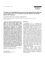
[51]. It also improves pre-operative staging of respectable
lung metastasis (See Figure 3). In Small cell Lung cancer,
the role of PET is not completely established. According to
Hauber et al [52], PET was equivalent to the battery of
Advantages of PET/CT in staging Lung cancerFigure 3
Advantages of PET/CT in staging Lung cancer. Coro-
nal slice of a PET/CT scan demonstrating a large left lung
mass showing peripheral hypermetabolism with central
necrosis (olive arrow), positive mediastinal disease, two liver
lesions, and previously unsuspected pelvic bone metastases
(red arrows). The presence of distant metastases changes the
treatment options for the patient.
Radiation Oncology 2008, 3:25 />Page 8 of 13
(page number not for citation purposes)
staging procedures done conventionally. Craig et al [53]
reported that patients were actually down staged based on
PET results. PET-CT plays a vital role in identifying mes-
othelioma patients who respond to treatment improved
over CT alone [54]. Ten studies pointed out the significant
implications of FDG-PET in staging lymph node involve-
ment.
FDG-PET is also useful in the noninvasive evaluation of
distant metastatic disease in lung cancer. Erasmus et al, at
Duke University [55], studied 27 patients with known
SCLC and an adrenal mass shown on conventional imag-
ing (mean size, 3 cm). FDG-PET identified metastatic dis-
ease in 25 of 33 lesions, "23 of which were confirmed
positive by biopsy. All lesions negative by PET were also
negative histologically (sensitivity, 100%). In a cohort of
94 patients at the University Hospital, Zurich, prospec-
tively evaluated by FDG-PET imaging for mediastinal
involvement, 4~14% were found to have distant meta-
static disease that was not shown by conventional CT.
These findings are supported by data in the literature,
showing an advantage of FDG-PET in lung cancer staging
over CT [56]. PET is thus a promising imaging modality
for patients with extensive disease and poor prognoses,
making treatment more efficacious.
Gastro-intestinal tumors
The advent of PET imaging has also led to significant
advances in staging of GI malignancies. FDG-PET plays a
vital role in detecting metastatic disease in esophageal
cancer with overall accuracy of 82% and high specificity
and sensitivity levels exceeding other conventional stag-
ing modalities [57].
It has maximum benefit for patients with locally advanced
disease in whom a curative surgery can treat the patient. It
also has great potential in predicting histopathological
response to neo-adjuvant therapy and in monitoring the
radiofrequency ablation success soon after intervention
[49].
In gastric cancer, FDG-PET helps in detecting distant
metastasis such as to liver, lung, adrenals, ovaries and
skeleton [58].
With advent in research, 18F-FDG-PET detects metastases
in colorectal cancer patients and helps decide a better
treatment plan to prolong their survival. Early 18F FDG-
Restaging of colorectal cancerFigure 4
Restaging of colorectal cancer. Sagittal (left) and coronal (right) PET/CT slices of patient with prior surgery and increasing
carcinoembryonic antigen show increased FDG uptake in multiple liver lesions(red arrows), as well as recurrence of local dis-
ease in the presacral space (yellow arrow).
Radiation Oncology 2008, 3:25 />Page 9 of 13
(page number not for citation purposes)
PET can predict pathological response to pre-operative
treatment [40] (See Figure 4). Also, automated segmenta-
tion of PET signal from rectal cancer may allow immediate
and sufficiently accurate definition of a preliminary work-
ing planning target volume(PTV) for pre-op radiotherapy
[41].
PET has not proved of much assistance in diagnosis of
pancreatic malignancy but it can help in detection of
metastases [59]. FDG-PET helps identify two distinct scin-
tigraphic patterns of focal and uniform uptake that predict
the presence of diffuse or nodular Peritoneal Carcinoma-
tosis [60].
Brain tumors
A main challenge in the management of brain tumors lies
in the localization of the extent of tumor and assessment
of the functional status of the surrounding brain. Carbon-
11-labeled methionine (MET), iodine-123-labeled α-
methyl-tyrosine (IMT) and fluorine-18-labeled O-(2)
fluoroethyl-L-tyrosine (FET) are the most important
amino acids playing a major role in detection of Gliomas.
C-11 Methionine PET improves the target volume deline-
ation of meningiomas treated with stereotatic fractionated
radiotherapy [24]. Also, the use of PET and PET-CT in con-
junction with functional MRI has greatly aided in the
management of different brain tumors. Herholz et al.
showed a sensitivity and specificity of MET-PET in differ-
entiating between non tumoral tissue and low-grade glio-
mas of 76% and 87%, respectively [61]. FDG PET is of
limited use in brain tumors as the uptake of FDG by nor-
mal brain tissue is high, making it indistinguishable from
the tumor tissue. But still, DiChiro et al [62] and Alavi et
al [63] showed that the amount of FDG uptake in the
tumor tissue correlates to the histological grading of the
tumor and has prognostic implications. FDG-PET has
been evaluated in the planning of radiation with Intensity
modulated radio-surgery and radiotherapy with Simulta-
neous Energy Boost (SEB). FET-PET reliably distinguishes
between post therapy benign lesions and tumor recur-
rence after initial treatment of low- and high-grade glio-
mas [64]. For meningiomas, which usually occur in the
tentorium, orbit, sella, falx cerebri, there is a problem in
defining the tumor extension as the normal tissue in these
areas gives the same contrast enhancement as the tumor
tissue. Recently, it was demonstrated that by using MET-
PET/CT fused images, meningioma borders can be more
accurately defined in correlation to critical normal organs
[65,66].
Gynecological tumors
Gynecological malignancies often present a challenge due
to their late presentations and insidious nature of symp-
toms. PET has been shown to be superior to CT alone in
staging of cervical cancer [67]. Whole-body FDG PET is a
sensitive and specific tool for the detection of recurrent
cervical cancer in patients who have clinical findings sus-
picious for recurrence [68]. Reinhardt et al. found a posi-
tive predictive value (PPV) for nodal involvement of 90%
with FDG-PET compared to 64% with MRI in non treated
patients with cervical cancer [69]. More recently, Deh-
dashti et al. were the first to demonstrate in 14 cervical
cancer patients, an NPV of enhanced Cu-ATSM uptake for
the response to treatment [70]. FDG PET was also found
to be superior to CT in the evaluation of pelvic and para-
aortic lymph nodes. CT-PET guided IMRT has been used
to develop treatment plans to deliver radiotherapy to pos-
itive para-aortic region lymph nodes [71]. In Gestational
trophoblastic neoplasia, FDG-PET is potentially useful for
providing precise metastatic mapping of tumor extent,
monitoring response and localizing viable tumors after
chemotherapy [72]. In ovarian cancer, Bristow et al [73],
evaluated uses of PET in detecting clinically occult but sur-
gically resectable disease. They found that its ability to
localize persistent disease and failure to identify small vol-
ume disease was useful in selecting patients who are can-
didates for cytoreductive surgery. In vulvar cancer, a
prospective PET study evaluating the detection of groin
metastases has been reported [74]. PET-CT thus may alter
the management of patients with a variety of gynecologic
malignancies.
Renal and urological tumors
Currently FDG-PET has a limited role in diagnosis of pros-
tate cancer mainly because of the low uptake of FDG in
the tumor and normal excretion of FDG through urine.
Visualization of prostate cancer with current imaging
methods (CT, MRI, and ultrasonography) is severely
impaired [75]. The low glucose uptake, the significant
overlap of tracer uptake in tumor and in the benign pros-
tate hyperplasia, and the renal excretion of FDG into the
bladder limit the diagnosis of prostate cancer using FDG-
PET [76-78]. FDG-PET appears to be promising in the
assessment of lymph nodes and bone metastases [79].
Morris et al. showed that FDG-PET can differentiate
osseous metastases from scintigraphically quiescent
lesions [80]. However, the results of FDG-PET in early
stages of prostate cancer are not satisfactory for tumor
detection, and other tracers have been intensively evalu-
ated in the recent past. The development of new tracers
and technical improvements will probably make PET
imaging a viable diagnostic tool in prostate cancer and
renal cell carcinoma [81]. C-11 acetate and C-11 choline
seem to be the two promising tracers playing an impor-
tant role in Prostate cancer. In patients with primary tes-
ticular cancer, PET can be used in conjunction with
conventional imaging techniques to diagnose retroperito-
neal masses. FDG-PET has shown very encouraging results
in a limited number of studies, and has also demonstrated
a good sensitivity for initial staging. FDG-PET seems to be
Radiation Oncology 2008, 3:25 />Page 10 of 13
(page number not for citation purposes)
superior to conventional imaging modalities for detecting
local disease and recurrence, and distant metastases [79].
Incorporation of functional information into the radiation
medicine treatment planning
Though formal radiation therapy treatment planning
techniques date from the earliest days of the 20
th
century,
the current era of reliable dosimetry and treatment plan-
ning can conveniently be bookmarked beginning with the
rise of the mini-computer and micro-computer and the
associated software developed in the 1970s. Rather than
the rough dosimetric approximations and look-up tables
previously used for "ballpark" dosimetric analyses, we are
now in an era of physical rigor going far beyond the initial
impressionistic estimates. As 3-D target localization expe-
rience grew, the original question of target volume projec-
tion into a series of planar two-dimensional spaces was
replaced by a much more sophisticated hierarchy of delib-
erately planned target volumes including the "surgical" or
"gross" target volume (GTV), the "pathologic" or
"expanded clinical" target volume (CTV) and the real-
world "corrected" or "planning" target volume (PTV).
Each of these enlarging tissue volumes represented a finer-
tuned understanding of what one must do to make the
radiation dose deposition matrix correspond with the
known and expected clonogenically viable tumor regions.
These target sub-volumes included the dosimetric impact
of various poorly visualized "microscopic disease" regions
(included within the CTV) and dosimetric uncertainties
due to expected target movement and radiation edge
effects (seen within the PTV).
The addition of the PET information allows a new, more
realistic target volume to be defined based on a kind of
probability envelope indicating the tissue region undergo-
ing the metabolic processes defining the "biologic target
volume" (BTV). This "BTV" indicates the region in which
the described physiology is readily demonstrable. In
oncology, the most common "BTV" represents the area of
abnormal glucose metabolism indicated by FDG-PET and
related processes. While very non-specific, many different
kinds of neoplasms have now been shown to display
markedly abnormal glucose metabolism and the sensitiv-
ity and specificity detectible in the non-invasive imaging
of this process is on the order of 80 to 90 percent for many
tumors. Surprisingly, this sensitivity and specificity may
rival or exceed that of CT or MRI for certain physiologi-
cally active tumors such as lung cancer. The recent popu-
larization of dual-platform PET-CT detectors now allows
sub-centimeter correlation between the source of the PET
signal and the anatomic region responsible for that signal
[82].
In designing appropriate radiotherapy target volumes, it is
apparent that the extra cost and difficulty of utilizing the
BTV to define the treatment volume will only be justified
if the clinical data show that the application of the BTV
approach will add information that is actually new (versus
simply redundant with anatomic imaging techniques).
This appears to be the case. The Agency for Healthcare Pol-
icy and Research (AHPR) investigated 16 studies incorpo-
rating information on over 1,000 patients and compared
staging data from PET or PET-CT to data obtained using
CT information alone for lung cancer patients. In 17 per-
cent of cases, the FDG-PET correctly modified tumor
stage. The use of this methodology to cancel or modify
potentially toxic surgical approaches in tumors which
later displayed occult metastatic spread was reduced by
over fifty percent. Multiple cost effectiveness analyses
based on this sort of data have concluded that the incre-
mental costs associated with the use of PET-CT were justi-
fiable and in accord with other well-accepted principles
used for medical economics [8,9]. For diagnostically diffi-
cult cases with CT-indicated enlargement of regional
lymph nodes, the use of functional imaging would be
especially useful if it proved reliable. However, the relative
lack of PET specificity in patients with other known causes
for physiologic inflammation makes this method too
unreliable to depend on. At present, it appears that PET-
based target volume definition is fraught with difficulty in
any circumstance with active inflammation. This unfortu-
nately includes many postoperative settings and situa-
tions with benign causes of immune system activation.
Conclusion
The field of radiation medicine and nuclear imaging are
both progressing rapidly with respect to technologic
sophistication and multi-platform interface capabilities.
Radiation oncology has previously incorporated multiple
imaging methodologies including: CT, ultrasound, and
MRI into the treatment planning process to allow highly
accurate and serially updated beam-direction instructions.
This field is now known as "image-guided radiation ther-
apy" (IGRT) and can be seen as further evolutionary pro-
gression in the quest to maximize dose delivered to true
target tissue and minimize the dose delivered to nearby
non-target tissues. A near-term future goal is now the
incorporation of functional imaging methods such as 18
FDG-PET in the same fashion. Multiple recent studies are
appearing in literature attesting to the value of incorporat-
ing PET-CT information in radiotherapy treatment plan-
ning [83-87]. This will allow a determination of the
degree of physiologic activity located within various sub-
components of presumed target tissue. As more and more
types of tumor targets are validated for this sort of func-
tional and predictive analysis, functional imaging is likely
to enter the main stream as a critical tool for radiation
medicine field design and as an accepted non-invasive
surrogate endpoint appropriate for clinical trial design
and clinical decision-making. All of these advances can be
Radiation Oncology 2008, 3:25 />Page 11 of 13
(page number not for citation purposes)
seen as way-stations on the road to the effective, non-inva-
sive, minimally toxic, and ultimately personalized cancer
medicine.
Competing interests
The authors declare that they have no competing interests.
Authors' contributions
RM and NS have contributed to conception and design,
acquisition, analysis and interpretation of data. RM, DN
and NS have been involved in drafting the manuscript and
revising it critically for important intellectual content. RM
and NS have given final approval of the version to be pub-
lished.
References
1. Adler L, Faulhaber P, Schnur K: Axillary lymph node metastases:
Screening with [F-18]2-deoxy-2-fluoro-D-glucose (FDG)
PET. Radiology 1997, 203:323-27.
2. Avril N, Dose J, Janicke : Assessment of axillary lymph node
involvement in breast cancer patients with positron emis-
sion tomography using radiolabeled 2-(fluorine-18)-fluoro-2-
deoxy-D-glucose. J Natl Cancer Inst 1996, 88:1204-9.
3. Adams S, Baum RP, Stuckensen T: Prospective comparison of
18
F-
FDG PET with conventional imaging modalities (CT, MR,
US) in lymph node staging of head and neck cancer. Eur J Nucl
Med 1998, 25(9):1255-1260.
4. Stokkel MP, Broek FW, vanRijk PP: The role of FDG PET in the
clinical management of head and neck cancer. Oral Oncol
1998, 34:466-71.
5. Boykin KN, Zibari GB, Lilien DL: The use of FDG-positron emis-
sion tomography for the evaluation of colorectal metastases
of the liver. Am Surg 1999, 65:1183-5.
6. Fong Y, Saldinger PF, Akhurst T: Utility of
18
F-FDG positron
emission tomography scanning on selection of patients for
resection of hepatic colorectal metastases. Am J Surg 1999,
178:282-7.
7. Gambhir SS, Czernin J, Schwimmer J, Silverman DH, Coleman RE,
Phelps ME: A tabulated summary of the FDG-PET literature.
J Nucl Med 2001, 42(5 Suppl):1S-93S.
8. Maurer AH, Urbain JLC, Malmud LS: Grainger & Allison's Diagnostic
Radiology: A textbook of Medical Imaging 4th edition. London: Churchill .
9. de Wit M, Bumann D, Beyer W: Whole-body positron emission-
tomography (PET) for diagnosis of residual mass in patients
withlymphoma. In Ann Oncol Volume 8. Livingstone Inc; 1997:57-60.
2001
10. Wiedmann E, Baican B, Hertel A: Positron emission tomography
(PET) for staging and evaluation of response to treatment in
patients with Hodgkin's disease. Leuk Lymphoma 1999,
34:545-51.
11. Zinzani PL, Magagnoli M, Chierichetti F: The role of positron emis-
sion tomography (PET) in the management of lymphoma
patients. Ann Oncol 1999, 10:1181-84.
12. Castellucci P, Nanni C, Farsad M, Alinari L, Zinzani P, Stefoni V,
Battista G, Valentini D, Pettinato C, Marengo M, Boschi S, Canini R,
Baccarani M, Monetti N, Franchi R, Rampin L, Fanti S, Rubello D:
Potential pitfalls of 18F-FDG PET in a large series of patients
treated for malignant lymphoma: prevalence and scan inter-
pretation. Nucl Med Commun 2005, 26(8):689-94.
13. Romer W, Hanauske AR, Ziegler S: Positron emission tomogra-
phy in non-Hodgkin's lymphoma: assessment of chemother-
apy with fluorodeoxyglucose. Blood 1998, 91:4464-71.
14. Wahl RL, Zasadny K, Helvie M: Metabolic monitoring of breast
cancer chemohormonotherapy using positron emession
tomography: Initial evaluation. J Clin Oncol 1993, 11:2101-2111.
15. Bassa P, Kim EE, Inoue T: Evaluation of preoperative chemothe-
raphy using PET with fluorine-18-fluorode-oxyglucose in
breast cancer. J Nucl Med 1996, 37:931-8.
16. Herrmann K, Krause B, Wester H: Change of tumor cell prolifer-
ation during R-CHOP chemotherapy of diffuse large B-cell
lymphoma (DLBCL) measured by
18
FLT-PET. Presented at:
Society of Nuclear Medicine 53rd Annual Meeting; June 3–7, 2006; San
Diego .
17. Kahn ST, Flowers C, Lechowicz MJ, Hollenbach K, Johnstone PA:
Value of PET restaging after CT for non Hodgkin's lym-
phoma : Implications for consolidation Radiotherapy. Int J
Radiat Oncol Biol Phys 2006, 66(4):961-5.
18. Bentzen SM: Theragnostic imaging for radiation oncology:
dose painting by numbers. Lancet Oncol 2005, 6:112-17.
19. Champman JD, Bradley JD, Eary JF, Haubner R, Larson SM, Michalski
JM, Okunieff PG, Strauss HW, Ung YC, Welch MJ: Molecular (func-
tional) imaging for radiotherapy applications: an RTOG sym-
posium. Int J Radiat Oncol Biol Phys 2003, 55(2):294-301.
20. Cheson BD, Pfistner B, Juweid ME, Gascoyne RD, Specht L, Horning
SJ, Coiffier B, Fisher RI, Hagenbeek A, Zucca E, Rosen ST, Stroobants
S, Lister TA, Hoppe RT, Dreyling M, Tobinai K, Vose JM, Connors JM,
Federico M, Diehl V: The International Harmonization Project
on Lymphoma. Revised response criteria for malignant lym-
phoma. J Clin Oncol 25(5):579-86. 2007 Feb 10
21. Juweid ME, Stroobants S, Hoekstra OS, Mottaghy FM, Dietlein M,
Guermazi A, Wiseman GA, Kostakoglu L, Scheidhauer K, Buck A,
Naumann R, Spaepen K, Hicks RJ, Weber WA, Reske SN, Schwaiger
M, Schwartz LH, Zijlstra JM, Siegel BA, Cheson BD: Imaging Sub-
committee of International Harmonization Project in Lym-
phoma. Use of positron emission tomography for response
assessment of lymphoma: consensus of the Imaging Sub-
committee of International Harmonization Project in Lym-
phoma. J Clin Oncol 25(5):571-8. 2007 Feb 10
22. Zanzonico P: PET-based biological imaging for radiation ther-
apy treatment planning. Crit Rev Eukaryot Gene Expr 2006,
16(1):61-101.
23. Schwartz DL, Ford E, Rajendran J, Yueh B, Coltrera MD, Virgin J,
Anzai Y, Haynor D, Lewellyn B, Mattes D, Meyer J, Phillips M, Leblanc
M, Kinahan P, Krohn K, Eary J, Laramore GE: FDG-PET/CT imag-
ing for pre radiotherapy staging of head-and-neck squamous
cell carcinoma. Int J Radiat Oncol Biol Phys 61(1):129-36. 2005 Jan 1
24. Grosu AL, Weber WA, Astner ST, Adam M, Krause BJ, Schwaiger M,
Molls M, Nieder C: 11C-methionine PET improves the target
volume delineation of meningiomas treated with stereo tac-
tic fractionated radiotherapy. Int J Radiat Oncol Biol Phys
66(2):339-44. 2006 Oct 1
25. Kitagawa Y, Sano K, Nishizawa S, Nakamura M, Ogasawara T, Sadato
N, Yonekura :
FDG-PET for prediction of tumor aggressive-
ness and response to intra-arterial chemotherapy and radio-
therapy in head and neck cancer. Eur J Nucl Med Mol Imaging
2003, 30(1):63-71. Epub 2002 Oct 26
26. Stuckensen T, Kovacs AF, Adams S, Baum RP: Staging of theneck
in patients with oral cavity squamous cell carcinomas: a pro-
spective comparison of PET, ultrasound, CT and MRI. J Crani-
omaxillofac Surg 2000, 28:319-24.
27. Benchaou M, Lehmann W, Slosman DO, Becker M, Lemoine R,
Rufenatch D, Donath A: The role of FDG-PET in the preopera-
tive assessment of N-staging in head and neck cancer. Acta
Otolaryngol (Stockh) 1996, 116:332-5.
28. Kau RJ, Alexiou C, Laubenbacher C, Werner M, Schwaiger M, Arnold
W: Lymph node detection of head and neck squamous cell
carcinomas by positron emission tomography with fluorode-
oxyglucose F18 in a routine clinical setting. Arch Otolaryngol
Head Neck Surg 1999, 125(12):1322-8.
29. Douglas JG, Stelzar KJ, Mankoff DA, Tralins KS, Krohn KA, Muzi M,
Silbergeld DL, Rostomily RC, Scharnhorst J, Spence AM: [F-18]-
fluorodeoxyglucose positron emission tomography fortar-
geting radiation dose escalation for patients with glioblast-
omamultiforme: clinical outcomes and patterns of failure.
Int J Radiat Oncol Biol Phys 64(3):886-91. 2006 Mar 1, Epub 2005 Oct
19
30. Schwartz DL, Ford EC, Rajendran J, Yueh B, Coltrera MD, Virgin J,
Anzai Y, Haynor D, Lewellyn B, Mattes D, Meyer J, Phillips M, Leblanc
M, Kinahan P, Krohn K, Eary J, Laramore GE: FDG-PET/CT-guided
intensity modulated head and neck radiotherapy: a pilot
investigation. Head Neck 2005, 27(6):478-87.
31. Lowe VJ, Boyd JH, Dunphy FR, Kim H, Dunleavy T, Collins BT, Martin
D, Stack BC Jr, Hollenbeak C, Fletcher JW: Surveillance for recur-
rent head and neck cancer using positron emission tomogra-
phy. J Clin Oncol 2000, 18:651-8.
Radiation Oncology 2008, 3:25 />Page 12 of 13
(page number not for citation purposes)
32. Kunkel M, Grotz KA, Forster GJ, Wahlmann U, Benz P, Kutzner J,
Rippins G, Wagner W: Therapy monitoring with 2-(18F)-FDG
positron emission tomography after neoadjuvant radiation
treatment of mouth carcinoma. Strahlenther Onkol 2001,
177:145-52.
33. Nishioka T, Shiga T, Shirato H, Tsukamoto E, Tsuchiya K, Kato T,
Ohomori K, Yamazaki A, Aoyama H, Hashimoto S, Chang TC, Miya-
saka K: Image fusion between 18FDG-PET and MRI/CT for
radiotherapy planning of oropharyngeal and nasopharyngeal
carcinomas. J Radiat Oncol Biol Phys 2002, 53:1051-7.
34. Heron DE, Smith RP, Andrade RS: Advances in image-guided
radiation therapy – the role of PET-CT. Med Dosim Spring 2006,
31(1):3-11.
35. Greven KM, Williams DW, Keyes JW, et al.: Can positron emis-
sion tomography distinguish tumor recurrence from irradia-
tion sequelae in patients treated for larynx cancer? Cancer J
Sci Am 1997, 3:353-7.
36. Farber LA, Benard F, Machtay M, et al.: Detection of recurrent
head and neck squamous cell carcinomas after radiation
therapy with 2-
18
F-fluoro-2-deoxy-D-glucose positron emis-
sion tomography. Laryngoscope 1999, 109:970-5.
37. Lowe VJ, Dunphy FR, Varvares M, et al.: Evaluation of chemother-
apy response with advanced head and neck cancer using [F-
18] fluorodeoxyglucose positron emission tomography.
Head Neck 1997, 19:666-674.
38. Kau RJ, Alexiou C, Laubenbacher C: Lymph node detection of
head and neck squamous cell carcinomas by positron emis-
sion tomography with [F-18] fluorodeoxyglucose in routine
clinical setting. Arch Otolaryngol Head Neck Surg 1999, 125:1322-8.
39. Wolfort RM, Li BD, Johnson LW, Turnage RH, Lilien D, Ampil F, Bur-
ton G, Chu QD: The role of whole-body fluorine-18-FDG pos-
itron emission tomography in the detection of recurrence in
symptomatic patients with stages II and III breast cancer.
World J Surg 2006, 30(8):1422-7.
40. Weir L, Worsley D, Bernstein V: The value of FDG positron
emission tomography in the management of patients with
breast cancer. Breast J 2005, 11(3):204-9.
41. Kumar R, Alavi A: Fluorodeoxyglucose-PET in the manage-
ment of breast cancer. Radiol Clin North Am 2004, 42(6):1113-22.
42. Port ER, Yeung H, Gonen M, Liberman L, Caravelli J, Borgen P, Larson
S: 18F-2-fluoro-2-deoxy-D-glucose positron emissiontomog-
raphy scanning affects surgical management in selected
patientswith high-risk, operable breast carcinoma. Ann Surg
Oncol 2006, 13(5):677-84. Epub 2006 Mar 16
43. Marshall C, Mustafa S, Wheatley DC, Eremin JE, El-Sheemy M, Jibril
JA, Eremin O, Griffiths PA: A comparison of 18F-FDG gamma
camera PET, mammography and ultrasonography in dem-
onstrating primary disease in locally advanced breast cancer.
Nucl Med Commun 2004, 25(7):721-5.
44. Kawada K, Murakami K, Sato T, Kojima Y, Ebi H, Mukai H, Tahara M,
Shimokata K, Minami H: Prospective Study of Positron Emission
Tomography for Evaluation of the Activity of Lapatinib, a
Dual Inhibitor of the ErbB1 and ErbB2 Tyrosine Kinases, in
patients with advanced tumors. Jpn J Clin Oncol 2007,
37(1):44-8.
45. Radan L, Ben-Haim S, Bar-Shalom R, Guralnik L, Israel O: The role
of FDG PET/CT in suspected recurrence of breast cancer.
Cancer 107(11):2545-51. 2006 Dec 1
46. Obedian E, Haffty BG: Internal mammary nodal irradiation in
conservatively-managed breast cancer patients: is there a
benefit? Int J Radiat Oncol Biol Phys 44(5):997-1003. 1999 Jul 15
47. Weber WA, Dietlein M, Hellwig D, Kirsch CM, Schicha H, Schwaiger
M: PET with (18) F-fluorodeoxyglucose for staging of non-
small cell lung cancer. Nuklearmedizin 2003, 42:135-44.
48. van Tinteren H, Hoekstra OS, Smit EF, Bergh JH van den, Schreurs AJ,
Stallaert RA, van Velthoven PC, Comans EF, Diepenhorst FW, Ver-
boom P, van Mourik JC, Postmus PE, Borres M, Teule GJ: Effective-
ness of positron emission tomography in the preoperative
assessment of patients with suspected non-small-cell lung
cancer: the PLUS multicenter randomized trial. Lancet 2002,
359(9315):1388-93.
49. Esteves FP, Schuster DM, Halkar RK: Gastrointestinal tract
malignancies and positron emission tomography: an over-
view. Semin Nucl Med 2006, 36(2):169-81.
50. Sonmezoglu K: The use of FDG-PET scanning in lung cancer.
Tuberk Toraks 2005, 53(1):95-114. Review Turkish
51. Schrevens L, Lorent N, Dooms C, Vansteenkiste J: The role of PET
scan in diagnosis, staging, and management of non-small cell
lung cancer. Oncologist 2004, 9(6):633-43. Review
52. Hauber HP, Bohuslavizki KH, Lund CH, Fritscher-Ravens A, Meyer A,
Pforte A: Positron Emission Tomography in the staging of
Small cell lung cancer: a preliminary study. Chest 2001,
119(3):950-4.
53. Craig MD, Rogers JS, Gupta N: Evaluation of PET scan in small
cell lung cancer.2004 ASCO Annual meeting proceedings. J
Clin Oncol 2004, 22:7214.
54. Steinert HC, Santos Dellea MM, Burger C, Stahel R: Therapy
response evaluation in malignant pleural mesothelioma with
integrated PET-CT imaging. Lung Cancer 2005, 49(Suppl
1):S33-5.
55. Erasmus JJ, McAdams HP: Evaluation of adrenal masses in
patients with bronchogenic carcinoma using
18
F-fluorodeox-
yglucose positron emission tomography. AJR Am J Roentgenol
1997, 168:1357-1360.
56. Weber WA, Petersen V, Schmidt B, Tyndale-Hines L, Link T, Peschel
C, Schwaiger M: Positron emission tomography in non-small-
cell lung cancer: prediction of response to chemotherapy by
quantitative assessment of glucose use. J Clin Oncol 2003,
21:2651-7.
57. Flamen P, Lerut A, Van Cutsem E, De Wever W, Peeters M, Stroo-
bants S, Dupont P, Bormans G, Hiele M, De Leyn P, Van Raemdonck
D, Coosemans W, Ectors N, Haustermans K, Mortelmans L: Utility
of positron emission tomography for the staging of patients
with potentially operable esophageal carcinoma. J Clin Oncol
2000, 18(18):3202-10.
58. Lim JS, Yun MJ, Kim MJ, Hyung WJ, Park MS, Choi JY, Kim TS, Lee JD,
Noh SH, Kim KW: CT and PET in stomach cancer: preopera-
tive staging and monitoring of response to therapy. Radio-
graphics 2006, 26(1):143-56. Review
59. Heinrich S, Goerres GW, Schaefer M, Sagmeister M, Bauerfeind P,
Pestalozzi BC, Hany TF, von Schulthess GK, Clavien PA: Positron
emission tomography/computed tomography influences on
the management of resectable pancreatic cancer and its
cost-effectiveness. Ann Surg 2005, 242(2):235-43.
60. Turlakow A, Yeung HW, Salmon AS, Macapinlac HA, Larson SM:
Peritoneal carcinomatosis: role of (18) F-FDG PET. J Nucl
Med 2003, 44(9):1407-12.
61. Herholz K, Holzer T, Bauer B, Schroder R, Voges J, Ernestus RI, Men-
doza G, Weber-Luxenburger G, Lottgen J, Thiel A, Wienhard K,
Heiss WD: 11C-methionine PET for differential diagnosis of
low grade gliomas. Neurology 1998, 50:1316-22.
62. DiChiro G: Positron emission tomography using F-18 fluoro-
deoxyglucose in brain tumors- a powerful diagnostic and
prognostic tool. Invest Radiol 1986, 22:360-71.
63. Alavi JB, Alavi A, Chawluk J, Kushner M, Powe J, Hickey W, Reivich
M: Positron emission tomography in patients with glioma. A
predictor of prognosis. Cancer 1988, 62:1074-8.
64. Solberg TD, Agazaryan N, Goss BW, Dahlbom M, Lee SP: A feasibil-
ity study of 18F-fluorodeoxyglucose positron emission tom-
ography targeting and simultaneous integrated boost for
intensity modulated radiosurgery and radiotherapy. J Neuro-
surg 2004, 101:381-9.
65. Grosu AL, Lachner R, Wiedenmann N, Stark S, Thamm R, Kneschau-
rek P, Schwaiger M, Molls M, Weber WA: Validation of a method
for automatic image fusion (BrainLAB System) of CT data
and 11C-methionine- PET data for stereotactic radiotherapy
using a LINAC: first clinical experience. Int J Radiat Oncol Biol
Phys 2003, 56:1450-63.
66. Sweeney RA, Bale RJ, Moncayo R, Seydl K, Trieb T, Eisner W, Burt-
scher J, Donnemiller E, Stockhammer G, Lukas P: Multimodality
cranial image fusion using external markers applied via vac-
uum mouthpiece and a case report. Strahlenther Onkol 2003,
179:254-60.
67. Grigsby PW, Siegel BA, Dehdashti F: Lymph node staging by pos-
itron emission tomography in patients with carcinoma of the
cervix. J Clin Oncol 2001, 19(17):3745-9.
68. Havrilesky LJ, Wong TZ, Secord AA, Berchuck A, Clarke-Pearson :
The role of PET scanning in the detection of recurrent cer-
vical cancer. Gynecol Oncol 2003, 90(1):186-90.
69. Reinhardt MJ, Ehritt-Braun C, Vogelgesang D, Ihling C, Hogerle S, Mix
M, Moser E, Krause TM: Metastatic lymph nodes in patients
Publish with Bio Med Central and every
scientist can read your work free of charge
"BioMed Central will be the most significant development for
disseminating the results of biomedical research in our lifetime."
Sir Paul Nurse, Cancer Research UK
Your research papers will be:
available free of charge to the entire biomedical community
peer reviewed and published immediately upon acceptance
cited in PubMed and archived on PubMed Central
yours — you keep the copyright
Submit your manuscript here:
/>BioMedcentral
Radiation Oncology 2008, 3:25 />Page 13 of 13
(page number not for citation purposes)
with cervical cancer: detection with MR imaging and FDG
PET. Radiology 2001, 218(3):776-82.
70. Dehdashti F, Grigsby PW, Mintun MA, Lewis JS, Siegel BA, Welch MJ:
Assessing tumor hypoxia in cervical cancer by positron emis-
sion tomography with 60Cu-ATSM: relationship to thera-
peutic response – a preliminary report. Int J Radiat Oncol Biol
Phys 2003, 55(5):1233-8.
71. Esthappan J, Mutic S, Malyapa RS, Grigsby PW, Zoberi I, Dehdashti F,
Miller TR, Bosch WR, Low DA: Treatment planning guidelines
regarding the use of CT/PET-guided IMRT for cervical carci-
noma with positive paraaortic lymph nodes. Int J Radiat Oncol
Biol Phys 58(4):1289-97. 2004 Mar 15
72. Yen TC, Lai CH: Positron emission tomography in gynecologic
cancer. Semin Nucl Med 2006, 36(1):93-104.
73. Bristow RE, Simpkins F, Pannu HK, Fishman EK, Montz FJ: Positron
emission tomography for detecting clinically occult surgi-
cally resectable metastatic ovarian cancer. Gynaecol Oncolol
2002, 85(1):196-200.
74. Cohn DE, Dehdashti F, Gibb RK, Mutch DG, Rader JS, Siegel BA, Her-
zog TJ: Prospective evaluation of positron emission tomogra-
phy for the detection of groin node metastases from vulvar
cancer. Gynaecol Oncol 2002, 85(1):179-84.
75. Moul JW: Prostate specific antigen only progression of pros-
tate cancer. J Urol 2000, 163:1632-42.
76. Effert PJ, Bares R, Handt S, Wolff JM, Bull U, Jakse G: Metabolic
imaging of untreated prostate cancer by positron emission
tomography with 18fluorine-labeled deoxyglucose. J Urol
1996, 155(3):994-8.
77. Hofer C, Laubenbacher C, Block T, Breul J, Hartung R, Schwaiger M:
Fluorine-18-fluorodeoxyglucose positron emission tomogra-
phy is useless for the detection of local recurrence after rad-
ical prostatectomy. Eur Urol 1999, 36(1):31-5.
78. Shreve PD, Grossman HB, Gross MD, Wahl RL: Metastatic pros-
tate cancer: initial findings of PET with 2-deoxy-2-[F-18]
fluoro-D-glucose. Radiology 1996,
199(3):751-6.
79. Kumar R, Zhuang H, Alavi A: PET in the management ofurologic
malignancies. Radiol Clin North Am 2004, 42(6):1141-53. Review
80. Morris MJ, Akhurst T, Osman I, Nunez R, Macapinlac H, Siedlecki K,
Verbel D, Schwartz L, Larson SM, Scher HI: Fluorinated deoxyglu-
cose positron emission tomography imaging in progressive
metastatic prostate cancer. Urology 2002, 59(6):913-8.
81. Mathews D, Oz OK: Positron emission tomography in prostate
and renal cell carcinoma. Curr Opin Urol 2002, 12(5):381-5.
82. Bybel B, Brunken RC, Shah SN, Guiyun W, Turbiner E, Neumann DR:
PET and PET/CT imaging: What clinicians need to know?
Cleve Clin J Med 2006, 73(12):1075-1087.
83. Juweid ME, Stroobants S, Hoekstra OS: Use of positron emission
tomography for response assessment of lymphoma: consen-
sus of the Imaging Subcommittee of International Harmoni-
zation Project in Lymphoma. J Clin Oncol 2007, 25:571-8.
84. Bradley J, Thorstad WL, Mutic S: Impact of FDG-PET onradia-
tion therapy volume delineation in non-small-cell lung can-
cer. Int J Radiat Oncol Biol Phys 2004, 59:78-86.
85. van Baardwijk A, Baumert BG, Bosmans G: The currentstatus of
FDG-PET in tumor volume definition in radiotherapy treat-
ment planning. Cancer Treat Rev 2006, 32:245-60.
86. Hutchings M, Loft A, Hansen M, Berthelsen AK, Specht L: Clinical
impact of FDG-PET/CT in the planning of radiotherapy for
early-stage Hodgkin lymphoma. Eur J Haematol 2007,
78:206-12.
87. Girinsky T, Maazen R van der, Specht L, et al.: Involved-node radi-
otherapy (INRT) in patients with early Hodgkin lymphoma:
concepts and guidelines. Radiother Oncol 2006, 79:270-7.