Báo cáo khoa học: "Recommendations for implementing stereotactic radiotherapy in peripheral stage IA non-small cell lung cancer: report from the Quality Assurance Working Party of the randomised phase III ROSEL study" pps
Bạn đang xem bản rút gọn của tài liệu. Xem và tải ngay bản đầy đủ của tài liệu tại đây (487.36 KB, 14 trang )
BioMed Central
Page 1 of 14
(page number not for citation purposes)
Radiation Oncology
Open Access
Study protocol
Recommendations for implementing stereotactic radiotherapy in
peripheral stage IA non-small cell lung cancer: report from the
Quality Assurance Working Party of the randomised phase III
ROSEL study
Coen W Hurkmans*
1
, Johan P Cuijpers
2
, Frank J Lagerwaard
2
,
Joachim Widder
3
, Uulke A van der Heide
4
, Danny Schuring
1
and
Suresh Senan
2
Address:
1
Department of Radiation Therapy, Catharina Hospital, Eindhoven, The Netherlands,
2
Department of Radiation Oncology, VU
University Medical Center, Amsterdam, The Netherlands,
3
Department of Radiation Oncology, University Medical Center Groningen, Groningen,
The Netherlands and
4
Department of Radiation Oncology, University Medical Center Utrecht, Utrecht, The Netherlands
Email: Coen W Hurkmans* - ; Johan P Cuijpers - ;
Frank J Lagerwaard - ; Joachim Widder - ; Uulke A van der Heide - ;
Danny Schuring - ; Suresh Senan -
* Corresponding author
Abstract
Background: A phase III multi-centre randomised trial (ROSEL) has been initiated to establish the
role of stereotactic radiotherapy in patients with operable stage IA lung cancer. Due to rapid
changes in radiotherapy technology and evolving techniques for image-guided delivery, guidelines
had to be developed in order to ensure uniformity in implementation of stereotactic radiotherapy
in this multi-centre study.
Methods/Design: A Quality Assurance Working Party was formed by radiation oncologists and
clinical physicists from both academic as well as non-academic hospitals that had already
implemented stereotactic radiotherapy for lung cancer. A literature survey was conducted and
consensus meetings were held in which both the knowledge from the literature and clinical
experience were pooled. In addition, a planning study was performed in 26 stage I patients, of which
22 were stage 1A, in order to develop and evaluate the planning guidelines. Plans were optimised
according to parameters adopted from RTOG trials using both an algorithm with a simple
homogeneity correction (Type A) and a more advanced algorithm (Type B). Dose conformity
requirements were then formulated based on these results.
Conclusion: Based on current literature and expert experience, guidelines were formulated for
this phase III study of stereotactic radiotherapy versus surgery. These guidelines can serve to
facilitate the design of future multi-centre clinical trials of stereotactic radiotherapy in other patient
groups and aid a more uniform implementation of this technique outside clinical trials.
Published: 12 January 2009
Radiation Oncology 2009, 4:1 doi:10.1186/1748-717X-4-1
Received: 24 September 2008
Accepted: 12 January 2009
This article is available from: />© 2009 Hurkmans et al; licensee BioMed Central Ltd.
This is an Open Access article distributed under the terms of the Creative Commons Attribution License ( />),
which permits unrestricted use, distribution, and reproduction in any medium, provided the original work is properly cited.
Radiation Oncology 2009, 4:1 />Page 2 of 14
(page number not for citation purposes)
Background
Until recently, conventionally fractionated high-dose
radiation therapy was the preferred treatment in patients
with stage I NSCLC who were unfit to undergo surgery or
declined surgery. This is, however, a poor alternative to
surgery in operable patients as the mean reported crude
local recurrence rates are as high as 40% (range 6–70%),
resulting in a three year overall and cause-specific survival
of only 34 and 39%, respectively [1].
Recently, stereotactic radiotherapy has gained much inter-
est in the treatment of medically inoperable patients with
stage I lung cancer, as local control rates are dramatically
improved with this technique compared to conventional
fractionation. In studies where schedules with a biologi-
cally effective dose (BED) larger than 100 Gy are used, typ-
ical local control rates are approximately 90%. The largest
series were reported from Japan [2,3], United States [4]
and the Netherlands [5], comprising experience in over
750 patients. Onishi et al. [6] retrospectively described the
results of 257 patients treated in 14 Japanese centres using
a number of different fractionation schedules and delivery
approaches. This Japanese study also included nearly 100
patients who refused surgery, and the 5-year overall sur-
vival rate of 70.8% observed after a BED of 100 Gy among
those patients is at least equivalent to the outcome after
surgery [7-9]. Currently, several phase II trials have started
in operable lung cancer patients [10] (RTOG 0618 and
JCOG 0403), however, to date no prospective multi-cen-
tre randomized studies have been performed to compare
stereotactic radiotherapy with surgery in patients with
operable lung cancer.
A randomized phase III trial of Radiosurgery Or Surgery
for operable Early stage (stage 1A) non-small cell Lung
cancer (ROSEL, ClinicalTrials.gov ID = NCT00687986)
has been opened for accrual in August 2008. The study is
initiated by the VU medical centre Amsterdam and the
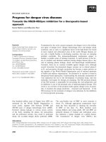
Dutch Lung Cancer Research Group. The primary study
objectives are to compare local and regional control, qual-
ity of life and treatment costs at 2 and 5 years in patients
who are randomized to either surgery or radiosurgery
(Figure 1). Treatment costs are a primary end-point, as the
costs associated with surgery for stage IA in The Nether-
lands are far higher than the present costs of stereotactic
radiotherapy [11]. These costs are expected to be even
more if the costs of post-operative revalidation and loss of
economic activity are taken into account. However,
patients treated with stereotactic radiotherapy could incur
costs for salvage treatment if a higher incidence of local or
regional recurrences is detected. Therefore, treatment costs
were considered to be a relevant end-point.
Secondary objectives include overall survival, pulmonary
function tests, quality adjusted life years and total costs
(both direct and indirect). In case of surgery, a lobectomy
should be carried out, but limited resections are accepta-
ble. Careful radiological follow-up is performed within
the trial in patients treated by SRT, as salvage surgery or
mediastinal radiation therapy might still be possible in
case of clinical, radiological or histological evidence of
local or hilar disease progression.
Accreditation and dosimetry guidelines have been previ-
ously developed for trials of stereotactic radiotherapy such
as RTOG 0236 and JCOG 0403 [12-14]. However, a reas-
sessment was considered necessary because a new patient
group was being treated with stereotactic radiotherapy,
namely patients who were fit to undergo both primary
and salvage surgery. As a result, normal tissue dose-con-
straints had to be more stringently defined in order to
minimize the risk of increased complications after salvage
surgery. Furthermore, IGRT technology from different
vendors has been rapidly adopted at various Dutch cen-
tres, which had to be taken into account. The resulting
guidelines include both minimum requirements that
must be met by each participating centre as well as recom-
mendations for possible further improvements. They are
presented here in order to facilitate the implementation of
future multi-centre studies, to stimulate and improve the
implementation of stereotactic techniques in clinical prac-
tice and to improve the comparability of results.
Methods
A ROSEL Quality Assurance Working Party was formed by
radiation oncologists and medical physicists from both
academic as well as non-academic hospitals that had
already implemented stereotactic radiotherapy for lung
cancer. Several working party meetings were organised in
which both the knowledge from literature and clinical
experience were shared and amalgamated. In support of
these meetings, a literature search was conducted by
searching MEDLINE with different key words and their
permutations such as stereotactic radiotherapy, stage I
lung cancer, treatment planning, CT scan, patient posi-
tioning and tumour mobility. Abstract books of the
ASTRO, ASCO, AAPM and ESTRO/ECCO from 2004 to
2008 were reviewed. It was recognized that there was little
data available in the literature about the influence of dif-
ferent planning algorithms on the planning of stereotactic
radiotherapy. Therefore, an additional planning study was
performed in 22 stage IA and 4 stage 1B non-small cell
lung cancer patients in order to develop and evaluate the
planning guidelines differentiated according to type of
dose calculation algorithm used. Patient characteristics
and treatment planning details have been reported previ-
ously [15].
In brief, a four-dimensional (4D)-CT was reconstructed in
ten equally spaced time bins using respiratory phase bin-
Radiation Oncology 2009, 4:1 />Page 3 of 14
(page number not for citation purposes)
ROSEL study designFigure 1
ROSEL study design.
Radiation Oncology 2009, 4:1 />Page 4 of 14
(page number not for citation purposes)
ning for each patient. From these phases, a maximum
intensity projection (MIP) was reconstructed [16]. The
datasets were then imported in the Pinnacle
3
treatment
planning system (Philips Medical Systems, Wisconsin).
Using the MIP dataset, an experienced radiation oncolo-
gist delineated the internal target volume (ITV). Organs at
risk were delineated on an average-density CT reconstruc-
tion. The PTV was created by expanding the ITV with a 3
mm margin. The treatment plans consisted of 9 equally
spaced coplanar 6 MV beams which were not allowed to
enter through the oesophagus, heart, spinal cord or con-
tralateral lung. The plans were inversely optimized using
the direct aperture optimization module of the Pinnacle
3
treatment planning system with the same objectives as
used in the ROSEL trial. Three different plans were cre-
ated; using an advanced (type A) dose calculation algo-
rithm, a less advanced (type B) algorithm and a plan
assuming all tissues within the body to have unit density,
in accordance with the RTOG study 0236 and 0618 proto-
cols [17,18].
In order to facilitate the clinical use of these recommenda-
tions, we divided the process of implementing high-dose
radiotherapy into the following headings: CT scanning
and patient positioning, target volume definition, organs
at risk definition, Dose calculation algorithms and frac-
tionation, dose prescription, coverage and constraints,
treatment planning and treatment execution.
Patient positioning and CT scanning
The patient should be scanned in the treatment position
which should be supine with both arms raised above the
head using an arm-rest or other fixation device. Positions
which are less comfortable for the patient should be
avoided so as to prevent the likelihood of uncontrolled
movement during scanning or treatment. Four-dimen-
sional (4D) CT scanning is strongly recommended in
order to account for an individualised assessment and
incorporation of tumour motion [19-21]. Preferably 10
but no less than 6 breathing phases should be recon-
structed in order to determine the tumour movement for
treatment planning. Using 10 phases, it was found that
generally the full amplitude of motion can be captured
[22]. Within the ROSEL trial, acquisition of a slow-CT
scan or multiple (at least 3) rapid planning scans covering
the entire range of tumour motion is also allowed, as 4D-
CT scanners are not widely available yet. However, target
volume delineation might be more difficult as the images,
and thus also the tumour volume, of slow-CT scans are
blurred [23,24]. All centres participating in the ROSEL
study will most likely be able to implement 4D-CT scan-
ning in the near future. Generally, intravenous contrast is
not necessary for planning CT scans for early stage lung
cancer, but contrast-enhanced CT images may still be used
for dose calculations. Although the effect of intravenous
(IV) contrast on dose calculations for lung patients is not
specifically studied, the influence of IV contrast in head
and neck intensity modulated radiotherapy plans was
proven to be insignificant [25]. The slice spacing between
reconstructed CT images should be ≤3 mm over the com-
plete tumour trajectory and ≤5 mm elsewhere. The scan
should encompass the entire lung volume in order to cal-
culate meaningful lung dose-volume parameters.
Target volume definition
The gross tumour volume (GTV) will generally be con-
toured using CT pulmonary windows; however, soft tissue
windows may be used to avoid inclusion of adjacent ves-
sels or chest wall structures within the GTV. The correct-
ness of the GTV delineation should be checked in axial,
sagittal and coronal views. The clinical target volume
(CTV) is assumed to be identical to the GTV, i.e. with no
margin for microscopic disease added, which appears to
be justified by the high local control rates observed in
patients undergoing careful post-treatment follow-up
[26]. This approach has also been accepted in the ASTRO-
ACR recommendations on stereotactic radiotherapy [27].
For PTV definition, two main treatment planning and exe-
cution techniques can be distinguished; planning and
irradiation based on the internal target volume (ITV) con-
cept or the time-averaged mean position of the tumour.
PTV based on the ITV concept
For 4D CT scans, the ITV can be derived from the union of
GTV delineations on all breathing phases or alternatively,
from contouring on a maximum intensity projection
(MIP) CT-dataset [28,29]. The appropriateness of the
MIP-delineation should at least be confirmed by a visual
inspection of the projected ITV contours on the CT-data-
sets of the end-inspiration and end-expiration phase bins
using axial, sagittal and coronal views. In addition to the
MIP contouring, the GTV should also be contoured in a
single phase (preferably the end-expiration phase,
because this is the most stable tumour position and the
phase with the least breathing artefacts) in all patients in
order to determine the GTV size. For checking the ITV con-
tour based on the MIP it is not necessary to delineate the
end-inspiration and end-expiration phase bins (visual
assessment suffices). Alternatively, the ITV may be con-
structed by the union of all delineations of the GTV in all
breathing phases. If only 3D CT data is available, the ITV
should be based on either multiple slow CT-scans cover-
ing the whole tumour trajectory or an additional margin
of 3–5 mm in all directions around the CTV determined
on a single slow CT-scan [30]. The ITV to PTV margin is
primarily meant to take into account patient set-up uncer-
tainties. However, small intra-fractional variations in the
tumour motion and mean position may be present. Also
inter-fractional variations may be present, but these might
Radiation Oncology 2009, 4:1 />Page 5 of 14
(page number not for citation purposes)
be corrected for using tumour based image guided posi-
tion verification and correction [31]. In addition, small
delineation uncertainties will exist. Thus, a minimum of 3
mm ITV to PTV margin is required in all dimensions, even
if a set-up error of <3 mm can be guaranteed. On the other
hand, the ITV to PTV margin should not exceed 5 mm, as
this would unnecessarily enlarge treated volumes. In case
an institution would need to apply a larger margin, e.g.
because of their set-up accuracy, it is advised to first
improve its (set-up) technique (see also paragraph about
treatment execution).
PTV based on the mean tumour position
As an alternative to the ITV concept, planning and irradi-
ation based on the time-averaged mean position of the
tumour has been developed [32]. In contrast to the ITV to
PTV margin discussed previously, the CTV to PTV margin
needed here should take the tumour motion into account.
However, similar to the reasoning given for the ITV to PTV
margin, a minimum margin of 3 mm should be used for
the incorporation of the other uncertainties.
Organs at risk definition
Dose volume criteria for organs at risk (OAR) given in a
next paragraph are all constraints to the highest doses
received by the OAR. As a consequence, the impact of dif-
ferences in delineation protocols between institutions is
not expected to be high, as these differences are likely to
be primarily of influence on the delineations located out-
side the high dose region. However, in order to support
future normal tissue complication probability (NTCP)
modelling studies, the OAR delineation guidelines as used
in the ROSEL protocol are given below.
When 4D-CT scans are used for treatment planning, the
critical OAR should be contoured on the relevant refer-
ence reconstruction (i.e. the scan used for dose calcula-
tions, see also paragraph about treatment planning). This
can generally be performed without taking into account
potential mobility of these organs, as current experience is
based on this type of delineations. However, extremes of
motion of organs such as the oesophagus may influence
the choice of beam arrangements in case of 'peripheral'
lesions located in the proximity of the mediastinum [33].
Also, patient set-up corrections due to tumour shifts lead
to a change in the dose given to the OAR. To avoid exces-
sive doses to OAR, it is recommended to evaluate the
impact of such shifts on the OAR dose during treatment
planning. This might be accomplished by using Planning
organ at Risk Volumes (PRV) [34].
The spinal cord and oesophagus should be contoured
starting at least 10 cm above the superior extent of the PTV
and continuing on every CT slice to at least 10 cm below
the inferior extent of the PTV. For patients with tumours
located in the mid- or lower zones of the lungs, the peri-
cardium and/or heart should be contoured as a single
structure. The superior aspect (or base) for purposes of
contouring will begin at the level of the inferior aspect of
the aortic arch (aorto-pulmonary window) and extend
inferiorly to the apex of the heart.
The defined ipsilateral brachial plexus originates from the
spinal nerves exiting the neural foramen on the involved
side from around C5 to T2 [35,36].
For peripheral tumours in the upper lobes, the major
trunks of the brachial plexus should be contoured, using
the subclavian and axillary vessels as surrogates. This neu-
rovascular complex will be contoured starting proximally
at the bifurcation of the brachiocephalic trunk into the
jugular/subclavian veins (or carotid/subclavian arteries)
and following along the route of the subclavian vein to
the axillary vein ending after the neurovascular structures
cross the 2nd rib.
The trachea and proximal bronchial tree are contoured as
two separate structures using mediastinal windows on CT
to correspond to the mucosa, submucosa and cartilage
rings and airway channels associated with these structures.
For this purpose, the trachea will be divided into two sec-
tions: the proximal trachea and the distal 2 cm of trachea.
The proximal trachea will be contoured as one structure,
and the distal 2 cm of trachea will be included in the struc-
ture identified as proximal bronchial tree (main carina,
right and left main bronchi, right and left upper lobe
bronchi, intermedius bronchus, right middle lobe bron-
chus, lingular bronchus, right and left lower lobe bron-
chi).
Delineation of the chest wall has not been regularly per-
formed. Little is known about chest wall morbidity in rela-
tion to dose in stereotactic radiotherapy, and therefore
delineation is not mandatory within the ROSEL trial [37].
However, it is recommended to delineate the chest wall in
case of tumours in close proximity to the chest wall. This
will aid the development of NTCP models concerning
chest wall toxicity.
Dose calculation algorithms and fractionation
A number of different dose fractionation schedules have
been reported for lung SRT [38,39], but the optimal dose
fractionation schedule may vary with tumour stage and
location. Although no randomized studies comparing dif-
ferent fractionation schedules have been conducted for
stage I tumours, most of the clinical experience is based
on schedules with 3 fractions of 20 Gy. In RTOG study
0236, RTOG study 0618 and in the ROSEL study, this frac-
tionation scheme is used. In all studies, eligibility for
inclusion was limited to lesions located ≥ 2 cm distal to
Radiation Oncology 2009, 4:1 />Page 6 of 14
(page number not for citation purposes)
the hilar structures. Within the ROSEL study, a more con-
servative fractionation scheme of 5 fractions of 12 Gy is
also allowed for patients with a tumour with broad con-
tact to the thoracic wall or adjacent to the heart or medi-
astinum. Lung function is not considered to affect the
scheduling or fractionation. The largest clinical experience
published thus far did not exclude any patient on the basis
of poor lung function [26], and did not observe excessive
lung toxicity when 'risk-adapted' SRT schemes were used
This is supported by 2 recent reviews [40,41]. A report by
Timmerman [42] which suggested that toxicity rates were
high for central tumors treated with SRT has been criti-
cized on the grounds of the toxicity definitions used [43].
However, it is recognized that differences between calcu-
lation algorithms in the various treatment planning sys-
tems may be as high as 30% in individual cases [15].
These differences are largest for lung tumour treatment
plans, and generally increase with decreasing field size,
which is especially relevant in stereotactic radiotherapy of
stage 1A lung tumours. Thus, depending on the treatment
planning algorithm used, one should actually use an alter-
native nominal fraction dose to deliver the same actual
dose to the patient. Unfortunately, extensive data compar-
ing all the calculation algorithms that are likely to be used
in the ROSEL study are not available. For the nominal
dose fractionation schedules allowed within the ROSEL
trial two main type of algorithms are distinguished
[15,44].
• Type A models: Models primarily based on electronic
path length (EPL) scaling for inhomogeneity corrections.
Changes in lateral transport of electrons are not (well)
modelled. The algorithms in this group are e.g. Eclipse/
ModBatho and Eclipse/ETAR from Varian, OMP/PB and
Plato/ETAR from Nucletron, PrecisePLAN from Elekta, I-
plan Dose/PB from BrainLAB, and XiO/Convolution from
CMS.
• Type B models: Models that in an approximate way con-
sider changes in lateral electron transport. The models in
this group are e.g. Pinnacle/CC from Philips Medical Sys-
tems, Eclipse/AAA from Varian, OMP/CC from Nucletron,
I-Plan-dose with XVMC Monte-Carlo algorithm from
BrainLAB and XiO/Superposition from CMS.
As a guideline, the fractionation schedule(s) and dose
constraints one wants to implement should be adapted to
the dose algorithm used. For example, within the ROSEL
trial, it was decided that for type A models, a standard frac-
tionation schedule of 3 fractions of 20 Gy or 3 fractions of
18 Gy and a conservative fractionation schedule of 5 frac-
tions of 12 Gy or 5 fractions of 11 Gy could be allowed.
For type B models, the standard fractionation should be 3
fractions of 18 Gy and the conservative fractionation
should be 5 fractions of 12 Gy or 5 fractions of 11 Gy. A 3
fractions of 20 Gy schedule is not allowed in combination
with type B models in the ROSEL trial, as this might lead
to dose levels being approximately 10% higher than the
dose levels with which extensive experience has been
gained in the VU Medical Centre Amsterdam, using a type
A algorithm. These higher dose levels might lead to
increased morbidity. The fractionation of 5 times 12 Gy is
still allowed with type B models since the errors of type A
algorithms in calculating dose to the thoracic wall, heart
or mediastinum are expected to be less significant.
Although this also would lead to approximately 10%
higher dose levels, the biologically effective dose for the
PTV will still be well below the BED of the 3 fractions
schedule. There are no indications in the literature that
this would lead to an unacceptable level of morbidity. It is
highly recommended to include dose algorithm specifics
in future reports about stereotactic radiotherapy for lung
tumours. If a more accurate algorithm becomes available
to the authors of such articles, one should also consider
the publication of the recalculated data. These data can be
used to improve our dose-effect models, which aid the fur-
ther improvement of stereotactic radiotherapy.
Dose prescription, coverage and constraints
In line with current multi-institutional trials and multiple
single-centre experiences, the dose prescription should be
based on 95% of the target volume (PTV) receiving at least
the nominal fraction dose (e.g., 20 Gy per fraction = 60 Gy
total), and 99% of the target volume (PTV) receiving a
minimum of 90% of the fraction dose. The dose maxi-
mum within the PTV should preferably not be less than
110% or exceed 140% of the prescribed dose, similar to
the criteria formulated in RTOG protocol 0618 [18]. The
location of the treatment plan normalization point,
which is in fact only influencing the display of the dose
distribution, can be left to the institutions preference.
RTOG trial 0236 defined a set of parameters to quantify
the conformity of the dose and PTV coverage. The same
parameters were used in RTOG trial 0618 and are used
here. However, the ROSEL trial requires the use of inho-
mogeneity corrections, whereas this is not allowed within
the RTOG trials. Consequently, the dose conformity
requirements in the ROSEL study differ from the RTOG
recommendations. Moreover, a distinction in these values
is made between type A and B algorithms, because of the
significant differences in calculation results between them
(Table 1).
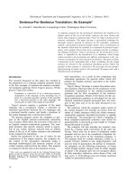
From Figure 2 it is clear that using a type B algorithm, it is
more difficult to conform the planned dose to the PTV
than using a type A algorithm, especially for a small PTV.
This is caused by the increased influence of lateral scatter
disequilibrium for smaller PTV, which is modelled better
Radiation Oncology 2009, 4:1 />Page 7 of 14
(page number not for citation purposes)
using a type B algorithm. Thus, a less strict conformity
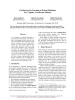
requirement was formulated. The difference between type
B and type A or unit density calculations is even more pro-
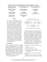
nounced for the R50% values (Figure 3). Also for the dose
at 2 cm from the PTV (Figure 4) and the percentage of the
lung receiving more than 20 Gy (Figure 5), it is clear that
a type B algorithm will result in higher values, due to the
fact that the change in lateral scattering in lung tissue is
taken into account much better. Again, the conformity
requirements for type B algorithms were relaxed for these
parameters. However, relaxation of these requirements
does not result in an actual inferior patient treatment. On
the contrary, because these more advanced algorithms
provide a better description of the actual dose distribu-
tion, the user has a greater opportunity to optimize the
dose distribution to the stated requirements. Therefore,
the use of these more advanced algorithms is strongly
encouraged. Please note that the figures presented here are
based on the treatment plans generated without recalcula-
tion with a more advanced algorithm, thus representing
treatment planning clinical practice within the ROSEL
trial, while in the article of Schuring and Hurkmans the
results were presented after recalculation, thus quantify-
ing the actual delivered dose differences arising from the
use of different algorithms [15]. To emphasize the
improvement that can be achieved using a more advanced
algorithm over a type A algorithm or a unit density calcu-
lation, the dose to the PTV after recalculation is given in
Figure 6 (reprinted with permission from Schuring and
Hurkmans [15]. The figure clearly shows that The EPL
plans (Type A algorithm) consistently overestimate the
dose to the PTV, resulting in an average D
95
of 48 Gy, 20%
lower than the prescribed value. The overestimation of the
dose increased with decreasing PTV size, although large
variations are observed between individual patients. For
the unit density calculations the recalculated D
95
ranged
between as much as 63 and 42 Gy for individual patients.
Dose-volume constraints for OAR within the ROSEL pro-
tocol are given in Table 2 and differ from the ones used in
RTOG 0236 and 0618 (for lung constraints, see previous
Table 1). A reassessment was considered necessary
because a new patient group will be treated with stereotac-
tic radiotherapy within the ROSEL trial, namely patients
who are fit to undergo both primary and salvage surgery.
As a result, normal tissue dose-constraints have to be
more stringently defined in order to minimize the risk of
increased complications after salvage surgery. Addition-
ally, new constraints were formulated to be used for the 5
fraction scheme. Furthermore, the constraints are based
on 1 cc volumes (except for the spinal cord), to prevent an
excessive dependency on the calculation grid size in the
evaluation of these parameters. Skin dose, with the con-
straint that no point within the skin should receive a dose
higher than 24 Gy as dictated in RTOG 0618 is not
included in Table 2, as dose calculations within this
region are often not very accurate and this dose parameter
is often very labour intensive to score. However, this will
be evaluated in a dummy run procedure planned before
trial participation for each institution.
Treatment planning
If treatment planning and irradiation are based on the ITV
concept, the PTV incorporates the complete respiratory
Table 1: Dose conformity requirements and definition of protocol deviations. R
100%
and R
50%
= ratio of respectively the 100% and 50%
Prescription Isodose Volume to the PTV. D
2 cm
= dose maximum at 2 cm from the PTV as percentage of the prescribed dose. V
20 Gy
=
Percent of lung receiving 20 Gy or more (both lungs minus GTV).
Type A models (standard algorithms)
R
100%
R
50%
D
2 cm
(%) V
20 Gy
(%) PTV (cc)
Deviation Deviation Deviation Deviation
None Minor None Minor None Minor None Minor
<1.15 1.15–1.25 <8 8–10 <55 55–60 <4 4–6 0–20
<1.15 1.15–1.25 <7 7–8 <65 65–70 <6 6–8 20–40
<1.10 1.10–1.20 <6 6–6.5 <65 65–75 <8 8–10 >40
Type B models (more advanced algorithms)
R
100%
R
50%
D
2 cm
(%) V
20 Gy
(%) PTV (cc)
Deviation Deviation Deviation Deviation
None Minor None Minor None Minor None Minor
<1.25 1.25–1.40 <12 12–14 <65 65–75 <5 5–8 0–20
<1.15 1.15–1.25 <9 9–11 <70 70–80 <6 6–10 20–40
<1.10 1.10–1.20 <6 6–8 <70 70–80 <10 10–15 >40
Radiation Oncology 2009, 4:1 />Page 8 of 14
(page number not for citation purposes)
tumour mobility. Several studies indicate that the use of
the ITV concept leads to the use of larger margins than
necessary to compensate for tumour motion due to
breathing [45-48]. This may in turn lead to the unneces-
sary exposure of relatively large volumes of organs at risk,
especially for patients with very mobile tumours. How-
ever, Lagerwaard et al. have shown that the incidence of
toxicity is low using this concept and a risk-adapted frac-
tionation schedule [26]. Therefore, the use of this concept
is accepted within the ROSEL trial. However, one might
want to avoid unnecessary exposure of organs at risk due
to breathing motion, and four techniques can be distin-
guished [49]: 1) adaptation of margin recipe [32,50,40],
2) tumour tracking, 3) gating and 4) reduction of breath-
ing motion [51]. These methods are not mutually exclu-
sive, for example, one might use abdominal compression
in combination with the mean-position margin recipe. It
must be emphasised that introduction of these techniques
is not needed for the majority of the patients. In a study
performed by Underberg and colleagues, it was shown
that only 15% of their patients would have a clinically rel-
evant PTV reduction (defined as 50% or more) using gat-
ing compared to the PTV based on the ITV concept [52].
They also showed that the PTV reduction correlated well
with the tumour mobility. Thus, the abovementioned
techniques should be primarily considered when treating
very mobile tumours or for example tumours close to the
stomach.
It has been shown that the use of a different margin recipe
leads to a similar reduction of the PTV as gating [45,50].
From a patients' perspective, the use of an adapted margin
recipe might be preferred, as gating significantly prolongs
the treatment time and this, in turn, leads to significantly
more intra-fractional changes in tumour position [53].
Also, the use of an abdominal compression plate or active
breathing control device might be less comfortable for a
patient. This less comfortable position might lead to
Ratio of Prescription Isodose Volume to the PTV (R
100%
) from a total of 22 patients with stage IA tumours and 4 patients with stage 1B tumours (with PTVs of 59 cc, 85 cc, 107 cc and 108 cc)Figure 2
Ratio of Prescription Isodose Volume to the PTV (R
100%
) from a total of 22 patients with stage IA tumours and
4 patients with stage 1B tumours (with PTVs of 59 cc, 85 cc, 107 cc and 108 cc).
0 25 50 75 100 125
0.9
1.0
1.1
1.2
1.3
1.4
Type A algorithm
Type B algorithm
RTOG
R
100%
[ - ]
PTV [cm
3
]
Radiation Oncology 2009, 4:1 />Page 9 of 14
(page number not for citation purposes)
increased patient movement and no data about this pos-
sible effect is available yet. Tumour tracking by means of
an external marker does not cause any patient discomfort
and might be seen as a patient friendly alternative. How-
ever, it is shown that variations in external/internal
motion correlation are present, making their use poten-
tially less accurate [54,55]. The use of internal markers is
considered more accurate, but is associated with an
increased risk of pneumothorax [56]. Furthermore, gating
and tracking are also technically challenging techniques.
They can only be used on a wide scale if existing technical
problems can be solved [57].
Due to the wide penumbra of high energy (≥ 15 MV)
beams, it is recommended to only use photon (x-ray)
beams with energies of 6–10 MV. Experience has been
gained with both coplanar and non-coplanar techniques,
with in general a 7–13 beam angles in case static beams
are used. Dynamic conformal arcs can be used, although
generally thoracic wall doses are larger than with multiple
static beams.
For ITV based treatment plans, dose calculations can be
performed on the 3D CT scan reconstruction generated
without breathing phase binning. (i.e. an average scan or
untagged scan reconstruction). This has proven to be a
good approximation of 4D dose calculations if combined
with a type B algorithm [47,58].
For mid-position based treatment plans, dose calculations
should be either performed on the CT reconstruction
phase which represents the time-averaged mean position
of the tumour or on scan reconstruction generated with-
out breathing phase binning.
Treatment execution
It is advised to keep the inter-fraction interval at a mini-
mum of 40 hours, in line with the RTOG protocol 0618.
Ratio of 50% Prescription Isodose Volume to the PTV (R
50%
) from a total of 22 patients with stage IA tumours and 4 patients with stage 1B tumours (with PTVs of 59 cc, 85 cc, 107 cc and 108 cc)Figure 3
Ratio of 50% Prescription Isodose Volume to the PTV (R
50%
) from a total of 22 patients with stage IA tumours
and 4 patients with stage 1B tumours (with PTVs of 59 cc, 85 cc, 107 cc and 108 cc).
0 25 50 75 100 125
0
4
8
12
16
20
Type A algorithm
Type B Algorithm
RTOG
R
50%
[ - ]
PTV [cm
3
]
Radiation Oncology 2009, 4:1 />Page 10 of 14
(page number not for citation purposes)
The maximum inter-fraction interval should be 4 days.
Within the ROSEL trial, the standard fractionation should
be given over 5–8 days, while the conservative fractiona-
tion should be given over 10–14 days. In general, it is rec-
ommended to keep the treatment time as short as possible
in order to limit possible patient movement and patient
discomfort. Longer sessions have been correlated with sig-
nificantly more inter-fractional changes in tumour posi-
tion [53].
Patient positioning should be determined by imaging at
the treatment unit itself by means of kV-CT imaging, MV-
CT imaging or orthogonal kV imaging. It is strongly rec-
ommended that the target position should be compared
to the target position in the images used for treatment
planning, and appropriate patient set-up corrections
should be applied when tumour shifts are detected [31].
As a minimum requirement within the ROSEL protocol,
an on-line set-up correction protocol based upon bony
anatomy should be applied.
Discussion
The ROSEL trial Quality Assurance Working Party in this
article has tried to present a broad overview of all the tech-
nical aspects of stereotactic radiotherapy for early stage
lung cancer. Our aim was to develop widely applicable
guidelines in view of the number of stereotactic radiother-
apy systems used at centres in The Netherlands which will
participate in the ROSEL trial. However, we also formu-
lated recommendations assuming the most advanced
technical possibilities are at ones disposal. Hopefully,
these recommended techniques can be implemented on a
large scale in the near future. As stereotactic radiotherapy
techniques are in general highly sophisticated, our paper
cannot possibly cover all areas in detail. As many aspects
of implementation depend on the available equipment,
we recommend that centres should familiarize themselves
Maximum dose 2 cm from PTV in any direction (D
2 cm
) as % of prescribed dose from a total of 22 patients with stage I tumours and 4 patients with stage 1B tumours (with PTVs of 59 cc, 85 cc, 107 cc and 108 cc)Figure 4
Maximum dose 2 cm from PTV in any direction (D
2 cm
) as % of prescribed dose from a total of 22 patients with
stage I tumours and 4 patients with stage 1B tumours (with PTVs of 59 cc, 85 cc, 107 cc and 108 cc).
0 25 50 75 100 125
40
50
60
70
80
90
Type A algorithm
Type B algorithm
RTOG
D
2cm
[%]
PTV [cm
3
]
Radiation Oncology 2009, 4:1 />Page 11 of 14
(page number not for citation purposes)
with technical details of the equipment to be used. Appro-
priate quality assurance systems should also be imple-
mented. A comprehensive overview of quality assurance
issues can be found in a special edition about quality
assurance of the Int. J. Radiat. Oncol. Biol. Phys. (71S,
2008).
To the best of our knowledge, this is the first trial in stere-
otactic lung radiotherapy which makes a distinction in
dose prescription and dose to OAR criteria based on the
calculation algorithm used. As was clearly shown, the
dosimetric differences from the use of different algo-
rithms can be large, and it is more difficult to plan a con-
formal dose distribution using a more advanced
algorithm. Without making a distinction based on type of
algorithm, this might lead to the incorrect assumption
that centres with such algorithms use less conformal tech-
niques. However, it is shown that the actually delivered
dose using type A algorithms can deviate as much as 30%,
which is highly dependent on the patient specific anat-
omy and in general the deviation increases with decreas-
ing target volume [15]. Therefore, relationships between
treatment outcome and dose generated from stereotactic
lung cancer trials which not primarily applied type B cal-
culation algorithms should be interpreted with caution.
Conclusion
Guidelines and recommendations have been formulated
to aid the implementation of stereotactic radiotherapy for
early stage lung cancer patients in both individual centres
as in future multi-institutional trials. They are formulated
such that stereotactic treatment can safely and effectively
be implemented in clinical practice in a wide variety of
hospitals and treatment results become better compara-
ble.
Competing interests
The authors declare that they have no competing interests.
Percent of lung (both lungs minus GTV) receiving 20 Gy or more (V
20 Gy
) from a total of 22 patients with stage I tumours and 4 patients with stage 1B tumours (with PTVs of 59 cc, 85 cc, 107 cc and 108 cc)Figure 5
Percent of lung (both lungs minus GTV) receiving 20 Gy or more (V
20 Gy
) from a total of 22 patients with stage
I tumours and 4 patients with stage 1B tumours (with PTVs of 59 cc, 85 cc, 107 cc and 108 cc).
0 25 50 75 100 125
0
4
8
12
16
20
Type A algorithm
Type B algorithm
RTOG
V
20%
[%]
PTV [cm
3
]
Radiation Oncology 2009, 4:1 />Page 12 of 14
(page number not for citation purposes)
Authors' contributions
CH drafted the manuscript, coordinated and participated
in the Quality Assurance Working Party designing the
guidelines, and participated in performing the calcula-
tions comparing dose calculation algorithms. JC, FL, JW
and UH were all members of the Quality Assurance Work-
ing Party. DS participated in performing the calculations
comparing dose calculation algorithms. SS conceived of
the study, and participated in its design and coordination.
All authors read and approved the final manuscript.
Acknowledgements
The ROSEL study is supported by a grant from ZonMW. Grants for the
ROSEL radiotherapy quality assurance work from Elekta, Philips Medical
Systems and Promis Electro-Optics are gratefully acknowledged.
Dose to 95% of the PTV as a function of the PTV after recalculation using a type B algorithm (Collapsed Cone (CC) algorithm, Pinnacle 8.0 h) from a total of 22 patients with stage IA tumours and 4 patients with stage 1B tumours (with PTVs of 59 cc, 85 cc, 107 cc and 108 cc) (reprinted with permission from ref 20)Figure 6
Dose to 95% of the PTV as a function of the PTV after recalculation using a type B algorithm (Collapsed Cone
(CC) algorithm, Pinnacle 8.0 h) from a total of 22 patients with stage IA tumours and 4 patients with stage 1B
tumours (with PTVs of 59 cc, 85 cc, 107 cc and 108 cc) (reprinted with permission from ref 20). Plans were opti-
mized using a type A algorithm (EPL), a unit density calculation (UD) or a type B algorithm (CC).
0 20406080100120
0
10
20
30
40
50
60
70
CC plan
UD plan
EPL plan
95
PTV [cm
3
]
Table 2: Dose constraints for organs at risk and definition of protocol deviations.
Organ Volume (cc) Deviation given as cumulative absolute dose (Gy)
3 fraction scheme 5 fraction scheme
None Minor None Minor
Spinal Cord Any point 18 > 18 to 22 25 > 25 to 28
Oesophagus 1 24 > 24 to 27 27 > 27 to 28.5
Ipsilateral Brachial Plexus 1 24 > 24 to 26 27 > 27 to 29
Heart 1 24 > 24 to 26 27 > 27 to 29
Trachea and main stem bronchus 1 30 > 30 to 32 32 > 32 to 35
Radiation Oncology 2009, 4:1 />Page 13 of 14
(page number not for citation purposes)
References
1. Qiao X, Tullgren O, Lax I, Sirzen F, Lewensohn R: The role of radi-
otherapy in treatment of stage I non-small cell lung cancer.
Lung Cancer 2003, 41:1-11.
2. Nagata Y, Takayama K, Matsuo Y, Norihisa Y, Mizowaki T, Sakamoto
T, Sakamoto M, Mitsumori M, Shibuya K, Araki N, Yano S, Hiraoka M:
Clinical outcomes of a phase I/II study of 48 Gy of stereotac-
tic body radiotherapy in 4 fractions for primary lung cancer
using a stereotactic body frame. Int J Radiat Oncol Biol Phys 2005,
63:1427-1431.
3. Onishi H, Shirato H, Nagata Y, Hiraoka M, Fujino M, Gomi K, Niibe
Y, Karasawa K, Hayakawa K, Takai Y, Kimura T, Takeda A, Ouchi A,
Hareyama M, Kokubo M, Hara R, Itami J, Yamada K, Araki T: Hypof-
ractionated stereotactic radiotherapy (HypoFXSRT) for
stage I non-small cell lung cancer: updated results of 257
patients in a Japanese multi-institutional study. J Thorac Oncol
2007, 2:S94-100.
4. McGarry RC, Papiez L, Williams M, Whitford T, Timmerman RD:
Stereotactic body radiation therapy of early-stage non-
small-cell lung carcinoma: phase I study. Int J Radiat Oncol Biol
Phys 2006, 63(4):1010-1015.
5. Lagerwaard FJ, Haasbeek CJA, Smit EF, Slotman BJ, Senan S: Out-
come After Stereotactic Radiotherapy in 'High-Risk'
Patients With Stage I Non-small Cell Lung Cancer
(NSCLC). Int J Radiat Oncol Biol Phys 2007, 69:S87-S88.
6. Onishi H, Araki T, Shirato H, Nagata Y, Hiraoka M, Gomi K, Yamas-
hita T, Niibe Y, Karasawa K, Hayakawa K, Takai Y, Kimura T,
Hirokawa Y, Takeda A, Ouchi A, Hareyama M, Kokubo M, Hara R,
Itami J, Yamada K: Stereotactic hypofractionated high-dose
irradiation for stage I nonsmall cell lung carcinoma. Cancer
2004, 101:1623-1631.
7. El-Sherif A, Gooding WE, Santos R, Pettiford B, Ferson PF, Fernando
HC, Urda SJ, Luketich JD, Landreneau RJ: Outcomes of sublobar
resection versus lobectomy for stage I non-small cell lung
cancer: a 13-year analysis. Ann Thorac Surg 2006, 82:408-415.
8. Strand TE, Rostad H, Moller B, Norstein J: Survival after resection
for primary lung cancer: a population based study of 3211
resected patients. Thorax 2006, 61:710-715.
9. Rami-Porta R, Ball D, Crowley J, Giroux DJ, Jett J, Travis WD, Tsuboi
M, Vallieres E, Goldstraw P: The IASLC Lung Cancer Staging
Project: proposals for the revision of the T descriptors in the
forthcoming (seventh) edition of the TNM classification for
lung cancer. J Thorac Oncol 2007, 2:593-602.
10. Timmerman RD, Park C, Kavanagh BD:
The North American
experience with stereotactic body radiation therapy in non-
small cell lung cancer. J Thorac Oncol 2007, 2:S101-S112.
11. Pasic A, Brokx HA, Vonk NA, Paul RM, Postmus PE, Sutedja TG:
Cost-effectiveness of early intervention: comparison
between intraluminal bronchoscopic treatment and surgical
resection for T1N0 lung cancer patients. Respiration 2004,
71:391-396.
12. Timmerman RD, Paulus R, Galvin J, Michalski J, Straube WL, Bradley
J, Fakiris A, Bezjak A, Videtic G, Choy H: Toxicity Analysis of
RTOG 0236 Using Stereotactic Body Radiation Therapy to
Treat Medically Inoperable Early Stage Lung Cancer
Patients. Int J Radiat Oncol Biol Phys 2007, 69:S86.
13. Xiao Y, Straube WL, Bosch WR, Timmerman RD, Galvin JM: Dosi-
metric Evaluation of Heterogeneity Corrections for RTOG
0236: Hypofractionated Radiotherapy of Inoperable Stage I/
II Non-small Cell Lung Cancer. Int J Radat Oncol Biol Phys 2007,
69:S46-S47.
14. Hiraoka M, Ishikura S: A Japan clinical oncology group trial for
stereotactic body radiation therapy of non-small cell lung
cancer. J Thorac Oncol 2007, 2:S115-S117.
15. Schuring D, Hurkmans CW: Developing and evaluating stereo-
tactic lung RT trials: What we should know about the influ-
ence of inhomogeneity corrections on dose. Radiat Oncol 2008,
3:21.
16. Underberg RW, Lagerwaard FJ, Slotman BJ, Cuijpers JP, Senan S: Use
of maximum intensity projections (MIP) for target volume
generation in 4DCT scans for lung cancer. Int J Radiat Oncol Biol
Phys 2005, 63:253-260.
17. Timmerman RD, Michalski J, Galvin J, Fowler JF, Choy H, Gore E,
Johnstone D: RTOG 0236 : A Phase II Trial of Stereotactic
Body Radiation Therapy (SBRT) in the Treatment of
Patients with Medically Inoperable Stage I/II Non-Small Cell
Lung Cancer. RTOG 2007.
18. Timmerman RD, Galvin J, Gore E, Pass H, Edelman MJ, Kong FP:
RTOG 0618 A phase II trial of stereotactic body radiation
therapy (SBRT) in the treatment of patients with operable
stage I/II non-small cell lung cancer. RTOG 2007.
19. Bosmans G, Buijsen J, Dekker A, Velders M, Boersma L, De Ruysscher
D, Minken A, Lambin P: An "in silico" clinical trial comparing
free breathing, slow and respiration correlated computed
tomography in lung cancer patients. Radiother Oncol 2006,
81:73-80.
20. Chen GT, Kung JH, Beaudette KP: Artifacts in computed tomog-
raphy scanning of moving objects. Semin Radiat Oncol 2004,
14:19-26.
21. Keall P: 4-dimensional computed tomography imaging and
treatment planning. Semin Radiat Oncol 2004, 14:81-90.
22. Rietzel E, Pan T, Chen GT: Four-dimensional computed tomog-
raphy: image formation and clinical protocol. Med Phys 2005,
32:874-889.
23. Seki S, Kunieda E, Takeda A, Nagaoka T, Deloar HM, Kawase T,
Fukada J, Kawaguchi O, Uematsu M, Kubo A: Differences in the
definition of internal target volumes using slow CT alone or
in combination with thin-slice CT under breath-holding con-
ditions during the planning of stereotactic radiotherapy for
lung cancer. Radiother Oncol 2007, 85:443-449.
24. Wurstbauer K, Deutschmann H, Kopp P, Sedlmayer F: Radiother-
apy planning for lung cancer: slow CTs allow the drawing of
tighter margins. Radiother Oncol 2005, 75:165-170.
25. Letourneau D, Finlay M, O'sullivan B, Waldron JN, Cummings BJ, Rin-
gash J, Kim JJ, Bayley AJ, Dawson LA: Lack of influence of intrave-
nous contrast on head and neck IMRT dose distributions.
Acta Oncol 2008, 47:90-94.
26. Lagerwaard FJ, Haasbeek CJA, Smit EF, Slotman BJ, Senan S: Out-
comes of Risk-Adapted Fractionated Stereotactic Radio-
therapy for Stage I Non-Small-Cell Lung Cancer. Int J Radiat
Oncol Biol Phys 2008, 70:685-692.
27. Potters L, Steinberg M, Rose C, Timmerman R, Ryu S, Hevezi JM,
Welsh J, Mehta M, Larson DA, Janjan NA: American Society for
Therapeutic Radiology and Oncology and American College
of Radiology practice guideline for the performance of ster-
eotactic body radiation therapy. Int J Radiat Oncol Biol Phys 2004,
60:1026-1032.
28. Bradley JD, Nofal AN, El Naga IM, Lu W, Liu J, Hubenschmidt J, Low
DA, Drzymala RE, Khullar D: Comparison of helical, maximum
intensity projection (MIP), and averaged intensity (AI) 4D
CT imaging for stereotactic body radiation therapy (SBRT)
planning in lung cancer. Radiother Oncol 2006, 81:264-268.
29. Riegel AC, Chang JY, Vedam SS, Johnson V, Chi PC, Pan T: Cine
Computed Tomography Without Respiratory Surrogate in
Planning Stereotactic Radiotherapy for Non-Small-Cell
Lung Cancer. Int J Radiat Oncol Biol Phys 2008. DOI: 10.1016/
j.ijrobp.2008.04.047
30. van Sornsen de Koste JR, Lagerwaard FJ, Nijssen-Visser MR, Grave-
land WJ, Senan S: Tumor location cannot predict the mobility
of lung tumors: a 3D analysis of data generated from multi-
ple CT scans. Int J Radiat Oncol Biol Phys 2003, 56:348-354.
31. Sonke JJ, Lebesque J, van Herk M: Variability of four-dimensional
computed tomography patient models. Int J Radiat Oncol Biol
Phys 2008, 70:590-598.
32. Wolthaus JWH, Schneider C, Sonke JJ, van Herk M, Belderbos JSA,
Rossi MMG, Lebesque JV, Damen EFM: Mid-ventilation CT scan
construction from four-dimensional respiration-correlated
CT scans for radiotherapy planning of lung cancer patients.
Int J Radat Oncol Biol Phys 2006, 65:1560-1571.
33. Dieleman EM, Senan S, Vincent A, Lagerwaard FJ, Slotman BJ, van
Sornsen de Koste JR: Four-dimensional computed tomo-
graphic analysis of esophageal mobility during normal respi-
ration. Int J Radiat Oncol Biol Phys 2007, 67:775-780.
34. ICRU report 62 prescribing, recording and reporting photon
beam therapy. (Supplement to ICRU report 50) 1999, 33:1-51.
35. Hall WH, Guiou M, Lee NY, Dublin A, Narayan S, Vijayakumar S,
Purdy JA, Chen AM: Development and Validation of A Stand-
ardized Method for Contouring THE Brachial Plexus: Pre-
liminary Dosimetric Analysis Among Patients Treated with
IMRT for Head-and-Neck Cancer. Int J Radiat Oncol Biol Phys
2008, 72;1:S385.
Publish with BioMed Central and every
scientist can read your work free of charge
"BioMed Central will be the most significant development for
disseminating the results of biomedical researc h in our lifetime."
Sir Paul Nurse, Cancer Research UK
Your research papers will be:
available free of charge to the entire biomedical community
peer reviewed and published immediately upon acceptance
cited in PubMed and archived on PubMed Central
yours — you keep the copyright
Submit your manuscript here:
/>BioMedcentral
Radiation Oncology 2009, 4:1 />Page 14 of 14
(page number not for citation purposes)
36. Madu CN, Quint DJ, Normolle DP, Marsh RB, Wang EY, Pierce LJ:
Definition of the supraclavicular and infraclavicular nodes:
implications for three-dimensional CT-based conformal
radiation therapy. Radiology 2001, 221:333-339.
37. Collins BT, Erickson K, Reichner CA, Collins SP, Gagnon GJ, Diet-
erich S, McRae DA, Zhang Y, Yousefi S, Levy E, Chang T, Jamis-Dow
C, Banovac F, Anderson ED: Radical stereotactic radiosurgery
with real-time tumor motion tracking in the treatment of
small peripheral lung tumors. Radiat Oncol 2007, 2:39.
38. Koto M, Takai Y, Ogawa Y, Matsushita H, Takeda K, Takahashi C,
Britton KR, Jingu K, Takai K, Mitsuya M, Nemoto K, Yamada S: A
phase II study on stereotactic body radiotherapy for stage I
non-small cell lung cancer. Radiother Oncol 2007, 85:429-434.
39. Sinha B, McGarry RC: Stereotactic body radiotherapy for bilat-
eral primary lung cancers: the Indiana university experience.
Int J Radiat Oncol Biol Phys 2007, 66:1120-1124.
40. Haasbeek CJ, Senan S, Smit EF, Paul MA, Slotman BJ, Lagerwaard FJ:
Critical review of nonsurgical treatment options for stage I
non-small cell lung cancer. Oncologist 2008, 13:309-319.
41. Brock J, Ashley S, Bedford J, Nioutsikou E, Partridge M, Brada M:
Review of Hypofractionated Small Volume Radiotherapy for
Early-stage Non-small Cell Lung Cancer. Clin Oncol (R Coll
Radiol) 2008, 20:666-676.
42. Timmerman R, McGarry R, Yiannoutsos C, Papiez L, Tudor K,
DeLuca J, Ewing M, Abdulrahman R, DesRosiers C, Williams M,
Fletcher J: Excessive toxicity when treating central tumors in
a phase II study of stereotactic body radiation therapy for
medically inoperable early-stage lung cancer. J Clin Oncol 2006,
24:4833-4839.
43. Senan S, Haasbeek NJ, Smit EF, Lagerwaard FJ: Stereotactic radio-
therapy for centrally located early-stage lung tumors. J Clin
Oncol 2007, 25:464.
44. Knöös T, Wieslander E, Cozzi L, Brink C, Fogliata A, Albers D, Nys-
tröm H, Lassen S: Comparison of dose calculation algorithms
for treatment planning in external photon beam therapy for
clinical situations. Phys Med Biol 2006, 51:5785-5807.
45. Wolthaus JW, Sonke JJ, van Herk M, Belderbos JS, Rossi MM, Leb-
esque JV, Damen EM: Comparison of different strategies to use
four-dimensional computed tomography in treatment plan-
ning for lung cancer patients. Int J Radiat Oncol Biol Phys 2008,
70:1229-1238.
46. Guckenberger M, Krieger T, Baier K, Richter A, Polat B, Flentje M:
Four Dimensional Target Volume Generation in Pulmonary
Stereotactic Body Radiotherapy. Int J Radiat Oncol Biol Phys 2007,
69(1):S191.
47. Admiraal MA, Schuring D, Hurkmans CW: Dose calculations
accounting for breathing motion in stereotactic lung radio-
therapy based on 4D-CT and the internal target volume.
Radiother Oncol 2008, 86:55-60.
48. Mutaf YD, Brinkmann DH: Optimization of internal margin to
account for dosimetric effects of respiratory motion. Int J
Radiat Oncol Biol Phys 2008, 70:1561-1570.
49. Giraud P, Yorke E, Jiang S, Simon L, Rosenzweig K, Mageras G:
Reduction of organ motion effects in IMRT and conformal
3D radiation delivery by using gating and tracking tech-
niques. Cancer Radiother 2006, 10:269-282.
50. Burnett SS, Sixel KE, Cheung PC, Hoisak JD: A study of tumor
motion management in the conformal radiotherapy of lung
cancer. Radiother Oncol 2008, 86:77-85.
51. Heinzerling JH, Anderson JF, Papiez L, Boike T, Chien S, Zhang G,
Abdulrahman R, Timmerman R: Four-dimensional computed
tomography scan analysis of tumor and organ motion at var-
ying levels of abdominal compression during stereotactic
treatment of lung and liver. Int J Radiat Oncol Biol Phys 2008,
70:1571-1578.
52. Underberg RWM, Lagerwaard FJ, Slotman BJ, Cuijpers JP, Senan S:
Benefit of respiration-gated stereotactic radiotherapy for
stage I lung cancer: an analysis of 4DCT datasets. Int J Radiat
Oncol Biol Phys 2005, 62:554-560.
53. Purdie TG, Bissonnette JP, Franks K, Bezjak A, Payne D, Sie F, Sharpe
MB, Jaffray DA: Cone-beam computed tomography for on-line
image guidance of lung stereotactic radiotherapy: localiza-
tion, verification, and intrafraction tumor position. Int J Radiat
Oncol Biol Phys 2007, 68:243-252.
54. Korreman SS, Juhler-Nottrup T, Boyer AL: Respiratory gated
beam delivery cannot facilitate margin reduction, unless
combined with respiratory correlated image guidance. Radi-
other Oncol 2008, 86:61-68.
55. Ionascu D, Jiang SB, Nishioka S, Shirato H, Berbeco RI: Internal-
external correlation investigations of respiratory induced
motion of lung tumors. Med Phys 2007, 34:3893-3903.
56. Geraghty PR, Kee ST, McFarlane G, Razavi MK, Sze DY, Dake MD:
CT-guided transthoracic needle aspiration biopsy of pulmo-
nary nodules: needle size and pneumothorax rate. Radiology
2003, 229:475-481.
57. Jiang SB: Radiotherapy of mobile tumors. Semin Radiat Oncol
2006, 16:239-248.
58. Guckenberger M, Wilbert J, Krieger T, Richter A, Baier K, Meyer J,
Flentje M: Four-dimensional treatment planning for stereo-
tactic body radiotherapy. Int J Radiat Oncol Biol Phys 2007,
69:276-285.