Báo cáo khoa học: "Radiation-induced cancer after radiotherapy for non-Hodgkin''''s lymphoma of the head and neck: a retrospective study" ppsx
Bạn đang xem bản rút gọn của tài liệu. Xem và tải ngay bản đầy đủ của tài liệu tại đây (755.67 KB, 7 trang )
BioMed Central
Page 1 of 7
(page number not for citation purposes)
Radiation Oncology
Open Access
Research
Radiation-induced cancer after radiotherapy for non-Hodgkin's
lymphoma of the head and neck: a retrospective study
Kazuma Toda*
1
, Hitoshi Shibuya
1
, Keiji Hayashi
1
and Fumio Ayukawa
2
Address:
1
Department of Radiology, Tokyo Medical and Dental University, 5–45, Yushima 1-chome, Bunkyo-ku, Tokyo 113-8519, Japan and
2
Department of Radiology, Niigata Cancer Center Hospital, Niigata, Japan
Email: Kazuma Toda* - ; Hitoshi Shibuya - ; Keiji Hayashi - ;
Fumio Ayukawa -
* Corresponding author
Abstract
Background: survivors of non-Hodgkin's lymphoma (NHL) are well known to be at an increased
risk of second malignancies. In this study, we evaluated the incidence and clinical features of head
and neck cancer (HNC) occurring after radiotherapy (RT) for NHL.
Materials and methods: We investigated the clinical records of 322 patients who had received
RT for early-stage NHL of the head and neck at our institute between 1952 and 2000.
Results: There were 4 patients with a second HNC developing in the irradiated field, consisting
of 2 patients with gum cancer, 1 case with tongue cancer and 1 case with maxillary sinus cancer.
The pathological diagnosis in all the 4 patients was squamous cell carcinoma (SCC). Two of the
patients (one with gum cancer and one with maxillary sinus cancer) died of the second HNC, while
the remaining 2 patients are still living at the time of writing after therapy for the second HNC,
with neither recurrence of the second tumor nor relapse of the primary tumor. The ratio of the
observed to the expected number (O/E ratio) of a second HNC was calculated to be 12.7 (95%CI,
4.07–35.0), and the absolute excess risk (AER) per 10,000 person-years was 13.3. The median
interval between the RT and the diagnosis of the second HNC was 17.0 years (range, 8.7 to 22.7
years).
Conlusion: The risk of HNC significantly increased after RT for early-stage NHL. These results
suggest that second HNC can be regarded as one of the late complications of RT for NHL of the
head and neck.
Background
Carcinogenesis associated with exposure to radiation is
widely known, first reported in the early 20th century,
when skin cancer was noted in radiation workers. The risk
of carcinogenesis following low-dose radiation exposure
was estimated to be 0.05–0.1 Sv based on the results of
follow-up of atomic bomb survivors in Japan, however,
that associated with exposure to much lower doses, such
as that associated with diagnostic X-ray examinations, is
debatable [1,2]. Exposure to therapeutic doses of radia-
tion has also been shown to be associated with an
increased risk of a second cancer, although the precise risk
remains unknown. For selected cancers with a high cure
rate, the benefits of treatment need to be weighed against
the potential risk of treatment-related second malignancy.
Published: 10 July 2009
Radiation Oncology 2009, 4:21 doi:10.1186/1748-717X-4-21
Received: 17 March 2009
Accepted: 10 July 2009
This article is available from: />© 2009 Toda et al; licensee BioMed Central Ltd.
This is an Open Access article distributed under the terms of the Creative Commons Attribution License ( />),
which permits unrestricted use, distribution, and reproduction in any medium, provided the original work is properly cited.
Radiation Oncology 2009, 4:21 />Page 2 of 7
(page number not for citation purposes)
Progress of therapeutic modalities in recent decades have
considerably improved the prognosis of malignant lym-
phoma, on the other hand, development of therapy-
related second cancer as a late complication of treatment
has became obvious [3-6]. As compared with the case in
HL, RT still occupies a more important position in the
treatment of NHL, especially early-stage NHL. Although
the head and neck area is one of the most frequent sites of
NHL, the risk of a second HNC after RT for NHL still
remains unclear. We investigated the incidence and clini-
cal features of a second HNC occurring after RT for early-
stage NHL.
Materials and methods
We conducted a retrospective review of a total of 322
patients who received RT with/without chemotherapy as
initial therapy for early-stage NHL (stage I or stage II) of
the head and neck at our institute between 1952 and 2000
[7]. The patient parameters investigated were the sex, age
at the time of RT, the chemotherapy regimen employed,
the clinical stage and location of the lymphoma, the irra-
diated field, the dose and type of RT, and the cause of
death.
For the patients in whom a second HNC developed in the
irradiated field after RT, we investigated the site and path-
ological diagnosis, the interval from the time of RT to the
diagnosis of the second HNC, and the clinical course of
the second cancer. We calculated the expected numbers of
second cancers by using the person-years method [8,9].
We used the age-, sex-, and calendar year-specific cancer
incidence rates in the general population of Japan [10]. O/
E ratio was then calculated with the 95%CI from the Pois-
son distribution. These results were statistically analyzed
by the SPSS for Windows (SPSS Inc. Chicago, Illinois).
Results
The patient characteristics are listed in Table 1. In all, 96
patients had NHL lesions in the Waldeyer's ring. Extran-
odal lesions were seen in 124 patients. The most frequent
site of NHL was the oral cavity (n = 48). Neoadjuvant and/
or adjuvant chemotherapy was administered in 144
patients (44.7%), and the most frequently administered
regimen was cyclophosphamide, doxorubicin, vincris-
tine+ prednisolone (CHOP) or a CHOP-like regimen (n =
88).
RT was administered with high-voltage X-rays from a lin-
ear accelerator in 150 patients, with γ-rays from Co-60 in
89 patients, with orthovoltage X-rays in 55 patients, with
either high-voltage X-rays or γ-rays plus electrons in 15
patients, with electrons alone in 9 patients, with high-
voltage X-rays plus γ-rays in 2 patients, with γ-rays plus
orthovoltage X-rays in 1 patient, and orthovoltage X-rays
plus brachytherapy in 1 patient. RT was administered with
conventional RT techniques, therefore 1 field, 2 opposed
fields and a combination of them were mostly used.
The median total dose of RT was 40.8 Gy (range, 5.5–78
Gy), and the dose per fraction was 1.5–3 Gy (2 Gy in most
cases). The total radiation dose was unknown in the
patient who received low-dose-rate intracavitary brachy-
therapy in addition to orthovoltage X-rays for NHL of the
tonsil. The total dose employed was 5.5–19.8 Gy in 9
patients (2.8%), 21–30 Gy in 64 patients (19.9%), 30.8–
40 Gy in 87 patients (27.1%), 40.8–50 Gy in 143 patients
(44.5%), 50.6–60 Gy in 16 patients (5.0%), and over 60
Gy in 2 patients (0.6%).
The overall 2-, 5- and 10-year survival rates of the patients
calculated by the Kaplan-Meier method were 77.6%,
Table 1: Characteristics of all the patients (n = 322)
n%
Sex
Male 191 59.3
Female 131 40.7
Age at the time of RT (median, 53 years{range,4 – 91})
<60 years 200 62.1
м 60 years 122 37.9
Stage
I 200 62.1
II 122 37.9
Chemotherapy
+ 144 44.7
- 178 55.3
Follow-up duration after RT
Average(range) 8.6 years(0 – 35.1)
Abbreviations RT: radiotherapy
Radiation Oncology 2009, 4:21 />Page 3 of 7
(page number not for citation purposes)
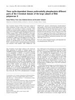
65.5% and 54.7%, respectively. The median survival time
was 14.9 years (95%CI, 8.2–21.5 years). The lymphoma-
related 2-, 5- and 10-year survival rates were 80.1%,
70.4% and 63.7%, respectively. There was a significant
difference in the overall survival rate between NHL
patients with clinical stage I and those with clinical stage
II (p < 0.05) (Fig. 1).
Of the patients, 19 (5.9%) developed a second malig-
nancy, which was metachronous in 16 cases and synchro-
nous in 3 cases (table 2). In 4 patients, the second HNC
occurred in the irradiated field. The clinical outlines of
these 4 patients are shown in [table S1; Additional file 1].
Two of the 4 patients had also received chemotherapy (3
cycles of CHOP). The pathological diagnosis of the sec-
ond HNC in all the 4 cases (2 cases of cancer of the gum,
1 case of tongue cancer, and 1 case of maxillary sinus can-
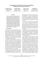
cer) was SCC (fig. 2). The median interval after the RT to
the development of the second cancer was 13.9 years
(range, 8.7 to 22.7 years). Two of the patients (1 with gum
cancer and 1 with maxillary sinus cancer) died of the sec-
ond cancer. The remaining 2 patients are still living at the
time of writing, with neither recurrence of the second
HNC nor relapse of the primary NHL, or indeed any
severe complications during the follow-up. The patient
with gum cancer is still living, 3.3 years after surgery for
SCC of the right upper gum, and the patient with tongue
cancer is also still living, 8.9 years after RT for SCC of the
tongue. The latter case received 90.5 Gy as brachytherapy
for tongue cancer by Au-198 grain implantation.
During the 2776 person-years (PYs) of observation, the
expected number of a second HNC in the general popula-
tion was 0.31, so that the O/E ratio was 12.7 (95%CI,
4.07–35.0, p < 0.01). The absolute excess risk (AER) of a
second HNC per 10,000 PYs was 13.3. When the analysis
was limited to the 192 patients who could be followed up
for over 5 years, the expected number was 0.28 during
2544 PYs, the O/E ratio was 14.1 (95%CI, 4.5–38.7, p <
0.01), and the AER was 14.6. Furthermore, the O/E ratio
was 12.0 (95%CI, 2.1–48.4, p < 0.01) during 1600 PYs in
the 178 patients who did not receive chemotherapy, and
13.5 (95%CI, 2.3–54.5, p < 0.01) during 1176 PYs in
the144 patients who received chemotherapy.
Of the 19 patients with a second cancer, 2 cases of second
cancer arose near the previous radiation field: one of
laryngeal cancer developing 14 years after RT for NHL of
the nasal cavity, and one of esophageal cancer developing
16 years after RT and chemotherapy for NHL of the oral
cavity and neck.
Discussion
Some definitions of radiation-induced malignancy have
been proposed. We removed the 2 second cancers (one
each of laryngeal cancer and esophageal cancer) which
arose near the radiation field from the analysis of radia-
tion-induced cancer according to the criteria that Sakai et
al. proposed, even though these cases might well have had
a relation to scattered radiation [11]. Cahan et al. reported
their criteria for the diagnosis of radiation-induced oste-
osarcoma in the middle of last century [12]. According to
their criteria, the primary lesion for which RT was admin-
istered must be a benign disease. In the early part of the
last century, RT was widely used for benign diseases such
as tuberculous lymphadenitis, skin diseases, thyroid dis-
eases and spondylitis, however, at present, RT is mainly
used to treat malignancies. The limitation of the prior dis-
ease treated by RT to a benign disease might thus be
impractical. Sakai et al. argued the criteria for the diagno-
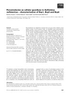
(a) Overall survival and lymphoma-specific survival rates in NHL patientsFigure 1
(a) Overall survival and lymphoma-specific survival rates in NHL patients. (b) Overall survival of NHL patients by
stage.
Radiation Oncology 2009, 4:21 />Page 4 of 7
(page number not for citation purposes)
sis of radiation-induced cancer, except leukemia, and their
results suggested that the reliability of the diagnosis of
radiation-induced cancer depends on the pathological
diagnosis, the organ of origin, the follow-up duration
after RT (over 5 years) and on whether the lesion is located
in the irradiated field [11]. These criteria were based on
the criteria of double primary cancer proposed by Warren
et al. [13].
A limitation of our study is that our study population was
small. A long latency period of radiation-induced malig-
nancies except leukemia would make it difficult to ana-
lyze these malignancies [11,12,14]. Therefore, a large
number of patients who have been under observation for
a long time after RT would be necessary to correctly assess
a radiation-induced cancer. Tward et al reported that the
O/Eratio of a second HNC among 77823 NHL patients
was 1.28 (95%CI, 1.12–1.46) [5]. British group reported
that the O/Eratio of a second HNC among 5519 HL
patients was 2.8(95%CI, 1.1–5.8) and that among 2456
NHL patients was 2.6(95%CI, 0.8–6.0) [4,6]. These stud-
ies showed that RT alone did not significantly relate to a
second HNC, although the relationship between the
details of RT and a site of second malignancies was not
considered. In this study, O/Eratio of a second HNC was
higher than those previously reported and significantly
increased even among the patients who received RT alone.
The possibility that those large-scale studies underesti-
mated carcinogenicity of RT because of the lack of consid-
eration for the details of RT could not be ruled out,
although our study population was smaller than that in
previous studies.
Chemotherapy for NHL is held to be associated with a cer-
tain risk of carcinogenesis, especially of leukemia, lung
cancer, bladder cancer and colorectal cancer [4]. No cases
of second leukemia and second lung cancer were observed
in this study. The lack may be affected by a strong relation-
ship between these second malignancies and chemother-
apy. Chemotherapy occupied a relatively lower place in
the therapy for NHL than in that for HL, at least especially
in the earlier decades. For example, only about 27% of all
the patients received CHOP that is now standard regimen
for B-cell NHL in combination with rituximab and
CHOP-like regimens in this study. The increased risk of
second malignancies in the synergy of radiation and
chemotherapy is also known, although how the synergy
affected induction of second HNC was unknown.
The risk of certain malignancies is significantly associated
with smoking, habitual alcohol consumption, immuno-
suppressive conditions, and some genetic disorders. It
would appear that the higher risk of a second cancer in
patients with HNC remains even after they stop smoking
[15]. Moertel et al. described multicentric cancer develop-
ment associated with carcinogenic stimulation of large
areas of tissues [16,17]. Slaughter et al. proposed "field
cancerization" in oral SCC [18]. These reports underscore
the difficulty of distinguishing radiation-induced malig-
nancy from not only recurrence of the first malignancy,
but also from multicentric primary tumors in the head
and neck area [19]. We did not have sufficient data about
the smoking, alcohol drinking habit, and genetic disor-
ders of all the patients. We do know, though, that only 1
of the 4 patients with a second HNC had a smoking his-
tory, and that none of them engaged in habitual alcohol
consumption.
A dose-response relationship is known in the develop-
ment of leukemia in experimental animals. The incidence
of leukemia was reported to increase with the radiation
dose in the dose range between 3 and 10 Gy [20]. The
explanation for the decrease in the incidence at higher
doses is that the number of surviving cells decreases at
these doses. A similar relationship was suggested between
sarcoma induction and the radiation dose employed, with
the maximum dose levels for malignant transformation
and decreased cell survival being higher than those for
leukemia [21]. This is held to be one of the reasons why
sarcomas are likely to be induced in heavily irradiated tis-
sues. A relationship between initial RT doses and second
head and neck malignancies was unknown in this study.
The RT doses employed in our study were relatively lower
than those used for other solid tumors, and likely to be
higher than those used for NHL in today. To be concrete,
about half of all the patients including 4 second HNC
patients received 40 Gy and over. No other pathological
diagnosis than SCC was seen in second head and neck
malignancies and this result was similar to previous stud-
ies [22,23]. In contrast, Sale et al. reported 13 second
Table 2: Characteristics of the second tumor(n = 19)
Type of second tumor n O/Eratio 95%CI AER*
Synchronous 3
Esophagus 1
Stomach 1
Cervix 1
Metachronous 16 0.8 0.47–1.33 -14.6
Head and neck
In irradiated field 4 12.7 4.07–35.0 13.3
Out of irradiated field 1
Esophagus 2 3.24 0.56–13.1 4.95
Stomach 2 0.39 0.07–1.55 -11.4
Colon 3 1.55 0.40–4.93 3.80
Breast 1
Gallbladder 1
Soft-tissue sarcoma(buttocks) 1
Myeloma(thoracic vertebra) 1
* Absolute excess risk per 10,000 person-years
Radiation Oncology 2009, 4:21 />Page 5 of 7
(page number not for citation purposes)
malignancies of the head and neck after RT, with the most
frequent histological diagnosis being sarcoma, followed
in frequency by SCC [24]. And Patel et al. reported 10
patients of radiation-induced sarcoma of the head and
neck, and malignant fibrous histiocytoma was the com-
monest pathological diagnosis (4 patients) in their
patient series [25]. The difference of the pathological diag-
nosis among these studies might be related to the differ-
ence of RT doses, nevertheless a correct relationship
between initial RT doses and second head and neck malig-
nancies is unclear because of a small number of these
malignancies.
Equipment and techniques mainly used for RT in today
are likely to differ from those used for our patients. About
half of our patients were treated with a linear accelerator
which is now standard RT equipment, and almost all the
patients were treated with conventional RT techniques.
However, how advance of radiation techniques affects sec-
ond malignancies is held to be debatable. Intensity mod-
ulated radiation therapy (IMRT) which is one of the
advanced RT techniques is concerned to increase the risk
of a second cancer compared with three-dimensional con-
formal radiotherapy (3D-CRT) [26,27]. The change from
3D-CRT to IMRT involves a bigger volume of normal tis-
sue irradiated by lower doses as a result of the increase of
fields, of monitor units and of scattered radiation. In con-
trast, Ruben at al. argued that the risk of radiation-induced
cancer did not significantly differ between IMRT and 3D-
CRT concerning the body in totality, and the risk of sec-
ond cancer was regarded to be influenced by RT equip-
ment [28]. At least, it must be inappropriate to simply
apply our results to NHL patients treated with modern RT
equipment and techniques.
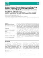
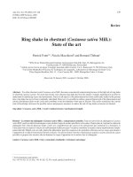
(a) Dose distribution of RT for NHL of the maxillary sinusFigure 2
(a) Dose distribution of RT for NHL of the maxillary sinus. (b) and (c) PET-CT showing second SCC infiltrating the
bone.
Radiation Oncology 2009, 4:21 />Page 6 of 7
(page number not for citation purposes)
It is debatable whether the prognosis of radiation-induced
malignancy might differ from that of spontaneously
occurring tumors. Previous studies on radiation-induced
sarcoma suggested a poor prognosis of these patients and
also the beneficial effects of surgery for these tumors
[21,25,29-33]. In addition, the poor prognosis of radia-
tion-induced sarcoma of the head and neck might be
related to the difficulty in complete resection of these
tumors due to post-radiation changes [25]. It was held
that surgery should be conducted prior to RT in the treat-
ment of radiation-induced cancer, because of the lowered
tolerance of the tissues to re-radiation and the oxygen
effect of the second tumor [19]. McHugh et al. compared
the characteristics of radiation-induced craniofacial oste-
osarcoma with those of the corresponding primary
tumors, and proposed that the poorer prognosis of radia-
tion-induced osteosarcoma was related to the higher
expression of adverse prognostic markers, such as p53,
TP53 mutations, ezrin expression, and the higher prolifer-
ative activity [34]. In contrast, there are some reports of
laryngeal and pharyngeal cancer after radiation for thyro-
toxicosis and tuberculous lymphadenitis being success-
fully treated by radiation from a linear accelerator [35,36].
The choice of the therapeutic modality for radiation-
induced cancer is affected not only by the nature of the
tumor, but also by several patient factors, mainly the
extent of the existing tissue damage. Because of the small
number of cases, we could not estimate the prognosis of
second HNC after RT for NHL. However, we assumed that
patients with a second cancer after RT for NHL would have
a little advantage over those with a radiation-induced can-
cer after the treatment of other solid tumors. Because rel-
atively lower radiation doses given for lymphomas than
those for solid tumors would lead to a lower extent of
damage of the surrounding tissue, patients with a second
cancer after RT for NHL might show better tolerance to
treatment for the second tumor. Therefore, an early detec-
tion of second HNC may aid in a better choice of a thera-
peutic modality. Of course, an irradiated area is ought to
be under careful observation. In addition, observations of
NHL patients ordinarily include systemic follow-up that
may encourage a detection of second primary cancer even
distant from an irradiated area.
Conclusion
The risk of HNC significantly increased after RT for early-
stage NHL, although a precise relationship between RT
and second head and neck malignancies remains unclear
because of a small number of cases. Anyway, we propose
to regard second HNC as one of the late complications
after RT for NHL of head and neck.
Competing interests
The authors declare that they have no competing interests.
Authors' contributions
KT and HS designed/conducted analysis and wrote the
manuscript. KH and FA assisted in the acquisition and
analysis of data. All authors have read and approved the
final manuscript.
Additional material
References
1. Pierce DA, Preston DL: Radiation-related cancer risks at low
doses among atomic bomb survivors. Radiat Res 2000,
154:178-186.
2. Berrington de Gonzalez A, Darby S: Risk of cancer from diagnos-
ticX-rays: estimates for UK and 14 other countries. Lancet
2004, 363:345-351.
3. Coleman CN: Secondary neoplasms in patients treated for
cancer: Etiology and perspective. Radiat Res 1982, 92:188-200.
4. Mudie NY, Swerdlow AJ, Higgins CD, Smith P, Qiao Z, Hancock BW,
Hoskin PJ, Linch DC: Risk of second malignancy after non-
Hodgkin's lympnoma: a british cohort study. J Clin Oncol 2006,
24:1568-74.
5. Tward JD, Wendland MM, Shrleve DC, Szabo A, Gaffney DK: The
risk of secondary malignancies over 30 years after the treat-
ment of non-Hodgkin's lymphoma. Cancer 2006, 107:108-115.
6. Swerdlow AJ, Barder JA, Hudson GV, Cunningham D, Gupta RK,
Hancock BW, Horwich A, Lister TA, Linch DC: Risk of Second
Malignancy After Hodgkin's Disease in a Collaborative Brit-
ish Cohort: The Relation to Age at Treatment. J Clin Oncol
2000, 18:498-509.
7. Lister TA, Crowther D, Sutcliffe SB, Glatstein E, Canellos GP, Young
RC, Rosenberg SA, Coltman CA, Tubiana M: Report of a commit-
tee convened to discuss the evaluation and staging of
patients with Hodgkin's disease; Cotswolds meeting. J Clin
Oncol 1989, 7:1630-1636.
8. Schoenberg BS, Myers MH: Statistical methods for studying
multiple primary malignant neoplasms. Cancer 1977,
40:1892-1898.
9. Breslow NE, Day NE: Statistical methods in cancer research. The design
and analysis of cohort studies Volume II. Lyon, France: International
Agency for Research on cancer; 1987. IARC Sci Publ 82
10. Oshima A, Kuroishi T, Tajima K, Eds: Gan toukeihakusho rikan/sibou/
yogo 2004 (statistical white paper of cancer. Incidence/death/prognosis)
Tokyo, Japan: Shinoharashinsha Inc; 2004.
11. Sakai K, Kitamura T, Hinata H, Yamashita H: Second cancers fol-
lowing RT for malignant tumors. the second mail study in
japan. Nippon Igaku Hoshasen Gakkai Zasshi 1986, 46(6):811-818.
12. Cahan WG, Woodard HQ, Higinbotham NL, Stewart FW, Coley BL:
Sarcoma arising in irradiated bone: Report of 11 cases. Can-
cer 1948, 1:3-29.
13. Warren S, Gates O: Multiple primary malignant tumors: a sur-
vey of the literature and a statistical study. Am J Cancer 1932,
16:1358-1414.
14. Goolden AWG: Radiation cancer, A review with special refer-
ence to radiation tumours in the pharynx, larynx, and thy-
roid. Brit J Radiol 1957, 30:626-640.
15. Leon X, Quer M, Diez S, Orus C, Lopez-Pousa A, Burgues J: Second
neoplasm in patients with head and neck cancer. Head Neck
1999, 21:204-210.
16. Moertel CG, Dockerty MB, Baggenstoss AH: Multiple Primary
Malignant Neoplasms. Cancer 1961, 14:238-248.
Additional file 1
Table S1. Characteristics of the radiation-induced head and neck cancer
patients (n = 4).
Click here for file
[ />717X-4-21-S1.doc]
Publish with BioMed Central and every
scientist can read your work free of charge
"BioMed Central will be the most significant development for
disseminating the results of biomedical research in our lifetime."
Sir Paul Nurse, Cancer Research UK
Your research papers will be:
available free of charge to the entire biomedical community
peer reviewed and published immediately upon acceptance
cited in PubMed and archived on PubMed Central
yours — you keep the copyright
Submit your manuscript here:
/>BioMedcentral
Radiation Oncology 2009, 4:21 />Page 7 of 7
(page number not for citation purposes)
17. Moertel CG: Multiple primary malignant neoplasms. Cancer
1977, 40:1786-1792.
18. Slaughter DP, Southwick HW, Smejkal W: "Field cancerization" in
oral stratified squamous epithelium. Cancer 1953, 6:963-968.
19. Amemiya K, Shibuya H, Yoshimura R, Okada N: The risk of radia-
tion-induced cancer inpatients with squamous cell carci-
noma of the head and neck and its results of treatment. Brit
J Radiol 2005, 78:1028-1033.
20. Upton AC: The dose-response relation in radiation-induced
cancer. Cancer Res 1961, 21(6):717-729.
21. Murray EM, Werner D, Greeff EA, Deryck AT: Postradiation sar-
comas:20 cases and a literature review. Int J Radiat Oncol Biol
Phys. 1999, 45(4):951-961.
22. Miyahara H, Sato T, Yoshino K: Radiation-induced cancer of the
head and neck region. Acta Otolaryngol 1998, 533:60-64.
23. Umatani K, Satoh T, Yoshino K, Takagi T, Fujii T, Hatta C, Maetani C,
Lu B: Radiation-induced cancers of the head and neck(III).
Nihon Kikan Shokudoka Gakkai Kaiho 1989, 40(4):313-319.
24. Sale KA, Wallace DI, Girod DA, Tsue TT: Radiation-induced
malignancy of the head and neck. Otolaryngol Head Neck Surg
2004, 131:643-645.
25. Patel SG, See AC, Williamson PA, Archer DJ, Evans PH: Radiation
induced sarcoma of the head and neck. Head neck 1999,
21(4):346-354.
26. Hall EJ, Wuu CS: Radiation-induced second cancers: the
impact of 3D-CRT and IMRT. Int J Radiat Oncol Biol Phys. 2003,
56(1):83-88.
27. Kry SF, Followill D, White RA, Stovall M, Kuban DA, Salehpour M:
Uncertainty of calculated risk estimates for secondary
malignancies after radiotherapy. Int J Radiat Oncol Biol Phys.
2007, 68(4):1265-1271.
28. Ruben JD, Davis S, Evans C, Jones P, Gagliardi F, Haynes M, Hunter A:
The effect of intensity-modulated radiotherapy on radiation-
induced second malignancies. Int J Radiat Oncol Biol Phys. 2008,
70(5):1530-1536.
29. King AD, Ahuja AT, Teo P, Tse GMK, Kew J:
Radiation induced
sarcomas of the head and neck following RT for nasopharyn-
geal carcinoma. Clin Radiol 2000, 55:684-689.
30. Mark RJ, Bailet JW, Poen J, Tran LM, Calcaterra TC, Abemayer E, Fu
YS, Parker RG: Postirradiation Sarcoma of the Head and
Neck. Cancer 1993, 72(3):887-893.
31. Lagrange JL, Ramaioli A, Chateau MC, Marchal C, Resbeut M, Richaud
P, Lagarde P, Rambert P, Tortechaux J, Seng SH, de la Fontan B,
Reme-Saumon M, Bof J, Ghnassia JP, Coindre JM: Sarcoma after
radiation therapy: retrospective multiinstitutional study of
80 histologically confirmed cases. Radiology 2000,
216(1):197-205.
32. Thijssens KM, van Ginkel RJ, Suurmeijer AJ, Pras E, van der Graaf WT,
Hollander M, Hoekstra HJ: Radiation-Induced Sarcoma: A Chal-
lenge for the Surgeon. Ann Surg Oncol 2005, 12(3):237-245.
33. Weatherby RP, Dahlin DC, Ivins JC: Postradiation sarcoma of
bone: review of 78 Mayo Clinic cases. Mayo Clin Proc 1981,
56:294-306.
34. McHugh JB, Thomas DG, Herman JM, Ray ME, Baker LH, Adsay NV,
Rabah R, Lucas DR: Primary versus radiation-associated
craniofacial osteosarcoma. Cancer 2006, 107(3):554-562.
35. Garrett M: Eight further cases of radiation-induced cancer.
Brit Med J 1959, 1:1329-1331.
36. Goolden AW, Morgan RL: Radiation cancer of the pharynx. Acta
Radiol Ther Phys Biol 1965, 3:353-360.