Báo cáo khoa học: " The radiosensitizer 2-benzoyl-3-phenyl-6,7-dichloroquinoxaline 1,4-dioxide induces DNA damage in EMT-6 mammary carcinoma cells" potx
Bạn đang xem bản rút gọn của tài liệu. Xem và tải ngay bản đầy đủ của tài liệu tại đây (775.09 KB, 10 trang )
BioMed Central
Page 1 of 10
(page number not for citation purposes)
Radiation Oncology
Open Access
Research
The radiosensitizer 2-benzoyl-3-phenyl-6,7-dichloroquinoxaline
1,4-dioxide induces DNA damage in EMT-6 mammary carcinoma
cells
Joelle Haykal
1
, Fady Geara
2
, Makhluf J Haddadin
3
, Colin A Smith
1
and
Hala Gali-Muhtasib*
1
Address:
1
Department of Biology, American University of Beirut, Beirut, Lebanon,
2
Department of Radiation Oncology, American University of
Beirut, Beirut, Lebanon and
3
Department of Chemistry, American University of Beirut, Beirut, Lebanon
Email: Joelle Haykal - ; Fady Geara - ; Makhluf J Haddadin - ;
Colin A Smith - ; Hala Gali-Muhtasib* -
* Corresponding author
Abstract
Background: DCQ (2-benzoyl-3-phenyl-6,7-dichloroquinoxaline 1,4-dioxide), a synthetic
quinoxaline 1,4-dioxide, enhances the cytotoxic effect of ionizing radiation (IR) in vivo and in vitro.
We sought to clarify whether increased radiation-induced DNA damage, decreased rate of damage
repair, and the generation of reactive oxygen species (ROS) contribute to DCQ enhancement of
IR.
Methods: Murine mammary adenocarcinoma EMT-6 cells were treated with DCQ for 4 h before
exposure to 10 Gy IR. Treated cells were monitored for modulations in cell cycle, induction of
DNA damage, and generation of ROS.
Results: Combined DCQ and IR treatments (DCQ+IR) induced rapid cell-cycle arrests in EMT-6
cells, particularly in S and G
2
/M phases. Alkaline comet assays revealed high levels of DNA damage
in cells after exposure to DCQ+IR, consistent with damage-induced arrest. Unlike IR-only and
DCQ-only treated cells, the damage induced by combined DCQ+IR was repaired at a slower rate.
Combined treatment, compared to separate DCQ and IR treatments, activated DNA-protein
kinase and induced more p-ATM, supporting a role for double strand breaks (DSBs), which are
more toxic and difficult to repair than single strand breaks (SSBs). Contributing factors to DCQ
radiosensitization appear to be the induction of ROS and DSBs.
Conclusion: Collectively, our findings indicate that radiosensitization by DCQ is mediated by
DNA damage and decreased repair and that ROS are at least partially responsible.
Background
Eukaryotic cells have evolved DNA damage checkpoints
that control the fate of an insulted cell by inducing cell-
cycle arrest, repair of the damage, and cell death. Many
malignant cells have incompetent cell-cycle controls, and
hence, DNA synthesis and replication may proceed
despite the presence of unrepaired DNA damage, leading
eventually to unviable daughter cells [1,2]. Thus, such
malignant cells are sensitive to therapies that induce DNA
damage [3].
Published: 14 July 2009
Radiation Oncology 2009, 4:25 doi:10.1186/1748-717X-4-25
Received: 12 March 2009
Accepted: 14 July 2009
This article is available from: />© 2009 Haykal et al; licensee BioMed Central Ltd.
This is an Open Access article distributed under the terms of the Creative Commons Attribution License ( />),
which permits unrestricted use, distribution, and reproduction in any medium, provided the original work is properly cited.
Radiation Oncology 2009, 4:25 />Page 2 of 10
(page number not for citation purposes)
Some aromatic N-oxides such as quinoxalines induce
DNA damage in cancer cells. The hypoxic cytotoxin 7-
chloro-3-[(N, N-dimethylamino) propyl]amino]-2-qui-
noxalinecarbonitrile 1,4-dioxide hydrochloride (Q-85
HCl) has been shown to induce DNA damage under
hypoxic conditions in CaCo-2 cells by producing reactive
oxygen species (ROS) [4,5]. The mechanism of action of
such compounds is not yet clear. However, studies on qui-
noxaline 1,4-dioxide has shown that it is reduced enzy-
matically into an active, oxygen-sensitive radical
responsible for DNA cleavage [6].
A similar quinoxaline, 2-benzoyl-3-phenyl 6,7-dichloro-
quinoxaline 1,4-dioxide (DCQ) has been shown to be
cytotoxic and a radiosensitizer on several cancer cell lines,
including colon cancer cells. The radiosensitization effect
was also shown in vivo, using C57BL/6 mouse model [7].
Combined treatment with DCQ and radiation delayed the
growth of LLC tumors injected in the mice and reduced
the mean tumor volume by 80% [7]. Recent results have
shown that DCQ causes DNA damage in DLD-1 colon
cancer cells [8]. Despite data in vitro and in vivo confirming
that DCQ is a radiosensitizer little is known about its
mechanism of action. In this study, we first assessed the
effects of DCQ ± IR on cell cycle progression at early time-
points. Then, we tested whether DCQ radiosensitization
is associated with an enhancement in radiation-induced
DNA damage or with a decrease in the rate of damage
repair. Finally, we investigated the possible involvement
of ROS in the mechanism of DCQ toxicity.
Methods
Chemicals
RPMI 1640 with 25 mm HEPES and L-glutamine, Dul-
becco's modified eagle medium nutrient mixture F12,
fetal bovine serum, trypsin, penicillin-streptomycin and
Dulbecco's Phosphate Buffered Saline (PBS) were pur-
chased from Gibco BRL Life Technologies (Gaithersburg,
Maryland, US). The Cytotox non-radioactive cytotoxicity
assay kit and the Cell Titer 96 non-radioactive cell prolif-
eration assay kit were purchased from Promega Corp
(Madison, Wisconsin, US). Propidium iodide (PI),
YOYO-1 dye, fluorescein isothiocyanate (FITC) goat anti-
mouse IgG (H+L), and 5-(and-6)-chloromethyl-2',7'-
dichlordihydrofluorescein diacetate, acetyl ester (CM-
H
2
DCFDA) were purchased from Molecular Probes
(Eugene, Oregon, US). RNase A, dimethylsulfoxide
(DMSO) and N-acetyl cysteine (NAC) were obtained from
Sigma Chemical Company (St. Louis, Missouri, US). ATM
kinase phosphoser1981 antibody was obtained from
Chemicon International (California, US). DCQ was syn-
thesized from 5,6-dichlorobenzofurazan oxide and
dibenzoylmethane by the Beirut Reaction [9].
Cell Culture, Drug and Irradiation Treatment
The murine mammary adenocarcinoma cell line EMT-6
was cultured in growth media containing RPMI 1640 with
L-glutamine and 25 mm HEPES, supplemented with 10%
FBS and 1% penicillin-streptomycin (50 μg/mL), and
incubated in a humidified incubator (95% air 5% CO
2
) at
37°C (Forma Scientific Inc. Ohio, US).
DCQ was dissolved in DMSO at a concentration of 10
mg/mL. Prior to treatment, it was diluted in media con-
taining FBS. EMT-6 cells were plated at a density of 16 ×
10
3
cells/cm
2
. At 50% confluency, they were incubated
with DCQ (0–10 μM) for 4 h prior to irradiation (0–10
Gy).
Cells were irradiated at room temperature using a high
dose rate Cesium-137 Laboratory Irradiator (JL Shepherd)
that delivers gamma-irradiation at a dose rate of 174 cGy/
min. After irradiation, cells were replenished with fresh
media containing no drug and incubated for different
times.
The murine mammary epithelial cell line SCp2 (kindly
provided by R. Talhouk, Biology Department, American
University of Beirut, Lebanon) was used as a model for
normal, slowly proliferating cells [10]. SCp2 cells were
grown in normal growth media composed of DMEM: F12
supplemented with 5% FBS, 1% Penicillin-Streptomycin,
and 0.1% insulin (5 μg/mL, Sigma, St. Louis). To induce
differentiation of the SCp2 cells, the cells were plated in
growth media and 12 later the media was replaced with
differentiation medium lacking FBS [10]. The differentia-
tion medium consisted of DMEM: F12 supplemented
with 0.1% insulin (5 μg/mL), 0.1% hydrocortisone (1 μg/
mL), and 0.1% prolactin (3 μg/mL). For a more differen-
tiated state, a growth factor reduced basement membrane
derived from Engelbreth-Holm-Swarm tumor was added
12 h after plating. A basement membrane is known to
induce differentiation in SCp2 cells by making their envi-
ronment more similar to that of normal cells [10].
Proliferation and Cytotoxicity Assay
Cells were plated at a density of 10
5
cells/mL in 96-well
plates. After 24 h, cells were treated in triplicates with dif-
ferent DCQ concentrations. In some experiments, EMT-6
cells were pre-treated with either NAC (5 mM) or Tiron (1
mM) for 2 h prior to DCQ treatment.
Cytotoxicity was performed after 4 h of DCQ treatment
using the Cell Titer 96 non-radioactive cytotoxicity kit.
Briefly, supernatants were mixed with a substrate mix con-
taining tetrazolium salt that interacts with lactate dehy-
drogenase, a stable cytosolic enzyme that is released into
the supernatant upon cell lysis. The interaction results in
Radiation Oncology 2009, 4:25 />Page 3 of 10
(page number not for citation purposes)
the conversion of the tetrazolium salt into a red formazan
product, the absorbance of which is recorded at 492 nm.
As for the proliferation assays, cells were replenished with
drug-free media after the 4 h-DCQ treatment, and were
incubated for 20 h before the assay was performed using
the Cell Titer 96 non-radioactive cell proliferation. This
assay measures the ability of metabolically-active cells to
convert tetrazolium salt into a blue formazan product that
can be measured by its absorbance at 595 nm.
Flow Cytometry
Cells were either treated with 0.1% DMSO (control),
DCQ (0–10 μM) for 4 h, irradiation (10 Gy), or combina-
tions. Immediately after radiation or drug treatment, cells
were replenished with fresh media containing no drug
and incubated for 0 h, 2 h, and 4 h. Subsequently, cells
were harvested and fixed in ice-cold 70% ethanol and
stored at -20°C. On the day of DNA staining, cells were
incubated in 0.2 mg/mL RNase A at 37°C, and stained
with 6.25 μg/mL PI for 30 min in the dark at room tem-
perature. Finally, cell cycle analysis was performed using a
Fluorescence Activated Cell Sorter (Becton Dickinson,
Research Triangle, NC), and the percentages of cells in
sub-G
1
(< 2n), G
0
/G
1
, S and G
2
/M phases were deter-
mined using the Cell Quest program (BD Biosciences,
California, USA).
DNA Damage Detection by the Alkaline Comet Assay
The alkaline comet assay used is a modification of the
method developed by Singh that detects the frequency of
SSBs and alkaline-labile lesions in DNA [11]. Microscope
slides were coated with 1% normal melting agarose, and
left overnight to dry. Cells suspended in media were
mixed with 75 μL of 0.5% low-melting-point agarose
(LMPA) and were distributed on the coated slide. The
slides were left to gel for 10 min at 4°C, before a third
layer of 80 μL 0.5% LMPA was added to the slide and left
for 10 min at 4°C. The slides were then dipped in cold lys-
ing solution (1.25 M NaCl, 50 mM EDTA, 100 mM Tris
base and 0.01% sodium lauroyl sarcosine; pH 10) for a
minimum of 2 h at 4°C. Before proceeding, the slides
were incubated in pre-warmed lysing buffer containing
DNAse-free proteinase K for 1 h at 37°C. The slides were
transferred to an electrophoresis unit filled with electro-
phoresis buffer (300 mM NaOH, 1 mM EDTA, 0.2%
DMSO, and 0.1% 8-hydroxyquinoline; pH ~12.3), and
were left immersed in the solution for 20 min, before
being subjected to electrophoresis. Electrophoresis was
carried out for 20 min at a voltage of 0.5 V/cm and a cur-
rent of 250 mA. Next, the slides were rinsed with neutral-
ization buffer (20 mM Tris, 1 mg/mL spermine, and 50%
ethanol; pH 7.4) for 10 min. Finally, each slide was
stained with 50 μL of YOYO-1 stain (0.25 μM YOYO-1,
2.5% DMSO and 0.5% sucrose). YOYO-stained nuclei
were observed and photographed using a fluorescence
microscope (AXIOVERT 200, ZEISS Fluorescence and
optical microscope with ZEISS AXIOCAM HRC (Ger-
many) and KS 300 V3 image analysis software) illumi-
nated with blue light (490 nm). Images of a minimum of
50 cells per treatment were analyzed using the Comet-
Score™ software. In the present study, percentage of DNA
in the tail region, and tail moment (%DNA in tail × by tail
length (μm)) were used as parameters to assess DNA dam-
age.
Immunocytochemistry Detected by Flow Cytometry
Ser-1981-phosphorylated ATM (p-ATM) was detected
immunocytochemically by multiparameter cytometry
with respect to the cell cycle phases, using the method
developed by Huang and Darzynkiewicz [12]. Cells were
collected by trypsinization, centrifuged, washed with PBS,
and fixed with ice-cold 70% ethanol for a minimum of 2
h at -20°C. Ethanol was discarded by centrifugation at a
speed of 10000 rpm for 5 min, and the pellets were
washed with BSA-T-PBS containing 1% BSA and 0.2% Tri-
ton X-100 dissolved in PBS. The pellets were blocked in
BSA-T-PBS for 5 min at room temperature. After removal
of the 1% BSA solution by centrifugation, the cells were
incubated with the primary antibody Ser-1981-p-ATM at
a dilution of 1:100 overnight at 4°C. The cells were
washed twice with BSA-T-PBS, and the pellets were then
incubated in the dark with fluorescein isothiocyanate
(FITC)-conjugated secondary anti-mouse antibody (1:30)
for 1 h at room temperature. A volume of 5 mL of BSA-T-
PBS was added to the cell suspension and kept for 2 min
before centrifugation at 12000 rpm for 4 min. Finally, the
cells were counterstained with PI (5 μg/mL) solution con-
taining RNase A (0.1 mg/mL) for 30 min at room temper-
ature in the dark. Both the fluorescence of PI and FITC of
10
4
cells/treatment were measured using the FACS cytom-
eter, and analyzed using Cell Quest.
Detection of ROS by DCFDA assay
Cells were plated at a density of 16 × 10
3
cells/cm
2
and
treated at 50% confluency with 10 μM DCQ for 30 min.
Control and treated cells were collected by trypsinization,
centrifuged, washed with PBS, and incubated in 500 μl of
media (with 2% FBS) containing 10 μM of DCFDA for
20–30 min at 37°C. DCFDA is a chemically-stable, non-
fluorescent molecule that is hydrolyzed to DCFH inside
the cell. DCFH interacts with ROS to form a fluorescent
complex. Samples were then centrifuged, washed with
PBS, and then resuspended in 500 μl of PBS. The fluores-
cence of DCF was immediately measured by flow cytome-
try.
Chromatin Immunoprecipitation Followed by Western
Blot
Chromatin immunoprecipitaion was performed using
Chromatin Immunoprecipitation (CHIP) Assay Kit
(Upstate, New York, USA) according to the manufac-
Radiation Oncology 2009, 4:25 />Page 4 of 10
(page number not for citation purposes)
turer's protocol. Briefly, EMT-6 cells were treated at 70%
confluency. DNA-binding proteins were cross-linked to
DNA by adding 1% formaldehyde for 10 min at 37°C.
Cells were washed twice with ice-cold PBS containing pro-
tease inhibitors (1 mM PMSF, 1 μg/mL aprotinin and 1
μg/mL pepstatin A). Cells were collected and centrifuged
at a speed of 2000 rpm for 4 min at 4°C. Pellets (of 10
6
cells) were lysed with SDS Lysis buffer (provided by the
kit) containing protease inhibitors. The chromatin,
including bound proteins, was sonicated into smaller
fragments (200–1000 base pairs) using Misonix Sonicator
3000 at 10% power (3 W) for seven 10-second pulses sep-
arated by a 5 second-pause. Samples were centrifuged for
10 min at 13000 rpm at 4°C, and the supernatants were
diluted 10 fold in CHIP dilution buffer (provided by the
kit) and pre-cleared with protein A agarose/salmon sperm
DNA (50% slurry). DNA-PK antibody (1.5 mg/mL) was
used to co-immunoprecipitate the protein-DNA complex
which was then washed with different buffers: low salt
immune complex, high salt immune complex, LiCl
immune complex wash buffers, as well as two washes with
TE (Tris-EDTA) buffer. Proteins were dissolved in 25 μL 1×
sample buffer, boiled for 10 min, and resolved on a 5%
acrylamide gel to detect the level of DNA-PK by western
blotting.
Western Blot
Proteins were resolved by sodium dodecyl sulfate-polyacr-
ylamide gel electrophoresis (SDS-PAGE) on a 5% polyacr-
ylamide gel, and transferred onto an activated
polyvinylidene difluoride (PVDF) membrane in cold
transfer buffer (14.4 g of glycine, 3 g Tris base, and 1 g SDS
dissolved in 1 L of 20% methanol) at 30 volts overnight.
The membrane was then blocked for 1 h with 5% non-fat
milk dissolved in Tris-buffered saline (TBS) containing
0.1% Tween-20, and probed with DNA-PK antibody
diluted in 1% blocking buffer overnight at 4°C. The mem-
brane was incubated with horseradish peroxidase-conju-
gated secondary antibody 1 h at room temperature. The
membrane was exposed to X-ray film (Hyperfilm ECL)
using chemiluminescent substrate (Amersham).
Results
DCQ Induces S Phase and G2/M Arrest in EMT-6 Cells
Previous work has shown that DCQ, in combination with
IR, induces apoptosis in EMT-6 cells 24 h post-treatment,
and decreases their clonogenic survival [13]. To determine
the direct effects of DCQ ± IR on cell cycle progression of
EMT-6, cells were treated with 10 μM DCQ for 4 h fol-
lowed by irradiation with 10 Gy IR, or separately treated.
Treated cells were collected for flow cytometry either
directly (0 h), or at 2 h or 4 h after IR exposure (Figure 1).
IR induced cell-cycle arrest in the S phase at 2 h post-expo-
sure and this arrest increased at 4 h with 27% of the pop-
ulation accumulated in the S phase. DCQ alone also
caused an accumulation of cells in the late S phase imme-
diately after drug treatment, and this accumulation
increased to 26% after 2 h. Although IR alone and DCQ
alone caused similar level of arrests at the S + G
2
/M
phases, they induced distinct cell distribution profiles
where IR caused more intra-S phase arrest, while DCQ
induced more G
2
/M arrest suggesting differences in their
mechanisms of action. The combination treatment of
DCQ+IR resulted in a strong arrest at 4 h where 61% of
the population accumulated in the S+G
2
/M phases. More-
over, a significant increase in cell death represented by the
sub-G
1
population was associated with DCQ+IR (12.1%
at 4 h versus 2.5% in non-treated cells). Even at early time-
points this cytotoxic effect of the combination treatment
appears to be at least additive. These results corroborate
previous results that DCQ is anti-proliferative. We
hypothesize that DCQ and IR act via different mecha-
nisms. DCQ may cause DNA damage such as double-
strand breaks (DSBs) or bulky adducts, which are known
to induce S and G
2
/M arrest [14].
DCQ Induces DNA Damage in EMT-6 Cells
Since DNA damage is the primary cause of arrest at S or
G
2
/M phases, we tested whether DCQ induces DSBs in
EMT-6 cells by using neutral comet assay. Although both
treatments were observed to induce DSBs, the fluores-
cence intensity was too low to detect significant difference
in the level of DSBs between DCQ and IR treatments (data
not shown). The alkaline comet assay detects SSBs and
alkaline-labile DNA damage, such as abasic sites. Using
the alkaline comet assay, we detected the level of damage
induced by DCQ ± IR in exponentially growing EMT-6
(Figure 2A, B). Cells were treated at 50% confluency with
10 μM DCQ and the assay was directly performed after a
4 h-incubation with DCQ, IR treatment, or combination
treatment. Treatment with DCQ alone induced significant
levels of damage, similar to that induced by 10 Gy IR. In
response to combined DCQ and IR treatment, higher lev-
els of damage were observed: tail moment (%DNA in tail
× tail length) increased by 19.6-fold in comparison with
untreated cells.
DCQ Activates ATM and DNA-PK in Irradiated EMT-6
Cells
The nuclear kinase ATM is rapidly phosphorylated in the
presence of low levels of DSBs [15]. The immunocyto-
chemical detection of p-ATM thus provides a sensitive
approach to detect double-strand breaks (DSBs) gener-
ated following drug treatment in cells [16]. Cells were
treated with DCQ (10 μM), IR (10 Gy) or combinations
followed by replenishment with drug-free media. After 2
h, cells were collected and the level of p-ATM in relation
to the cell cycle was assayed in EMT-6 cells for each treat-
ment by subjecting the samples to immunocytochemistry
(Figure 3A, B). As expected, control cells showed the basal
level of p-ATM expression was higher in G
2
/M population
due to the role of ATM in mitosis. Exposure of EMT-6 cells
Radiation Oncology 2009, 4:25 />Page 5 of 10
(page number not for citation purposes)
to 10 μM DCQ triggered the activation of ATM by phos-
phorylation at Ser-1981; this phosphorylation level was
higher than that of 10 Gy-treated cells, reflecting higher
amounts of DSBs generated by DCQ than IR. The combi-
nation treatment had the highest levels of p-ATM over
untreated cells reaching 2 fold only in the G
2
/M phase.
DCQ and DCQ+IR induced the activation of ATM in all
phases of the cell cycle similar to the Topoisomerase II
inhibitor mitoxantrone [17].
Another major kinase activated by DNA damage is DNA-
PK, which is activated by binding to the damaged sites on
DNA [18]. The binding of DNA-PK to DNA was evaluated
by DNA-PK chromatin immunoprecipitation followed by
western blotting with an antibody against DNA-PK. We
observed that untreated cells had no significant DNA-PK
bound to the DNA, but a moderate signal was detected in
EMT-6 cells after 10 μM DCQ or 10 Gy IR, and a highly
significant increase in the active DNA-PK level was
induced in response to DCQ+IR (Figure 3C).
Slow Repair of Damage Observed in EMT-6 Exposed to
DCQ+IR
The time required to repair DNA depends on the type of
damage. SSBs are usually repaired much faster than DSB
after induction [19]. To assess whether DCQ toxicity is
due to the extent of damage induced or to slow repair fol-
lowing treatment, the extent of damage was assessed by
the alkaline comet assay at 0 h, 4 h and 16 h post-treat-
ments. Although a large extent of the damage induced by
IR alone and DCQ alone is repaired in less than 4 h, we
observed dramatically slowed repair of the damage
induced by DCQ+IR. Even at 16 h, significant DNA dam-
age remained unrepaired as evidenced by tail moments.
Damage was significantly higher (P-value < 0.01) in
response to DCQ+IR as compared to untreated and singly-
treated cells (Figure 4).
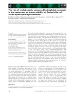
S and G2/M arrest induced by DCQ ± IR in EMT-6 cells at 0, 2, and 4 h post-treatmentFigure 1
S and G2/M arrest induced by DCQ ± IR in EMT-6 cells at 0, 2, and 4 h post-treatment. EMT-6 cells were treated
at 50% confluency with DCQ (10 μM) for 4 h, and/or irradiated (10 Gy). Immediately after the 4 h-drug incubation or IR expo-
sure, cells were replenished with media containing no drug. Cells were collected at 0 h, 2 h, and 4 h after the refreshment of
media and subjected to flow cytometry. Percentages of cells in the sub-G1 (A), S (B), and G2/M (C) phases of the cell cycle
were determined by CellQuest and the averages ± SD are plotted for each treatment. Dashed lines represent the % of control
cells in each phase of the cell cycle. SD: standard deviation.
Radiation Oncology 2009, 4:25 />Page 6 of 10
(page number not for citation purposes)
DCQ Generates Reactive Oxygen Species in EMT-6 cells
N-oxides undergo redox-cycling producing reactive oxy-
gen species (ROS) [20]. We hypothesized that DCQ may
cause DNA damage by ROS induction due to its redox-
cycling. Indeed, DCQ treatment alone, either directly or
indirectly, induced the generation of ROS in EMT-6 cells
after 30 min of treatment as measured by the DCFH assay
(Figure 5A). To determine if ROS play a role in the radio-
sensitizing effect of DCQ in EMT-6 cells, strong anti-oxi-
dants such as Tiron and NAC were added before treatment
with DCQ alone or in combination with IR, to scavenge
any DCQ-generated ROS. Cells pretreated with anti-oxi-
dants were more resistant to the anti-proliferative effect of
DCQ. However, the anti-oxidants did not completely
abolish the anti-neoplastic effect of DCQ whether alone
or in combination with IR (Figure 5B, C). These results
indicate that ROS play at least a partial role in the radio-
sensitizing effect of DCQ in EMT-6 cells.
DCQ Targets Rapidly-Proliferating Cells
Because DCQ appears to slow repair, we expected that tox-
icity would depend on proliferation rate. We assessed
whether reducing proliferation would decrease DCQ tox-
icity by culturing murine mammary epithelial cell line
SCp2 under conditions to induce differentiation and
thereby slow proliferation. When cultured in differentia-
tion media, SCp2 proliferation rate was reduced to
approximately 50% compared to cells cultured in normal
growth media, and an even stronger decrease in prolifera-
tion was observed with cells supplemented with basement
membrane (Figure 6A). After 4 h of DCQ treatment,
slowly proliferating SCp2 cells were more resistant to
toxic concentrations of 10 μM DCQ, suggesting selective
toxicity to proliferating cells (Figure 6B).
Discussion
Several mechanisms of radiosensitization are known,
including redox modulators [21], inhibitors of DNA dam-
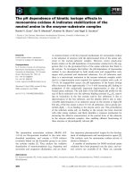
DNA damage induced by DCQ+IR in EMT-6 cellsFigure 2
DNA damage induced by DCQ+IR in EMT-6 cells. A. Representative images of comets induced by DCQ± IR in EMT-6
cells subjected to the alkaline comet assay. EMT-6 cells were treated with IR (10 Gy), 4 h of DCQ (10 μM), or in combination.
Cells were directly collected after the treatment and subjected to the alkaline comet assay. Images were taken using a fluores-
cent microscope at 40× (oil immersion) magnification. The tail lengths of the comets observed by each treatment are propor-
tional to the amount of DNA damage induced. B. The mean of the parameters (% DNA in comet's tail and tail moment) are
shown in the graphs above. The histograms summarize the averages of two independent experiments ± SE and show the mean
of the %DNA in comet's tail and tail moments. More than 50 cells per treatment were photographed and quantified using
TriTek CometScore software. SE: standard error.
Radiation Oncology 2009, 4:25 />Page 7 of 10
(page number not for citation purposes)
age repair [22], and regulators of growth factor receptors
and other signaling molecules [23,24]. Misrepair of DNA
damage causes mutation, and extensive damage may
cause cell cycle arrest, or death if irreparable or too slowly
repaired. The role ROS can play in cellular response to
radiation has been well established [25].
Here, we show for the first time that DCQ induces DSBs
in EMT-6 cells, in addition to SSBs and alkaline-labile
lesions detected by the alkaline comet assay. DCQ causes
more G2/M arrest than IR. Exposure of EMT-6 cells to 10
μM DCQ produced damage detected by the alkaline
comet assay, and DSBs evaluated by p-ATM level, almost
equivalent to that produced by 10 Gy IR. The combina-
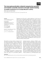
Phosphorylation of ATM and activation of DNA-PK by DCQ ± IR in EMT-6 cells at 2 h post-treatmentFigure 3
Phosphorylation of ATM and activation of DNA-PK by DCQ ± IR in EMT-6 cells at 2 h post-treatment. A. EMT-
6 cells were treated with 10 μM DCQ, 10 Gy IR, or combination treatments, fixed and subjected to immunocytochemical
detection of ATM phosphorylated on Ser1981, and stained with PI to detect at the same time p-ATM in each phase of the cell
cycle. B. The mean of the FL-1 intensity (reflecting the level of p-ATM expression) at each phase of the cell cycle are plotted.
C. Anti-DNA-PK was immunoprecipitated with DNA from lysates of 10
6
EMT-6 cells treated with 10 μM DCQ, 10 Gy IR, or
combination treatments using CHIP assay. The immunoprecipitate was resolved on a 5% gel by electrophoresis, transferred to
nitrocellulose and probed with anti-DNA-PK. The bands were quantified using LabWorks 4.0 software. CHIP: chromatin
immunoprecipitation.
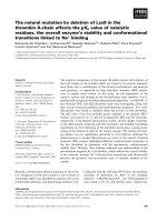
Slow repair of DNA damage in DCQ+IR-treated cellsFigure 4
Slow repair of DNA damage in DCQ+IR-treated cells. Treated cells were either collected directly after treatment (0 h)
or refreshed with drug-free media and incubated for another 4 or 16 h. The mean of the parameters (% DNA in comet's tail
and tail moment) are shown in the graphs above.
Radiation Oncology 2009, 4:25 />Page 8 of 10
(page number not for citation purposes)
tion of DCQ+IR induced significantly higher SSBs than
each treatment alone. Radiosensitization of DCQ not
only correlates with higher induction of DNA damage, but
also with slower repair of this damage. Alkaline comet
assays 4-hours post treatment revealed dramatically
slowed repair of damage in DCQ+IR treated cells com-
pared to separate IR or DCQ treatments. Little damage
remained 4 h after separate treatments with DCQ or IR,
supporting a model in which radiosensitization involves
the generation of more difficult-to-repair DSBs. These
results suggest combination treatment may have thera-
peutic value.
DNA damage, in particular DSBs, imposes a critical threat
to the survival of cells if left unrepaired [26]. As a response
to the damage, cells activate the DNA damage checkpoint.
DSBs are detected by two main players in the DNA dam-
age checkpoint: ATM and DNA-PK. Signal transduction,
induced by the activation of these two signals, can cause
cell-cycle arrest, repair, and cell death. Moreover, both are
activated at very early stages of the DNA damage response,
and are involved in DNA repair [27]. DNA-PK was acti-
vated in response to DCQ alone more than IR alone. The
combination treatment induced the highest amount of
active DNA-PK. ATM plays a critical role in S and G
2
/M
phase arrest. Activated by DSBs, ATM becomes phosphor-
ylated at Ser-1981 [15]. We show that ATM was activated
in all phases of the cell cycle in response to the damage
induced by all treatments. In the combination treatment
the expression of p-ATM in G
2
/M phase was twice that of
untreated cells.
Following IR treatment, EMT-6 cells arrest in S phase.
Such an arrest is mainly caused by the activation of the
intra-S-phase checkpoint due to significant amount of
DSBs [28]. It is responsible for inhibition of DNA replica-
tion at late origins of replication. In addition to cell cycle
arrest, the intra-S-phase checkpoint induces a cascade of
reactions, that either attempt to repair the damage, mainly
by homologous recombination, or induce cell death,
depending on the extent of the damage induced. If dam-
age is not repaired before the end of the S phase, cells
would arrest at the G
2
/M DNA-damage checkpoint [28].
The G
2
/M arrest induced by DCQ and the S-phase accu-
mulation induced by IR appear together in combination
treatments. Despite our intriguing findings that the com-
bination treatment DCQ+IR induces DNA damage,
including DSBs, and slows repair, the precise mechanisms
are still not clear.
ROS generation by DCQ in EMT-6 cellsFigure 5
ROS generation by DCQ in EMT-6 cells. A. Cells were treated with 10 μM DCQ for 30 min before measuring ROS
release by the DCFH-DA assay as described in the materials and methods. B & C. EMT-6 cells were pretreated for 2 h with
the strong anti-oxidants Tiron and NAC, then with DCQ (2.5, 5 and 10 μM), and later subjected to 0 Gy (-IR) or 10 Gy (+IR)
radiation. Afterwards, cells were replenished with drug-free media and incubated for an additional 20 h before the determina-
tion of cell proliferation. The values plotted represent an average (± SD) of two independent experiments. ROS: Reactive oxy-
gen species.
Radiation Oncology 2009, 4:25 />Page 9 of 10
(page number not for citation purposes)
The slow repair of DNA damage caused by DCQ+IR may
have multiple contributing factors. DCQ appears to cause
more DSBs than IR, as evidenced by the increase in p-ATM
and DNA-PK levels. DCQ could create DSBs by generating
closely opposed SSBs via ROS. Our observation of ROS
generation upon DCQ treatment and the decrease in the
sensitivity of cells to DCQ upon addition of anti-oxidants,
support a role for redox cycling of DCQ. Although, the
ROS scavengers did not completely reverse the effect of
DCQ alone or in combination with IR, this does not elim-
inate the possibility that the radiosensitizing effect of
DCQ may only involve ROS, because they may be mainly
short-lived hydroxyl radicals that are not quenched by the
anti-oxidants.
One possible mechanism of DCQ radiosensitization is
that IR induces a higher concentration of the free radical
character of DCQ, which is translated into increased SSBs
and DSBs. The increased levels of SSBs, in combination
with increased levels of difficult-to-repair DSBs could
overwhelm cellular DNA repair pathways. The proposed
mechanism was observed to be selective for rapidly prolif-
erating cells, presumably because slowly dividing cells
have more time to repair DNA damage. This finding sug-
gests clinical potential.
Conclusion
This study presents evidence that the radiosensitizing
effects of DCQ are associated with an increase in DNA
damage, including DSBs, the activation of the key DNA
damage markers, p-ATM and DNA-PK, and the generation
of ROS. The significant levels of unrepaired damage
detected by alkaline comet assay in EMT-6 cells following
treatment by DCQ+IR indicate that decreased DNA repair
contributes to the mechanism of DCQ radiosensitization.
Competing interests
The authors declare that they have no competing interests.
Authors' contributions
JH carried out the experiments in the study and drafted
the manuscript. FG and CAS were involved in revising the
manuscript critically for important intellectual content
and CAS helped in preparing the final draft of the manu-
script. MH provided the compound and reviewed the
manuscript. HGM conceived of the study, and partici-
pated in its design and coordination and drafting of the
manuscript. All authors read and approved the final man-
uscript.
Acknowledgements
This study was supported by the University Research Board of the Ameri-
can University of Beirut and the Lebanese National Council for Scientific
Research.
References
1. Shapiro GI, Harper JW: Anticancer drug targets: cell cycle and
checkpoint control. J Clin Investig 1999, 104:1645-1653.
2. Hartwell LH, Kastan MB: Cell cycle control and cancer. Science
1994, 266:1821-1828.
3. Gasser S: DNA damage response and development of tar-
geted cancer treatments. Ann Med 2007, 39:457-464.
4. Azqueta A, Pachon G, Cascante M, Creppy EE, Lopez de Cerain A:
DNA damage induced by a quinoxaline 1,4-di-N-oxide deriv-
ative (hypoxic selective agent) in Caco-2 cells evaluated by
the comet assay. Mutagenesis 2005, 20:165-171.
5. Azqueta A, Arbillaga L, Pachon G, Cascante M, Creppy EE, López de
Cerain A: A quinoxaline 1,4-di-N-oxide derivative induces
DNA oxidative damage not attenuated by vitamin C and E
treatment. Chem Biol Interact 2007, 168:95-105.
6. Ganley B, Chowdhury G, Bhansali J, Daniels JS, Gates KS: Redox
activated, hypoxia-selective DNA cleavage by quinoxaline
1,4-di-N-oxide. Bioorg Med Chem 2001, 9:2395-2401.
7. Gali-Muhtasib H, Sidani M, Geara F, Assaf-Diab M, Al-Hmaira J, Hadd-
adin M, Zaatari G: Quinoxaline 1,4-dioxides are novel angio-
genesis inhibitors that potentiate antitumor effects of
ionizing radiation. Int J Oncol 2004, 24:1121-1131.
8. Itani W, Geara F, Haykal J, Haddadin M, Gali-Muhtasib H: Radiosen-
sitization by 2-benzoyl-3-phenyl-6,7-dichloroquinoxaline 1,4-
dioxide under oxia and hypoxia in human colon cancer cells.
Radiat Oncol 2007, 2:1.
Resistance of slow proliferating cells to DCQFigure 6
Resistance of slow proliferating cells to DCQ. A. After
plating SCp2 cells in growth media (GM), they were sub-
jected to different conditions. Cells were either kept in GM,
or shifted 12 h after plating to differentiation media (DM) or
DM+BM (basement membrane) for a more differentiated
state. SCp2 cells were incubated for one day then assayed for
proliferation using Cell Titer 96 non-radioactive cell prolifer-
ation. The proliferation rate of cells grown in GM was con-
sidered as 100%. B. The viability of SCp2 cells at different
differentiating states were measured by Cytotox 96 non-
radioactive cytotoxicity assay directly after 4 h of DCQ
treatment.
Publish with BioMed Central and every
scientist can read your work free of charge
"BioMed Central will be the most significant development for
disseminating the results of biomedical research in our lifetime."
Sir Paul Nurse, Cancer Research UK
Your research papers will be:
available free of charge to the entire biomedical community
peer reviewed and published immediately upon acceptance
cited in PubMed and archived on PubMed Central
yours — you keep the copyright
Submit your manuscript here:
/>BioMedcentral
Radiation Oncology 2009, 4:25 />Page 10 of 10
(page number not for citation purposes)
9. Haddadin M, Issidorides C: The Beirut Reaction. Heterocycles
1993, 35:1503-1525.
10. Talhouk RS, Mroue R, Mokalled M, Abi-Mosleh L, Nehme R, Ismail A,
Khalil A, Zaatari M, El-Sabban ME: Heterocellular interaction
enhances recruitment of alpha and beta-catenins and ZO-2
into functional gap-junction complexes and induces gap junc-
tion-dependant differentiation of mammary epithelial cells.
Exp Cell Res 2008, 314:3275-3291.
11. Singh NP: Sodium ascorbate induces DNA single-strand
breaks in human cells in vitro. Mutat Res 1997, 375:195-203.
12. Huang X, Darzynkiewicz Z: Cytometric assessment of histone
H2AX phosphorylation: a reporter of DNA damage. Methods
Mol Biol 2006, 314:73-80.
13. Haykal J, Fernainy P, Itani W, Haddadin M, Geara F, Smith CA, Gali-
Muhtasib H: Radiosensitization of EMT6 mammary carcinoma
cells by 2-benzoyl-3-phenyl-6,7-dichloroquinoxaline 1,4-diox-
ide. Radiother Oncol 2008, 86:412-418.
14. Orren DK, Peterson LN, Bohr VA: Persistent DNA damage
inhibits S-phase and G2 progression, and results in apoptosis.
Mol Biol Cell 1997, 8:1129-1142.
15. Bakkenist CJ, Kastan MB: DNA damage activates ATM through
intermolecular autophosphorylation and dimer dissociation.
Nature 2003, 421:499-506.
16. Tanaka T, Kurose A, Huang X, Dai W, Darzynkiewicz Z: ATM acti-
vation and histone H2AX phosphorylation as indicators of
DNA damage by DNA topoisomerase I inhibitor topotecan
and during apoptosis. Cell Prolif 2006, 39:49-60.
17. Kurose A, Tanaka T, Huang X, Halicka HD, Traganos F, Dai W,
Darzynkiewicz Z: Assessment of ATM phosphorylation on Ser-
1981 induced by DNA topoisomerase I and II inhibitors in
relation to Ser-139-histone H2AX phosphorylation, cell
cycle phase, and apoptosis. Cytometry Part A 2005, 68A:1-9.
18. Collis SJ, DeWeese TL, Jeggo PA, Parker AR: The life and death of
DNA-PK. Oncogene 2005, 24:949-961.
19. Olive PL: The role of DNA single- and double-strand breaks in
cell killing by ionizing radiation. Radiat Res 1998, 150:
S42-S51.
20. Cerecetto H, Gonzalez M: N-oxides as hypoxia selective cyto-
toxins. Mini Rev Med Chem 2001, 1:219-231.
21. Rosenberg A, Knox S: Radiation sensitization with redox mod-
ulators: a promising approach. Int J Radiat Oncol Biol Phys 2006,
64:343-354.
22. Lawrence TS, Blackstock AW, McGinn C: The mechanism of
action of radiosensitization of conventional chemotherapeu-
tic agents. Semin Radiat Oncol 2003, 13:13-21.
23. Katz D, Ito E, Liu FF: On the path to seeking novel radiosensi-
tizers. Int J Radiat Oncol Biol Phys. 2009, 73(4):988-996.
24. Wardman P: Chemical Radiosensitizers for use in radiother-
apy. Clin Oncol (R Coll Radiol) 2007, 19:397-417.
25. Valerie K, Yacoub A, Hagan MP, Curiel DT, Fisher PB, Grant S, Dent
P: Radiation-induced cell signaling: inside-out and outside-in.
Mol Cancer Ther 2007, 6:789-801.
26. Vilenchik MM, Knudson AG: Endogenous DNA double-strand
breaks: production, fidelity of repair and induction of cancer.
Proc Natl Acad Sci USA 2003, 100:12871-12876.
27. Yang J, Yu Y, Hamrick HE, Duerksen-Hughes PJ: ATM, ATR and
DNA-PK: initiators of the cellular genotoxic stress
responses. Carcinogenesis 2003, 24:1571-1580.
28. Bartek J, Lukas C, Lukas J: Checking on DNA damage in S phase.
Nat Rev Mol Cell Biol 2004, 5:792-804.