Báo cáo khoa học: "cExternal beam radiation results in minimal changes in post void residual urine volumes during the treatment of clinically localized prostate cancer" ppt
Bạn đang xem bản rút gọn của tài liệu. Xem và tải ngay bản đầy đủ của tài liệu tại đây (471.38 KB, 9 trang )
BioMed Central
Page 1 of 9
(page number not for citation purposes)
Radiation Oncology
Open Access
Research
cExternal beam radiation results in minimal changes in post void
residual urine volumes during the treatment of clinically localized
prostate cancer
PeterFOrioIII
1
, Gregory S Merrick*
2
, Zachariah A Allen
2
, Wayne M Butler
2
,
Kent E Wallner
3
, Brian S Kurko
2
and Robert W Galbreath
2
Address:
1
Brooke Army Medical Center Department of Radiation Oncology, Ft. Sam, Houston, TX 78234, USA,
2
Schiffler Cancer Center and
Wheeling Jesuit University 1 Medical Park Wheeling, WV 26003, USA and
3
Puget Sound Healthcare Corporation Group Health Cooperative
University of Washington Seattle, WA 98108, USA
Email: Peter F Orio - ; Gregory S Merrick* - ;
Zachariah A Allen - ; Wayne M Butler - ;
Kent E Wallner - ; Brian S Kurko - ; Robert W Galbreath -
* Corresponding author
Abstract
Background: To evaluate the impact of external beam radiation therapy (XRT) on weekly
ultrasound determined post-void residual (PVR) urine volumes in patients with prostate cancer.
Methods: 125 patients received XRT for clinically localized prostate cancer. XRT was delivered
to the prostate only (n = 66) or if the risk of lymph node involvement was greater than 10% to the
whole pelvis followed by a prostate boost (n = 59). All patients were irradiated in the prone
position in a custom hip-fix mobilization device with an empty bladder and rectum. PVR was
obtained at baseline and weekly. Multiple clinical and treatment parameters were evaluated as
predictors for weekly PVR changes.
Results: The mean patient age was 73.9 years with a mean pre-treatment prostate volume of 53.3
cc, a mean IPSS of 11.3 and a mean baseline PVR of 57.6 cc. During treatment, PVR decreased from
baseline in both cohorts with the absolute difference within the limits of accuracy of the bladder
scanner. Alpha-blockers did not predict for a lower PVR during treatment. There was no significant
difference in mean PVR urine volumes or differences from baseline in either the prostate only or
pelvic radiation groups (p = 0.664 and p = 0.458, respectively). Patients with a larger baseline PVR
(>40 cc) had a greater reduction in PVR, although the greatest reduction was seen between weeks
one and three. Patients with a small PVR (<40 cc) had no demonstrable change throughout
treatment.
Conclusion: Prostate XRT results in clinically insignificant changes in weekly PVR volumes,
suggesting that radiation induced bladder irritation does not substantially influence bladder residual
urine volumes.
Published: 22 July 2009
Radiation Oncology 2009, 4:26 doi:10.1186/1748-717X-4-26
Received: 9 April 2009
Accepted: 22 July 2009
This article is available from: />© 2009 Orio et al; licensee BioMed Central Ltd.
This is an Open Access article distributed under the terms of the Creative Commons Attribution License ( />),
which permits unrestricted use, distribution, and reproduction in any medium, provided the original work is properly cited.
Radiation Oncology 2009, 4:26 />Page 2 of 9
(page number not for citation purposes)
Introduction
Increasingly sophisticated conformal radiotherapy deliv-
ery technologies and organ localization protocols have
resulted in significant changes in treatment paradigms
offered to patients with clinically localized prostate can-
cer. These technologies allow physicians to offer dose
escalations to the targets while respecting normal tissue
tolerances of surrounding organs [1-4]. Simultaneously,
smaller treatment margins are employed to minimize side
effects and potential complications. As a result, the precise
evaluation of internal organ movement has become
extremely important to ensure optimal dose to the target
area. Three-dimensional conformal radiotherapy (3D-
CRT) led to significant sparing of normal tissue by con-
forming the dose to the prostate gland. As a result, 3D-
CRT was the first modality to generate widespread con-
cern about prostate gland motion during treatment.
Intensity modulated radiation therapy (IMRT) produces
much steeper dose gradients than 3D-CRT and may result
in tighter margins between the clinical target volume
(CTV) and the planning target volume (PTV). Internal
organ displacement of even a few millimeters may result
in geographic miss of the target volume. Methods to mon-
itor prostate motion have become increasingly important
in the era of dose escalation. The use of computerized
tomography (CT) has been the gold standard for in vivo
imaging as well as structure identification, and has been
emphasized in numerous internal organ motion studies
[5-9]. Although cone beam CT has been integrated into
linear accelerator systems, most CT studies are performed
in a manner simulating treatment. For this reason, many
institutions implant intraprostatic gold fiducial markers
for identification on electronic portal imaging. This pro-
vides three dimensional information regarding prostate
position in relation to the treatment isocenter [10,11].
Other technologies, such as the BAT ultrasound system
and intraprostatic electromagnetic transponders are also
solutions to account for daily variations in prostate posi-
tioning [12,13].
Variables with the potential to influence prostate motion
are an important aspect of clinical research in the delivery
and treatment of prostate cancer. The two organs receiving
the greatest scrutiny are the bladder and rectum secondary
to the close proximity to the prostate gland. The literature
demonstrates a robust relationship between the influence
of rectal filling on prostate displacement, where as the
influence of the bladder is a little more controversial
[6,7,9,14,15]. Researchers who have reported displace-
ment of the prostate by the bladder have typically demon-
strated movement to be in the posterior and inferior
direction [6-8,10,16]. Conversely other researchers have
reported no or a minimal influence of bladder filling on
prostate motion [5,9,15,17,18]. Techniques in patient
immobilization, treatment position and instructions to
maintain a full or empty bladder during treatment may
influence the bladder and prostate interaction [9,11,18].
This body of research specifically addresses the influence
of daily whole pelvic or prostate only daily radiation treat-
ments on weekly ultrasound determined post-void resid-
ual (PVR) urine volumes in patients with clinically
localized prostate cancer treated prone with an empty
bladder. This analysis helps to provide insight into PVR
urine volume variations as patient's progress through
radiation treatments to determine if such changes are clin-
ically significant.
Methods
One hundred and twenty five patients were treated for
clinical stage T1b-T3a (2002 AJCC) prostate cancer with
either definitive external beam radiation therapy to the
prostate only (n = 68) or to the whole pelvis followed by
a prostate boost (n = 59) [19]. For patients with < 10%
risk of pelvic lymph node involvement, the target volume
consisted of the prostate gland and seminal vesicles with
margin [20]. For patients with > 10% risk of pelvic lymph
node involvement, the pelvic lymph nodes were included
in the initial target volume. Intensity Modulated Radia-
tion Therapy (IMRT) was utilized in all treatments. Patient
treated with prostate only radiation received 81 Gy.
Patients who were treated with pelvic radiation received
45 Gy to the prostate and regional nodes followed by a 36
Gy boost to the prostate.
All patients were irradiated in the prone position and
immobilized in a custom aquaplast hip-fix immobiliza-
tion device with an empty bladder and rectum at the time
of simulation and treatment. Patients were instructed to
urinate immediately prior to initial CT simulation and
daily during external beam radiation therapy. Patients
were instructed to defecate prior to simulation and daily
radiation if the urge was felt.
At the time of CT simulation, PVR volumes were meas-
ured within 10 minutes of voiding by transabdominal
ultrasonography (Bladder Scan BVI 3000, Diagnostic
Ultrasound, Brothel, Washington). PVR determinations
were obtained weekly throughout treatment. These values
were compared to the baseline PVR volume from the time
of simulation. PVR urine volumes determined by ultra-
sound were not compared to CT scan as previous investi-
gators have determined there is a high degree of
correlation between bladder scanner volumes and Com-
puted Tomography volumes and more importantly
weekly changes from baseline were measured by ultra-
sound and not computed tomography [18,21-23]. Previ-
ously published correlations for the BVI model 3000
range from 0.86–0.95 [21,23]. The bladder scanner is
reported to operate within a margin of accuracy of ± 20 cc
Radiation Oncology 2009, 4:26 />Page 3 of 9
(page number not for citation purposes)
in the range of 0 to 699 ml of urine volume. Accuracy of
the bladder scanner is reported by the manufacturer
within the operator's manual and as based on scanning
diagnostic ultrasound tissue equivalent phantoms [24].
The BVI 3000 bladder scanner is a portable Ultrasound
originally developed to measure residual urine volumes
after micturition. The scanning head is positioned on the
patient's body 2 cm above the pubic symphysis in a mid-
line position. The bladder volume is calculated from a 2
MHz transducer which automatically rotates in 15 degree
increments to provide a 3-dimensional model of the blad-
der to estimate the urine volume. Two highly experienced
nurses, specifically trained and competent in the use of
the BVI 3000, performed all scans analyzed in this study.
An alpha-blocker was initiated in 56 patients at a mean of
4.7 weeks ± 2.2 weeks into treatment. Alpha blockers were
initiated for urinary irritative or obstructive symptoms.
Alpha-blockers consisted of either tamsulosin hydrochlo-
ride (0.4 – 0.8 mg daily) or terazosin hydrochloride (5–10
mg daily).
One-way ANOVA, t-tests, and Fisher's exact chi-squared
were applied to the clinical and treatment parameters of
the two treatment cohorts (prostate only and pelvic radia-
tion patients). All data was analyzed using SPSS version
14.0 software (SPSS, Inc., Chicago, IL). Statistical signifi-
cance was set at a p < 0.05 for all analyses. In scatter plots
of PVR over time, various empirical regression functions
were tested for an optimum fit to the data, and either a
quadratic function, y = a + b * time + c * time
2
or a logistic
regression function, y = a/1+ b * exp
-c * Time
where y is
either the PVR urine volume or the difference between the
PVR urine volume and the baseline PVR urine volume,
consistently outperformed linear regression by resulting
in a larger correlation coefficient and therefore were used
uniformly throughout.
Results
Table 1 summarizes the clinical and treatment parameters
of the study population, stratified by treatment cohort.
The mean patient age was 73.9 ± 8.0 years with a mean
pre-treatment prostate volume of 53.3 ± 33.5 cubic cen-
timeters, a mean PVR urine volume of 57.6 ± 77.3 cubic
centimeters. Patients treated with prostate only external
beam radiation therapy compared with patients treated
with whole pelvic radiation therapy had statistically lower
pre-treatment PSA (p = 0.011); lower Gleason Score (p <
0.001); lower percent positive biopsies (p < 0.001; earlier
staged disease (p < 0.001); a lower incidence of perineural
invasion (p = 0.001) and were less likely to have received
androgen deprivation therapy (ADT) (p < 0.001). No sta-
tistical differences were demonstrated between the groups
concerning patient age at treatment, prostate volume,
post-void residual urine (PVR) volumes and the use of
alpha-blockers during treatment.
Table 1: Clinical and treatment parameters of the study population stratified by treatment cohort.
Continuous Variables Prostate only (n = 66) Pelvis (n = 59) All Patients (n = 125)
Mean ± SD Median Mean ± SD Median p* Mean ± SD Median
Age at treatment (years) 73.9 ± 7.8 75.5 73.9 ± 8.3 76.3 0.971 73.9 ± 8.0 75.8
Pre-treatment IPSS 10.9 ± 7.0 10.0 11.8 ± 8.3 11.5 0.526 11.3 ± 7.6 10.0
Pre-treatment PSA (ng/mL) 7.7 ± 5.3 6.5 29.6 ± 68.4 10.3 0.011 18.0 ± 48.0 7.3
Gleason Score 6.5 ± 0.7 6. 7.8 ± 1.2 8.0 < 0.001 7.1 ± 1.2 7.0
% positive biopsies 29.0 ± 23.7 18.5 62.3 ± 32.8 62.5 < 0.001 44.2 ± 32.6 33.3
Prostate volume (cm
3
) 54.8 ± 33.8 47.3 51.6 ± 33.2 42.0 0.597 53.3 ± 33.5 46.5
Post void residual (cc) 57.3 ± 67.0 30.0 58.0 ± 88.0 27.0 0.961 57.6 ± 77.3 28.0
BMI 27.8 ± 3.9 27.4 29.2 ± 5.0 28.5 0.950 28.4 ± 4.4 28.2
Categorical Variables Count (%) Count (%) p
∀
Count (%)
Stage (median) T1b-T2b 65 (98.5) 46 (78.0) < 0.001 111 (88.8)
≥ T2c 1 (1.5) 13 (22.0) 14 (11.2)
ADT none 55 (83.3) 19 (32.2) < 0.001 74 (59.2)
≤ 6 months 7 (10.6) 2 (3.4) 9 (7.2)
> 6 months 4 (6.1) 38 (64.4) 42 (33.6)
Diabetes 11 (16.9) 14 (23.7) 0.236 25 (20.0)
Hypertension 43 (65.2) 39 (66.1) 0.531 82 (65.6)
Alpha blocker 53 (80.3) 43 (72.9) 0.221 96 (76.8)
Perineural invasion 13 (19.7) 28 (47.5) 0.001 41 (32.8)
* p values calculated by one-way ANOVA
∀ p values determined by 2-sided Fisher's Exact Test
Radiation Oncology 2009, 4:26 />Page 4 of 9
(page number not for citation purposes)
Of the 125 patients, 96 were exposed to alpha blockers
during their treatment [Table 2, 3]. A total of 56 patients
were placed on alpha blockers during treatment. Forty
patients were actively treated with alpha-blockers prior to
radiation. Thirty patients were started on alpha blockers at
a mean of 4.70 ± 2.2 weeks in the prostate only group and
twenty-six patients initiated alpha-blockers at a mean of
4.7 ± 2.4 weeks in the pelvic radiation group (p = 0.941)
[Table 2].
Table 3 summarizes the variation in PVR urine volume
readings over the course of the study, stratified by therapy
cohort, alpha-blocker use, baseline PVR volume group
and radiation cohort. Of the 125 patients included for
analysis, 66 patients were treated with prostate only radi-
ation and 59 patients were treated with whole pelvic radi-
ation therapy followed by a prostate boost. Seventy-six
patients had a PVR urine volume at the time of simulation
measured to be less than or equal to 40 cc. Forty-nine
patients had PVR urine volumes greater than 40 cc. For the
overall population, the mean PVR urine volume over the
entire course of radiation treatment was 48.2 cc. The mean
individual PVR urine volume over all weeks of the study
for patients with a baseline PVR > 40 cc was 86.9 cc verses
in comparison to 23.2 cc in the patient group with base-
line PVR ≤ 40 cc (p < 0.001). No significant difference was
found between the mean individual PVR urine volume
over all weeks of treatment in patients treated with pros-
tate only versus pelvic radiation with values of 46.3 cc and
50.2 cc, respectively (p = 0.725).
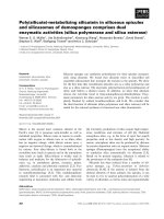
Figure 1a demonstrates that the mean PVR urine volume
between the two treatment cohorts were not significantly
different from each other (p = 0.664) over the duration of
therapy. The mean PVR urine volumes demonstrated the
greatest decreased over the first three weeks in both pros-
tate only and pelvic radiation groups, although became
variable with time and demonstrated an increase towards
the end of therapy back to baseline measurements. The
magnitude of difference is less than 20 cc in both cohorts,
which are at the limit of accuracy of the bladder scanner.
Figure 1b graphs the mean difference in baseline PVR
urine volumes as a function of weeks of external beam
radiation therapy. Both cohorts of patients demonstrated
a decrease from baseline measurements with the greatest
trend seen over the first three weeks of treatment. No sig-
nificant differences were demonstrated concerning the
magnitude of change from baseline PVR urine volumes
when comparing pelvic radiation to prostate only radia-
tion.
Larger baseline PVR allows for a greater absolute volume
changes as radiation induced bladder irritability increases,
therefore patients were stratified into two groups based on
initial PVR. A cut-off of 40 cc was chosen as previous stud-
ies have demonstrated that bladder volumes greater than
40 cc in addition to rectal filling had the potential to influ-
ence daily prostate position while treated in the prone
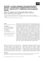
position [9]. Figure 2 shows the distribution of PVR
cohort by week and is stratified by pre-treatment PVR ≤ 40
or > 40 cc. Patients were defined as having a worse PVR if
they moved from a lower PVR category (≤ 40 cc) to the
higher category (> 40 cc), while patients in the higher cat-
egory who moved to a lower category were defined as bet-
ter. During subsequent points in time, only a small
fraction of patients with an initial PVR ≤ 40 cc exceeded 40
cc, while patients with an initial PVR > 40 cc had a high
probability of a subsequent PVR < 40 cc.
Figure 3 graphically demonstrates the mean PVR urine
volumes by week of radiation treatment for both prostate
only and pelvic radiation patients to two standard devia-
tions. Over two standard deviations the mean PVR
reported are similar between the two groups albeit varia-
ble due to the intrinsic accuracy of ± 20 cc of the bladder
scanner. The radiation field utilized did not appear to
greatly influence the mean PVR compared to one another.
Figure 4a graphically represents the mean PVR urine vol-
umes versus weeks of radiation treatment, stratified by
treatment group and baseline PVR urine volumes ≤40 or
>40 cc. The greatest changes over time in mean PVR were
demonstrated in both treatment cohorts with base line
volumes greater than 40 cc. As demonstrated in previous
graphs the greatest and most consistent change is over the
first three weeks of treatment. Very little change in the
mean PVR is demonstrated in the group of patients treated
with prostate only radiation with baseline PVR urine vol-
Table 2: Week of alpha-blocker initiation relative to start of external beam therapy, stratified by radiation cohort.
XRT Therapy Cohort Number. of patients* Alpha-blocker Initiated (weeks) p
†
Mean ± SD Median
Prostate Only 30 4.7 ± 2.2 4.5 0.941
Pelvis 26 4.7 ± 2.4 4.5
Overall 56 4.7 ± 2.3 4.5
* – Does not include patients who started alpha-blockers prior to treatment.
†
p-value determined by one-way ANOVA
Radiation Oncology 2009, 4:26 />Page 5 of 9
(page number not for citation purposes)
umes less than or equal to 40 cc. The same is demon-
strated in the pelvic radiation group with slightly greater
variability, although within the limits of the bladder scan-
ner. Figure 4b graphically represents the mean difference
from baseline PVR urine volumes versus weeks of radia-
tion treatment, stratified by treatment group and baseline
PVR urine volumes <40 or >40 cc. The data continues to
demonstrate very little change in PVR volumes over time
for both treatment cohorts when baseline PVR urine vol-
umes are less than or equal to 40 cc. Both cohorts of
patients continue to demonstrate greater differences in
mean PVR urine volumes from baseline over time in
patients with baseline urine volumes greater than 40 cc.
The mean differences from baseline are greater in the less
than or equal to 40 cc group in both treatment cohorts
and the converse is found in the greater than 40 cc group.
Discussion
In an era which is rapidly becoming defined by increas-
ingly sophisticated treatment planning and radiation
delivery techniques, the basic tenant of irradiating what is
intended to be treated while respecting normal tissue tol-
erance has never been more important. To achieve these
goals it is necessary to treat a dynamic and moving target,
which is exemplified in prostate radiotherapy [14,25].
With dose escalation, strategies must be refined to
decrease prostate treatment margins to minimize toxicity
to normal structures. Therefore, an investigation of all fac-
tors with the potential to influence prostate motion is crit-
ical. The bladder and rectum are regarded as the two most
important structures in terms of daily prostate motion.
This study details the post void residual urine volume
prior to daily radiation treatments and the influence of
external beam radiation therapy on PVR urine volumes
throughout treatment.
If a patient is asked to empty his bladder prior to simula-
tion and then prior to radiation treatment, bladder filling
should influence the prostate's position to a lesser degree
as previously reported by Zelefsky et al [9]. Although PVR
urine volumes were recently explored in cervical cancer
treatments, little data is available concerning PVR urine
volumes as patients progress through external beam radi-
ation therapy for prostate cancer treated with an empty
bladder and in a prone position [22]. Posterior and infe-
rior movement of the prostate gland due to bladder filling
was first described by Ten Haken and colleagues, and
reproduced by several investigators in subsequent studies
[6-8,10]. Melian et al. have reported that bladder filling
influenced the position of the prostate in patients treated
in the prone position[8]. Zelefsky et al. also demonstrated
that bladder volumes greater than 40 cm
3
could predict
for greater than 3 mm deviations of the prostate and sem-
inal vesicles while in the prone treatment position when
the rectal volume is greater than 60 cc [9]. Zellars et al.
reported that patients who were treated in the supine posi-
tion and instructed to have a full bladder prior to treat-
ment demonstrated an associated posterior displacement
of the prostate when evaluated 4 to 5 weeks after initiation
of therapy [7]. Conversely, other researchers have not seen
a relationship between bladder filling and prostate posi-
tion, although these patients were treated in the supine
treatment position [5,15,17].
Bladder filling is more easily controlled on a daily basis
than rectal filling, assuming that the patient voids imme-
diately prior to treatment. This strategy is simple and
should help to remove the potential influence of the blad-
der on prostate motion. This paper specifically reports the
influence of external beam radiation therapy on serial
PVR urine volumes as patients proceed through treatment.
Several strategies currently exist for daily image guidance
for prostate treatment, therefore the purpose of this paper
is not to correlate specific PVR urine volumes with pros-
tate motion, but rather determine the influence of exter-
nal beam radiation therapy on PVR urine volumes as
Table 3: Variation in individual post-void residual (PVR) volume readings over the course of the study, stratified by therapy cohort,
alpha-blocker use, baseline PVR volume group, and radiation cohort.
Parameter Group Number of patients Mean of N weeks of PVR
readings
p-value Mean Std. Dev. of N PVR
readings
p-value
Mean ± SD Mean ± SD
Alpha-blocker use No 29 26.5 ± 29.3 0.027 22.3 ± 18.8 0.011
Yes 96 54.7 ± 65.6 35.7 ± 36.9
Baseline PVR volume ≤ 40 cc 76 23.2 ± 31.7 < 0.001 23.4 ± 32.7 <0.001
> 40 cc 49 86.9 ± 72.7 46.7 ± 31.4
Radiation cohort Prostate only 66 46.3 ± 55.4 0.725 29.7 ± 21.6 0.317
Pelvis 59 50.2 ± 65.6 35.8 ± 43.9
Overall population 125 48.2 ± 60.2 32.5 ± 34.0
* The median number N of PVR readings was 10.
p-values were calculated by independent samples t-test.
Radiation Oncology 2009, 4:26 />Page 6 of 9
(page number not for citation purposes)
patients proceed through treatment [25]. If PVR urine vol-
umes remain relatively stable throughout external beam
radiation treatment than there would be little correlation
to prostate motion from the original planning CT simula-
tion.
Our study population consisted of patients treated with
external beam radiation for prostate cancer. Two common
types of radiation treatments were studied, pelvic radia-
tion followed by a cone down to the prostate and prostate
only radiation. As such the effects of PVR urine volumes
could be compared in patients receiving whole pelvic
radiation therapy for a portion of their treatment com-
pared to prostate only radiation therapy. These two
cohorts provide insight in the potential for PVR urine vol-
ume changes in the most common clinical scenarios for
definitive external radiation therapy for prostate. Patients
in the whole pelvic cohort had larger portions of their
bladder irradiated and presumably had the potential for a
greater degree of radiation induced bladder irritation.
There were significant differences in the clinical presenta-
tion between the two cohorts of patients within the two
radiation groups. These differences are attributable to our
selection criteria. Importantly, these two groups of
patients allowed us to study different treatment strategies,
depending on risk of lymph node involvement, on PVR
urine volumes as patients progressed through external
beam radiation treatment for prostate cancer. Patients
(A). Mean post-void residual volume as a function of week of external beam radiation therapy (XRT) treatment, stratified by radiation groupFigure 1
(A). Mean post-void residual volume as a function of
week of external beam radiation therapy (XRT)
treatment, stratified by radiation group. The bladder
scanner operates within a margin of accuracy of ± 20 cc. (B)
Mean difference from baseline in post-void residual (PVR)
volume as a function of week of external beam radiation
therapy (XRT) treatment, stratified by radiation group. The
best-fit lines were determined by quadratic regression analy-
sis. The bladder scanner operates within a margin of accuracy
of ± 20 cc.
Distribution of PVR cohort by week and stratified by pre-treatment post-void residual volumeFigure 2
Distribution of PVR cohort by week and stratified by
pre-treatment post-void residual volume. Patients
moving from the lower PVR category (≤ 50 cc) to the higher
category (> 50 cc) were labeled as worse, while patients in
the higher category who moved to the lower were labeled as
better. The number of patients in each baseline category var-
ies over time based upon treatment length.
Radiation Oncology 2009, 4:26 />Page 7 of 9
(page number not for citation purposes)
treated with prostate only radiation were determined to
have lower pre-treatment PSA, lower percent positive
biopsies, lower Gleason Scores and clinical stage than
patients treated with pelvic radiation. This finding is
expected as higher PSA, Gleason Score and clinical stage
predicts for a greater probability of lymph node involve-
ment [20]. Our policy was to treat lymph nodes if the risk
of involvement was greater than 10%.
The mean individual PVR urine volume over all weeks of
treatment in the pelvic and prostate radiation groups was
not statistically different with values of 46.3 cc and 50.2 cc
respectively. However, mean PVR urine volumes stratified
by week in both groups demonstrated the patients treated
with whole pelvic radiation had larger baseline PVR urine
volumes at the beginning of treatment. Larger baseline
PVR theoretically would allows for greater absolute vol-
ume changes as radiation induced bladder irritability
increased. Although higher baseline mean PVR urine vol-
umes predicted for greater mean PVR urine volumes dur-
ing treatment, PVR decreased from baseline in both
cohorts with the absolute difference within the limits of
accuracy of the bladder scanner. Such small differences are
unlikely to result in any clinical significance in prostate
motion. Also of interest is that the difference from base-
line PVR urine volumes in both cohorts appeared to have
the greatest change during the first three weeks of treat-
ment and then became well within the limits of accuracy
of the bladder scanner. It is likely that patient attention to
detail (i.e. bladder emptying) accounted for the changes
during the first three weeks of treatment. As such, it is
Plot of mean post-void residual volume ± 2 standard error versus week of XRT treatment, stratified by pre-treatment (baseline) post-void residual volumeFigure 3
Plot of mean post-void residual volume ± 2 standard
error versus week of XRT treatment, stratified by
pre-treatment (baseline) post-void residual volume.
The number of patients in each baseline category varies over
time based upon treatment length.
(A) Plot of mean post-void residual volume versus week of XRT treatment, stratified by treatment group and pre-treat-ment (baseline) post-void residual volumeFigure 4
(A) Plot of mean post-void residual volume versus
week of XRT treatment, stratified by treatment
group and pre-treatment (baseline) post-void resid-
ual volume. The number of patients in each baseline cate-
gory varies over time based upon treatment length. (B) Plot
of mean difference from baseline in post-void residual vol-
ume versus week of XRT treatment, stratified by treatment
group and pre-treatment (baseline) post-void residual vol-
ume. The number of patients in each baseline category varies
over time based upon treatment length.
Radiation Oncology 2009, 4:26 />Page 8 of 9
(page number not for citation purposes)
probable that PVR volume determinations early in the
course of treatment may be sufficient with subsequent
weekly determinations omitted.
Previous research by Zelefsky's group has demonstrated
that bladder volumes greater than 40 cc had the potential
to influence daily prostate position while treated in the
prone position when rectal filling was greater than 60 cc
[9]. As such patients were stratified by radiation treatment
group and a baseline PVR cutoff of 40 cc. Patients with a
baseline PVR = 40 cc did not experience any appreciable
change in PVR during treatment while patients with a
baseline PVR > 40 cc were most likely to experience
changes (i.e. decrease) from the baseline PVR. This
marked decline could result in a smaller degree of prostate
motion but also in the setup being different from what
was initially simulated. Although, patients who are iden-
tified with a higher PVR urine volume at the time of sim-
ulation may require attention to bladder filling depending
on the technologies of daily prostate localization
employed. A shortcoming of our study is that patients
with substantial decreases in serial PVR's were not re-
planned via CT simulation (all patients however were
treated with daily cone beam CT guidance).
On average, alpha-blockers were prescribed 4.7 weeks
into treatment. Alpha-blockers were not demonstrated to
influence PVR in either treatment cohort. This, however, is
not surprising since the vast majority of changes in PVR
occurred in the first three weeks or therapy. Alpha-block-
ers were initiated primarily for irritative symptoms.
Conclusion
External beam radiation therapy results in a clinically
insignificant change in weekly post-void residual urine
volumes (especially when PVR urine volumes are less than
40 cc), suggesting that radiation induced bladder irritabil-
ity does not substantially influence bladder residual urine
volumes.
Competing interests
The authors declare that they have no competing interests.
Authors' contributions
PFO has done statistical analysis as well as drafted the
manuscript. GSM has made the selection of patients,
involved with the study design, has been involved with
writing and revising the manuscript, statistical analysis
and final approval of the version to be published. ZAA has
been involved with the statistical analysis and design of
the tables/figures. WMB has been involved with the statis-
tical analysis. KEW has been involved in manuscript revi-
sion and review of the intellectual content. BSK has been
involved with statistical analysis. RWG has been involved
with statistical analysis and design of the tables/figures.
All authors read and approved the final manuscript.
References
1. Zelefsky MJ, Leibel SA, Gaudin PB, Kutcher GJ, Fleshner NE, Venkat-
ramen ES, Reuter VE, Fair WR, Ling CC, Fuks Z: Dose escalation
with three-dimensional conformal radiation therapy affects
the outcome in prostate cancer. Int J Radiat Oncol Biol Phys 1998,
41:491-500.
2. Kuban DA, Tucker SL, Dong L, Starkschall G, Huang EH, Cheung MR,
Lee AK, Pollack A: Long-term results of the M.D. Anderson
randomized dose-escalation trial for prostate cancer. Int J
Radiat Oncol Biol Phys 2008, 70:67-74.
3. Pollack A, Zagars GK, Smith LG, Lee JJ, von Eschenbach AC, Antolak
JA, Starkschall G, Rosen I: Preliminary results of a randomized
radiotherapy dose-escalation study comparing 70 Gy with 78
Gy for prostate cancer. J Clin Oncol 2000, 18:3904-3911.
4. Zietman AL, DeSilvio ML, Slater JD, Rossi CJ Jr, Miller DW, Adams
JA, Shipley WU: Comparison of conventional-dose vs high-
dose conformal radiation therapy in clinically localized ade-
nocarcinoma of the prostate: A randomized controlled trial.
JAMA 2005, 294:1233-1239.
5. Pinkawa M, Asadpour B, Gagel B, Piroth MD, Holy R, Eble MJ: Pros-
tate position variability and dose-volume histograms in radi-
otherapy for prostate cancer with full and empty bladders.
Int J Radiat Oncol Biol Phys 2006, 64:856-861.
6. Schild SE, Casale HE, Bellefontaine LP: Movement of the prostate
due to rectal and bladder distention: implications for radio-
therapy. Med Dosim 1993, 18:13-15.
7. Zellars RC, Roberson PL, Strawderman M, Zhang D, Sandler HM, Ten
Haken RK, Osher D, McLaughlin PW: Prostate position late in
the course of external beam therapy: patterns and predic-
tors. Int J Radiat Oncol Biol Phys 2000, 47:655-660.
8. Melian E, Mageras GS, Fuks Z: Variation in prostate position
quantitation and implications for three-dimensional confor-
mal treatment planning. Int J Radiat Oncol Biol Phys 1997,
38:73-81.
9. Zelefsky MJ, Crean D, Mageras GS, Lyass O, Happersett L, Ling CC,
Leíble SA, Fuks Z, Bull S, Koov HM, van Herk M, Kutcher GJ: Quan-
tification and predictors of prostate position variability in 50
patients evaluated with multiple CT scans during conformal
radiotherapy.
Radiother Oncol 1999, 50:225-234.
10. Crook JM, Raymond Y, Salhani D, Yan H, Esche B: Prostate motion
during standard radiotherapy as assessed by fiducial mark-
ers. Radiother Oncol 1995, 37:35-42.
11. Bayley AJ, Catton CN, Haycocks T, Kelly V, Alasti H, Bristow R, Cat-
ton P, Crook J, Gospodarowicz MK, McLean M, Milosevic M, Warde
P: A randomized trial of supine vsprone positioning in
patients undergoing escalated dose conformal radiotherapy
for prostate cancer. Radiother Oncol 2004, 70:37-44.
12. Lattanzi J, McNeeley S, Pinover W, Horwitz E, Das I, Schultheiss TE,
Hanks GE: A comparison of daily CT localization to a daily
ultrasound-based system in prostate cancer. Int J Radiat Oncol
Biol Phys 1999, 43:719-725.
13. Balter JM, Wright JN, Newell LJ, Friemel B, Dimmer S, Cheng Y,
Wong J, Vertatschitsch E, Mate TP: Accuracy of a wireless locali-
zation system for radiotherapy. Int J Radiat Oncol Biol Phys 2005,
61:933-937.
14. Ghilezan MJ, Jaffray DA, Siewerdsen JH, Van Herk M, Shetty A, Sharpe
MB, Zafar Jafri S, Vicini FA, Matter RC, Brabbins DS, Martinez AA:
Prostate gland motion assessed with cine-magnetic reso-
nance imaging (cine-MRI). Int J Radiat Oncol Biol Phys 2005,
62:406-417.
15. Antolak JA, Rosen II, Childress CH, Zagars GK, Pollack A: Prostate
target volume variation during a course of radiotherapy. Int
J Radiat Oncol Biol Phys 1998, 42:661-672.
16. Ten Haken RK, Forman JD, Heimburger DK, Gerhardsson A, McShan
DL, Perez-Tamayo C, Schoeppel SL, Lichter AS: Treatment plan-
ning issues related to prostate movement in response to dif-
ferential filling of the rectum and bladder. Int J Radiat Oncol Biol
Phys 1991, 20:1314-1324.
17. Beard CJ, Kijewski P, Bussiere M, Gelman R, Gladstone D, Shaffer K,
Plunkett M, Castello P, Coleman CN: Analysis of prostate and
seminal vesicle motion: Implications for treatment planning.
Int J Radiat Oncol Biol Phys 1996, 34:451-458.
18. Stam MR, Th. Van Lin EN, Vight LP Van Der, Kaanders JH, Visser AG:
Bladder filling variations during radiation treatment of pros-
tate cancer: Can the use of a bladder ultrasound scanner and
biofeedback optimize bladder filling? Int J Radiat Oncol Biol Phys
2006, 65:371-377.
Publish with BioMed Central and every
scientist can read your work free of charge
"BioMed Central will be the most significant development for
disseminating the results of biomedical research in our lifetime."
Sir Paul Nurse, Cancer Research UK
Your research papers will be:
available free of charge to the entire biomedical community
peer reviewed and published immediately upon acceptance
cited in PubMed and archived on PubMed Central
yours — you keep the copyright
Submit your manuscript here:
/>BioMedcentral
Radiation Oncology 2009, 4:26 />Page 9 of 9
(page number not for citation purposes)
19. Greene FL, Balch CM, Fleming I, Fritz A, Haller DG, Morrow M, Page
DL: AJCC Cancer Staging Manual 6th edition. Springer-Verlag New
York, LLC; 2002.
20. Partin AW, Mangold LA, Lamm DM, Walsh PC, Epstein JI, Pearson JD:
Contemporary update of prostate cancer staging nomo-
grams (Partin Tables) for the new millennium. Urology 2001,
58:843-848.
21. O'Doherty UM, McNair HA, Norman AR, Miles E, Hooper S, Davies
M, Lincoln N, Balyckvi J, Childs P, Dearnaley DP, Huddart RA: Vari-
ability of bladder filling in patients receiving radical radio-
therapy to the prostate. Radiotherapy and Oncology 2006,
79:335-340.
22. Ahmad R, Hoogeman MS, Quint S, Mens JW, de Pree I, Heijmen BJ:
Inter-fraction bladder filling variations and time trends for
cervical cancer patients assessed with a portable 3-dimen-
sional ultrasound bladder scanner. Radiotherapy and Oncology
2008, 89:172-179.
23. Byun SS, Kim HH, Lee E, Paick JS, Kamg W, Oh SJ: Accuracy of
bladder volume determinations by ultrasonography: Are
they accurate over the entire bladder volume range? Urology
2003, 62:656-660.
24. BladderScan BVI 3000 Noninvasive Bladder Volume Instru-
ment Operator's Manual. C 2004 by Diagnostic Ultrasound Corpo-
ration .
25. Kupelian PA, Langen KM, Willoughby TR, Zeidan OA, Meeks SL:
Image-guided radiotherapy for localized prostate cancer:
Treating a moving target. Semin Radiat Oncol 2008, 18:58-66.