Báo cáo khoa học: "Conformal radiotherapy for lung cancer: interobservers'''' variability in the definition of gross tumor volume between radiologists and radiotherapists" pot
Bạn đang xem bản rút gọn của tài liệu. Xem và tải ngay bản đầy đủ của tài liệu tại đây (904.45 KB, 8 trang )
BioMed Central
Page 1 of 8
(page number not for citation purposes)
Radiation Oncology
Open Access
Research
Conformal radiotherapy for lung cancer: interobservers' variability
in the definition of gross tumor volume between radiologists and
radiotherapists
Chiang J Tyng*
1
, Rubens Chojniak
1
, Paula NV Pinto
1
, Marcelle A Borba
1
,
Almir GV Bitencourt
1
, Ricardo C Fogaroli
2
, Douglas G Castro
2
and
Paulo E Novaes
2
Address:
1
Department of Diagnostic Imaging, Hospital A C Camargo, Rua Prof. Antônio Prudente, 211, São Paulo SP, Brazil and
2
Department of
Radiotheraphy, Hospital A C Camargo, Rua Prof. Antônio Prudente, 211, São Paulo SP, Brazil
Email: Chiang J Tyng* - ; Rubens Chojniak - ; Paula NV Pinto - ;
Marcelle A Borba - ; Almir GV Bitencourt - ;
Ricardo C Fogaroli - ; Douglas G Castro - ; Paulo E Novaes -
* Corresponding author
Abstract
Background: Conformal external radiotherapy aims to improve tumor control by boosting
tumor dose, reducing morbidity and sparing healthy tissues. To meet this objective careful
visualization of the tumor and adjacent areas is required. However, one of the major issues to be
solved in this context is the volumetric definition of the targets. This study proposes to compare
the gross volume of lung tumors as delineated by specialized radiologists and radiotherapists of a
cancer center.
Methods: Chest CT scans of a total of 23 patients all with non-small cell lung cancer, not
submitted to surgery, eligible and referred to conformal radiotherapy on the Hospital A. C.
Camargo (São Paulo, Brazil), during the year 2004 were analyzed. All cases were delineated by 2
radiologists and 2 radiotherapists. Only the gross tumor volume and the enlarged lymph nodes
were delineated. As such, four gross tumor volumes were achieved for each one of the 23 patients.
Results: There was a significant positive correlation between the 2 measurements (among the
radiotherapists, radiologists and intra-class) and there was randomness in the distribution of data
within the constructed confidence interval.
Conclusion: There were no significant differences in the definition of gross tumor volume
between radiologists and radiotherapists.
Background
Lung cancer is becoming increasingly frequent in both
genders worldwide. Three-dimensional conformal radio-
therapy has been utilized for non-small-cell lung cancer,
especially for those in advanced stage or for the inopera-
ble early-stage diseases. Conformal external radiotherapy
is based on the extensive use of modern medical imaging
techniques, efficient dosimetric software, accurate patient
Published: 5 August 2009
Radiation Oncology 2009, 4:28 doi:10.1186/1748-717X-4-28
Received: 4 April 2009
Accepted: 5 August 2009
This article is available from: />© 2009 Tyng et al; licensee BioMed Central Ltd.
This is an Open Access article distributed under the terms of the Creative Commons Attribution License ( />),
which permits unrestricted use, distribution, and reproduction in any medium, provided the original work is properly cited.
Radiation Oncology 2009, 4:28 />Page 2 of 8
(page number not for citation purposes)
positioning methods, stringent verification and quality
control of procedures, aiming to increase tumor control
by boosting tumor dose, reducing morbidity and sparing
healthy tissues. Refined visualization of the tumor and
adjacent areas is required to attain this objective.
Computerized planning has to calculate with accuracy
and show the dose throughout the irradiated volume of
the patient, taking into account the shape of the field and
the modification devices of the beams used to obtain a
conformal and homogeneous dose in the target volume.
The idea of giving shape to the radiation fields, in order to
shape only the target volume, is referred to as "target-
driven planning" and is the primary difference between
conformal (3D) and conventional (2D) radiotherapy.
Conformal radiotherapy permits better adaptation of the
dosimetric distribution to the tumor volume, reduction of
healthy organs exposure, and on the long term, higher
dose of tumor irradiation [1-5].
The volume of the tumor mass (gross tumor volume) rep-
resents the area of greatest concentration of tumor cells. It
is usually defined as the tumor clinically evident and visi-
ble in imaging studies, such as computed tomography or
magnetic resonance. The appropriate use of the imaging
study is crucial upon definition of tumor volume.
In the majority of cases, toxicities of degrees 3 to 5 are
lower than 10% in patients tested with higher doses, using
three-dimensional conformal radiation therapy tech-
niques [6-8].
However, one of the most difficult problems to solve in
this context is the volumetric definition of targets [5,9,10].
The high precision of this radiotherapic technique
demands a stringent and qualified approach by means of
therapeutic preparation procedures [11,12]. Methodolog-
ical rules should be established for volumetric definition
of targets, taking into account the difficulties in delineat-
ing the macroscopic volume of the target and its micro-
scopic involvement [5,13-15].
Delineation is generally performed in many centers by
radiotherapists who often have no training or experience
in radiology, making it harder to accurately identify the
details of anatomic structures in computed tomography
imaging. With the more generalized use of conformal
radiotherapy and other new technologies, the immediate
need of assuring the quality control in the definition of
gross tumor volume was evidenced [16].
On the other hand, although radiologists are better quali-
fied to interpret radiological anatomy, they are not always
familiar with the natural history of the disease. Differ-
ences in delineation can, therefore, be observed among
physicians due to imprecise tomographic data or diver-
gent planning. These differences have already been
reported in literature for delineation of prostate, lungs,
central nervous system or esophagus tumors [9,17-23],
but the magnitude of all these differences is still not com-
pletely assessed.
The objective of this study is to compare the delineation
of gross tumor volume of lung tumors among experienced
radiologists and radiotherapists from an oncology refer-
ence center on Brazil.
Methods
Chest CT scans of all the patients with non-small-cell lung
cancer, not submitted to surgery and referred to confor-
mal radiotherapy of Hospital A. C. Camargo (São Paulo,
Brazil) during the year 2004 were analyzed.
All the tomographic exams were performed in the ade-
quate position for treatment in the same tomography
equipment (GE HiSPEED), with identical acquisition
parameters and injection of endovenous contrast
medium. Each acquisition was carried out in patients with
apnea, in the helicoidal mode, with pitch of 1 and slice
thickness of 7 mm reconstructed every 5 mm.
A total group of 23 patients was analyzed, of which 9 were
females and 14 males. The average age was 69 years, rang-
ing from 53 to 85 years. At the time of the diagnosis, 5
were in clinical stage IB; 5, in IIB; 6, in IIIA; 6, in IIIB; and
1, in IV.
The 23 cases were delineated by two radiologists and two
radiotherapists from Hospital A. C. Camargo.
Each physician has received a written summary of the
medical records of each patient. Only the gross tumor vol-
ume (i.e., the visible primary tumor and the enlarged
lymph nodes) was delineated. According to definitions of
the International Commission on Radiation Units and
Measurements-ICRU (1993, 1999) the gross tumor vol-
ume is the visible or palpable tumor extension. As regards
lymph nodes, those whose smaller axis diameter is larger
than or equal to 1 cm are considered compromised. The
lymph nodes were included in the delineation of the gross
tumor volume, when located close to the primary tumor,
or were delineated separately, if distant. We analyzed the
gross tumor volume as a whole: both the primary tumor
and the enlarged lymph nodes in each section. The opti-
mal visualization parameters were defined in a prior
study, with -600/1600 UH for the pulmonary window
and +20/400 for the mediastinal window considered
mandatory for delineation [24]. The magnification factor
was chosen by the physician. The previous delineation
was recorded, but was not made available to the other
Radiation Oncology 2009, 4:28 />Page 3 of 8
(page number not for citation purposes)
physicians. For gross tumor volume calculation, delinea-
tion was performed with the ECLIPSE
®
software from
VARIAN with the electronic cursor in each tomographic
section, being thus the tumor area multiplied by the slice
thickness, and the total volume resulted from the sum of
the tumor volume of all slices. In this manner, we
obtained 4 gross tumor volumes for each one of the 23
patients.
The measurements were initially analyzed descriptively by
means of the averages calculation, as well as the standard
deviations and medians and the observation of minimum
and maximum values.
The statistical methods utilized were Pearson's correlation
coefficient, the Bland-Altman plot, the intraclass correla-
tion coefficient described by Fleiss and the coefficient of
variation. The level of significance utilized for the tests
was 5%.
Results
Table 1 shows the average, standard deviation, median,
minimum and maximum values observed by the radiolo-
gists and radiotherapists.
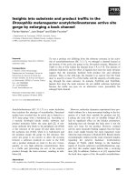
Analyzing the measurements of the radiotherapists, we
can see represented in figure 1, the two measurements of
the radiotherapists and the value of Pearson's correlation
coefficient, in which we observe significant and positive
correlation between the two measurements. The intraclass
correlation coefficient for the radiotherapist is 0.989 (p <
0.001) with confidence interval of 95% equal to (0.974;
0.995).
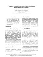
We can also evaluate this concordance by the Bland-Alt-
man method. The graph representing this analysis is
showed in Figure 2. The differences between the measure-
ments ranged from 42.88 to 37.74, with average of 3.10
and standard deviation of 21.15. Thus we obtained a con-
fidence interval of 95% equal to (-39.20; 45.41).
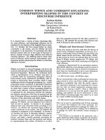
Analyzing the radiologists' findings, Figure 3 shows the
measurements they attained and the value of Pearson's
correlation coefficient, in which we observe significant
and positive correlation between the two measurements.
The intraclass correlation coefficient is equal to 0.762 (p <
0.001) with confidence interval of 95% equal to (0.522;
0.891).
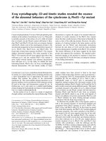
We can also evaluate this congruity by the Bland-Altman
method. Figure 4 shows the graph representing this anal-
ysis. The differences between the measurements ranged
from -466.27 to 26.23 with average of -31.35 and stand-
ard deviation of 101.27, thus we obtained a confidence
interval of 95% equal to (-233.88; 171.18).
Analyzing radiotherapists and radiologists findings, we
utilized the average between the measurements of the
radiologists and the average of the measurements of the
radiotherapists.
Figure 5 represents the measurements of the radiologists
and of the radiotherapists and the value of Pearson's cor-
relation coefficient, in which we observe significant and
positive correlation between the two measurements. The
intraclass correlation coefficient is equal to 0.942 (p <
0.001) with confidence interval of 95% equal to (0.869;
0.975). Hence an excellent correlation between the two
measurements has been found.
We can also evaluate this congruity by the Bland-Altman
method. The graph representing this analysis is contained
in figure 6. The differences between the measurements
ranged from -192.09 to 50.14 with average of -3.51 and
standard deviation of 48.73, thus we obtained a confi-
dence interval of 95% equal to (-100.98; 93.95).
In table 2, we calculate the coefficient of variation among
the 4 measurements, to wit: those of the 2 radiotherapists
and those of the 2 radiologists, which once again indicates
good congruity among them, with the exception of only
one value.
Discussion
Inoperable lung cancer prognosis remains very poor.
Besides the alternate fractionated schemes and combined
Table 1: Values of average, standard deviation, median, minimum and maximum of the values observed by the radiologists and
radiotherapists
Observer Average SD Median Minimum Maximum
Radiotherapist 1 140.84 136.29 83.56 13.03 516.85
Radiotherapist 2 137.74 141.68 78.81 11.22 496.39
Average 139.29 138.61 74.44 12.13 496.26
Radiologist 1 127.13 128.03 72.36 13.87 450.26
Radiologist 2 158.48 169.21 65.36 12.09 547.91
Average 142.80 141.24 68.27 12.98 465.35
Radiation Oncology 2009, 4:28 />Page 4 of 8
(page number not for citation purposes)
therapies, new planning strategies, including conformal
radiotherapy and dose increase, are under investigation
[25-29].
It is known that the general survival rate, cause-specific
survival and local tumor control are directly correlated
with the gross tumor volume in cm3. In the multivariate
analysis the most predictive independent survival variable
is the gross tumor volume [30].
Recently, the data mentioned by LEUNENS et al. (1993),
evidenced that the gross tumor volume definition is not
that simple, and there can be risks in excessive confidence
in the medical capacity to estimate the tumor extension
with the imaging approaches [17].
Due to the number of uncertainties and of phenomena
related to the tumor, the definition of gross tumor volume
in thoracic radiotherapy could result in greater volume
variations [22,27,31,32], focused on the definition of
lung cancer gross tumor volume as part of a delineation
protocol. The three authors concluded that there is signif-
icant variation in target volume definition.
VAN DE STEENE et al. (2002), showed unexpected major
interobservers' variability, with tumor delineation varying
by several centimeters, due to:
1) difficulty in discriminating between tumor and atel-
ectasia;
2) difficulty in distinguishing normal and pathological
structures of the tumor;
3) use of different tomographic windows and partial vol-
ume effects;
Measurements of the radiotherapists and the value of Pear-son's correlation coefficientFigure 1
Measurements of the radiotherapists and the value of
Pearson's correlation coefficient.
Measurements of the radiotherapists and the graph by the Bland-Altman methodFigure 2
Measurements of the radiotherapists and the graph
by the Bland-Altman method.
Measurements of the radiologists and the value of Pearson's correlation coefficientsFigure 3
Measurements of the radiologists and the value of
Pearson's correlation coefficients.
Measurements of the radiologists and the graph by the Bland-Altman methodFigure 4
Measurements of the radiologists and the graph by
the Bland-Altman method.
Radiation Oncology 2009, 4:28 />Page 5 of 8
(page number not for citation purposes)
4) insufficient anatomic knowledge [33].
Another study carried out by GIRAUD et al. (2002) that
compared the delineation of gross tumor volume per-
formed by radiologists and radiotherapists, showed sig-
nificant differences between the two groups: radiologists
tended to delineate lower and more homogeneous vol-
umes than radiotherapists, especially in the "difficult"
cases [34]. The delineation of the target volume and high-
risk organ constitutes a critical stage in conformal radio-
therapy [5,9,10,13,35] and the subsequent steps are
dependent on correct gross-volume delineation. Field
shaping and dose planning are based exclusively on the
tumor volumes and critical normal tissue delineated.
GIRAUD et al. (2002) suggested that the correct definition
of the gross tumor volume can be attained, when radio-
therapists are well trained in chest imaging [34]. SUNDAR
and SYMONDS (2002) suggest a compulsory period of
structured training in section imaging diagnosis for radio-
therapists [36].
According to the recommendations of ICRU 50 (1991,
1993) and later on, of ICRU 62 (1999), gross tumor vol-
ume delineation should be performed as close as possible
to the tumor and/or lymph node, without adding any
safety margin [37-39]. Successive additional volumes are
designated taking into account other treatment uncertain-
ties. A second attitude adopted by the majority consists of
attempting to distinguish between the tumor tissue and
the surrounding collapsed parenchyma. This choice calls
for perfect tomographic acquisition with rapid injection
of the contrast medium and a first series of slices per-
formed immediately after the injection [18].
Our results were incompatible with those of VAN DE
STEENE et al. (1996), SENAN et al. (1999) and GIRAUD
et al. (2002), which exhibited significant differences in
lung tumor delineation [22,34,40].
In our study there was excellent intraclass correlation
(Pearson's correlation coefficient) in the case of the radio-
therapists and good correlation in the case of the radiolo-
gists. In the latter, the correlation was slightly lower due to
a single point at which there was greater discrepancy
between the first and the second measurement. In the
analysis of radiotherapists and radiologists, we also
observed excellent correlation between the two measure-
ments.
Congruity was also evaluated by the Bland-Altman
method, while randomness was observed in the distribu-
tion of data within the constructed confidence interval,
and only one point fell outside the interval, indicating
that the error among the measurements does not tend to
increase when the measurement values are higher. More-
over, the average of the differences was close to zero, indi-
cating good concordance between the two measurements.
The discrepant measurement of gross tumor volume of
one of the radiologists resulted from associated atelectasis
that constitutes the main cause of error in tumor volume
delineation. We emphasize that in our study, we observed
one case of tumor with mediastinal invasion, two with
invasion of the thoracic wall and two causing lung atel-
ectasis.
Some peculiarities of Hospital A. C. Camargo might have
contributed to these results, such as integration of the
radiotherapy and diagnostic imaging departments, intern-
ship of the radiotherapy residents in diagnostic imaging
with learning of sectional anatomy, and geographical
proximity of the radiology and radiotherapy departments,
Measurements of the radiotherapists and the radiologists and the value of Pearson's correlation coefficientFigure 5
Measurements of the radiotherapists and the radiol-
ogists and the value of Pearson's correlation coeffi-
cient.
Measurements of the radiotherapists and the radiologists and the graph by the Bland-Altman methodFigure 6
Measurements of the radiotherapists and the radiol-
ogists and the graph by the Bland-Altman method.
Radiation Oncology 2009, 4:28 />Page 6 of 8
(page number not for citation purposes)
which are located in the same building, on adjacent
floors.
Despite of there are no statistically significant differences
in the definition of gross tumor volume between radiolo-
gists and radiotherapists in this study, in nine of twenty-
three evaluated patients there was a difference greater than
20%, which can be clinically relevant. Most of these cases
involved primary tumors located close to the mediasti-
num or chest wall, which hindered the proper measure-
ment of the lesions. Regardless of the overlapping
volumes have not been assessed, in neither case the
observers considered different structures to delineate the
target volumes.
Recently some authors have shown that delineation accu-
racy can be improved by using fluorodeoxyglucose-posi-
tron emission tomography (FDG-PET)/CT information.
FDG-PET/CT is a functional study that has proved to be
more accurate than CT in determining extent of non-
small-cell lung cancer. Integration of FDG-PET/CT on the
volume delineation can reduce interobserver variation
compared with CT based delineation and alter gross
tumor volume in about 50% of the cases. [41,42] FDG-
PET/CT images are particularly useful in defining the tar-
get volume in the presence of atelectasis and in defining
involved lymph nodes. [43]
Conclusion
Radiotheraphy plays an important role in the manage-
ment of inoperable lung cancer patients, A precise and
consistent delineation of target volumes is needed to
improve treatment and avoid complications. Although
some authors have found large rates of interobserver vari-
ability on volume delineation for lung cancer, in this sur-
vey, there was no statistically significant difference in the
definition of gross tumor volume between radiotherapists
and radiologists or intraclasses. Some institutional charac-
teristics should be responsible for this finding, such as
integration between radiotherapy and diagnostic imaging
departments.
Competing interests
The authors declare that they have no competing interests.
Authors' contributions
CJT conceived of the study, and participated in its design,
acquisition, analysis and interpretation of data, and
helped to draft the manuscript.
RC and PEN conceived of the study, and participated in its
design and coordination, and helped to draft the manu-
script.
PNVP, RCF and DGC conceived of the study, and partici-
pated in its design, acquisition of data, and helped to draft
the manuscript.
MAB and AGVB have been involved in literature review,
drafting the manuscript and revising it critically for publi-
cation.
Table 2: Coefficient of variation (COV) among the 4 measurements (2 radiologists and 2 radiotherapists)
Patient Radiotherapist 1 Radiotherapist 2 Radiologist 1 Radiologist 2 Average COV
1 108.55 127.42 127.29 155.28 129.64 0.15
2 369.29 370.13 391.36 387.46 379.56 0.03
3 125.05 121.29 125.97 130.56 125.72 0.03
4 13.03 11.22 13.87 12.09 12.55 0.09
5 52.13 33.13 35.87 27.78 37.23 0.28
6 68.45 78.81 72.54 65.36 71.29 0.08
7 83.56 65.31 72.36 64.18 71.35 0.12
8 35.90 27.40 46.20 27.02 34.13 0.26
9 77.58 49.27 67.62 41.39 58.97 0.28
10 135.91 109.46 81.64 547.91 218.73 1.01
11 65.30 65.82 83.64 116.94 82.93 0.29
12 186.47 199.94 178.92 317.77 220.78 0.30
13 453.51 496.39 382.78 466.78 449.87 0.11
14 33.94 32.31 28.72 28.98 30.99 0.08
15 195.41 215.96 157.15 172.20 185.18 0.14
16 71.31 62.83 64.20 67.13 66.37 0.06
17 53.26 52.13 52.19 51.76 52.34 0.01
18 516.85 475.67 450.26 480.44 480.81 0.06
19 266.67 302.54 303.24 308.28 295.18 0.06
20 95.41 94.10 50.23 46.40 71.54 0.38
21 121.23 83.49 59.19 56.56 80.12 0.37
22 73.29 53.71 43.71 47.11 54.46 0.24
23 37.24 39.65 34.97 25.58 34.36 0.18
Radiation Oncology 2009, 4:28 />Page 7 of 8
(page number not for citation purposes)
All authors have given final approval of the version to be
published.
References
1. Emami B, Lyman J, Brown A, Coia L, Goitein M, Munzenrider JE, Shank
B, Solin LJ, Wesson M: Tolerance of normal tissue to therapeu-
tic irradiation. Int J Radiat Oncol Biol Phys 1991, 21:109-22.
2. Emami B, Purdy JA, Manolis J, Barest G, Cheng E, Coia L, Doppke K,
Galvin J, LoSasso T, Matthews J, Munzenrider J, Shank B: Three-
dimensional treatment planning for lung cancer. Int J Radiat
Oncol Biol Phys 1991, 21:217-27.
3. Emami B: Three-dimensional conformal radiation therapy in
bronchogenic carcinoma. Semin Radiat Oncol 1996, 6:92-7.
4. Armstrong J, Zelefsky M, Leibel S, Burman C, Han C, Harrison LB,
Kutcher GJ, Fuks ZY: Strategy for dose escalation using 3-
dimensional conformal radiation therapy for lung cancer.
Ann Oncol 1995, 6:693-7.
5. Armstrong J: Target volume definition for three-dimensional
conformal radiation therapy of lung cancer. Br J Radiol 1998,
71:587-94.
6. Graham MV, Purdy JA, Emami B, Matthews JW, Harms WB: Prelim-
inary results of a prospective trial using three dimensional
radiotherapy for lung cancer. Int J Radiat Oncol Biol Phys 1995,
33:993-1000.
7. Sibley GS, Mundt AJ, Shapiro C, Jacobs R, Chen G, Weichselbaum R,
Vijayakumar S: The treatment of stage III nonsmall cell lung
cancer using high dose conformal radiotherapy. Int J Radiat
Oncol Biol Phys 1995, 33:1001-7.
8. Armstrong J, Raben A, Zelefsky M, Burt M, Leibel S, Burman C,
Kutcher G, Harrison L, Hahn C, Ginsberg R, Rusch V, Kris M, Fuks Z:
Promising survival with three-dimensional conformal radia-
tion therapy for non-small cell lung cancer. Radiother Oncol
1997, 44:17-22.
9. Beckendorf V, Elles S, Madelis G: Postoperative radiation ther-
apy in lung cancer: clinical target volume delineation in a
workshop including radiation oncologist and radiologists
[abstract]. Radiother Oncol 2000, 56(Supl 1):S39. [Presented at
19th Annual ESTRO Meeting, 2000 Sept 1923; Istanbul]
10. Antolak JA, Rosen II: Planning target volumes for radiotherapy:
how much margin is needed? Int J Radiat Oncol Biol Phys 1999,
44:1165-70.
11. Perez CA: The critical need for accurate treatment planning
and quality control in radiation therapy. Int J Radiat Oncol Biol
Phys 1977, 2:815-8.
12. Perez CA, Stanley K, Grundy G, Hanson W, Rubin P, Kramer S, Brady
LW, Marks JE, Perez-Tamayo R, Brown GS, Concannon JP, Rotman
M: Impact of irradiation technique and tumour control and
survival of patients with unresectable non-oat cell carcinoma
of the lung. Cancer 1982, 50:1091-9.
13. Ekberg L, Holmberg O, Wittgren L, Bjelkengren G, Landberg T:
What margins should be added to the clinical target volume
in radiotherapy treatment planning for lung cancer? Radiother
Oncol 1998, 48:71-7.
14. Giraud Ph, Antoine M, Larrouy A, Milleron B, Callard P, De Rycke Y,
Carette MF, Rosenwald JC, Cosset JM, Housset M, Touboul E: Eval-
uation of microscopic tumor extension in non-small-cell lung
cancer for three-dimensional conformal radiotherapy plan-
ning. Int J Radiat Oncol Biol Phys 2000, 48:1015-24.
15. Jaffray DA, Yan D, Wong JW: Managing geometric uncertainty
in conformal intensity-modulated radiation therapy. Semin
Radiat Oncol 1999, 9:4-19.
16. McNee SG, Rampling R, Dale AJ, Gregor A: An audit of 3D treat-
ment planning facilities and practice in the UK. Clin Oncol
1998, 10:18-23.
17. Leunens G, Menten J, Weltens C, Verstraete J, Schueren E van der:
Quality assessment of medical decision making in radiation
oncology: variability in target volume delineation for brain
tumours. Radiother Oncol 1993, 29:169-75.
18. Satoh K, Kobayashi T, Kawase Y, Mitani M, Takahashi K, Takashima
H, Ohkawa M, Tanabe M, Kojima K: CT-pathologic correlation in
the tumour margins of lung cancer. Radiat Med 1996,
14:167-72.
19. Cazzaniga LF, Marinoni MA, Bossi A, Bianchi E, Cagna E, Cosentino D,
Scandolaro L, Valli M, Frigerio M: Interphysician variability in
defining the planning target volume in the irradiation of
prostate and seminal vesicles. Radiother Oncol 1998, 47:293-6.
20. Oozeer R, Chauvet B, Toy BJ, Berger C, Garcia R, Felix-Faure C, Le
Thanh H, Reboul F: Definition of prostatic contours using
tomodensitometric slices: study of differences among radia-
tion oncologists and between examinations. Cancer Radiother
1999, 3:333-40.
21. Rasch C, Barillot I, Remeijer P, Touw A, van Herk M, Lebesque JV:
Definition of the prostate in CT and MRI: a multi-observer
study. Int J Radiat Oncol Biol Phys 1999, 43:57-66.
22. Senan S, van Sornsen de Koste JS, Samson M, Tankink H, Jansen P,
Nowak PJ, Krol AD, Schmitz P, Lagerwaard FJ: Evaluation of a tar-
get contouring protocol for 3D conformal radiotherapy in
non-small cell lung cancer. Radiother Oncol 1999, 53:247-55.
23. Tai P, Van Dyk J, Yu E, Battista J, Stitt L, Coad T: Variability of tar-
get volume delineation in cervical esophageal cancer. Int J
Radiat Oncol Biol Phys 1998, 42:277-88.
24. Giraud Ph, Dubray B, Gaboriaud G, Rosenwald JC, Cosset JM: Influ-
ence of CT images visualization parameters for target vol-
ume delineation in lung cancer [abstract]. Radiother Oncol
2000, 56(Supl 1):S39. [Presented at 19th Annual ESTRO Meeting,
2000 Sept 1923; Istanbul]
25. Armstrong J, Burman C, Leibel S, Fontenla D, Kutcher G, Zelefsky M,
Fuks Z: Three-dimensional conformal radiation therapy may
improve the therapeutic ratio of high dose radiation therapy
for lung cancer. Int J Radiat Oncol Biol Phys 1993, 26:685-9.
26. Graham MV, Matthews JW, Harms WB Sr, Emami B, Glazer HS,
Purdy JA: Three-dimensional radiation treatment planning
study for patients with carcinoma of the lung. Int J Radiat Oncol
Biol Phys 1994, 29:1105-17.
27. Graham MV, Purdy JA, Emami B, Matthews JW, Harms WB: Prelim-
inary results of a prospective trial using three dimensional
radiotherapy for lung cancer. Int J Radiat Oncol Biol Phys 1995,
33:993-1000.
28. Vijayakumar S, Chen GT: Implementation of three dimensional
conformal radiation therapy: prospects opportunities, and
challenges. Int J Radiat Oncol Biol Phys 1995, 33:979-83.
29. Robertson JM, Ten Haken RK, Hazuka MB, Turrisi AT, Martel MK, Pu
AT, Littles JF, Martinez FJ, Francis IR, Quint LE, Lichter AS: Dose
escalation for non-small cell lung cancer using conformal
radiation therapy. Int J Radiat Oncol Biol Phys 1997, 37:
1079-85.
30. Bradley JD, Leumwananonthachai N, Purgy JA, Wasserman TH, Lock-
ett MA, Graham MV, Perez CA: Gross tumor volume, critical
prognostic factor in patients treated with three-dimensional
conformal radiation therapy for non-small-cell lung carci-
noma. Int J Radiat Oncol Bio 2002, 52:49-57.
31. Hamilton CS, Denham JW, Joseph DJ, Lamb DS, Spry NA, Gray AJ,
Atkinson CH, Wynne CJ, Abdelaal A, Bydder PV, Chapman PJ, Mat-
thews JHL, Stevens G, Ball D, Kearsley J, Ashcroft JB, Janke P, Gut-
mann A: Treatment and planning decisions in non-small cell
carcinoma of the lung: an Australasian patterns of practice
study. Clin Oncol R Coll Radiol 1992, 4:141-7.
32. Valley JF, Mirimanoff RO: Comparison of treatment techniques
for lung cancer. Radiother Oncol 1993, 28:168-73.
33. Steene J Van de, Linthout N, de Mey J, Vinh-Hung V, Classens C, Nop-
pen M, Bel A, Storme G: Definition of gross tumour volume in
lung cancer: inter-observer variability. Radiother Oncol 2002,
62:37-49.
34. Giraud P, Elles S, Helfre S, De Rycke Y, Servois V, Carette MF, Alzieu
C, Bondiau PY, Dubray B, Touboul E, Housset M, Rosenwald JC, Cos-
set JM: Conformal radiotherapy for lung cancer: different
delineation of the gross tumor volume (GTV) by radiologists
and radiation oncologists. Radiother Oncol 2002, 62:27-36.
35. Logue JP, Sharrock CL, Cowan RA, Read G, Marrs J, Mott D: Clinical
variability of target volume description in conformal radio-
therapy planning. Int J Radiat Oncol Biol Phys 1998, 41:929-31.
36. Sundar S, Symonds RP: Diagnostic radiology for radiotherapist:
the case for structured training in cross-sectional imaging
(CT and MRI). Clin Oncol 2002, 14:413-4.
37. [ICRU] International Commission on Radiation Units and Measure-
ments: Prescribing, recording and reporting photon beam
therapy. Washington, DC: ICRU; 1991. (ICRU Report 50)
38. [ICRU] International Commission on Radiation Units and Measure-
ments: Prescribing, recording, and reporting photon beam
therapy. Bethesda MD: ICRU; 1993. (ICRU Report 50)
Publish with BioMed Central and every
scientist can read your work free of charge
"BioMed Central will be the most significant development for
disseminating the results of biomedical research in our lifetime."
Sir Paul Nurse, Cancer Research UK
Your research papers will be:
available free of charge to the entire biomedical community
peer reviewed and published immediately upon acceptance
cited in PubMed and archived on PubMed Central
yours — you keep the copyright
Submit your manuscript here:
/>BioMedcentral
Radiation Oncology 2009, 4:28 />Page 8 of 8
(page number not for citation purposes)
39. [ICRU] International Commission on Radiation Units and Measure-
ments: Prescribing, recording, and reporting photon beam
therapy (supplement to ICRU Report 50). Bethesda. MD:
ICRU; 1999. (ICRU Report 62)
40. Steene J Van de, Linthout N, de Mey J, Vinh-Hung V, Claassens C,
Noppen M, Bel A, Storme G: Definition of gross tumor volume
in lung cancer: inter-observer variability. Radiother Oncol 2002,
62:37-49.
41. Faria SL, Menard S, Devic S, Sirois C, Souhami L, Lisbona R, Freeman
CR: Impact of FDG-PET/CT on radiotherapy volume deline-
ation in non-small-cell lung cancer and correlation of imag-
ing stage with pathologic findings. Int J Radiat Oncol Biol Phys
2008, 70:1035-1038.
42. Van Baardwijk A, Bosmans G, Boersma L, Buijsen J, Wanders S, Hoch-
stenbag M, van Suylen RJ, Dekker A, Dehing-Oberije C, Houben R,
Bentzen SM, van Kroonenburgh M, Lambin P, De Ruysscher D: PET-
CT-based auto-contouring in non-small-cell lung cancer cor-
relates with pathology and reduces interobserver variability
in the delineation of the primary tumor and involved nodal
volumes. Int J Radiat Oncol Biol Phys 2007, 68:771-778.
43. Greco C, Rosenzweig K, Cascini GL, Tamburrini O: Current status
of PET/CT for tumour volume definition in radiotherapy
treatment planning for non-small cell lung cancer (NSCLC).
Lung Cancer 2007, 57:125-134.