Báo cáo khoa học: " RapidArc, intensity modulated photon and proton techniques for recurrent prostate cancer in previously irradiated patients: a treatment planning comparison study" doc
Bạn đang xem bản rút gọn của tài liệu. Xem và tải ngay bản đầy đủ của tài liệu tại đây (1.35 MB, 11 trang )
BioMed Central
Page 1 of 11
(page number not for citation purposes)
Radiation Oncology
Open Access
Research
RapidArc, intensity modulated photon and proton techniques for
recurrent prostate cancer in previously irradiated patients: a
treatment planning comparison study
Damien C Weber*
1,5
, Hui Wang
1
, Luca Cozzi
2
, Giovanna Dipasquale
1
,
Haleem G Khan
3
, Osman Ratib
4,5
, Michel Rouzaud
1
, Hansjoerg Vees
1
,
Habib Zaidi
4
and Raymond Miralbell
1,5
Address:
1
Department of Radiation Oncology, University Hospital of Geneva, Geneva, Switzerland,
2
Oncology Institute of Southern Switzerland,
Medical Physics Unit, Bellinzona, Switzerland,
3
Institute of Radiology Jean Violette, Geneva, Switzerland,
4
Department of Nuclear Medicine,
University Hospital of Geneva, Geneva, Switzerland and
5
Faculty of medicine, UNIGE, University of Geneva, Switzerland
Email: Damien C Weber* - ; Hui Wang - ; Luca Cozzi - ;
Giovanna Dipasquale - ; Haleem G Khan - ; Osman
Ratib - ; Michel Rouzaud - ; Hansjoerg Vees - ;
Habib Zaidi - ; Raymond Miralbell -
* Corresponding author
Abstract
Background: A study was performed comparing volumetric modulated arcs (RA) and intensity
modulation (with photons, IMRT, or protons, IMPT) radiation therapy (RT) for patients with
recurrent prostate cancer after RT.
Methods: Plans for RA, IMRT and IMPT were optimized for 7 patients. Prescribed dose was 56
Gy in 14 fractions. The recurrent gross tumor volume (GTV) was defined on
18
F-fluorocholine PET/
CT scans. Plans aimed to cover at least 95% of the planning target volume with a dose > 50.4 Gy.
A maximum dose (D
Max
) of 61.6 Gy was allowed to 5% of the GTV. For the urethra, D
Max
was
constrained to 37 Gy. Rectal D
Median
was < 17 Gy. Results were analyzed using Dose-Volume
Histogram and conformity index (CI
90
) parameters.
Results: Tumor coverage (GTV and PTV) was improved with RA (V
95%
92.6 ± 7.9 and 83.7 ± 3.3%),
when compared to IMRT (V
95%
88.6 ± 10.8 and 77.2 ± 2.2%). The corresponding values for IMPT
were intermediate for the GTV (V
95%
88.9 ± 10.5%) and better for the PTV (V
95%
85.6 ± 5.0%). The
percentages of rectal and urethral volumes receiving intermediate doses (35 Gy) were significantly
decreased with RA (5.1 ± 3.0 and 38.0 ± 25.3%) and IMPT (3.9 ± 2.7 and 25.1 ± 21.1%), when
compared to IMRT (9.8 ± 5.3 and 60.7 ± 41.7%). CI
90
was 1.3 ± 0.1 for photons and 1.6 ± 0.2 for
protons. Integral Dose was 1.1 ± 0.5 Gy*cm
3
*10
5
for IMPT and about a factor three higher for all
photon's techniques.
Conclusion: RA and IMPT showed improvements in conformal avoidance relative to fixed beam
IMRT for 7 patients with recurrent prostate cancer. IMPT showed further sparing of organs at risk.
Published: 9 September 2009
Radiation Oncology 2009, 4:34 doi:10.1186/1748-717X-4-34
Received: 2 June 2009
Accepted: 9 September 2009
This article is available from: />© 2009 Weber et al; licensee BioMed Central Ltd.
This is an Open Access article distributed under the terms of the Creative Commons Attribution License ( />),
which permits unrestricted use, distribution, and reproduction in any medium, provided the original work is properly cited.
Radiation Oncology 2009, 4:34 />Page 2 of 11
(page number not for citation purposes)
Background
Biochemical failures (BF) of prostate cancer after external
beam radiation therapy (RT) is not an unusual event and
is observed in a substantial number of prostate cancer
patients [1,2]. CapSURE™ (Cancer of the Prostate Strategic
Urologic Research Endeavor) data have demonstrated a
biochemical failure rate following radiation therapy as
high as 63% [3]. Up to 70% of these patients will have evi-
dence of recurrent or residual disease within the prostate
gland [4]. Although curative treatment is still an option if
the patient presents organ-confined disease only, no con-
sensus exists however on the optimal salvage therapy
modality for these patients. Therapeutic management of
these patients includes salvage radical prostatectomy, cry-
otherapy, brachytherapy or high-intensity focused ultra-
sound, with or without hormonal deprivation therapy.
Re-irradiation with conformal techniques is yet another
strategy with potential curative intent. Re-irradiation tech-
niques must however minimally deliver radiation dose to
pre-irradiated organ at risk (OARs) in the direct vicinity of
the target volume.
The demonstration of organ-confined only recurrent dis-
ease in patients with BF is not easily done with conven-
tional radiology. Identifying precisely the target recurrent
volume is of paramount importance when delivering
focused high-radiation dose in a pre-irradiated area.
Recent progress in imaging with PET tracers such as ace-
tate or choline labelled with
11
C or
18
F have improved sig-
nificantly the accuracy in diagnosing the site of relapse
[5]. Local tracer uptake within the gland may correspond
to the locally recurring gross-tumor volume (GTV) and
can be contoured in the RT treatment planning system.
RapidArc (RA), is a novel technique which may achieve
several objectives: i) improve organ at risks (OARs) and
non-target tissue sparing compared to other intensity
modulated RT (IMRT) techniques; ii) maintain or
improve the same degree of target coverage; iii) reduce sig-
nificantly the treatment time per fraction. Dose compara-
tive studies using RA, have been published in prostate
[6,7], cervix uteri [8] and anal canal cancer [9], showing
significant improvements when compared to non-RA
techniques. This technique could be thus used to treat
geometrically complex partial recurrent tumor volumes
within the prostate gland after RT.
The present study was undertaken to assess the treatment
planning inter-comparison between photon and proton
RT, namely IMRT and IMPT, to RA, as applied to a total of
7 recurrent pre-irradiated prostate cancer patients
Methods
The institutional
18
F-Choline database containing 47
prostate cancer patients was queried to identify individu-
als with: 1) biochemically recurrence; 2) local relapse
only; 3) previous high-dose (≥ 70 Gy) RT and 4) endorec-
tal MRI. Seven of such patients were identified (median
age, 77 years; Table 1). They all underwent previous cura-
tive 3D conformal RT (median dose, 74 Gy; HDR brachy-
therapy boost 14 Gy in 2 fractions, 2 patients), 4.8 to 7.6
(median, 5.9) years before biological recurrence (Table 1).
Table 1: Patients characteristics
No of patient 1 2 3 4 5 6 7
Age (years) 81637969777869
Recurrence time (years) 5.86 4.82 6.75 5.16 5.85 5.82 7.55
PSADT (month) 13.9 9.0 10.2 8.5 10.2 5.5 25.8
Tumour stage (at relapse) T2c T3b T2c T2c T3a T2c T3b
PSA at recurrence (ng/ml) 5.11 6.76 2.80 5.14 6.32 5.95 13.00
Gleason score at recurrence 3+4 - 3+4 3+3 4+3 4+3 3+3
GTV (cm
3
) 0.61 1.09 3.48 5.08 5.75 10.36 19.93
CTV (cm
3
) 2.59 3.29 9.72 12.84 15.65 20.91 38.61
PTV(cm
3
) 6.68 8.13 22.13 26.67 30.42 39.47 64.20
Abbreviations: PSA = prostate-specific antigen; PSADT = prostate-specific antigen doubling time; PET-CT = Positron Emission Tomography and
Computed Tomography; GTV = gross tumour volume; CTV = Clinical Target Volume; PTV = Planning Target Volume.
Radiation Oncology 2009, 4:34 />Page 3 of 11
(page number not for citation purposes)
The median dose received by 50%/1% of the rectum and
bladder by this prior treatment were 44.1 (range, 60.0 -
38.5)/71.0 (range, 74.5 - 62.4) and 59.0 (range, 67.2 -
43.4)/74.0 (range, 78.0 - 64.4) Gy, respectively. The
median rectal volume receiving 35 Gy was 79.4%, and
range from 56.0 to 96.0%. Local relapse was proven by
PET-CT examination with
18
F-choline; failures were con-
firmed by sextant biopsy in all but one patient. A positive
correlation between
18
F-choline uptake and the location
of the histological proven recurrence was observed in all 6
patients. Table 2 details the radiological and pathological
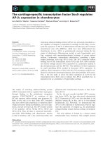
correlation of these recurrences. PET/CT imaging was per-
formed on the Biograph 16 scanner (Siemens Medical
Solution, Erlangen, Germany) operating in 3D mode (Fig.
1). An endorectal MRI, with spectroscopy and contrast
enhancement, was acquired for all patients [10]. The main
organs at risk (OARs) considered for all patients were the
urethra (defined on the base of MR imaging and verified
by an experienced radiologist), bladder, rectum, penile
bulb and femoral heads The non-target tissue was defined
as the patient's volume covered by the CT scan minus the
planning target volume (PTV).
For all patients, GTV was outlined using the signal-to-
background ratio-based adaptive thresholding technique
described in [11] and adapted to our PET/CT scanner
characteristics. Data acquisition and processing protocols
are described elsewhere [12]. The clinical applicability of
detecting prostate recurrence with 18F-Choline PET has
been demonstrated in our previous series [13]. Fig. 1
depicts the PET GTV for 1 patient. Clinical target volume
(CTV) was defined adding a 3D anisotropic margin of 3
mm (CTV was however limited to the prostate and semi-
nal vesicles and could not be stretched beyond these struc-
tures), excluding the urethra in all cases. PTV was defined
adding a 3D anisotropic margin of 3 mm (2 mm in prox-
imity of the urethra) to the CTV. A summary of the sizes
of the GTVs, PTVs and OARs are detailed in Tables 1 and 2.
Dose prescription of 56 Gy to PTV was delivered accord-
ing to a hypofractionated radiation schedule consisting of
14 daily fractions of 4 Gy, twice weekly (overall treatment
time, 7 weeks) [14]. All plans were normalized to the
mean dose of the PTV.
Plans aimed to cover at least 95% of the PTV with a dose
greater than 90% of the dose prescription. An over-dosage
of maximum 61.6 Gy (110%) was allowed to 5% of both
CTV and PTV. For the urethra, the maximum dose was
constrained to 37 Gy. A dose lower than 28 Gy delivered
to 50% of the volume of the bladder, penile bulb and fem-
oral heads was required for these OARs; likewise, a dose <
17 Gy was constraint to 30% of the rectal volume.
Four sets of plans were compared in this study, all
designed on the Varian Eclipse treatment planning system
(version 8.6.10) with 6 MV photon beams from a Varian
Clinac equipped with either a Millennium Multileaf Col-
limator (MLC) with 120 leaves (RA_M120; spatial resolu-
tion of 5 mm at isocentre) or a High Definition MLC with
120 leaves (RA_HD120; spatial resolution of 2.5 mm at
isocentre). Plans for RA were optimized selecting a maxi-
mum DR of 600 MU/min and a fixed DR of 600 MU/min
was selected for IMRT.
The Anisotropic Analytical Algorithm photon dose calcu-
lation algorithm was used for all photon cases [15]. The
dose calculation grid was set to 2.5 mm.
Table 2: Prostate cancer recurrence on MRI, PET and biopsy
Recurrent Site
No of patient MRI PET CT Biopsy (Number of positive cores)
1 L, R L L (1/7); R (0/6)
2SVSV ND
3 L, R L, R L (1/3); R (3/4)
4 R R L (0/4); R (3/4)
5 L L, R L (2/3); R (1/3)
6 L, R L, R L (3/3); R (4/4)
7 L, R, SV L, R, SV L (4/4); R (3/4)
Abbreviations: L, left prostate lobe; R, right prostate lobe; SV, seminal vesicle; ND, not done.
Radiation Oncology 2009, 4:34 />Page 4 of 11
(page number not for citation purposes)
RA
RA uses continuous variation of the instantaneous dose
rate, MLC leaf positions and gantry rotational speed to
optimize the dose distribution. Details about RA optimi-
zation process have been published elsewhere [8]. To
minimize the contribution of tongue and groove effect
during the arc rotation and to benefit from leaves trajecto-
ries non-coplanar with respect to patient's axis, the colli-
mator rotation in RA remains fixed to a value different
from zero. In the present study collimator was rotated to
~30° depending on the patient.
For the study, two sets of plans were optimized, each with
a single arc 360°. The first set (RA_M120) was created
using the Millennium MLC, the second set (RA_HD120)
with the High Definition MLC.
IMRT
Plans were designed according to the dynamic sliding
window method [16] with five fixed gantry beams. One
single isocentre was located at the target center of mass.
All beams were coplanar with collimator angle set to 0°.
The Millennium MLC was used for the study.
IMPT
Intensity modulated proton plans were obtained for a
generic proton beam through a spot scanning optimiza-
tion technique implemented in the Eclipse treatment
planning system from Varian. The optimization process
has been detailed elsewhere [17]. Spot spacing was set to
3 mm, circular lateral target margins were set to 5 mm,
proximal margin to 5 mm and distal margin to 2 mm. In
all cases coplanar beam arrangement was adopted using 3
fields, one with posterior and two with anterior oblique
incidence.
Quantitative evaluation of plans was performed by means
of standard Dose-Volume Histogram (DVH). For GTV and
PTV, the values of D
98%
and D
2%
(dose received by the
98% and 2% of the volume) were defined as metrics for
minimum and maximum doses and thereafter reported.
To complement the appraisal of minimum and maximum
dose, V
95%
and V
107%
(the volume receiving at least 95% or
at most 107% of the prescribed dose) were reported. The
homogeneity of the treatment was expressed in terms of
the standard deviation and of D
5%
-D
95%
. The conformal-
ity of the plans was measured with a Conformity Index,
CI
90%
defined as the ratio between the patient volume
receiving at least 90% of the prescribed dose and the vol-
ume of the PTV.
For OARs, the analysis included the mean dose, the max-
imum dose expressed as D
1%
and a set of appropriate vol-
ume (V
X
) and dose (D
Y
) metrics.
For non-target tissue, the integral dose, (Dose
Int
) is
defined as the integral of the absorbed dose extended to
over all voxels excluding those within the target volume
(Dose
Int
dimensions are Gy*cm
3
). This was reported
together with the observed mean dose and some repre-
sentative V
x
values.
To visualize the global difference between techniques,
average cumulative DVH for GTV and PTV, OARs and
healthy tissue, were built from the individual DVHs.
These DVHs were obtained by averaging the correspond-
ing volumes over the whole patient's cohort for each dose
bin of 0.05 Gy.
To appraise the difference between the techniques, the
paired, two-tails Student's t-test was applied whenever
applicable. Data were considered statistically significant
for p < 0.05.
GTV in the axial (
A), coronal (B) and sagital (C) simulation CT with PET fusion and
18
F-choline PET slice, respectivelyFigure 1
GTV in the axial (A), coronal (B) and sagital (C) simu-
lation CT with PET fusion and
18
F-choline PET slice,
respectively.
A
B
C
Radiation Oncology 2009, 4:34 />Page 5 of 11
(page number not for citation purposes)
Results
The mean prostate volume was 35.4 ± 7.8 cm
3
and the
average GTV and PTV volumes are reported in Table 3. The
mean ratio between PTV and prostate volume was 0.77 ±
0.50 with a range from 0.19 to 1.76.
For the GTV and PTV, the RA_HD120 and IMRT tech-
niques produced the best and worst dose homogeneity,
respectively (Table 3). The GTV coverage was optimal with
RA (mean V
95%
92%; Table 3). The PTV coverage (V
95%
)
was better with IMPT, intermediate with RA and worse
with IMRT (Table 3).
The GTV and PTV V
95%
-difference observed between
RA_HD120 and RA_M120 (Table 3) is due to different
MLC characteristics, namely spatial resolution and trans-
mission. IMPT showed a moderate improvement com-
pared to IMRT (V
107
and V
95
; Table 3). Interestingly, IMPT
did not reach the performance of RA_HD120 for V
107
for
both the GTV and PTV (Table 3). None of the techniques
achieved the planning objective on minimum PTV dose
(Table 3). IMRT failed to reach the objective on D
5%
for
PTV while all others met the condition (Table 3).
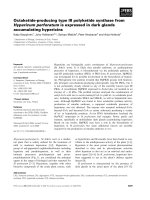
The rectal dose was significantly decreased with IMPT and
RA, respectively (Fig. 2, 3). For the intermediate dose
level, these two techniques more than halved the percent-
age of rectal volume receiving 35 and 45 Gy (Table 4). For
the high-dose level, IMPT delivered a decreased dose
when compared to the other two photons techniques
(Table 4).
For the urethra, none of the techniques was able to keep
the maximum dose below the threshold of 37 Gy (Table
4). IMPT violated this dose level by approximately 1 Gy,
while RA and IMRT exceeded this metric by 2.3 - 2.8 and
3 Gy, respectively. For the intermediate dose level, IMPT
and RA approximately halved the percentage of urethral
volume receiving 35 and 45 Gy (Table 4), respectively.
Since the urethra was included in the PTV in a majority (5/
7) of patients, the observed values were expected.
Table 3: Dosimetric results for GTV and PTV
Parameter IMRT IMPT RA_HD120 RA_M120 p
GTV Volume [cm3] 6.7 ± 6.8 [0.6-19.9]
Mean [Gy] 58.9 ± 2.2 56.5 ± 1.0 57.2 ± 0.6 57.3 ± 0.8 e
D
5
-D
95
[Gy] 12.4 ± 6.9 12.5 ± 6.0 8.5 ± 5.3 10.2 ± 5.3 a,b,c,d,e,f
D
2
[Gy] 64.6 ± 1.2 61.9 ± 2.7 60.7 ± 2.0 61.5 ± 1.6 a,b,c,d
D
98
[Gy] 49.3 ± 7.7 46.6 ± 6.9 49.2 ± 6.6 48.2 ± 6.3 d,e,f
V
95
[%] 88.6 ± 10.8 88.9 ± 10.5 92.6 ± 7.9 91.4 ± 8.5 d,e,f
V
107
[%] 52.3 ± 27.8 21.1 ± 14.9 9.1 ± 12.1 19.3 ± 14.2 b,f
PTV Volume [cm3] 27.7 ± 19.6 [6.7-64.2]
Mean [Gy] 56.0 ± 0.0 56.0 ± 0.0 56.0 ± 0.0 56.0 ± 0.0
D
5
-D
95
[Gy] 15.0 ± 2.0 13.6 ± 4.3 11.8 ± 2.7 13.2 ± 3.2 a,b,c,d,f
D
2
[Gy] 63.6 ± 0.9 61.4 ± 1.6 60.7 ± 1.5 61.5 ± 1.3 a,b,c,d,f
D
5
[Gy] 62.3 ± 0.9 60.7 ± 1.4 60.0 ± 1.2 60.7 ± 1.2 a,b,c,d,f
D
98
[Gy] 43.8 ± 2.8 42.4 ± 5.4 44.1 ± 4.0 43.5 ± 4.5 d,e,f
V
95
[%] 77.2 ± 2.2 85.6 ± 5.0 83.7 ± 3.3 81.8 ± 4.2 a,b,e,f
V
107
[%] 18.2 ± 2.6 12.6 ± 8.5 6.9 ± 6.4 12.5 ± 8.6 b,d,f
a = IMRT vs IMPT b = IMRT vs RA_HD120 c = IMRT vs RA_M120
d = IMPT vs RA_HD120 e = IMPT vs RA_M120 f = RA_HD120 vs RA_M120
Radiation Oncology 2009, 4:34 />Page 6 of 11
(page number not for citation purposes)
Table 4: Dosimetric results for OARs and non target tissues
Parameter IMRT IMPT RA_HD120 RA_M120 p
Rectum. Volume [cm3] 48.6 ± 17.6 [28.4-72.5]
D
50
[Gy] 10.1 ± 6.2 4.1 ± 4.0 8.2 ± 3.9 9.1 ± 4.2 a,b,d,e,f
D
1
[Gy] 49.6 ± 6.8 45.1 ± 9.2 45.2 ± 8.3 46.5 ± 7.8 a,b,c
V
35 Gy
[%] 9.8 ± 5.3 3.9 ± 2.7 5.1 ± 3.0 5.9 ± 3.3 a,b,c,e
V
45 Gy
[%] 3.6 ± 2.4 1.6 ± 1.3 1.6 ± 1.1 1.9 ± 1.3 a,b,c
Urethra. Volume [cm3] 0.7 ± 0.1 [0.6-0.8]
D
50
[Gy] 31.4 ± 13.1 26.8 ± 11.7 28.6 ± 11.4 28.6 ± 10.9 a,b,c,d,e
D
1
[Gy] 40.1 ± 3.3 38.1 ± 2.4 39.8 ± 3.5 39.3 ± 3.3 a,c,d,f
V
35 Gy
[%] 60.7 ± 41.7 25.1 ± 21.1 38.0 ± 25.3 36.0 ± 24.0 a,b,c
V
40 Gy
[%] 11.0 ± 12.8 0.6 ± 1.1 5.1 ± 5.4 4.0 ± 5.6
Left femoral head Volume [cm3] 60.1 ± 4.4 [54.8-67.6]
D
50
[Gy] 3.9 ± 2.6 0.1 ± 0.1 3.3 ± 2.1 3.5 ± 2.1 a,b,d,e,f
D
1
Gy] 14.6 ± 7.2 2.3 ± 2.0 7.4 ± 1.5 7.6 ± 1.3 a,b,c,d,e
Right femoral head Volume [cm3] 60.9 ± 5.8 [54.6-71.6]
D
50
[Gy] 3.9 ± 2.7 0.1 ± 0.1 3.2 ± 2.3 3.4 ± 2.1 a,d,e
D
1
Gy] 15.3 ± 7.5 2.5 ± 3.0 8.0 ± 1.8 8.0 ± 1.7 a,b,c,d,e
Bladder. Volume [cm3] 109.8 ± 63.6 [32.7-234.2]
D
50
[Gy] 4.9 ± 3.2 0.7 ± 0.9 4.6 ± 2.6 5.2 ± 3.0 a,d,e,f
D
1
[Gy] 42.3 ± 17.0 38.8 ± 19.6 41.3 ± 16.3 42.1 ± 15.8
V
35 Gy
[%] 6.4 ± 6.3 3.9 ± 4.3 4.1 ± 4.1 4.5 ± 4.2 a
V
50 Gy
[%] 1.9 ± 2.7 1.4 ± 2.1 1.3 ± 2.1 1.3 ± 2.1
Penile bulb. Volume [cm3] 7.2 ± 3.2 [3.0-13.2]
D
50
[Gy] 2.0 ± 1.5 0.9 ± 1.4 2.5 ± 1.7 3.2 ± 2.5 a,b,c,d,e
D
1
[Gy] 7.6 ± 9.4 7.1 ± 9.0 5.8 ± 4.6 7.7 ± 7.4
Non Target Tissue
Mean [Gy] 2.0 ± 0.8 0.7 ± 0.3 1.8 ± 0.7 1.9 ± 0.7 a,b,d,e,f
V
10 Gy
[%] 6.0 ± 2.6 2.8 ± 1.3 4.7 ± 2.5 5.1 ± 2.8 a,b,c,d,e
CI
90
1.3 ± 0.1 1.6 ± 0.2 1.3 ± 0.1 1.3 ± 0.1 a,d,e
Dose
Int
[Gy*cm
3
10
4
] 3.3 ± 1.6 1.1 ± 0.5 2.9 ± 1.3 3.1 ± 1.4 a,b,d,e,f
a = IMRT vs IMPT b = IMRT vs RA_HD120 c = IMRT vs RA_M120
d = IMPT vs RA_HD120 e = IMPT vs RA_M120 f = RA_HD120 vs RA_M120
Radiation Oncology 2009, 4:34 />Page 7 of 11
(page number not for citation purposes)
IMPT resulted in an almost complete avoidance of femo-
ral heads (Fig. 2; median inferior to 0.1 Gy; Table 4) while
both RA reduced maximum dose of about 50% compared
to IMRT.
IMPT was the best technique to spare the penile bulb (Fig.
3). For the bladder, all non-IMPT techniques were identi-
cal (Table 4; Fig 3).
Non target tissue irradiation was limited for all techniques
and the mean dose was kept under the Gy unit for the
majority of patients (Table 4). IMPT showed a Dose
Int
of
approximately a factor 3 lower than all the photon tech-
niques. The CI was however better with photons tech-
niques (mean CI improvement: 18%), because of the
wider lateral and distal spread induced by spot size, spac-
ing and margins used to achieve sufficient target coverage
(Table 4).
For all but one OARs (urethra), RA_HD120 results were
better than those observed with RA_M120 (Table 4). This
observed OAR's sparing derives from the superior spatial
resolution and inferior transmission through leaves with
the former when compared to the latter technique.
RA_M120 generally improved OARs sparing compared to
IMRT suggesting, given the usage of same MLC, a superior
modulation capability (Table 4). The only exception in
this pattern is represented by the penile bulb (D
1
7.7 vs.
7.6; Table 4). This OAR is moderately distant from the tar-
get and affected by higher scattering, mostly compensated
if the High Definition HD_120 MLC is used instead of the
Millennium M120.
Discussion
More than one out of four patients presenting a BF after
definitive RT will have clinical evidence of local recurrence
within 5 years [18]. Failure to control the prostate is not
only a cause of local disease progression but provides pos-
sibly a nidus for systemic spread, as shown by the distant
metastasis rate in this population [18]. A body of litera-
ture predicts however that complications, not limited to
but including, the rectum [19,20] and urethra [21,22],
after any salvage local therapy in a post-RT setting, is sig-
nificant. As such, rectal and urethral toxicity is a major
concern when using external beam RT as salvage local
therapy [23]. We have undertaken a treatment plan com-
parative study to assess the dose deposition to these OARs,
using intensity modulated photons and protons tech-
niques. Overall, IMPT and RA techniques substantially
decreased the dose in the intermediate range level to the
rectum and urethra (Fig. 3). All the volume and dose met-
rics for these OARs were substantially decreased with
IMPT and RA when compared to IMRT (Table 4). As such,
these findings might have bearing on clinical practice for
recurrent prostate cancer after RT. RA or IMPT might be an
alternative to salvage prostatectomy, cryosurgery or brach-
ytherapy in a selected number of patients.
Non conventional RT, be it IMRT, IMPT or RA, was simu-
lated essentially to capitalize the prerequisite tight dose
conformation necessary to administer radiation to these
heavily pre-treated prostates. This conformal ability was
coupled with the theoretical advantage of hypo fractiona-
tion in prostate cancer, while respecting the dose-toler-
ance of pre-irradiated OARs in the vicinity of the prostate.
An increasing body of data now suggests that the α/β ratio
for prostate is low, possibly in the range of 1-3 Gy [24]. If
this metric is accurately low, then hypo fractionated radi-
ation schedules should improve the therapeutic ratio [25].
It was chosen to elect a hypo fractionated radiation sched-
ule for this treatment plan comparison as the dose limit-
ing OARs in vicinity of the GTV was a major issue and may
have α/β ratios exceeding that for prostate cancer, thus
decreasing the probability of toxicity and increasing the
probability of cure. Assuming a complete inter-fraction
complete repair and no time factor, the total equivalent
Color wash IMRT, IMPT, RA_HD120 and RA_M120 dose distributions for the planning target volume (PTV) for two patients with recurrent prostate cancerFigure 2
Color wash IMRT, IMPT, RA_HD120 and RA_M120
dose distributions for the planning target volume
(PTV) for two patients with recurrent prostate can-
cer.
IMRT
IMPT
RA_HD120
RA_M120
Radiation Oncology 2009, 4:34 />Page 8 of 11
(page number not for citation purposes)
dose of 56 Gy delivered in 14 fractions would be about 88
Gy if the α/β ration is 1.5 if delivered at 1.8 Gy/fraction,
according to the presumed α/β ratio for prostate cancer
using the linear quadratic model.
Biochemical control of prostate cancer patients with
recurrent disease may ultimately not be achieved for two
main reasons. First, the biochemical failure might be
related to the presence of occult metastasis at salvage treat-
ment. It is therefore of paramount importance to appro-
priately choose patients who are most likely to have local
disease only, not limited to but including, interval PSA
failure > 3 years, positive re-biopsy, low Gleason score at
re-biopsy, low PSA values at relapse, PET positive intra-
prostatic tumor, negative bone scan/pelvic imaging stud-
ies and PSA-DT > 8 months. All our patients presented
these characteristics for the 6 former factors (1 re-biopsy
medically contra-indicated) and all but 1 had a PSA-DT >
8 months [26,27] (Table 1). Second, the local disease may
be inadequately addressed by conventional radiology.
Unfortunately, approximately half of all patients will have
extraprostatic disease [28] and it is thus critical to opti-
mally define the target volume. It is axiomatic that any
suboptimal GTV and PTV delineation may ultimately
translate into local failure. For all patients, we have used
metabolic imaging in conjunction with endo-rectal MRI.
PET imaging with the non-FDG tracers, such as
11
C-
choline,
11
C-acetate, and
18
F-fluorocholine have shown
promising results [29]. Notwithstanding the spatial limi-
tation of PET for the staging of prostate cancer (i.e. capsule
invasion, cT
3
),
18
F-choline PET has shown an overall sen-
sitivity of 86% in detecting local recurrent disease in a
recent series [30]. Likewise, Reske et al. [31] assessed the
value of choline PET/CT for localizing occult relapse of
prostate cancer after radical prostatectomy in 49 patients.
Focally increased
11
C-choline uptake in the prostatic fossa
was observed in 70% of patients with histological verifica-
tion of recurrence. As such, any re-irradiation techniques
should deliver radiation to small morphologically and
metabolically defined GTV.
Mean DVHs for CTV, PTV and OARsFigure 3
Mean DVHs for CTV, PTV and OARs.
Radiation Oncology 2009, 4:34 />Page 9 of 11
(page number not for citation purposes)
Patient selection for re-irradiation according to clinical
and biochemical factors is of critical importance as dis-
cussed earlier. First, the physicians have to comprehen-
sively assess the type of failure of her/his recurrent
prostate cancer. Second, the site of local failure has to be
defined precisely using biopsy and PET CT. Of note, in our
small cohort, all patients had a morphological-metabolic
and -pathological correlation (Table 2). None less central
to treatment success are the tumor geometrical character-
istics and localization within the prostate. All our patients
presented with small local recurrences, with a mean GTV
and PTV of 6.6 and 28.2 cm
3
, respectively (Table 1). The
smaller the tumor, the easier it will be to meet appropri-
ately the OAR's dose constraints for re-irradiation. The 3-
D locations of these recurrent tumors were however chal-
lenging. The urethra was in all but two cases fully sur-
rounded by the GTV. Huang et al. have reported on 47
salvage prostatectomies performed in prostate cancer
patients treated with primary RT. Sixty-seven % of patients
had recurrent cancer ≤ 5 mm from the urethra [28]. This
OAR, and not the rectum, was the dose limiting structure
in a recent HDR brachytherapy series [23]. This necessi-
tates the application of the most advanced radiation tech-
niques to guarantee satisfactory OAR's conformal
avoidance.
All techniques were able to deliver high-dose hypo-frac-
tionated re-irradiation. Cumulatively, IMRT, compared to
IMPT or RA, appeared to be less optimal, when certain but
not all dosimetric parameters are analyzed (Table 3, 4).
The magnitude of the clinical benefit of these latter tech-
niques remains however to be demonstrated. The less
favorable IMRT plan comparison metrics results of infe-
rior OAR sparing and of higher target dose heterogeneity
and significantly higher GTV and PTV hot spots (Fig. 3).
As expected, IMPT, presented a significantly better sparing
of non target tissues but did not offered a substantial
improvement of target coverage compared to RA. The
usage of the High Definition MLC for RA is somehow
advantageous compared to the Millennium MLC for both
target and OARs. This fact is noticeable and logical, given
the very small size of the GTVs and PTVs. This observed
difference between RA_HD120 and RA_M120 may also
be clinically not pertinent. RA, with the most generally
available Millennium MLC might therefore be considered
appropriate also for very small GTVs, offering this modal-
ity to a wider number of patients.
Another objective was to assess the capability of the differ-
ent radiation techniques to manage demanding and
opposite planning objectives such as PTV coverage vs. ure-
thra sparing. Such a dosimetric challenge, given the rela-
tive position of the two volumes, requires the generation
of very steep dose gradients to create in an ideally uniform
dose distribution of 56 Gy a donut hole with a maximum
dose of about 67% (a step of about 20 Gy in 2-3 mm, i.e.
6-10 Gy/mm). Although all techniques have failed these
paradoxical dose-constraints, IMPT and RA techniques
could be considered appropriate for these challenging
patients (Table 4; Fig. 2). These data are supportive of the
sophisticated modulation capabilities of RA with one sin-
gle arc, despite recent criticisms raised on the basis of
over-simplified geometrical assumptions [32].
There were several limitations of our study. First, the small
sample size limits the applicability of our conclusions to
all prostate cancer patients with recurrent local disease
after RT. As only 25% of these patients could be eligible to
local curative treatment [33], clinical judgment (i.e.
patient's overall health, morbidity from the local treat-
ment, recurrent tumor characteristics) should always
supersede any institutional re-treatment protocols applied
indiscriminately to this population. Second, it is axio-
matic that any high-dose re-irradiation of the prostate
should be undertaken only with appropriate treatment
positioning protocols, not limited but including image
guidance radiation delivery, robotic couch positioning
and prostatic implants for optimal radiation targeting.
These issues were purposely not addressed in this dose-
comparative study. Third, the localization of the urethra
on the planning CT can be problematic, even with the
help of an experienced radiologist and CT-MRI fusion. It
may be appropriate to catheterize these challenging
patients with small catheters during RT simulation.
Fourth, only generically dose constraints for OARs were
implemented for the RT planning of recurrent prostate
cancer in this series. At this juncture, given the potential
re-irradiation-induced toxicity, consideration could be
given to the prior individual RT plan to adapt each re-
treatment plans. As such, given the dosimetric metrics of
the prior RT, some patients could possibly not be retreated
with these techniques. Finally, the issue of delivering radi-
ation with a high dose gradient (i.e. 6 - 10 Gy/mm) to PET
defined GTVs has not been addressed in this study. This
concern will be developed in a future publication.
Conclusion
RA, IMPT and IMRT techniques were compared for sal-
vage local treatment in patients with recurrent prostate
cancer after RT. All techniques proved to be dosimetrically
adequate, with IMPT offering the best sparing of OARs
and RA a slightly superior coverage of GTV with an OAR
sparing intermediate between IMRT and IMPT. Given lim-
ited accessibility of proton facility, RA appears to be a
promising treatment solution for particularly small recur-
rent prostate tumors.
Abbreviations
RA: volumetric modulated arcs radiation therapy; IMRT:
intensity modulated radiation therapy; RT: radiation ther-
apy; IMPT: intensity modulated proton therapy; GTV:
Radiation Oncology 2009, 4:34 />Page 10 of 11
(page number not for citation purposes)
recurrent gross tumor volume; PET: positron emission
tomography; BF: biochemical failure; DVH: dose volume
histogram; CI: conformity index.
Competing interests
LC acts as Scientific Advisor to Varian Medical Systems
and is Head of Research and Technological Development
to Oncology Institute of Southern Switzerland, IOSI, Bell-
inzona. Other authors have no conflict of interest.
Authors' contributions
RM, LC and DCW were responsible for the primary con-
cept and the design of the study; HW, HV, HZ and LC per-
formed the data capture and analysis; LC performed the
statistical analysis; DCW and LC drafted the manuscript;
DCW and HW reviewed patient data; all authors revised
and approved the final manuscript.
Acknowledgements
This work was supported in part by Grant No. SNSF 3100A0-116547 from
the Swiss National Foundation.
References
1. Kupelian PA, Thakkar VV, Khuntia D, Reddy CA, Klein EA,
Mahadevan A: Hypofractionated intensity-modulated radio-
therapy (70 Gy at 2.5 Gy per fraction) for localized prostate
cancer: long-term outcomes. Int J Radiat Oncol Biol Phys 2005,
63:1463-1468.
2. Kuban DA, Thames HD, Levy LB, Horwitz EM, Kupelian PA, Martinez
AA, Michalski JM, Pisansky TM, Sandler HM, Shipley WU, Zelefsky MJ,
Zietman AL: Long-term multi-institutional analysis of stage
T1-T2 prostat cancer treated with radiotherapy in the PSA
era. Int J Radiat Oncol Biol Phys 2003, 57:915-928.
3. Agarwal PK, Sadetsky N, Konety BR, Resnick MI, Carroll PR, Cancer
of the Prostate Strategic Urological Research Endeavor (CaPSURE):
Treatment failure after primary and salvage therapy for
prostate cancer: likelihood, patterns of care, and outcomes.
Cancer 2008, 112:307.
4. Zagars GK, Pollack A, von Eschenbach AC: Prostate cancer and
radiation therapy the message conveyed by serum pros-
tate-specific antigen. Int J Radiat Oncol Biol Phys 1995, 33:23-35.
5. Langsteger W, Heinisch M, Fogelman I: The role of fluorodeoxy-
glucose, 18F-dihydroxyphenylalanine, 18F-choline, and 18F-
fluoride in bone imaging with emphasis on prostate and
breast. Semin Nucl Med 2006, 36:73-92.
6. Palma D, Vollans E, James K, Nakano S, Moiseenko V, Shaffer R,
McKenzie M, Morris J, Otto K: Volumetric modulated arc ther-
apy for delivery of prostate radiotherapy. Comparison with
intensity modulated radiotherapy and three-dimensional
conformal radiotherapy. Int J Radiat Oncol Biol Phys 2008,
72(4):996-1001.
7. Kjær-Kristoffersen F, Ohlhues L, Medin J, Korreman S: RapidArc
volumetric modulated therapy planning for prostate cancer
patients. Acta Oncol 2009, 48(2):227-32.
8. Cozzi L, Dinshaw KA, Shrivastava SK, Mahantshetty U, Engineer R,
Deshpande DD, Jamema SV, Vanetti E, Clivio A, Nicolini G, Fogliata
A: A treatment planning study comparing volumetric arc
modulation with RapidArc and fixed field IMRT for cervix
uteri radiotherapy. Radiother Oncol 2008, 89:180-91.
9. Clivio A, Fogliata A, Franzetti-Pellanda A, Nicolini G, Vanetti E, Wyt-
tenbach R, Cozzi L: Volumetric arc modulated radiotherapy
for carcinoams of the anal canal. A treatment planning com-
parison with fixed field IMRT. Radiother Oncol 2009,
92(1):118-24.
10. Miralbell R, Vees H, Lozano J, Khan H, Mollà M, Hidalgo A, Linero D,
Rouzaud M: Endorectal MRI assessment of local relapse after
surgery for prostate cancer: A model to define treatment
field guidelines for adjuvant radiotherapy in patients at high
risk for local failure. Int J Radiat Oncol Biol Phys 2007, 67:356-361.
11. Daisne JF, Sibomana M, Bol A, Doumont T, Lonneux M, Grégoire V:
Tri-dimensional automatic segmentation of PET volumes
based on measured source-to-background ratios: influence
of reconstruction algorithms. Radiother Oncol 2003, 69:247-250.
12. Vees H, Senthamizhchelvan S, Miralbell R, Weber DC, Ratib O, Zaidi
H: Assessment of various strategies for 18F-FET PET-guided
delineation of target volumes in high-grade glioma patients.
Eur J Nuc Med Mol Imaging 2009, 36:182-193.
13. Steiner C, Vees H, Zaidi H, Wissmeyer M, Berrebi O, Kossovsky MP,
Khan HG, Miralbell R, Ratib O, Buchegger F: Three-phase 18F-
fluorocholine PET/CT in the evaluation of prostate cancer
recurrence. Nuklearmedizin 2009, 48:1-9. quiz N2-3
14. Casanova N, Zilli T, Rouzaud M, Dipasquale G, Nouet P, Wang H,
Escudé L, Mollà M, Linero D, Miralbell R: Sequential dose escala-
tion study with two different hypofractionated IMRT tech-
niques for localized prostate cancer: acute toxicity. Int J Radiat
Oncol Biol Phys 2008, 72(1):. S289 (abstract 2268)
15. Ulmer W, Pyyry J, Kaissl W: A 3D photon superposition/convo-
lution algorithm and its foundation on results of Monte Carlo
calculations. Phys Med Biol 2005, 50:1767-90.
16. Chui C, LoSasso T, Spirou S: Dose calculation for photon beams
with intensity modulation generated by dynamic jaw or mul-
tileaf collimations. Med Phys 1994, 21:1237-1244.
17. Ulmer W: Theoretical aspects of energy range relations, stop-
ping power and energy straggling of protons. Radiat Phys and
Chem 2007, 76:1089-1107.
18. Lee WR, Hanks GE, Hanlon A: Increasing prostate-specific anti-
gen profile following definitive radiation therapy for local-
ized prostate cancer: clinical observations. J Clin Oncol 1997,
15:230-238.
19. Donnelly BJ, Saliken JC, Ernst DS, Weber B, Robinson JW, Brasher
PM, Rose M, Rewcastle J: Role of transrectal ultrasound guided
salvage cryosurgery for recurrent prostate carcinoma after
radiotherapy. Prostate Cancer Prostatic Dis 2005, 8:235-242.
20. Nguyen PL, Chen MH, D'Amico AV, Tempany CM, Steele GS, Albert
M, Cormack RA, Carr-Locke DL, Bleday R, Suh WW: Magnetic res-
onance image-guided salvage brachytherapy after radiation
in select men who initially presented with favorable-risk
prostate cancer: a prospective phase 2 study. Cancer 2007,
110:1485-1492.
21. Han KR, Cohen JK, Miller RJ, Pantuck AJ, Freitas DG, Cuevas CA, Kim
HL, Lugg J, Childs SJ, Shuman B, Jayson MA, Shore ND, Moore Y, Zis-
man A, Lee JY, Ugarte R, Mynderse LA, Wilson TM, Sweat SD, Zincke
H, Belldegrun AS: Treatment of organ confined prostate can-
cer with third generation cryosurgery: preliminary multi-
center experience. J Urol 2003, 170:1126-1130.
22. Sanderson KM, Penson DF, Cai J, Groshen S, Stein JP, Lieskovsky G,
Skinner DG: Salvage radical prostatectomy: quality of life out-
comes and long-term oncological control of radiorecurrent
prostate cancer. J Urol 2006, 176:2025-2031. discussion 2031-
2022
23. Lee B, Shinohara K, Weinberg V, Gottschalk AR, Pouliot J, Roach M
3rd, Hsu IC: Feasibility of high-dose-rate brachytherapy sal-
vage for local prostate cancer recurrence after radiotherapy:
the University of California-San Francisco experience. Int J
Radiat Oncol Biol Phys 2007, 67:1106-1112.
24. Miles EF, Lee WR: Hypofractionation for prostate cancer: a
critical review. Semin Radiat Oncol 2008, 18:41-47.
25. Wong GW, Palazzi-Churas KL, Jarrard DF, Paolone DR, Graf AK,
Hedican SP, Wegenke JD, Ritter MA: Salvage hypofractionated
radiotherapy for biochemically recurrent prostate cancer
after radical prostatectomy. Int J Radiat Oncol Biol Phys 2008,
70:449-455.
26. Zagars GK, Pollack A: Kinetics of serum prostate-specific anti-
gen after external beam radiation for clinically localized
prostate cancer. Radiother Oncol 1997, 44:213-221.
27. Zelefsky MJ, Ben-Porat L, Scher HI, Chan HM, Fearn PA, Fuks ZY,
Leibel SA, Venkatraman ES: Outcome predictors for the increas-
ing PSA state after definitive external-beam radiotherapy
for prostate cancer. J Clin Oncol 2005, 23:826-831.
28. Huang WC, Kuroiwa K, Serio AM, Bianco FJ Jr, Fine SW, Shayegan B,
Scardino PT, Eastham JA: The anatomical and pathological char-
acteristics of irradiated prostate cancers may influence the
Publish with BioMed Central and every
scientist can read your work free of charge
"BioMed Central will be the most significant development for
disseminating the results of biomedical research in our lifetime."
Sir Paul Nurse, Cancer Research UK
Your research papers will be:
available free of charge to the entire biomedical community
peer reviewed and published immediately upon acceptance
cited in PubMed and archived on PubMed Central
yours — you keep the copyright
Submit your manuscript here:
/>BioMedcentral
Radiation Oncology 2009, 4:34 />Page 11 of 11
(page number not for citation purposes)
oncological efficacy of salvage ablative therapies. J Urol 2007,
177:1324-1329.
29. Scher B, Seitz M, Albinger W, Tiling R, Scherr M, Becker HC, Souvat-
zogluou M, Gildehaus FJ, Wester HJ, Dresel S: Value of 11C-
choline PET and PET/CT in patients with suspected prostate
cancer. Eur J Nucl Med Mol Imaging 2007, 34:45-53.
30. Husarik DB, Miralbell R, Dubs M, John H, Giger OT, Gelet A,
Cservenyàk T, Hany TF: Evaluation of [(18)F]-choline PET/CT
for staging and restaging of prostate cancer. Eur J Nucl Med Mol
Imaging 2008, 35:253-263.
31. Reske SN, Blumstein NM, Glatting G: [(11)C]choline PET/CT
imaging in occult local relapse of prostate cancer after radi-
cal prostatectomy. Eur J Nucl Med Mol Imaging 2008, 35:9-17.
32. Bortfeld T, Webb S: Single-arc IMRT? Phys Med Biol 2009, 54:9-20.
33. Sylvester J, Grimm P, Blasco J, Meier R, Spiegel J, Heaney C, Cavanagh
W: The role of androgen ablation in patients with biochemi-
cal or local failure after definitive radiation therapy: a survey
of practice patterns of urologists and radiation oncologists in
the United States. Urology 2001, 58:65-70.