Báo cáo khoa học: " Intensity modulated radiotherapy (IMRT) in the treatment of children and Adolescents - a single institution''''s experience and a review of the literature" pps
Bạn đang xem bản rút gọn của tài liệu. Xem và tải ngay bản đầy đủ của tài liệu tại đây (813.13 KB, 10 trang )
BioMed Central
Open Access
Page 1 of 10
(page number not for citation purposes)
Radiation Oncology
Methodology
Intensity modulated radiotherapy (IMRT) in the treatment
of children and Adolescents - a single institution's experience and a
review of the literature
Florian Sterzing*
1
, Eva M Stoiber
1
, Simeon Nill
2
, Harald Bauer
3
,
Peter Huber
2
, Jürgen Debus
1
and Marc W Münter
1
Address:
1
Department of Radiation Oncology, University of Heidelberg, Heidelberg, Germany,
2
Clinical Cooperation Unit Radiation Oncology,
German Cancer Research Center (dkfz), Heidelberg, Germany and
3
Department of Anaesthesiology, University of Heidelberg, Heidelberg,
Germany
Email: Florian Sterzing* - ; Eva M Stoiber - ;
Simeon Nill - ; Harald Bauer - ; Peter Huber - ;
Jürgen Debus - ; Marc W Münter -
* Corresponding author
Abstract
Background: While IMRT is widely used in treating complex oncological cases in adults, it is not
commonly used in pediatric radiation oncology for a variety of reasons. This report evaluates our
9 year experience using stereotactic-guided, inverse planned intensity-modulated radiotherapy
(IMRT) in children and adolescents in the context of the current literature.
Methods: Between 1999 and 2008 thirty-one children and adolescents with a mean age of 14.2
years (1.5 - 20.5) were treated with IMRT in our department. This heterogeneous group of patients
consisted of 20 different tumor entities, with Ewing's sarcoma being the largest (5 patients),
followed by juvenile nasopharyngeal fibroma, esthesioneuroblastoma and rhabdomyosarcoma (3
patients each). In addition a review of the available literature reporting on technology, quality,
toxicity, outcome and concerns of IMRT was performed.
Results: With IMRT individualized dose distributions and excellent sparing of organs at risk were
obtained in the most challenging cases. This was achieved at the cost of an increased volume of
normal tissue receiving low radiation doses. Local control was achieved in 21 patients. 5 patients
died due to progressive distant metastases. No severe acute or chronic toxicity was observed.
Conclusion: IMRT in the treatment of children and adolescents is feasible and was applied safely
within the last 9 years at our institution. Several reports in literature show the excellent
possibilities of IMRT in selective sparing of organs at risk and achieving local control. In selected
cases the quality of IMRT plans increases the therapeutic ratio and outweighs the risk of potentially
increased rates of secondary malignancies by the augmented low dose exposure.
Published: 23 September 2009
Radiation Oncology 2009, 4:37 doi:10.1186/1748-717X-4-37
Received: 23 May 2009
Accepted: 23 September 2009
This article is available from: />© 2009 Sterzing et al; licensee BioMed Central Ltd.
This is an Open Access article distributed under the terms of the Creative Commons Attribution License ( />),
which permits unrestricted use, distribution, and reproduction in any medium, provided the original work is properly cited.
Radiation Oncology 2009, 4:37 />Page 2 of 10
(page number not for citation purposes)
Background
In more than a decade of clinical Intensity Modulated
Radiation Therapy (IMRT) this method of high precision
radiotherapy has proven remarkable advances in target
conformity, dose escalation in the target volume and spar-
ing of neighbouring organs at risk [1-14]. These qualities
permit the irradiation of patients with complex shaped
tumors at problematic locations which could not be
treated successfully with conventional radiation methods.
Within IMRT again different technical solutions are being
used. They all have the principle in common that radia-
tion beams with different intensities are used depending
on how much tumor or organ at risk is located within dif-
ferent areas of the beam. This way dose distributions can
be adapted to irregular tumor geometries close to organs
at risk. It is a rather difficult task to produce irregular
intensity maps with a linear accelerator that is designed to
produce beams of homogeneous intensity. A very com-
mon approach is segmental MLC-IMRT (step-and-shoot-
IMRT) [1]. The irregular fields are created as a summation
of many small fields resulting in a pulsed dose applica-
tion. Another way to modulate intensity is the dynamic
movement of collimator leaves during beam application
which is called dynamic MLC-IMRT or sliding window
technique [15]. A third common technique is helical
tomotherapy that uses a rotational beam delivery in a hel-
ical fashion together with a binary collimator [16]. With
all these devices excellent treatment options can be
opened for the most challenging cases in radiation oncol-
ogy. Examples are parotid gland sparing in head-and-neck
tumors or spinal cord sparing for tumors of the vertebral
column.
The history of IMRT for children is markedly different to
the history of IMRT for adult patients. While IMRT for
adults is a widely used as a standard of care for many indi-
cations meanwhile, for several reasons IMRT was used
with great caution in the paediatric population. Among
these are increased fraction time, necessity for exact
immobilization with tailor-made steep dose gradients
present and the fear of increased secondary malignancy
induction by changes in low dose spillage or integral dose
[17-21].
This study describes experience and outcome of IMRT for
children and adolescents in our institution. In addition a
review of the available literature reporting on technology,
quality, toxicity, outcome and concerns of IMRT is given.
Methods
When radiotherapy is required for children within a mul-
timodal study protocol, in our institution first planning
with conventional techniques is performed. If problems
with target coverage or sparing of close organs at risk
occur, IMRT is evaluated for potential benefits in this
regard.
From 1999 through 2008, at the German Cancer Research
Center, 31 children and adolescents with a mean age of
14.2 years (range 1.5 - 20.5 years) were treated using
IMRT. 17 patients were female, 14 were male. 21 patients
were less than 18 years old. In total, the treated group con-
sisted of twenty different tumor histologies, with Ewing's
sarcoma being the largest group (n = 5), followed by juve-
nile nasopharyngeal angiofibroma, esthesioneuroblast-
oma and rhabdomyosarcoma with three patients each.
Table 1 shows more detailed information about the
patients' characteristics. Treatment location was head and
neck in 50% of the treated sites (n = 17), other treatment
locations were abdominopelvic (n = 5), intracranial (n =
3), thoracic wall (n = 5) and spine (n = 4). 28 patients
were treated with curative intent despite most patients
having advanced or even metastatic (cases #2, #4, #23,
#30) disease. Eighteen patients underwent IMRT as part of
multimodality therapy, e.g. as part of a protocol. Eleven
patients received adjuvant radiotherapy and two patients
radiotherapy only (cases #29, #7). One boy with alveolar
rhabdomyosarcoma of the nasal cavity was treated twice
due to local relapse (case #23). One adolescent with a
desmoplastic small cell tumor was treated three times at
different sites (case #12).
Three patients had previously received standard external
beam radiation (cases #2, #10, #14), including a girl with
metastatic Ewing's sarcoma, after definitive treatment
with multiagent chemotherapy and radiotherapy of the
pelvis. This girl received IMRT for tumor recurrence
involving the cervical spine. The second patient, a 19-year
old male with aggressive fibromatosis of the thoracic wall
started radiation treatment two years ago, but declined
further treatment after an administered total dose of 28.8
Gy at that time. He received IMRT to the previously treated
site. The third patient, a 16-year old boy underwent radi-
otherapy of the neurocranium (total dose 5.4 Gy) six years
ago as part of multimodality treatment of an acute lym-
phoblastic leukaemia. About four years later he presented
with an anaplastic astrocytoma and therefore received
external beam radiation to the right hemisphere (total
dose 54 Gy). IMRT was delivered sixteen months later for
recurrent astrocytoma.
One girl with malignant optical nerve glioma was treated
with an iodine seed implantation four years prior to IMRT
(case #15).
Administered doses varied according to whether IMRT
was definitive, postoperative, delivered to a previously
treated tumor site, or part of a treatment protocol (e.g.
Ewing's sarcoma) and depended on the proximity of crit-
ical organs.
Follow up examinations including MRI scans were per-
formed six weeks after completing radiotherapy and after
Radiation Oncology 2009, 4:37 />Page 3 of 10
(page number not for citation purposes)
Table 1: Patient characteristics
Case Diagnosis Location Age
[years, months]
# fields median Dose
[Gy]
number of
fractions
Previous RT
1 Ewing's sarcoma orbita 14, 7 9 54 30
2 Ewing's sarcoma spine (cervical) 15, 0 7 45 25 RT pelvis 45 Gy
3 Ewing's sarcoma infratemporal fossa 15, 4 7 54 30
4 Ewing's sarcoma pelvis 16, 10 8 54 30
5 Ewing's sarcoma scapula 19, 9 9 45 25
6 Myoepithelial Parotis
Ca
parotid gland 19, 1 7 66 33
7 Giant cell tumor os sacrum 20, 6 7 66 33
8 Meningeoma intracranial 12, 4 7 57.6 32
9 Desmoid Tumor spine (cervical) 17, 7 7 54 30
10 Aggressive
fibromatosis
thoracic wall 19, 8 5 45 25 RT thoracic wall 28.8
Gy
11 Angiofibromatous
tumor
spine (cervical) 19, 1 7 56 28
12 Desmoplastic small
cell tumor
abdomen 17, 3 7 56 28
abdomen 18, 1 7 45 25
thoracic wall 19, 3 7 50.4 28
13 Adenoid cystic
carcinoma
parotid gland 17, 0 7 66 33
14 Astrocytoma WHO
III
intracranial 16, 0 8 30.6 17 RT neurocranium 5.4
Gy + TBI 12Gy,
RT right hemisphere
54 Gy
15 Malignant opticus
glioma
optic nerve 4, 5 7 50 25 previous iodine seed
implantation
16 Lymphoepithelial
Carcinoma
nasopharynx 17, 11 9 66 30
17 Melanoma orbita 7, 6 8 60 30
18 Juvenile
nasopharyngeal
fibroma
nasopharynx 10, 11 7 50.4 28
19 Juvenile
nasopharyngeal
fibroma
nasopharynx 15, 11 7 50.4 28
Radiation Oncology 2009, 4:37 />Page 4 of 10
(page number not for citation purposes)
that in intervals of three to six months for the first two
years. Further follow-up visits usually took place annually.
Radiotherapy
Inverse treatment planning for stereotactic-guided IMRT
was realized by the KonRad treatment planning system,
developed at our institute [8,22]. The KonRad system is
connected to the 3D treatment planning system VIR-
TUOS, which allows calculation and visualization of the
dose distribution. 3D planning based on contrast
enhanced MRI and CT imaging was performed, using
individually manufactured rigid scotch masks for head
immobilization. Thoracic and abdominopelvic targets
were positioned with a vacuum bag and a scotch cast mask
fixation. Definition of the planning target volume was
performed on the basis of image fusion techniques. In
most patients IMRT was administered using a simultane-
ous integrated boost concept.
A Siemens linear accelerator (Medical Solutions Siemens,
Erlangen, Germany) with 6 MV photons was used for
treatment. It is equipped with an integrated motorized
multileaf collimator, which allows a sequential step-and-
shoot technique. In three patients (cases #17, #26, #27) a
miniature-multileaf collimator (ModuLeaf MLC, MRC-
Systems GmbH, Heidelberg, Germany) with a leaf width
of 2.75 mm at isocenter was used. This collimator is
attached to an accessory holder of the Siemens accelerator.
During treatment all patients were evaluated at least on a
weekly basis to assess acute toxicity.
Results
Median follow up time was 34 (1 - 68) months; mean
administered dose was 51.6 Gy (21.6 - 66), including the
patients that received concomitant chemotherapy. The
two patients previously treated with standard external
beam radiation on the IMRT treatment site, were treated
up to a total dose of 45 Gy and 30.6 Gy respectively (cases
#10, #14).
Intravenous sedation with propofol during radiotherapy
session was necessary in 6 children (cases #15, #21, #23,
#27, #30, #31). These children were all younger than 6
years at the time of treatment This was tolerated well with-
out severe side-effects and with fast recovery after treat-
ment. No general anaesthesia with intubation was
necessary.
side effects
Reported acute side effects of radiotherapy were low grade
skin erythema (CTC grade I-II), mucositis (CTC grade I-
20 Juvenile
nasopharyngeal
fibroma
nasopharynx 18, 5 7 50.4 28
21 Rhabdomyosarcoma thoracic wall 5, 0 7 21.6 12
22 Rhabdomyosarcoma abdomen 18, 2 7 45 25
23 Rhabdomyosarcoma neck 4, 9 7 45 25
neck (re-Rt) 7, 4 7 36 20
24 Esthesioneuroblasto
ma
15, 10 10 60 30
25 Esthesioneuroblasto
ma
17, 10 7 54 30
26 Esthesioneuroblasto
ma
18, 6 7 63 32
27 PNET thoracic wall 1, 6 7 41.4 23
28 PNET thoracic wall/spine 19, 5 7 54 30
29 Chondrosarcoma scull 16, 3 7 64 32
30 Neuroblastoma adrenal gland 3, 4 8 39.6 22
31 Hypopharynx-ca neck 4, 9 5 60 30
Table 1: Patient characteristics (Continued)
Radiation Oncology 2009, 4:37 />Page 5 of 10
(page number not for citation purposes)
II), local alopecia, mild nausea, mild diarrhoea, loss of
taste and epistaxis (case #19). Pancytopenia occurred in
four patients (cases #1, #2, #4, #28) who received con-
comitant chemotherapy. In two of them pancytopenia
(CTC grade III) resulted in treatment interruption for two
days. No other severe acute side effects were observed.
One patient developed thoracic scoliosis two years follow-
ing spine irradiation (case #27, figure 1). One adolescent,
who was also treated with chemotherapy, claims of hypo-
aesthesia in his right forearm, two years after upper tho-
racic wall irradiation (case #28). One girl developed slight
enophthalmia after irradiation for a Ewing's sarcoma of
the orbit, visual acuity though is not impaired (case #1).
No other late toxicity was observed so far among survi-
vors.
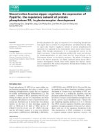
Figure 1 displays the treatment plan for a 18 months old
boy (case #27) with primitive neuroectodermal tumor
(PNET) of the right thoracic wall. He received chemother-
apy according to the Euro Ewing 99 protocol followed by
tumor resection with positive pathological margins. Post-
operative IMRT was delivered in order to decrease the
dose to the nearby spinal cord and lungs with a median
prescribed dose of 30.6 Gy to the PTV and 41.4 Gy to the
boost. During the radiation course regular CT-scans with
an in-room CT-Scanner were performed to confirm cor-
rect patient position. Thirty-eight months after finishing
treatment he underwent surgery for straightening of tho-
racic scoliosis. This occurred inspite of inclusion of the
complete vertebral body in the PTV. An asymmetric
growth of the thoracic wall is a possible explanation for
this.
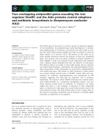
Figure 2 shows the IMRT plan for a 14 year old girl (case
# 1) with a Ewing's sarcoma of the left orbit, infiltrating
the dura mater and the left ethmoid sinus. The patient
received multiagent chemotherapy (7 cycles VIDE (vinc-
ristine, ifosfamide, doxorubicin, etoposide) followed by 6
cycles VAC (vincristine, adriamycin, cyclophosphamide))
and tumor resection (R1) prior to IMRT treatment. IMRT
was delivered in order to spare the lacrimal gland, optic
nerve and eyeball. At present, there are no signs of tumor
recurrence with an actuarial follow up of four and a half
years. Visual acuity is 1.0 on both eyes, though the patient
developed slight enophthalmia on the treated site.
local control and survival
Local failure occurred in 10 of 31 patients (table 2), time
to local failure was 4 - 53 months. In the event of local
tumor progression patients received chemotherapy or sur-
gical tumor resection, one patient with carcinoma of the
hypopharynx was reirradiated using IMRT (case #31). No
local relapse occurred among patients with juvenile
nasopharyngeal fibroma and esthesioneuroblastoma. So
far, 5 patients died due to distant metastases (cases #30,
#31, #21, #5, #4).
Discussion
We present a very heterogeneous group of children and
adolescents with 20 different tumor entities. All of these
31 patients have a very complex oncological constellation
IMRT-Plan for treatment of a 1.5 year old boy with a primitive neuroectodermal tumor (PNET) of the right thoracic wallFigure 1
IMRT-Plan for treatment of a 1.5 year old boy with a primitive neuroectodermal tumor (PNET) of the right
thoracic wall. A: A prescribed dose of 30.6 Gy to the PTV. B: 41.4 Gy prescribed to the boost. IMRT-Plan in colour wash
shows the 90% isodose region (dotted line).
Radiation Oncology 2009, 4:37 />Page 6 of 10
(page number not for citation purposes)
in common that made the application of a sufficient radi-
ation dose extremely difficult with conventional radio-
therapy techniques. Here the possible benefits of IMRT
like the sparing of organs at risk and the possibility of dose
escalation were considered to be more important for the
treatment success than the potentially increased risk of
secondary malignancies. We tried to increase chances of
cure the patients accepting possible risks in a matter of
decades in case of success. IMRT was feasible even if
anaesthesia was necessary and resulted in good local con-
trol rates for this group of children who represents a selec-
tion of extraordinary and difficult cases.
IMRT-plan for treatment of a 14 year old girl with Ewing sarcoma of the left orbit with a median prescribed dose of 54 GyFigure 2
IMRT-plan for treatment of a 14 year old girl with Ewing sarcoma of the left orbit with a median prescribed
dose of 54 Gy. A: Axial view of the dose distribution in colour wash shows the 90% isodose region (dotted line). B: Coronal
view of the dose distribution with sparing of the eye.
Table 2: Local failure after IMRT
Case Diagnosis Time to local failure [months] Dose
[Gy]
Treatment following failure
2 Ewing sarcoma 7 45 chemotherapy
4 Ewing sarcoma 9 54 chemotherapy
6 myoepithelial Parotis-carcinoma 7 66 surgery
8 Meningeoma 53 57.6 surgery
9 Desmoid tumor 14 54 surgery
11 Angiofibromatous tumor 7 56 surgery
15 Optic nerve glioma 36 50 surgery
21 Rhabdomyosarcoma 8 21.6 chemotherapy
23 Rhabdomyosarcoma 29 45 chemotherapy
31 Hypopharynx-Carcinoma 4 60 Re-irradiation (IMRT)
Radiation Oncology 2009, 4:37 />Page 7 of 10
(page number not for citation purposes)
IMRT could be applied with only few low grade acute tox-
icities and hardly any long term side effects so far. It is
important to note that the follow up is still quite short to
assess secondary malignancies. This radiotherapy tech-
nique allows reirradiations in difficult localisation that
could not be performed safely before.
In contrast to the big amount of publications in treating
adult patients with IMRT, there is only few data in litera-
ture about the use of IMRT in the paediatric population.
Good experiences with the treatment of twenty-two chil-
dren with IMRT have been reported by Bhatnagar et al.
[23]. They described substantial sparing of surrounding
critical structures in cranial, abdominopelvic or spinal
lesions, altogether a selection of very difficult oncological
situations. Conventional treatment technologies would
have resulted in a markedly higher dose to organs at risk
or would have required compromises regarding the possi-
ble target dose.
Penagaricano et al. summarized their experience of 5 chil-
dren treated with IMRT with a high degree of conformality
[24]. The dose distribution could be adapted to arc shaped
volumes in contrast to conventional therapy where
treated volumes are usually box shaped and encompass
big areas of treated normal tissue. Similar conclusions are
drawn by Paulino et al. in their synopsis of this method
for children [24,25]. They summarize that IMRT is a valu-
able alternative to conventional treatment techniques for
paediatric cancer patients. The improved dose distribu-
tions coupled with the ease of delivery of the IMRT fields
make this technique very attractive, especially in view of
the potential to increase local control and possibly
improve on survival. A third survey of a heterogeneous
group of children treated with IMRT is given by Teh et al.
within a general article about decreased treatment related
morbidity with IMRT [26]. Experiences with 185 patients
treated with IMRT in general are presented, among these
forty children suffering from different tumors. Similar to
the conclusions by the authors described before they con-
clude that IMRT offers new options in escalating dose and
achieving better local control while simultaneously reduc-
ing toxicity.
Besides these compilations of composed cohorts a larger
number of articles provides data on special indications
and more predefined collectives. They specially deal with
intracranial or head-and-neck tumors since the sensitive
structures like eyes, brain stem, parotid glands or inner
ears represent an extraordinary challenge in the radiother-
apeutic management. Starting with the biggest of all cen-
tral nervous treatments the irradiation of the entire
craniospinal axis as required in medulloblastoma or ger-
minoma can be done with improved conformity and spar-
ing of sensitive structures as shown by Penagaricano et al.
[27]. In a retrospective planning evaluation they illustrate
the possibilities of helical tomotherapy (as one solution
of IMRT) to cover a target volume of this size avoiding the
problems of field junctions and the resulting dangers of
under or overdosage inherent in conventional techniques.
After treating the whole craniospinal axis the primary
tumor region is supposed to be irradiated with an extra
boost to the posterior fossa. Huang et al. describe reduced
ototoxicity when sparing the inner ear by IMRT compared
to conventional radiotherapy, where the cochlear region
receives the full therapeutic dose [28]. Thirteen percent of
the IMRT Group had grade 3 or 4 hearing loss, compared
to 64% of the conventional-RT group. The sparing of the
hearing apparatus is of special importance since several
modern combined chemotherapy regimens contain oto-
toxic agents like cisplatinum. Jain et al. showed that this
improvement of ototoxicity was not achieved at the cost
of increased neuropsychological changes [29].
Another challenging situation in that IMRT might sub-
stantially improve the treatment is retinoblastoma. Krasin
et al. presented a planning study comparing different con-
ventional photon, electron and IMRT techniques in the
treatment of intraocular retinoblastoma [30]. The best
sparing of the bony orbit was achieved with IMRT yielding
a promising potential of avoiding asymmetrical bone
growth after successful radiotherapy. The mean volume of
bony orbit treated with IMRT above 20 Gy (as a threshold
of bone growth disturbance) was 60% in contrast to 90%
in conventional technique. Schroeder et al. report on 22
children with localized intracranial ependymoma treated
with IMRT. They were able to achieve a three year local
control of 68% while enabling minimal rates of toxicity
(no visual or hearing impairment, no necrosis, no myeli-
tis) [31].
The irradiation of head-and-neck tumors is quite rare in
children. Nevertheless long term toxicity is a huge concern
and often impairs the quality of life. Special focus here is
xerostomia caused by a fibrotic atrophy of the parotid
glands. Consecutive dental damage, dysphagia, problems
of speach and taste are feared. In a study by Wolden et al.
twenty-eight patients with head-and-neck rhabdomyosar-
coma were treated with IMRT. The age ranged from 1-29
years, the thee year local control was 95% with minimal
side effects [9]. In a similar approach by the groups of
Atlanta (20 children) and Houston (19 children) head-
and-neck rhabdomyosarcomas could be treated with a 3
year local control of 100% and a four year local control of
92.9% respectively [32,33]. Combs et al. presented a
cohort of 19 children with rhabdomyosarcoma treated
with stereotactic radiotherapy (n = 14) or IMRT (n = 5)
[34]. The three and five-year local control rate was 89%,
no toxicity > CTC grade 2 were observed. An Indian anal-
ysis of IMRT for nasopharyngeal cancer (19 children)
Radiation Oncology 2009, 4:37 />Page 8 of 10
(page number not for citation purposes)
showed reduced toxicity in terms of xerostomia, skin reac-
tion and mucous membrane reaction compared to con-
ventional radiotherapy (17 children) [35]. Acute
xerostomia grade 2 occurred in 31.6% in IMRT vs. 88.2%
in conventional radiotherapy. Grade 2 dysphagia was also
significantly reduced with 42.0% vs. 94.1%. IMRT was
also able to provide superior target coverage and as a con-
sequence of the reduced toxicity an improved compliance.
Juvenile angiofibroma can be cured by radiotherapy in
unresectable or relapsing cases. They are difficult to treat
for because of the same surrounding risk structures as dis-
cussed above. Especially with respect to the benign nature
of these tumors a well balanced toxicity profile is vital as
described by Kuppersmith et al. and can be achieved by
the means of IMRT [36].
Another potential indication is the radiosurgical treat-
ment of arteriovenous malformations (avm). Lesions that
are unresectable and not accessible for interventional neu-
roradiology can be obliterated by high dose single course
radiotherapy. Fuss et al. presented the possibilities of
IMRT in seven children with avm of complex shape, that
could hardly be treated with conventional methods [37].
Two avm obliterated completely, three partially, while no
treatment related side effects occurred.
In the discussions about precautions of IMRT in children
the advantages are achieved at the cost of raised low dose
outside the target. With a higher number of monitor units
required the total body dose can increase significantly
[38]. However, in a study by Koshy et al. no increased
extra target dose to thyroid, breast, and testis was seen in
children treated with IMRT compared with a control
group of children treated with conventional radiotherapy
for cranial and abdominopelvic tumors [39].
The methods that allow the intensity modulation of the
radiation beams increase the volume of tissue receiving
low dose compared to conventional radiotherapy [40].
The effects in adult patients are the same, however, there
are 3 reasons for special consideration in the treatment of
children: higher sensitivity to radiation induced cancer,
relation of scattered dose to the small body volume and
genetic susceptibility due to germline mutations [18,41-
45]. While high dose to neighbouring structures can be
selectively decreased by the means of IMRT, low dose is
distributed in the rest of the body. Consequences of this
special treatment technique can only be estimated until
now.
Data of the childhood cancer survivor study (CCSS)
showed 5 year survival rates of 79% for all different tumor
entities [46]. With such a high number of long term survi-
vors secondary neoplasms become highly relevant. The
risk is especially increased in patients of very young age,
Hodgkin's disease, treatment with alkylating agents, radi-
ation therapy and female gender [47,48].
Secondary cancer induction is dose dependent and tissue
irradiated with doses below 6 Gy is known to be especially
endangered to develop secondary cancer [49]. The calcu-
lated risk of secondary malignancies after treatment with
IMRT was estimated to be doubled [17,19]. It is important
to note that these numbers are only estimations and cal-
culations with no fundament of clinical data due to the
lack of enough follow-up time. In addition integral dose
is often discussed to be potentially higher in IMRT com-
pared to conventional radiotherapy. This is not necessar-
ily true since the high dose region to normal tissue is
markedly reduced with the improved conformity [50]. As
stated above the characteristic new feature of dose expo-
sure in IMRT is a shift towards low dose spread out. Espe-
cially in the tissues with a high incidence of secondary
cancers the ability of IMRT to produce conformal avoid-
ance of these structures might limit the risk of these late
effects. Techniques like helical tomotherapy have the
potential of selectively sparing the thyroid gland and
breast tissue in craniospinal irradiation.
The number of children treated with IMRT and the hard
evidence for the benefit of this technology is limited [13].
However, waiting for this evidence would last for many
years. Many of the uncertainties cannot be answered by
simply transferring the standards of evidence based med-
icine in medical oncology one by one to radiation oncol-
ogy. Randomizing children or adults in two different
radiotherapy regimens knowing that one will definitely
inactivate the parotid glands, one kidney or affect bone
growth is simply unethical. Withholding children the pos-
sibility to reduce doses to organs at risk in difficult cases is
hard to justify. As long as proton treatment with its great
potential of decreased integral dose is not widely availa-
ble, IMRT provides an excellent tool in difficult situations.
Patient selection is absolutely crucial with regard to the
worries about potentially increased chances of secondary
malignancies. Reserved for complex cases with close prox-
imity of organs at risk IMRT represents a powerful and ver-
satile treatment option when used with the necessary
caution [25,51].
Conclusion
Intensity modulated radiotherapy is a feasible method of
radiotherapy for paediatric malignancies. It was applied
safely in 31 patients within the last eight years in difficult
oncologic situations. Conventional radiotherapy would
have been associated with limited dose to the target or
high normal tissue complication probability. In all the
presented patients it was decided that the benefit of
increased tumor control probabilities and improved spar-
Radiation Oncology 2009, 4:37 />Page 9 of 10
(page number not for citation purposes)
ing of organs at risk had a higher clinical impact than the
calculated increased risk of late side-effects.
As long as the risk of secondary cancer induction can only
be estimated IMRT for children should only be used with
caution. Longer follow up time is needed to quantify this
long term complication. Conventional radiotherapy
remains the standard of care in radiation oncology for
children and can be delivered with acceptable toxicity in
the majority of children.
Nevertheless, reserved to special cases with close proxim-
ity of sensitive structures, it can provide great benefit for
paediatric patients and should not be withheld because of
estimations based on a radiobiological model. It widens
the therapeutic window and reduces long term toxicity for
an increased number of long term cancer survivors.
Declaration of competing interests
The authors declare that they have no competing interests.
Authors' contributions
FS is responsible for data acquisition, literature research
and writing of the manuscript. ES is responsible for data
acquisition, statistical analysis and writing of the manu-
script. SN is responsible for the physical aspects of IMRT
planning and treatment of the children. HB is responsible
for the anaesthesia management of the children. PH is
responsible for the clinical treatment of the children as
head of the division of radiation oncology in the German
Cancer Research Center. JD is responsible for the clinical
treatment of the children as of the department of radia-
tion oncology in the University of Heidelberg. MM is
responsible for the medical aspects of treatment planning
and application, idea for this paper, literature research
and proof reading. All authors read and approved the final
manuscript.
Acknowledgements
The work was supported by the German Research foundation (DFG) and
the University of Heidelberg, Germany, through a young investigator
award.
References
1. Intensity Modulated Radiation Therapy Collaborative Working
Group: Intensity-modulated radiotherapy: current status and
issues of interest. Int J Radiat Oncol Biol Phys 2001, 51:880-914.
2. Eisbruch A: Clinical aspects of IMRT for head-and-neck can-
cer. Med Dosim 2002, 27:99-104.
3. Nutting C, Dearnaley DP, Webb S: Intensity modulated radiation
therapy: a clinical review. Br J Radiol 2000, 73:459-469.
4. Pirzkall A, Carol M, Lohr F, Hoss A, Wannenmacher M, Debus J:
Comparison of intensity-modulated radiotherapy with con-
ventional conformal radiotherapy for complex-shaped
tumors. Int J Radiat Oncol Biol Phys 2000, 48:1371-1380.
5. Zhen W, Thompson RB, Enke CA: Intensity-modulated radiation
therapy (IMRT): the radiation oncologist's perspective. Med
Dosim 2002, 27:155-159.
6. Munter MW, Nill S, Thilmann C, Hof H, Hoss A, Haring P, Partridge
M, Manegold C, Wannenmacher M, Debus J: Stereotactic inten-
sity-modulated radiation therapy (IMRT) and inverse treat-
ment planning for advanced pleural mesothelioma.
Strahlenther Onkol 2003, 179:535-541.
7. Schulz-Ertner D, Didinger B, Nikoghosyan A, Jakel O, Zuna I, Wan-
nenmacher M, Debus J: Optimization of radiation therapy for
locally advanced adenoid cystic carcinomas with infiltration
of the skull base using photon intensity-modulated radiation
therapy (IMRT) and a carbon ion boost. Strahlenther Onkol
2003, 179:345-351.
8. Munter MW, Debus J, Hof H, Nill S, Haring P, Bortfeld T, Wannen-
macher M: Inverse treatment planning and stereotactic inten-
sity-modulated radiation therapy (IMRT) of the tumor and
lymph node levels for nasopharyngeal carcinomas. Descrip-
tion of treatment technique, plan comparison, and case
study. Strahlenther Onkol 2002, 178:517-523.
9. Wolden SL, Wexler LH, Kraus DH, Laquaglia MP, Lis E, Meyers PA:
Intensity-modulated radiotherapy for head-and-neck rhab-
domyosarcoma. Int J Radiat Oncol Biol Phys 2005, 61:1432-1438.
10. Studer G, Lutolf UM, Davis JB, Glanzmann C: IMRT in Hypopha-
ryngeal Tumors. Strahlenther Onkol 2006, 182:331-335.
11. Cavey ML, Bayouth JE, Colman M, Endres EJ, Sanguineti G:
IMRT to
escalate the dose to the prostate while treating the pelvic
nodes. Strahlenther Onkol 2005, 181:431-441.
12. Guckenberger M, Flentje M: Intensity-Modulated Radiotherapy
(IMRT) of Localized Prostate Cancer: A Review and Future
Perspectives. Strahlenther Onkol 2007, 183:57-62.
13. Veldeman L, Madani I, Hulstaert F, De Meerleer G, Mareel M, De
Neve W: Evidence behind use of intensity-modulated radio-
therapy: a systematic review of comparative clinical studies.
Lancet Oncol 2008, 9:367-375.
14. Sterzing F, Schubert K, Sroka-Perez G, Kalz J, Debus J, Herfarth K:
Helical Tomotherapy: Experiences of the First 150 Patients
in Heidelberg. Strahlenther Onkol 2008, 184:8-14.
15. Boyer A, Xing L, Luxton G, Chen Y, Ma C: IMRT by dynamic MLC.
In The Use of Computers in Radiation therapy, XIIIth International Confer-
ence, Heidelberg, Germany, May 22-25 2000 Edited by: Schlegel W,
Bortfeld T. Berlin: Springer; 2000:160-163.
16. Mackie TR, Balog J, Ruchala K, Shepard D, Aldridge S, Fitchard E,
Reckwerdt P, Olivera G, McNutt T, Mehta M: Tomotherapy. Semin
Radiat Oncol 1999, 9:108-117.
17. Hall EJ, Wuu CS: Radiation-induced second cancers: the
impact of 3D-CRT and IMRT. Int J Radiat Oncol Biol Phys 2003,
56:83-88.
18. Hall EJ: Intensity-modulated radiation therapy, protons, and
the risk of second cancers. Int J Radiat Oncol Biol Phys 2006, 65:1-7.
19. Kry SF, Salehpour M, Followill DS, Stovall M, Kuban DA, White RA,
Rosen II: The calculated risk of fatal secondary malignancies
from intensity-modulated radiation therapy. Int J Radiat Oncol
Biol Phys 2005, 62:1195-1203.
20. Schneider U, Lomax A, Pemler P, Besserer J, Ross D, Lombriser N,
Kaser-Hotz B: The impact of IMRT and proton radiotherapy
on secondary cancer incidence. Strahlenther Onkol 2006,
182:647-652.
21. Mazonakis M, Zacharopoulou F, Kachris S, Varveris C, Damilakis J,
Gourtsoyiannis N: Scattered dose to gonads and associated
risks from radiotherapy for common pediatric malignancies:
a phantom study. Strahlenther Onkol 2007, 183:332-337.
22. Rhein B, Haring P, Debus J, Schlegel W: [Dosimetric verification
of IMRT treatment plans at the German Cancer Research
Center (DKFZ)]. Z Med Phys 2002, 12:122-132.
23. Bhatnagar A, Deutsch M: The Role for Intensity Modulated
Radiation Therapy (IMRT) in Pediatric Population. Technol
Cancer Res Treat 2006, 5:591-596.
24. Penagaricano JA, Papanikolaou N, Yan Y, Ratanatharathorn V: Appli-
cation of intensity-modulated radiation therapy for pediatric
malignancies. Med Dosim 2004, 29:247-253.
25. Paulino AC, Skwarchuk M: Intensity-modulated radiation ther-
apy in the treatment of children. Med Dosim 2002, 27:115-120.
26. Teh BS, Mai WY, Grant WH 3rd, Chiu JK, Lu HH, Carpenter LS, Woo
SY, Butler EB: Intensity modulated radiotherapy (IMRT)
decreases treatment-related morbidity and potentially
enhances tumor control. Cancer Invest 2002, 20:437-451.
27. Penagaricano JA, Yan Y, Corry P, Moros E, Ratanatharathorn V: Ret-
rospective evaluation of pediatric cranio-spinal axis irradia-
tion plans with the Hi-ART tomotherapy system. Technol
Cancer Res Treat 2007, 6:355-360.
Publish with BioMed Central and every
scientist can read your work free of charge
"BioMed Central will be the most significant development for
disseminating the results of biomedical research in our lifetime."
Sir Paul Nurse, Cancer Research UK
Your research papers will be:
available free of charge to the entire biomedical community
peer reviewed and published immediately upon acceptance
cited in PubMed and archived on PubMed Central
yours — you keep the copyright
Submit your manuscript here:
/>BioMedcentral
Radiation Oncology 2009, 4:37 />Page 10 of 10
(page number not for citation purposes)
28. Huang E, Teh BS, Strother DR, Davis QG, Chiu JK, Lu HH, Carpenter
LS, Mai WY, Chintagumpala MM, South M, Grant WH 3rd, Butler EB,
Woo SY: Intensity-modulated radiation therapy for pediatric
medulloblastoma: early report on the reduction of ototoxic-
ity. Int J Radiat Oncol Biol Phys 2002, 52:599-605.
29. Jain N, Krull KR, Brouwers P, Chintagumpala MM, Woo SY: Neu-
ropsychological outcome following intensity-modulated
radiation therapy for pediatric medulloblastoma. Pediatr Blood
Cancer 2008, 51:275-279.
30. Krasin MJ, Crawford BT, Zhu Y, Evans ES, Sontag MR, Kun LE, Mer-
chant TE: Intensity-modulated radiation therapy for children
with intraocular retinoblastoma: potential sparing of the
bony orbit. Clin Oncol (R Coll Radiol) 2004, 16:215-222.
31. Schroeder TM, Chintagumpala M, Okcu MF, Chiu JK, Teh BS, Woo
SY, Paulino AC: Intensity-modulated radiation therapy in
childhood ependymoma. Int J Radiat Oncol Biol Phys 2008,
71:987-993.
32. McDonald MW, Esiashvili N, George BA, Katzenstein HM, Olson TA,
Rapkin LB, Marcus RB Jr: Intensity-Modulated Radiotherapy
with Use of Cone-Down Boost for Pediatric Head-and-Neck
Rhabdomyosarcoma. Int J Radiat Oncol Biol Phys 2008,
72(3):884-891.
33. Curtis AE, Okcu MF, Chintagumpala M, Teh BS, Paulino AC: Local
Control After Intensity-Modulated Radiotherapy for Head-
and-Neck Rhabdomyosarcoma. Int J Radiat Oncol Biol Phys 2008,
73(1):173-177.
34. Combs SE, Behnisch W, Kulozik AE, Huber PE, Debus J, Schulz-Ertner
D: Intensity Modulated Radiotherapy (IMRT) and Fraction-
ated Stereotactic Radiotherapy (FSRT) for children with
head-and-neck-rhabdomyosarcoma. BMC Cancer 2007, 7:177.
35. Laskar S, Bahl G, Muckaden M, Pai SK, Gupta T, Banavali S, Arora B,
Sharma D, Kurkure PA, Ramadwar M, Viswanathan S, Rangarajan V,
Qureshi S, Deshpande DD, Shrivastava SK, Dinshaw KA: Nasopha-
ryngeal Carcinoma in Children: Comparison of Conven-
tional and Intensity-Modulated Radiotherapy. Int J Radiat Oncol
Biol Phys 2008, 72(3):728-736.
36. Kuppersmith RB, Teh BS, Donovan DT, Mai WY, Chiu JK, Woo SY,
Butler EB: The use of intensity modulated radiotherapy for
the treatment of extensive and recurrent juvenile angiofi-
broma. Int J Pediatr Otorhinolaryngol 2000, 52:
261-268.
37. Fuss M, Salter BJ, Caron JL, Vollmer DG, Herman TS: Intensity-
modulated radiosurgery for childhood arteriovenous malfor-
mations. Acta Neurochir (Wien) 2005, 147:1141-1149.
38. Verellen D, Vanhavere F: Risk assessment of radiation-induced
malignancies based on whole-body equivalent dose esti-
mates for IMRT treatment in the head and neck region. Radi-
other Oncol 1999, 53:199-203.
39. Koshy M, Paulino AC, Marcus RB Jr, Ting JY, Whitaker D, Davis LW:
Extra-target doses in children receiving multileaf collimator
(MLC) based intensity modulated radiation therapy (IMRT).
Pediatr Blood Cancer 2004, 42:626-630.
40. Welsh JS, Limmer JP, Howard SP, Diamond D, Harari PM, Tome W:
Precautions in the use of intensity-modulated radiation ther-
apy. Technol Cancer Res Treat 2005, 4:203-210.
41. Hall EJ: The inaugural Frank Ellis Lecture latrogenic cancer:
the impact of intensity-modulated radiotherapy. Clin Oncol (R
Coll Radiol) 2006, 18:277-282.
42. Blatt J, Olshan A, Gula MJ, Dickman PS, Zaranek B: Second malig-
nancies in very-long-term survivors of childhood cancer. Am
J Med 1992, 93:57-60.
43. Gold DG, Neglia JP, Dusenbery KE: Second neoplasms after
megavoltage radiation for pediatric tumors. Cancer 2003,
97:2588-2596.
44. Paulino AC, Fowler BZ: Secondary neoplasms after radiother-
apy for a childhood solid tumor. Pediatr Hematol Oncol 2005,
22:89-101.
45. Lin HM, Teitell MA: Second malignancy after treatment of
pediatric Hodgkin disease. J Pediatr Hematol Oncol 2005,
27:28-36.
46. Robison LL: Treatment-associated subsequent neoplasms
among long-term survivors of childhood cancer: the experi-
ence of the Childhood Cancer Survivor Study. Pediatr Radiol
2009, 39(Suppl 1):S32-37.
47. Armstrong GT, Liu Q, Yasui Y, Neglia JP, Leisenring W, Robison LL,
Mertens AC: Late mortality among 5-year survivors of child-
hood cancer: a summary from the Childhood Cancer Survi-
vor Study. J Clin Oncol 2009, 27:2328-2338.
48. Nguyen F, Rubino C, Guerin S, Diallo I, Samand A, Hawkins M, Ober-
lin O, Lefkopoulos D, De Vathaire F: Risk of a second malignant
neoplasm after cancer in childhood treated with radiother-
apy: correlation with the integral dose restricted to the irra-
diated fields. Int J Radiat Oncol Biol Phys 2008, 70:908-915.
49. Dorr W, Herrmann T: Second primary tumors after radiother-
apy for malignancies. Strahlenther Onkol 2002, 178:357-362.
50. Parker W, Filion E, Roberge D, Freeman CR: Intensity-modulated
radiotherapy for craniospinal irradiation: target volume con-
siderations, dose constraints, and competing risks. Int J Radiat
Oncol Biol Phys 2007, 69:251-257.
51. Rembielak A, Woo TC: Intensity-modulated radiation therapy
for the treatment of pediatric cancer patients. Nat Clin Pract
Oncol 2005, 2:211-217.