Báo cáo khoa học: " Late treatment with imatinib mesylate ameliorates radiation-induced lung fibrosis in a mouse model" docx
Bạn đang xem bản rút gọn của tài liệu. Xem và tải ngay bản đầy đủ của tài liệu tại đây (998.37 KB, 9 trang )
BioMed Central
Page 1 of 9
(page number not for citation purposes)
Radiation Oncology
Open Access
Research
Late treatment with imatinib mesylate ameliorates
radiation-induced lung fibrosis in a mouse model
Minglun Li
1,5
, Amir Abdollahi
1,4
, Hermann-Josef Gröne
2
,
Kenneth E Lipson
3,6
, Claus Belka
5
and Peter E Huber*
1,4
Address:
1
Department of Radiation Oncology German Cancer Research Center (DKFZ), Im Neuenheimer Feld 280, Heidelberg 69120, Germany,
2
Molecular Pathology, German Cancer Research Center (DKFZ), Im Neuenheimer Feld 280, Heidelberg 69120, Germany,
3
3M Pharmaceuticals,
St. Paul, MN 55144, USA,
4
Department of Radiation Oncology, University of Heidelberg Medical School, Heidelberg 69120, Germany,
5
Department of Radiation Oncology, University of Munich medical school, Munich 81377, Germany and
6
Fibrogen, Inc., South San Francisco,
CA 94080, USA
Email: Minglun Li - ; Amir Abdollahi - ; Hermann-Josef Gröne - h ;
Kenneth E Lipson - ; Claus Belka - ; Peter E Huber* -
* Corresponding author
Abstract
Background: We have previously shown that small molecule PDGF receptor tyrosine kinase
inhibitors (RTKI) can drastically attenuate radiation-induced pulmonary fibrosis if the drug
administration starts at the time of radiation during acute inflammation with present but limited
effects against acute inflammation. To rule out interactions of the drug with acute inflammation, we
investigated here in an interventive trial if a later drug administration start at a time when the acute
inflammation has subsided - has also beneficial antifibrotic effects.
Methods: Whole thoraces of C57BL/6 mice were irradiated with 20 Gy and treated with the RTKI
imatinib starting either 3 days after radiation (during acute inflammation) or two weeks after
radiation (after the acute inflammation has subsided as demonstrated by leucocyte count). Lungs
were monitored and analyzed by clinical, histological and in vivo non-invasive computed tomography
as a quantitative measure for lung density and lung fibrosis.
Results: Irradiation induced severe lung fibrosis resulting in markedly reduced mouse survival vs.
non-irradiated controls. Both early start of imatinib treatment during inflammation and late imatinib
start markedly attenuated the development of pulmonary fibrosis as demonstrated by clinical,
histological and qualitative and quantitative computed tomography results such as reduced lung
density. Both administration schedules resulted in prolonged lifespans. The earlier drug treatment
start resulted in slightly stronger beneficial antifibrotic effects along all measured endpoints than
the later start.
Conclusions: Our findings show that imatinib, even when administered after the acute
inflammation has subsided, attenuates radiation-induced lung fibrosis in mice. Our data also indicate
that the fibrotic fate is not only determined by the early inflammatory events but rather a complex
process in which secondary events at later time points are important. Because of the clinical
availability of imatinib or similar compounds, a meaningful attenuation of radiation-induced lung
fibrosis in patients seems possible.
Published: 21 December 2009
Radiation Oncology 2009, 4:66 doi:10.1186/1748-717X-4-66
Received: 24 August 2009
Accepted: 21 December 2009
This article is available from: />© 2009 Li et al; licensee BioMed Central Ltd.
This is an Open Access article distributed under the terms of the Creative Commons Attribution License ( />),
which permits unrestricted use, distribution, and reproduction in any medium, provided the original work is properly cited.
Radiation Oncology 2009, 4:66 />Page 2 of 9
(page number not for citation purposes)
Background
Radiotherapy is a mainstay of treating neoplasm in lungs
[1-4]. Fibrosis is the main chronic side effect of radiother-
apy that potentially prevents to deliver the necessary dose
to benefit cancer patients [5-7]. Although modern tech-
niques of radiotherapy (stereotactic radiotherapy, intra-
operative radiotherapy, interstitial brachytherapy etc.) are
now increasingly used to improve dose distribution and
reduce side effects, radiation induced fibrotic lesions still
occur [8-10]. There has been remarkably little progress in
the development of effective antifibrotic therapies [7,11].
Recent studies indicated that pulmonary fibrosis is not a
unique pathologic process but rather an excess of the
same biologic events involved in normal tissue repair with
persistent and exaggerated wound healing ultimately
leading to an excess of fibroblast replication and matrix
deposition [7,11]. A cascade of many cytokines leading to
lung fibrosis after radiation injury has been described
[12,13]. Recently, a number of new regulators in radia-
tion-induced lung injury such as intercellular adhesion
molecules (ICAM-1) and the CD95 ligand system have
been reported [14,15]. Typical fibrogenic mediators
include TGF-β, IL-1, TNF-α, bFGF, and thrombin, but also
PDGF has been implicated to demonstrate profibrotic
activities [16-21]. Thus the PDGF/PDGFR system can be
considered as a promising target for treating fibrotic dis-
eases [22-26].
Recently, we have shown that PDGF receptor tyrosine
kinase inhibitors (RTKI) including imatinib can attenuate
radiation-induced pulmonary fibrosis if the drug admin-
istration starts before the toxic event or within three days
after the insult [27]. PDGF RTKI prolonged survival and
protected mice from lung fibrosis, presented as reduced
lung density measured by computed tomography exami-
nations, although the radiation-induced acute inflamma-
tion was not significantly abrogated. Thus we
hypothesized that fibrogenesis is a separate process after
acute inflammation, correlated but not dependent to
acute inflammation [27].
However in this previous study PDGF RTKI (SU9518) was
administrated during RT-induced acute inflammation (1
day before and 3 days after radiation) showing no marked
but certain effects on the acute inflammation. Therefore,
acute inflammation is affected to some extent by concur-
rent drug administration and, even if the measurable
extent of inflammation had not been significantly
affected, the administration during inflammation could
still be a prerequisite for the later antifibrotic effects. Thus,
in the present study we chose a late drug treatment start-
ing at the time when the primary inflammation has sub-
sided, to rule out direct and indirect effects associated with
acute inflammation,
Therefore we report here the full results of the imatinib
experiments including the data on the late imatinib
administration arm starting two weeks after radiation, at a
time when the acute inflammation has already completely
subsided. These experiments thus investigate i) the role of
acute inflammation in the development of radiation
induced lung fibrosis, ii) if attenuation of the acute
inflammation is necessary to block fibrosis and iii) if a late
drug administration after radiation insult and after the
acute inflammation has subside still has antifibrotic
potential. To this end thorax of C57BL/6 mice were irradi-
ated with 20 Gy and mice were subsequently treated with
imatinib mesylate/Gleevec starting either three days after
radiation or two weeks after radiation. Longitudinal fol-
low-up of the mice lungs in vivo was performed by nonin-
vasive radiological monitoring using high resolution
computed-tomography [28].
Methods
Experimental protocol and animal model
All animal procedures were approved by institutional and
governmental authorities (Regierungspraesidium Karl-
sruhe, Germany). Fibrosis-prone mice (female C57BL/6J,
8 weeks old, approximate body weight 20 g, Charles River
Laboratories, Sulzfeld, Germany) were used. For thoracic
irradiation, mice were anesthetized by intraperitoneal
application of Domitor (Pfizer, Exton, USA; 0.2 mg/kg)
and Ketamin 10% (Park, Davis & Company, Berlin, Ger-
many; 100 mg/kg). Cobalt-60 gamma radiation (Siemens
Gammatron S, Erlangen, Germany) was administered as
single dose (20 Gy) to the entire thorax (0.441 Gy/min;
source surface distance: 0.7 m) using one standing field
anterior-posterior. Other organs, above and beyond the
thorax were shielded. Animals were supplied with diet
and water ad libitum.
Imatinib Mesylate was provided by SUGEN Inc. South San
Francisco, CA. To achieve clinically relevant doses [26,27],
Imatinib were formulated in standard mouse chow at 0.5
mg/g resulting in a dosage of 40 mg/kg/d. Imatinib
(Gleevec) is known to have high activity against three
kinases: Bcr/Abl, c-Kit, and PDGFR-α and -β [26,27]. In
irradiated animals, imatinib treatment was either started
three days after radiation or two weeks after radiation and
was continued until the end of observation, as stated in
our previous study [27]. Animals were checked three
times weekly, clinically examined and weighed.
Lung histology
Histological analysis from mice tissues was performed sys-
tematically at early and later time points after radiation as
described [27,28]. Briefly, lungs were fixed by intratra-
cheal instillation of 4% formalin, followed by overnight
fixation, embedding in paraffin, sectioned at 5 μm, and
stained with hematoxylin-eosin (H&E). The total count of
Radiation Oncology 2009, 4:66 />Page 3 of 9
(page number not for citation purposes)
leukocytes was determined by morphometric evaluation
(Q 600 Quantimet, Leica Microsystems, Wetzlar, Ger-
many) and the septal thickness was measured in 5 regions
of interest (ROI) for each mouse.
High-resolution computed tomography (HRCT) of mouse
lungs
To obtain an independent qualitative and quantitative
measure for lung fibrosis in the mice we used high-resolu-
tion computed tomography (CT). CT is the method of
choice for monitoring fibrosis in patients. This radiologi-
cal method allows non-invasive and repeated measure-
ments in the same mice in a longitudinal way [28]. CT
exams were performed in 5 randomly selected mice from
each group every second week during the entire observa-
tion period. CT images were captured on a Toshiba multi-
slice CT scanner (Aquilion 32). 120 kV with 100 mAS
were applied. 0.5 mm thin slices with 0.5 mm inter-slice
distance spanned the complete mouse chest (total acqui-
sition time 0.5 seconds). Multiplanar reconstructions
(MPR) were performed for semiquantitative analysis.
Hounsfield units (HU) of section slides from the upper
and lower lung region were determined. Eight regions of
interest (ROI) were defined in the following areas: the
right upper anterior and posterior regions, the left upper
anterior and posterior regions, the right lower anterior
and posterior regions and the left lower anterior and pos-
terior regions. Total arithmetic means ± SE of the HU were
calculated.
Statistics
Mouse survival curves after thoracic irradiation and imat-
inib treatments were calculated with the Kaplan-Meier
method and compared using the log-rank test. Other
quantitative data are given as mean values ± SD or as indi-
cated. For analysis of differences between the groups,
ANOVA followed by the appropriate post hoc test for indi-
vidual comparisons between the groups was performed.
All tests were two-tailed. P < 0.05 was considered statisti-
cally significant.
Results
Late imatinib interventive treatment prolongs mouse
survival
A single dose of 20 Gy thoracic irradiation induced
marked lung fibrosis and dramatically reduced animal
survival versus nonirradiated animals. Median survival
was 19 weeks after radiation vs. control mice which stayed
alive for more than one year (P < 0.0001). The Kaplan-
Meier curves are depicted in figure 1a. Imatinib treatment
starting 3 d after radiation prolonged median survival by
~11 weeks with a median survival of 30 weeks (P < 0.01
vs. radiation alone) as shown in a previous study [27].
Importantly to us, imatinib treatment starting 2 weeks
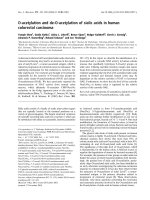
(a) Kaplan-Meier analysis of mouse survival following thoracic irradiation and imatinib treatmentFigure 1
(a) Kaplan-Meier analysis of mouse survival following thoracic irradiation and imatinib treatment. Death was
considered complete (cause-specific due to radiation) in all cases except those of planned euthanasia for histological assess-
ment, which were considered as censored. Radiation (20 Gy) reduced survival (P < 0.001 vs. the control as reported in (24)).
Imatinib treatment increased mouse survival if administration started as late as 2 weeks after radiation (P < 0.02 vs. radiation)
and if started early within 3 days after radiation (P < 0.01 as reported in (24)). The earlier start of drug treatment tended to be
more effective in prolonging survival than later start of drug treatment, but this difference was not significant (P > 0.1). (b)
Bodyweight follow-up after thoracic irradiation and imatinib treatment. Five mice were randomly selected in each group and
weighed every two weeks. Mean ± SE was presented. * P < 0.01 vs. the RT only group; # P < 0.01 vs. the control group.
Radiation Oncology 2009, 4:66 />Page 4 of 9
(page number not for citation purposes)
after radiation also prolonged median survival by ~8
weeks with a median survival of 27 weeks (P < 0.02 vs.
radiation alone). The difference in survival between ear-
lier and later treatment schedules was not statistically sig-
nificant (P > 0.1), but a tendency was present suggesting
that the earlier drug treatment start was beneficial.
To support the survival data we also analyzed the animals'
clinical status. We found that imatinib treatment in both
early and late treatment arms also attenuated radiation-
related clinical adverse effects such as weight loss (figure
1b, P < 0.02, at all time points after week 14) and other
clinical parameters, which were monitored weekly over
the entire observation period. In specific, imatinib early
and late improved clinical status including animal behav-
ior (worse after irradiation, improved by imatinib), tach-
ypnoea and heart rate (both higher after irradiation,
reduced by imatinib). Again, a marked difference of the
benefits caused by imatinib in the clinical animal param-
eters between the two imatinib schedules was not
observed.
Computed tomography of mice lungs
Computed tomography (CT) was used to obtain an inde-
pendent qualitative and quantitative measure of mice
lung fibrosis that could be repeated in the same animal
over time. As reported before [24], after week 16 typical
radiological features of lung fibrosis were visible after 20
Gy irradiation including irregular septal thickening,
patchy peripheral reticular abnormalities with intralobu-
lar linear opacities and subpleural honeycombing (figure
2a). The extent of fibrotic disease progression in CT
images correlated well with histology and clinical impair-
ment. Imatinib treatment was able to markedly reduce the
radiological/morphological signs of fibrosis after radia-
tion in both early and late imatinib treated mice. In addi-
tion to the morphological assessment, CT enabled
quantitation of fibrosis by an assessment of the lung den-
sity (quantified in Hounsfield units (HU)). We found that
lung density drastically increased during weeks 12 to 24
post radiotherapy (figure 2b) in irradiated mice only.
Imatinib in both early and late application arms strongly
inhibited this increase by approximately 50% (P < 0.001).
The earlier therapy start appeared to be slightly more
effective in reducing CT signs of lung fibrosis than a later
therapy, but this tendency did not reach statistical signifi-
cance (P > 0.1).
Histological assessment of lung fibrosis after irradiation
It is assumed that exposure of normal lung tissue to irra-
diation has two well-recognized adverse effects: acute/
subacute pneumonitis and fibrosis as long term sequelae
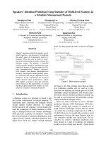
(a) High resolution computed tomography (CT) as a non-invasive tool for qualitative and quantitative longitudinal monitoring of pulmonary fibrosis progression in miceFigure 2
(a) High resolution computed tomography (CT) as a non-invasive tool for qualitative and quantitative longitu-
dinal monitoring of pulmonary fibrosis progression in mice. Representative CT scans showing progression of pulmo-
nary fibrosis in mice after 20 Gy whole thorax irradiation (RT), and treatment with imatinib treatment starting 3 days and 2
weeks after radiation (RT). Fibrosis is characterized by diffuse bilateral areas of "ground-glass" attenuation and intralobular
reticular opacities. (b) Quantitative lung density values derived from CT scans. The same 5 randomly chosen mice in each
treatment group were examined in a longitudinal study by CT every 2 weeks. 8 regions of interest (ROI) were randomly
selected in the lungs and the lung density (in Hounsfield Units (HU)) was determined for each ROI. Mean ± SE are presented. *
P < 0.01 vs. the RT only group; # P < 0.01 vs. the control group.
Radiation Oncology 2009, 4:66 />Page 5 of 9
(page number not for citation purposes)
[11,27-29]. To better understand the pathogenesis of the
radiation-induced lung fibrosis process, and to evaluate
the modulation after radiation and imatinib, mice were
selected for analysis of leukocyte infiltration, edema and
collagen-deposition with associated thickening of the
alveolar septum.
As described earlier a biphasic radiation response was
observed, initially consisting of acute and subacute pneu-
monitis, which was followed by the onset of fibrogenesis
[27]. The characteristic histologic findings in the pneumo-
nitis phase of the radiation response were prominent
inflammatory cell infiltrates in the alveoli and lung inter-
stitium with simultaneous interstitial edema (figure 3a).
Both parameters exhibited similar kinetics in the acute
phase, reaching their maxima about 72 hours after radia-
tion injury. After the acute radiation response, leukocyte
count spontaneously subsided within one week (figure
4a). When imatinib administration started early during
inflammation the treatment did not markedly decrease
this first, radiation-induced acute leukocyte peak (P >
0.2), although imatinib affected the inflammation to
some extent and, the tendency was seen that imatinib had
a nonsignificant tendency to reduce acute inflammation
in terms of reduced edema and leucocyte count.
Histological analysis of irradiated lungs further showed
the development of fibrosis by progressive collagen depo-
sition after week 12 (figure 3b). This fibrogenesis phase
was characterized by development of typical fibroblast
foci and exuberant deposition of extracellular matrix in
irradiated lungs (figure 3c). Both imatinib schedules
reduced collagen deposition and septal thickness (figure
4b), while the early administration appeared to be slightly
more effective than late administration. In irradiated
mice, the later fibrogenesis phase was accompanied by a
strong second onset of leukocyte infiltration that began
several weeks after irradiation and reached a peak at
approximately 20 weeks post irradiation.
At later time points (> 20 weeks) the fibrotic foci evolved
and coalesced into widespread fibrosis with remodeling
of the lung architecture. Moreover, in the irradiated lungs
the second onset of progressive fibrosis-related leukocyte
infiltration persisted until the morphologically described
fibrosis process was completed (after week 26). Figure 4a
also shows that this second inflammatory response was
also reduced in terms of reduced leucocyte count by both
early and late imatinib treatment (P < 0.05). Here again,
the earlier drug treatment start tended to be slightly more
effective than the later treatment start, but this difference
was not statistically significant (P > 0.5).
Discussion
Here we confirm that imatinib (Gleevec
©
) treatment is an
effective strategy to attenuate radiation-induced lung
fibrosis in mice. The beneficial drug effects are present
even when drug administration starts two weeks after
radiation when the acute radiation associated inflamma-
tion is completely subsided. The antifibrotic drug effectiv-
ity after the radiation-induced inflammation suggests that
the fibrotic fate after radiation is not completely deter-
mined by the early inflammatory events, but rather by
complex secondary signalling processes [27-31]. There-
fore the present paper confirms and extends previous pub-
lications on imatinib showing beneficial antifibrotic
effects in a radiation induced lung fibrosis model if the
drug treatment starts before or after the radiation insult
but within the acute inflammation [27].
Both early and late drug treatment start were able to atten-
uate the development of radiation induced lung fibrosis
as shown by histological analysis during the relative long
time course of lung fibrogenesis of up to 26 weeks. Imat-
inib markedly attenuated the development of fibroblast
foci and the subsequent remodeling of the lung architec-
ture. The morphological beneficial effects of imatinib
were in agreement with qualitative and quantitative high-
resolution computed tomography scans of mouse lungs.
Moreover, a significant survival benefit and reduced clini-
cal morbidity in imatinib treated mice was seen for both
treatment schedules.
When comparing the earlier (3 days) vs. the later imatinib
treatment start (2 weeks after radiation) we found that the
earlier therapy start was beneficial with respect to all end-
points tested (histology, survival, CT monitoring, clinical
behaviour), but this advantageous tendency for early
treatment did not reach statistical significance.
One assumption of the inflammation and fibrosis debates
were that fibrosis could be avoided if the early inflamma-
tion cascade was interrupted before irreversible tissue
injury occurred [7]. However, early anti-inflammatory
therapies, even in combination with potent immunosup-
pressives, fail to improve the disease outcome in the clin-
ical setting [7,11]. Therefore, acute inflammation is
probably not the only critical step in the development of
the fibrotic response. In our setting, although neither the
early imatinib start markedly reduced the acute inflamma-
tory response nor the later start did (which was scheduled
on purpose to not interfere with acute inflammation), the
drug still attenuated the onset and development of lung
fibrosis. Conversely, the second inflammatory response
occurring around 12 weeks and later after radiation, was
dramatically attenuated by both imatinib schedules. Thus
the inhibition of the later fibrogenesis consisting of stro-
mal cell migration, proliferation and extracellular matrix
deposition seems to be a principal drug target.
A viable hypothesis is that imatinib's antifibrotic effects in
the radiation lung model are conveyed via inhibition of
Radiation Oncology 2009, 4:66 />Page 6 of 9
(page number not for citation purposes)
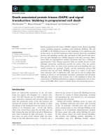
(a) Photomicrographs of H&E stained mouse lung tissue sections from a) control mice, b) irradiated mice (20 Gy, RT) and mice treated with imatinib starting 3 days (c) or 2 weeks (d) after thoracic irradiationFigure 3
(a) Photomicrographs of H&E stained mouse lung tissue sections from a) control mice, b) irradiated mice (20
Gy, RT) and mice treated with imatinib starting 3 days (c) or 2 weeks (d) after thoracic irradiation. Leukocytes
infiltration was marked with asterisk. (b) Photomicrographs of H&E stained mouse lung tissue sections at 16 weeks from a)
control mice, b) irradiated mice (20 Gy, RT) and mice treated with imatinib starting 3 days (c) or 2 weeks (d) after thoracic
irradiation. Fibroblasts were marked with arrow. (c) Photomicrographs of H&E stained lung tissue sections at 20 weeks from
a) control mice, b) irradiated mice (20 Gy, RT) and mice treated with imatinib starting 3 days (c) or 2 weeks (d) after thoracic
irradiation. Collagen depositions were marked with #.
Radiation Oncology 2009, 4:66 />Page 7 of 9
(page number not for citation purposes)
PDGF signalling, although imatinib has been demon-
strated to show marked activity against at least three
kinases: Bcr/Abl, c-Kit, PDGFR-α and -β which can all be
linked to fibrosis, in particular in conjunction with TGF-β
signalling [22-24]. The potential role of PDGF signalling
for the development of lung fibrosis, and in turn for the
treatment of fibrosis by inhibiting PDGF signalling is sup-
ported by data in idiopathic pulmonary fibrosis, asbestos-
, bleomycin- and radiation-induced lung fibrosis as well
as in fibrosis in other organs such as the kidneys, liver,
skin and heart [16-18,23-26]. Therefore, together with
data on radiation induced PDGF expression and phos-
phorylation of PDGFR in vitro and in vivo, and the inhibi-
tion by the kinase inhibitors, we believe that the
inhibition of PDGFR signalling is a key mechanism
behind our functional findings [27,30].
Nevertheless, one cannot exclude important primary roles
of Bcr/Abl, c-Kit, or TGF-beta pathways and one should
also keep in mind that ATP-competitive kinase inhibitors
rarely exhibit complete selectivity. Therefore many addi-
tional data towards a better mechanistic understanding of
our data could be obtained. At the same time, it will be
difficult to proof that one or more specific kinase/kinases
resulting in one or several protein expression event and no
other cascade is responsible for the beneficial role of imat-
inib here. For example, although others and we had previ-
ously shown that PDGF RTKI inhibited phosphorylation
of PDGFR in vivo and this likely contributed to the bene-
fits, it has also been reported that imatinib's c-kit effects
and its link to TGF-beta might be the responsible benefi-
cial antifibrotic pathway [22].
Moreover, the data should also be interpreted with the
understanding that there may also be additional potential
off-target effects. Such off-target effects may have well con-
tributed to the antifibrotic effects that these compounds
have. Rather than pointing out a single protein or gene, it
is conceivable that the development of fibrosis is not a
single step event, but rather an imbalance of an otherwise
physiological homeostatic system with many players
involved. In these terms fibrogenesis may be described as
a shift of the homeostatic system towards the profibrotic
state with the consequence that the entire process can only
be understood as a gene and protein network shift which
may call for systematic biology approaches for a deeper
and more correct understanding [32-34].
The network idea is perhaps fostered by the fact that e.g.
PDGF signalling alone is not an exclusive feature of fibro-
sis research but also known as a key signalling in cancer
research, since PDGF signalling is considered to be a driv-
ing force for cancer cells and known to be proangiogenic
[35]. Accordingly, the inhibition of PDGF signalling is
being investigated as anticancer drugs alone and in com-
bination with chemotherapy and radiotherapy [36-40].
Therefore, a two-fold rationale for the use of PDGF RTKI
in radiation oncology might unfold: first, employing the
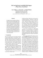
(a) Quantitative analysis of leukocyte numbers as inflammation parameterFigure 4
(a) Quantitative analysis of leukocyte numbers as inflammation parameter. Bars are mean ± SE. * p < 0.05 vs. con-
trols. §p < 0.05 vs. radiation only for both early and late imatinib schedules. (b) Quantitative analysis of septal thickness as
fibrotic parameter presenting deposition of extracellular collagen. Bars are mean ± SE. * p < 0.05 vs. controls. §p < 0.05 vs. radi-
ation only, for both early and late imatinib schedules.
Radiation Oncology 2009, 4:66 />Page 8 of 9
(page number not for citation purposes)
anticancer effects of PDGF inhibition while second,
simultaneously decreasing fibrosis as a common adverse
side effect in radiotherapy [41,42]. However, again, it is
unlikely that single pathway inhibition can completely
prevent lung fibrosis, considering the intricate genetic net-
working associated with this complex process. While our
data indicate that fibrosis can be attenuated or delayed, it
still progressed despite imatinib. Therefore, RTKI should
be considered in the context of other drug therapies, espe-
cially since, as for any other drug, potential side effects e.g.
cardiotoxicity have been reported for imatinib [43].
Conclusions
Taken together, we demonstrate here that drug treatment
using imatinib might be a useful therapeutic approach to
attenuate radiation-induced lung fibrosis or other types of
fibrosis, which exhibits benefits even after the damaging
insult and its acute inflammation has completely sub-
sided.
Competing interests
The authors declare that they have no competing interests.
Authors' contributions
ML performed experiments, analyzed data and partici-
pated in writing the manuscript. AA participated in
designing the study and analyzed data. KEL participated
in the study design and manuscript writing. HJG per-
formed and analyzed histology. PEH designed the study,
analyzed data, and wrote the manuscript. All authors
approved the final version of the manuscript.
Acknowledgements
We would like to thank Alexandra Tietz and Peter Peschke PhD. This work
was supported in part by grants from the Deutsche Krebshilfe 106997,
DFG National Priority Research Program the tumor-vessel interface
(SPP1190) and NASA/NSCOR NNJ04HJ12G, the Tumorzentum Heidel-
berg-Mannheim, and BMBF 03NUK004A-C.
References
1. Hurkmans CW, Cuijpers JP, Lagerwaard FJ, Widder J, Heide UA van
der, Schuring D, Senan S: Recommendations for implementing
stereotactic radiotherapy in peripheral stage IA non-small
cell lung cancer: report from the Quality Assurance Work-
ing Party of the randomised phase III ROSEL study. Radiat
Oncol 2009, 4:1.
2. Eckert F, Mueller AC: SCLC extensive disease - treatment guid-
ance by extent or/and biology of response? Radiat Oncol 2008,
3:33.
3. Uitterhoeve AL, Koolen MG, van Os RM, Koedooder K, Kar M van
de, Pieters BR, Koning CC: Accelerated high-dose radiotherapy
alone or combined with either concomitant or sequential
chemotherapy; treatments of choice in patients with Non-
Small Cell Lung Cancer. Radiat Oncol 2007, 2:27.
4. Collins BT, Erickson K, Reichner CA, Collins SP, Gagnon GJ, Diet-
erich S, McRae DA, Zhang Y, Yousefi S, Levy E, Chang T, Jamis-Dow
C, Banovac F, Anderson ED: Radical stereotactic radiosurgery
with real-time tumor motion tracking in the treatment of
small peripheral lung tumors. Radiat Oncol 2007, 2:39.
5. Milano MT, Constine LS, Okunieff P: Normal tissue toxicity after
small field hypofractionated stereotactic body radiation.
Radiat Oncol 2008, 3:36.
6. Yamashita H, Nakagawa K, Nakamura N, Koyanagi H, Tago M, Igaki
H, Shiraishi K, Sasano N, Ohtomo K: Exceptionally high incidence
of symptomatic grade 2-5 radiation pneumonitis after stere-
otactic radiation therapy for lung tumors. Radiat Oncol 2007,
2:21.
7. Kamp DW: Idiopathic pulmonary fibrosis: the inflammation
hypothesis revisited. Chest 2003, 124:1187-1190.
8. Niew Niewald M, Fleckenstein J, Licht N, Bleuzen C, Ruebe C: Intra-
operative radiotherapy (IORT) combined with external
beam radiotherapy (EBRT) for soft-tissue sarcomas - a ret-
rospective evaluation of the Homburg experience in the
years 1995-2007. Radiat Oncol 2009, 4:32.
9. Viani GA, Novaes PE, Jacinto AA, Antonelli CB, Pellizzon AC, Saito
EY, Salvajoli JV: High-dose-rate brachytherapy for soft tissue
sarcoma in children: a single institution experience. Radiat
Oncol 2008, 3:9.
10. Cornelissen R, Senan S, Antonisse IE, Liem H, Tan YK, Rudolphus A,
Aerts JG: Bronchiolitis obliterans organizing pneumonia
(BOOP) after thoracic radiotherapy for breast carcinoma.
Radiat Oncol 2007, 2:2.
11. Mason RJ, Schwarz MI, Hunninghake GW, Musson RA: NHLBI
Workshop Summary. Pharmacological therapy for idio-
pathic pulmonary fibrosis. Past, present, and future. Am J
Respir Crit Care Med 1999, 160:1771-1777.
12. McBride WH: Cytokine cascades in late normal tissue radia-
tion responses. Int J Radiat Oncol Biol Phys 1995, 33:233-234.
13. Hill RP, Rodemann HP, Hendry JH, Roberts SA, Anscher MS: Nor-
mal tissue radiobiology: from the laboratory to the clinic. Int
J Radiat Oncol Biol Phys 2001, 49(2):353-65.
14. Heinzelmann F, Jendrossek V, Lauber K, Nowak K, Eldh T, Boras R,
Handrick R, Henkel M, Martin C, Uhlig S, Köhler D, Eltzschig HK,
Wehrmann M, Budach W, Belka C: Irradiation-induced pneumo-
nitis mediated by the CD95/CD95-ligand system. J Natl Cancer
Inst 2006, 98(17):1248-51.
15. Hallahan DE, Geng L, Shyr YJ: Effects of intercellular adhesion
molecule 1 (ICAM-1) null mutation on radiation-induced
pulmonary fibrosis and respiratory insufficiency in mice. J
Natl Cancer Inst 2002, 94(10):733-41.
16. Ostman A, Heldin CH: Involvement of platelet-derived growth
factor in disease: development of specific antagonists. Adv
Cancer Res 2001, 80:1-38.
17. Rice AB, Moomaw CR, Morgan DL, Bonner JC: Specific inhibitors
of platelet-derived growth factor or epidermal growth factor
receptor tyrosine kinase reduce pulmonary fibrosis in rats.
Am J Pathol 1999, 155:213-221.
18. Hasselbalch HC, Bjerrum OW, Jensen BA, Clausen NT, Hansen PB,
Birgens H, Therkildsen MH, Ralfkiaer E, et al.: Imatinib mesylate in
idiopathic and postpolycythemic myelofibrosis. Am J Hematol
2003, 74:238-242.
19. Kumar S, Kolozsvary A, Kohl R, Lu M, Brown S, Kim JH: Radiation-
induced skin injury in the animal model of scleroderma:
implications for post-radiotherapy fibrosis. Radiat Oncol 2008,
3:40.
20. Bonner JC: Regulation of PDGF and its receptors in fibrotic
diseases. Cytokine Growth Factor Rev 2004, 15:255-273.
21. Simone NL, Soule BP, Gerber L, Augustine E, Smith S, Altemus RM,
Mitchell JB, Camphausen KA: Oral Pirfenidone in patients with
chronic fibrosis resulting from radiotherapy: a pilot study.
Radiat Oncol 2007, 2:19.
22. Daniels CE, Wilkes MC, Edens M, Kottom TJ, Murphy SJ, Limper AH,
Leof EB: Imatinib mesylate inhibits the profibrogenic activity
of TGF-beta and prevents bleomycin-mediated lung fibrosis.
J Clin Invest 2004, 114:1308-1316.
23. Vuorinen K, Gao F, Oury TD, Kinnula VL, Myllärniemi M: Imatinib
mesylate inhibits fibrogenesis in asbestos-induced interstitial
pneumonia. Exp Lung Res 2007, 33:357-73.
24. Azuma M, Nishioka Y, Aono Y, Inayama M, Makino H, Kishi J, Shono
M, Kinoshita K, Uehara H, Ogushi F, Izumi K, Sone S: Role of alpha1-
acid glycoprotein in therapeutic antifibrotic effects of imat-
inib with macrolides in mice. Am J Respir Crit Care Med 2007,
176(12):1243-50.
25. Chaudhary NI, Roth GJ, Hilberg F, Müller-Quernheim J, Prasse A, Zis-
sel G, Schnapp A, Park JE: Inhibition of PDGF, VEGF and FGF
signalling attenuates fibrosis. Eur Respir J 2007, 29(5):976-85.
26. Yamasaki Y, Miyoshi K, Oda N, Watanabe M, Miyake H, Chan J, Wang
X, Sun L, Tang C, McMahon G, Lipson KE: Weekly dosing with the
Publish with Bio Med Central and every
scientist can read your work free of charge
"BioMed Central will be the most significant development for
disseminating the results of biomedical research in our lifetime."
Sir Paul Nurse, Cancer Research UK
Your research papers will be:
available free of charge to the entire biomedical community
peer reviewed and published immediately upon acceptance
cited in PubMed and archived on PubMed Central
yours — you keep the copyright
Submit your manuscript here:
/>BioMedcentral
Radiation Oncology 2009, 4:66 />Page 9 of 9
(page number not for citation purposes)
platelet-derived growth factor receptor tyrosine kinase
inhibitor SU9518 significantly inhibits arterial stenosis. Circ
Res 2001, 88:630-636.
27. Abdollahi A, Li M, Ping G, Plathow C, Domhan S, Kiessling F, Lee LB,
McMahon G, Gröne HJ, Lipson KE, Huber PE: Inhibition of platelet
derived growth factor (PDGF) signaling attenuates pulmo-
nary fibrosis. J Exp Med 2005, 201:925-935.
28. Plathow C, Li M, Gong P, Zieher H, Kiessling F, Peschke P, Kauczor
HU, Abdollahi A, Huber PE: Computed Tomography Monitor-
ing of Radiation-Induced Lung Fibrosis in Mice. Invest Radiol
2004, 39:600-609.
29. Rubin P, Johnston CJ, Williams JP, McDonald S, Finkelstein JN: A per-
petual cascade of cytokines postirradiation leads to pulmo-
nary fibrosis. Int J Radiat Oncol Biol Phys 1995, 33:99-109.
30. Li M, Gong P, Plathow C, Trinh T, Lipson KE, Hauser K, Krempien R,
Debus J, Abdollahi A, Huber PE: Small molecule receptor tyro-
sine kinase inhibitor of platelet-derived growth factor signal-
ing (SU9518) modifies radiation response in fibroblasts and
endothelial cells. BMC Cancer 2006, 24;6(1):79.
31. Li M, Jendrossek V, Belka C: The role of PDGF in radiation
oncology. Radiat Oncol 2007, 2:5.
32. Huber PE, Hauser K, Abdollahi A: Genome Wide Expression
Profiling of Angiogenic Signaling and the Heisenberg Uncer-
tainty Principle. Cell Cycle 2004, 3:1348-1351.
33. Abdollahi A, Hahnfeldt P, Maercker C, Gröne HJ, Debus J, Ansorge
W, Folkman J, Hlatky L, Huber PE: Endostatin's antiangiogenic
signaling network. Mol Cell 2004, 13:649-663.
34. Abdollahi A, Schwager C, Kleeff J, Esposito I, Domhan S, Peschke P,
Hauser K, Hahnfeldt P, Hlatky L, Debus J, Peters JM, Friess H, Folk-
man J, Huber PE: Transcriptional network governing the ang-
iogenic switch in human pancreatic carcinoma. PNAS 2007,
104:12890-12895.
35. Pietras K, Sjöblom T, Rubin K, Heldin CH, Ostman A: PDGF recep-
tors as cancer drug targets.
Cancer Cell 2003, 3:439-443.
36. Oertel S, Krempien R, Lindel K, Zabel A, Milker-Zabel S, Bischof M,
Lipson KE, Peschke P, Debus J, Abdollahi A, Huber PE: Human
Glioblastoma and Carcinoma Xenograft Tumors Treated by
Combined Radiation and Imatinib (Gleevec
©
). Strahlenther
Onkol 2006, 182:400-7.
37. Abdollahi A, Lipson KE, Han X, Krempien R, Trinh T, Weber KJ, Hah-
nfeldt P, Hlatky L, Debus J, Howlett AR, Huber PE: SU5416 and
SU6668 attenuate the angiogenic effects of radiation-
induced tumor cell growth factor production and amplify the
direct anti-endothelial action of radiation in vitro. Cancer Res
2003, 63:3755-3763.
38. Huber PE, Bischof M, Jenne J, Heiland S, Peschke P, Saffrich R, Gröne
HJ, Debus J, Lipson KE, Abdollahi A: Trimodal cancer treatment:
beneficial effects of combined antiangiogenesis, radiation,
and chemotherapy. Cancer Res 2005, 65:3643-55.
39. Bischof M, Abdollahi A, Gong P, Stoffregen C, Lipson KE, Debus JU,
Weber KJ, Huber PE: Triple combination of irradiation, chem-
otherapy (pemetrexed), and VEGFR inhibition (SU5416) in
human endothelial and tumor cells. Int J Radiat Oncol Biol Phys
2004, 60:1220-1232.
40. Abdollahi A, Griggs DW, Zieher H, Roth A, Lipson KE, Saffrich R,
Gröne HJ, Hallahan DE, Reisfeld RA, Debus J, Niethammer AG,
Huber PE: Inhibition of integrin survival signaling enhances
antiangiogenic and antitumor response of radiation. Clin Can-
cer Res 2005, 11:6270-9.
41. Thilmann C, Nill S, Tücking T, Höss A, Hesse B, Dietrich L, Bendl R,
Rhein B, Häring P, Thieke C, Oelfke U, Debus J, Huber P: Correc-
tion of patient positioning errors based on in-line cone beam
CTs: clinical implementation and first experiments. Radiat
Oncol 2006, 24;1:16.
42. Münter MW, Schulz-Ertner D, Hof H, Nikoghosyan A, Jensen A, Nill
S, Huber P, Debus J: Inverse planned stereotactic intensity
modulated radiotherapy (IMRT) in the treatment of incom-
pletely and completely resected adenoid cystic carcinomas
of the head and neck: Initial clinical results and toxicity of
treatment. Radiat Oncol 2006, 6(1):17.
43. Kerkelä R, Grazette L, Yacobi R, Iliescu C, Patten R, Beahm C, Wal-
ters B, Shevtsov S, Pesant S, Clubb FJ, Rosenzweig A, Salomon RN,
Van Etten RA, Alroy J, Durand JB, Force T: Cardiotoxicity of the
cancer therapeutic agent imatinib mesylate. Nat Med 2006,
2:908-16.