Báo cáo khoa học: " Loratadine dysregulates cell cycle progression and enhances the effect of radiation in human tumor cell lines" pot
Bạn đang xem bản rút gọn của tài liệu. Xem và tải ngay bản đầy đủ của tài liệu tại đây (838.11 KB, 12 trang )
RESEA R C H Open Access
Loratadine dysregulates cell cycle progression
and enhances the effect of radiation in human
tumor cell lines
Benjamin P Soule
*
, Nicole L Simone, William G DeGraff, Rajani Choudhuri, John A Cook, James B Mitchell
Abstract
Background: The histamine receptor-1 (H1)-antagonist, loratadine has been shown to inhibit growth of human
colon cancer xenografts in part due to cell cycle arrest in G2/M. Since this is a radiation sensitive phase of the cell
cycle, we sought to determine if loratadine modifies radiosensitivity in several human tumor cell lines with
emphasis on human colon carcinoma (HT29).
Methods: Cells were treated with several doses of loratad ine at several time points before and after expo sure to
radiation. Radiation dose modifying factors (DMF) were determined using full radiation dose response survival
curves. Cell cycle phase was determined by flow cytometry and the expression of the cell cycle-associated proteins
Chk1, pChk1
ser345
, and Cyclin B was analyzed by western blot.
Results: Loratadine pre-treatment of exponentially growing cells (75 μM, 24 hours) increased radiation-induced
cytotoxicity yielding a radiation DMF of 1.95. However, treatment of plateau phase cells also yielded a DMF of 1.3
suggesting that mechanisms other than cell cycle arrest also contribute to loratadine-mediated radiation
modification. Like irradiation, loratadine initially induced G2/M arrest and activation of the cell-cycle associated
protein Chk1 to pChk1
ser345
, however a subsequent decrease in expression of total Chk1 and Cyclin B correlated
with abrogation of the G2/M checkpoint. Analysis of DNA repair enzyme expression and DNA fragmentation
revealed a distinct pattern of DNA damage in loratadine-treated cells in addition to enhanced radiation-induced
damage. Taken together, these data suggest that the observed effects of loratadine are multifactorial in that
loratadine 1) directly damages DNA, 2) activates Chk1 thereby promoting G2/M arrest making cells more
susceptible to radiation-induced DNA damage and, 3) downregulates total Chk1 and Cyclin B abrogating the
radiation-induced G2/M checkpoint and allowing cells to re-enter the cell cycle despite the persistence of
damaged DNA.
Conclusions: Given this unique possible mechanism of action, loratadine has potential as a chemotherapeutic
agent and as a modifier of radiation responsiveness in the treatment of cancer and, as such, may warrant further
clinical evaluation.
Background
It is well established that the effects of radiation varies
as a function of cell cycle position [1]. Specifically, cells
in G2/M phase are particularly susceptible to the effects
of radiation. Because of this, agents that alter cell cycle
progression are often potent radiation modifiers [2].
Normal cell cycle regulation is mediated by several
proteins that are responsive to both intra- and extracel-
lular stimuli. It has been demonstrated that th e com-
monly used antihistamine loratadine (ethyl4-( 8-chloro-
5,6-dihydro-11H-benzo[5,6]cyclohepta [1,2-b]pyridin-11-
ylidene)-1-piperidinecarboxylate), an antagonist of hista-
mine receptor-1, induces a cell cycle arrest in G2/M by
interfering with the activ ity of these regulatory proteins
[3]. Although a com prehensive mechanism was not elu-
cidated, in these prior studies loratadine treatment
resulted in anti-tumor effects.
* Correspondence:
Radiation Biology Branch, National Cancer Institute, National Institutes of
Health, 10 Center Drive, Building 10, Room B3B69, Bethesda, MD 20892, USA
Soule et al. Radiation Oncology 2010, 5:8
/>© 2010 Soule et al; licensee BioMed Central Ltd. This is an Open Access article distribut ed under the terms of the C reative Co mmons
Attribution License ( which permi ts unres tricted use, distribution, and reproduction in
any medium, pro vided the original work is prop erly cited.
Progression through the cell cycle is regulated by a
complex network of proteins that monitor the health of
the cell. This mechanism serves to protect cells from
potentially lethal stressors by temporarily halting cell
cycle progression to allow time for repair of damaged
cell components, especially damage involving DNA. For
example, it is well known that DNA damage induced by
radiation results in cell cycle block in G2/M during
which time the DNA repair machinery attempts to cor-
rect the damage. If the damage is repaired, cells are
released from the cell cycle block and are allowed to
divide. Persistent DNA damage may result in cell death
initiated by other surveillance mechanisms. In eukaryo-
tic cells, the G2/M checkpoint is controlled by several
proteins including cell division cycle 2 (Cdc2) and
Cyclin B [4]. Cdc2 is inactivated by phosphorylation
(Tyr-15, Thr-14) and activated by Cdc25C-mediated
dephospho rylation [5]. Cdc25C, in turn, is regulated by
14-3-3, which inhibits nuclear translocation of Cdc25C,
and Chk1 phospho rylation, which allows 14-3-3 binding
to occur [6]. Chk1 inhibition has been associated with
increased cytotoxicity of DNA damaging drugs [7-12],
and in our lab with increased sensitivity to the effects of
radiation (unpublished data). Recently, loratadine has
also been shown to cause Cdc2-associated G2/M arrest
by interfering with Chk1 and Cdc25C signaling [3]. It is
likely that the anti-tumor effects of loratadine observed
in other studies result, at least in part, from this activity.
Since G2/M is a particularly radiosensitive phase of
the cell cycle, it is logical to suggest that the induction
of a cell cycle block in G2/M by loratadine would
enhance radiation-induced cytotoxicity, however this has
not yet been studied. This study was initiated to deter-
mine whether loratadine modifies the effect of radiat ion
on cell survival and, if so, to elucidate the mechanism
underlying that effect.
Methods
Cell Culture Studies
HT29 (human colon carcinoma) and DU145 (human
prostate carcinoma) were purchased from American
Type Culture Collection (Manassas, VA). SF295 (human
glioblastoma) were a gift from Dr. Kevin Camphausen.
SF295 cells were grown in DMEM, and all other cell
lines were grown in RPMI 1640. All media contained
10% heat-inactivated fetal bovine serum and antibiotics.
For cell survival studie s, cells were plated (5 × 10
5
cells/
100 mm plastic petri dish) and incubated for 16 hours
at 37°C. Loratadine was dissolved in 0.1% DMSO then
added at various concentrations to the exponentia lly
growing cells in complete medium and the cells were
incubated at 37°C for 24 hour. DMSO (0.1%) was also
added to control cells. Most studies used a loratadine
concentration of 75 μM [3]; the only exception was
studies shown in Figure 1B where a range of loratadine
concentrations were used (10-450 μM). Some studies
involved the use of plateau phase cultures. For these
studies, cells were allowed to grow to confluence and
maintained in confluence without medium change for 3
days after which they were treated with loratadine (75
μM) as described above. Flow cytometery studies con-
firmed that these cultures were enriched in cells in G1
phase. Following incubation cells with or without lorata-
dine, cells were treated with varying do ses of radiatio n
using an Eldorado 8 cobalt-60 teletherapy unit (Thera-
tronics International Ltd. Kanata, Ontario, Canada) at
dose rates of 2.0-2.5 Gy/min. Control radiation survival
curves were conducted in parallel. Immediately after
irradiation, cells were trypsinized, counted, plated, and
incubated for 10-14 days for macroscopic colony forma-
tion. Colonies were then fixed wit h methanol/acetic acid
(3:1) and stained with crystal violet. Colonies with >50
cell s were scored and cell survival determined after cor-
recting for the plating efficiency and for loratadine cyto-
toxicity alone. For radiation studies, a dose modification
factor (DMF) was determined by taking the ratio of
radiation doses at the 10% survival level (control radia-
tion dose divided by the drug treated radiation dose).
DMF values > 1 indicate enhancement of radiosensitiv-
ity. Some studies involved cisp latin exposure to cells for
1 hour with or without a 24 hour pre-treatment with
loratadine. Following treatment, the cells were processed
for colony formation as described above.
Immunoblot Analysis for gH2AX
Cells were lysed in 10 mM HEPES pH 7.9, 1.5 mM
MgCl2, 10 mM KCl with 0.5 mM DTT and 1.5 mM
PMSF with complete protease inhibitor cocktail (Roche
Applied Science, Indianapolis, IN). Histones from the
nuclear pellet were extracted in 0.2 mol/L sulfuric acid
by incubating samples on ice for 4-6 hours. After centri-
fugation, acid-solu ble histones were transferred to fresh
tubes and 9 v olumes of ice cold acetone were added.
Histones were precipitated at -20°C overnight and were
pelleted by centrifugation at 14,000 rpm for 10 min at
4°C. Supernatant was discarded and pellets were air-
dried. Histones were solubilized in 4 mol/L urea and
protein concentration was determined by BioRad DC
protein assay. Histones were separated on 18% Tris-Gly-
cine gels (Invitrogen, Carlsbad, CA) by loading 20 μg
samples and transferred to nitrocellulose membrane
using iBlot Dry Blotting System from Invitrogen (Carls-
bad, CA). Membranes were incubated overnight at 4°C
with mouse monoclonal anti-phospho Histone H2AX
(Ser139), clone JBW301 (1:10,000) from Millipore (Bill-
erica, MA), washed 3 times with PBS-T and incubated
with HRP-conjugated anti-mouse antibody from Santa
Cruz Biotechnology, Inc. (Santa Cruz, CA). gH2AX was
visualized by ECL detection kit (Perkin Elmer, Waltham,
Soule et al. Radiation Oncology 2010, 5:8
/>Page 2 of 12
MA) using Fluor Chem SP imager (Alpha Innotech, San
Leandro, CA). Membranes were stripped using Re-Blot
Plus mild antibody stripping solution (Millipore; Biller-
ica, MA) and reprobed with 1:1000 rabbit antiserum to
histone H2A (acidic patch) from Millipore (Billerica,
MA) to ascertain uniform loading. Signal intensities
were normalized to thei r lo ading control H2A a nd
expressed as fold change compared to controls.
Pulsed-Field Gel Electrophoresis
DNA was prepared for electrophoresis by the methods
of Schwartz and Cantor [13] and Gardiner et al. [14] as
modified by Ager and Dewey [15] and Stamato and
Denko [16]. After loratidine t reatment (or x-irradiation
for a positive control), the cells were trypsinized, rinsed
in cold PBS, and resuspended in PBS at 10
7
per ml. An
equal volume of 1% low gelling temperature agarose was
added, and the cell suspensio n was drawn into
3
/
32
inch
(i.d.) silicone tubing with a syringe. Both ends of the
tubing were clamped, and the tubing was immersed in
an ice bath to rapidly solidify the agarose. The agarose
was then extruded f rom the tubing, cut into 5 mm
lengths, and the se “plugs” were placed into 1.5 ml ce n-
trifuge tubes. This procedure results in approximately
10
5
cells per 5 mm plug. DNA was purified by incubat-
ing at 55°C in ESP buffer (0.5 M EDTA, 1% Sarkosyl,
and 50 μg/ml proteinase K) for 24 hr. The plugs were
then rinsed in TE buffer (10 mM Tris, 1 mM EDTA)
for 24 hr with three buffer changes. RNA was digested
by incubation with 0.1 μg/ml boiled RNAse A in TE
buffer for 2 hr at 37°C.
0.8% agarose gels were cast in 0.5× TBE (1× TBE = 90
mM Tris, 90 mM boric acid, 2.5 mM EDTA with 0.5
μg/ml ethidium bromide). Agarose plugs were loaded
into 2 × 6 × 5 mm wells, and the wells were sealed with
melted agarose. Electrophoresis was carried out for 24
hr at 56 volts (4 volts/cm), with a 3:1 ratio of forward to
reverse pulse time. The initial forward pulse time was
7.5 seconds (reverse pulse 2.5 seconds), increasing to a
final forward pulse time of 90 seconds (final reverse
pulse 30 seconds). The running buffer (0.5× TBE) was
re-circulated and cooled to maintain a tem perature of
12-15°C. These electrophoresis conditions were chosen
based on methods of Stamato a nd Denko [16], and the
desire to keep the released DNA concent rated in a nar-
row band to facilitate quantification.
Quantification was done b y densitometry using a
FluorChem g el documentation system (Alpha Innotech,
San Leandro, CA) and AlphaEaseFC software (Alpha
Innotech, San Leandro, CA). Each band in the gel was
outlined manually and the density determined. The
results are expressed as “%DNA released,” determined
by dividing the density of the released DNA band by the
density of the total DNA in the l ane (the released DNA
band plus the unreleased DNA remaining in the well).
Cell Cycle Analysis
The effect of loratadine on cell cycle distribution was
analyzed by flow cytometry by propidium iodide staining
after treating cells with the drug for 24 hour. Briefly,
cells were trypsinized, washed with PBS and fixed in
70% ethanol overnight. Cells were pelleted and nuclei
were isolated by pepsin/HCl digestion followed by treat-
ment with 10 mmol/L bo rate (pH 8.6) to neutralize the
acid. Cells were then incubated with FITC-labeled anti-
human IgG and PI staining. Cell cycle data were col-
lected on BD FACSCalibur Flow Cytometer (San Jose,
CA) and analyzed using CellQuest/MOD-Fit software
(Verity Software House, Topsham, ME).
Western Blot Analysis
The cells were lysed in RIPAbuffer(SantaCruzBio-
technology, Inc. Santa Cruz, CA) containing protease
inhibitor cocktail and phosphatase inhibitors (Roche
Applied Science, Indianapolis, IN). T he samples were
incubated in the l ysis buffer on ice for 30 minutes, cen-
trifuged at 14000 rpm i n a refrigerated centrifuge for 30
minutes and the supernatant collected. The samples
were kept at 40°C if used on the same day or frozen at
-70°C for storage. Protein concentration was determined
with Dc Protein Asssay kit (Bio-Rad, Hercules, CA). 40
μg of protein was separated on 4-20% Tris-Glycine gels
(Invi trogen, Carlsbad, CA) and transferred to nitrocellu-
lose membrane using iBlot Dry Blotting System from
Invitrogen (Carlsbad, CA). Non specific protein binding
was blocked by incubating the membranes for 1 hour in
3% blocking grade non fat dry milk (Bio-Rad, Hercules,
CA) in TBST. The membranes were then left overnight
at 40°C in the primary antibody at a dilution of 1:1000
for rabbit monoclonal anti pChk1 (Ser 345) (Cell Signal-
ing Technology, Inc., Danvers, MA), 1:200 for mouse
monoclonal anti Chk1 (Santa Cruz Biotechnology, Inc.
CA), 1:1000 for mouse monoclonal anti Cyclin B (BD
Biosciences, Bedford MA); and 1:5000 for mouse mono-
clonal anti Actin (Millipore, Billerica, MA). The mem-
branes were washed thrice in TBST and incubated for 1
hour in horseradish p eroxidase conjugated secondary
antibody (Santa Cruz Biotechnology, Inc. Santa Cruz,
CA) at a dilution of 1:2000. The proteins were then
visualized by chemiluminescence (Western Lightning
Chemilumiscence Reagent Plus, Perkin Elmer, Waltham,
MA or ECL Advance Western Blotting Detection Kit,
GE Lifesciences, Pittsburg, PA) using Fluor Chem SP
imager (Alpha Innotech, San Leandro, CA). Fold change
in protein expression was expressed as a ratio calculated
by dividing the specific protein band density with the
actin band density (loading control), and then normal-
ized to the control.
Statistics
All experiments were performed a minimum of three
times. In some cases, the plots represent the average of
Soule et al. Radiation Oncology 2010, 5:8
/>Page 3 of 12
these experiments. For some experiments, representative
results are represented. Whether the plot represents the
average of several experiments or a representative
experiment has been indicated for each figure. When
present, error bars represent the standard error of the
mean. Dose modifying factors (DMF) were calculated
for clonogenic survival assays.
Results
Loratadine Dose Response and Time Course in Radiation-
treated Cells
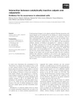
HT29 cells treated with loratadine (75 μM) 4, 8, 12, 18,
and 24 hours prior to irradiation (6 G y) demonstrated
that the radiation modifying effect of loratadine
increased with increasing exposure time prior to irradia-
tion (Figure 1A). The toxicity of loratadine alone was
minima l until exposure time exceeded 18 hours. For all
experiments, cell survival was asse ssed using a standard
clonogenic assay corrected for the toxicity of loratadine
alone. HT29 cells were then treated with loratadine (0,
10, 25, 50, 75, 150, 300, and 450 μM) for 24 hours prior
to irradiation (6 Gy). Loratadine decreased cell survi val
by one log after administration of a 75 μMdosebutno
effect was observed at lower doses (Figure 1B). The
cytotoxicity of loratadine alone increased with increasing
dose and was noted to increase markedly at the 75 μM
dose as well. Doses of loratadine higher than 75 μM
killed 100% of the cells (data not shown).
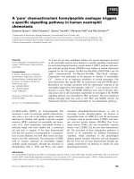
Effect of Loratadine on Radiation Dose Response
HT29 cells in log phase growth were treated with lorata-
dine (75 μM) for 24 hours prior to irradiation (0, 1.5, 3,
6, or 9 Gy, or 12 Gy for controls only). A radiation dose
response was clearly demonstrated with enhancement of
the radiation-induced cytotoxicity by lorat adine at all
radiation doses (Figure 2A) resulting in a radiation dose
modification factor (DMF) of 1.95 ± 0.07 compared to
cells not treated with loratadi ne. In contrast, HT29 cells
in log phase growth treated with loratadine (75 μM) for
24 hours after irradiation (0, 1.5, 3, 6, 9, or 12 Gy)
appeared to be minimally protected from radiat ion-
induced cytotoxicity (Figure 2B). HT29 cells were
allowed to re ach plateau phase in culture and were then
treated with loratadine (75 μM) for 24 hours prior to
irradiation (0, 1.5, 3, 6, or 9 Gy). This resulted in a
radiation DMF of 1.3 ± 0.16 compared to cells not trea-
ted with loratadine (Figure 2C).
Radiation Dose-modifying Effect of Loratadine in Other
Cell Types
HT29, SF295, and DU145 cells in log phase growth were
treated with loratadine (75 μM) for 24 hours prior to
irradiation (6 Gy) and cell survival was assessed using a
clonogenic assay. Loratadine alone was more toxic to
SF295 cells (17% survival) than HT29 cells (68% survi-
val) but enhanced the radiation response in both cell
lines (data not shown). Despite significant toxicity to
DU145 cells of loratadine alone (45% survival), no
increase in susceptibility to radiation-induced cytotoxi-
city was seen in loratadine-treated cells.
Effect of Loratadine on Cisplatin-treated Cells
HT29 cells treated with loratadine (75 μM) for 24 hours
prior to treatment with Cisplatin (7.5, 15, 30, or 45 μg/
ml for 1 hour). A Cisplatin dose response was clearly
demonstrated with enhancement of the cisplatin-
induced cytotoxicity by loratadine at all radiation doses
(Figure 2 D) resulting in a DMF of 2.6 ± 0.14 for
loratadine.
Effect of Histamine on Radiation Modification by
Loratadine
To establish whether the observed effects of loratadine
were being mediated by antagoni sm of the H1-receptor,
HT29 cells were treated with lorata dine with and with-
out exogenous histamine. Exposure to histamine (100 or
1000 μM) alone for 15 minutes did not alter survival
(data not shown). At both doses a cell survival of 99%
was observed by clonogenic assay. Likewise, histamine
did not modify the response to radiation as there was
no significant difference between cells exposed to 9 Gy
alone compared to those that were pretreated with
histamine.
Effect of Loratadine and Radiation on DNA-Repair
Proteins
gH2AX recruitment was measured by western blot in
HT29 cells treated with loratadine (75 μM) for 24 hours
prior to irradiation (6 Gy) t hen collected at 1, 6 and 24
hours after irradiation. One hour post-irradiation,
gH2AX was increased in radiation-treated samples com-
pared to unirradiated control (Figure 3). Commensurate
with DNA repair, this signal decreased over time and
returnedtobaselineby24hourspost-irradiation.At
one hour post-irradiation, the loratadine-treated irra-
diated sample demo nstrated more gH2AX signal than
the radiation-only sample. In contrast to the radiation-
only cells, the gH2AX signal in the cells treated with
loratadine and radiation remained elevated at 6 and 24
hours without evidence of diminution. Cells treated with
loratadine alone also demonstrated increased gH2AX
signal which increased at 6 hours but diminished by 24
hours after treatment. Using the same experimental
design, HT29 cells were analyzed by pulsed-field gel
electrophoresis at 0, 3, 6, and 24 hours post-irradiation.
As shown in Figure 4A, radiation-induced DNA frag-
ments (8 Gy) were evident within 1 hour following irra-
diation and resolved by 24 hours. Loratadine-treated
irradiated cells (LR+8 Gy) demonstrated increased DNA
fragmentation compared to radiation alone, and this
increase persisted through 24 hours. An additional band
corresponding to smaller DNA fragments (arrows) was
also seen in loratadine-treated cells. Densitometry
Soule et al. Radiation Oncology 2010, 5:8
/>Page 4 of 12
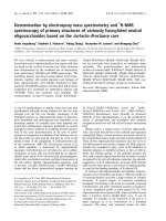
6Gy + LR10 + LR25 + LR 50 + LR 75
10
-2
10
-1
10
0
B
Surviving Fraction
6Gy + LR 4hr + LR 8hr + LR 12hr + LR 18hr + LR 24hr
10
-2
10
-1
10
0
A
Surviving Fraction
Figure 1 Effect of Loratadine Dose and Exposure Time on Response to Radiation. (A) Cells were treated with 75 μM loratadine for various
times prior to irradiation to 6 Gy. The radiation modifying effect increased with exposure time. Toxicity of loratadine alone was minimal until
exposure exceeded 18 hrs. (B) Cells were treated with loratadine (0, 10, 25, 50, 75, 150, 300, and 450 μM) for 24 hrs prior to irradiation. A
radiation modifying effect was only observed with a 75 μM dose. Toxicity of loratadine alone increased with dose and 100% of the cells were
killed at doses above 75 μM (data not shown). Cell survival is corrected for the toxicity of loratadine alone. The figure represents the mean ± SD
for 3 experiments.
Soule et al. Radiation Oncology 2010, 5:8
/>Page 5 of 12
analysis confirmed increased DNA fragments in irra-
diated cells, and a further increase in loratadine-treated
irradiated cells (Figure 4B). Loratadine alone (LR)
induced DNA fragmentation (Figure 4C) and also pro-
duced the additional band corresponding to smaller
DNA fragments (arrow).
Effect of Loratadine on In Vitro Cell Cycle Progression
HT29 cells treated with loratadine (75 μM) for 24 hours.
Loratadine was then washed off and cells were irradiated
(8 Gy). Cell cycle progression was analyzed by flow
cytometry after loratadine treatment, then again 6, 12,
and 18 hours after irradiation. After 24 hour treatment
with loratadine alone, cells exhibited a G2 block (from
14 to 37%) (Figure 5A). This G2 block persisted for 12
hours (hour 36) and returned to baseline by 18 h ours
after treatment (hour 42). Six hours after irradiation
alone (hour 30) cells also exhibited a G2 block (8 Gy)
which was similar in magnitude to loratadine-treated
cells. The radiation-induced G2 block increased from 14
to 74% by 12 hours after irradiation and began to
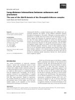
Figure 2 Effec t of Loratadine on Radiation or Cisplatin Dose Response. HT29 cells in log phase growth were treated with loratadine (75
μM) for 24 hrs prior to irradiation (A) or for 24 hrs after irradiation (B) to 0, 1.5, 3, 6 or 9 Gy or 12 Gy (controls only). A radiation DMF of 1.95 ±
0.07 was observed in cells pre-treated with loratadine. There was no significant radiation modification by loratadine treatment after irradiation.
(C) HT29 cells in plateau phase growth pre-treated with loratadine (75 μM, 24 hrs) demonstrated a radiation DMF of 1.3 ± 0.16. Solid circles =
loratadine + radiation, open circles = radiation alone. (D) HT29 cells in log phase growth were pre-treated with loratadine (75 μM, 24 hrs) prior
to treatment with Cisplatin (7.5, 15, 30, or 45 μg/ml for 1 hr). A DMF of 2.6 ± 0.14 was observed. Open circles = loratadine + cisplatin, solid
circles = cisplatin alone. Cell survival was assessed by clonogenic assay and corrected for the toxicity of loratadine alone. The figure represents
the mean ± SD for 3 experiments.
Soule et al. Radiation Oncology 2010, 5:8
/>Page 6 of 12
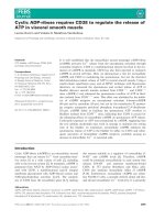
Figure 3 Effect of Loratadine and Radiation on DNA Repair Proteins. HT29 cells were either treated with loratadine (75 μM, 24 hrs) prior to
exposure to 8 Gy radiation, or treated with loratadine or radiation alone. gH2AX expression, determined by western blot at 1, 6, and 24 hrs after
irradiation, increased within 1 hr after irradiation and returned to baseline by 24 hrs. Loratadine treatment enhanced this expression at 1 hr and
resulted in persistent expression at 24 hrs. Loratadine alone also increased gH2AX expression with maximal expression at 6 hrs. The graph
represents the ratio of the densitometric value of the sample compared to control for a single representative experiment, LR = loratadine-
treated.
Soule et al. Radiation Oncology 2010, 5:8
/>Page 7 of 12
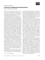
Figure 4 Effect of Loratadine and Radiation on DNA Damage. HT29 cells were either treated with loratadine (75 μM, 24 hrs) prior to
exposure to 8 Gy radiation, or treated with loratadine or radiation alone. Cells were analyzed by pulsed-field gel electrophoresis at 0, 3, 6, and
24 hrs. (A) Radiation-induced DNA fragments were evident immediately following irradiation and resolved by 24 hrs. Loratadine-treated irradiated
cells demonstrated increased and persistent DNA fragmentation and an additional band corresponding to smaller DNA fragments (arrows). (B)
Densitometry analysis confirmed increased DNA fragments in irradiated and loratadine-treated cells. (C) Loratadine alone induced DNA
fragmentation and an additional band corresponding to smaller DNA fragments (arrow). The graph represents the densitometric value of the
sample for a single representative experiment, LR = loratadine-treated.
Soule et al. Radiation Oncology 2010, 5:8
/>Page 8 of 12
decrease 18 hours after irradiation (from 74 to 58%).
Interestingly, despite inducing a G2 block, loratadine-
treated cells clearly dominated the cell cycle delay and
radiation with loratadine did not cause additional cell
cycle delays. As shown in Figure 5B, the percent of cells
in G2 did not increase following radiation in loratadine-
treated cells. In contrast to cells exposed to radiation
alone, the loratadine-treated irradiated cells had
returned to a more normal cell cycle distribution within
18 hours of the removal of loratadine.
Effect of Loratadine on Cell Cycle-associated Proteins
Western blots were performed to detect total Chk1,
phosphorylated Chk1 (pChk1
ser345
) and Cyclin B in
HT29 cells treated with loratadine (75 μM) for 24 hours
prior to irradiation (8 Gy). pChk1
ser345
increases in
response to loratadine (LR) within 6 hours after expo-
sure, peaks at 12 hours and returns to baseline by 36
hours (Figure 6). Chk1 pro gressively decreases after
exposure and remains depressed below baseline expres-
sion at 36 hours. In irradiated cells (8 Gy), both
pChk1
ser345
and Chk1 are increased at 8 and 16 hours
post-irradiation. Loratadine does not significantly a lter
the radiation-induced increase in pChk1
ser345
at 8 hour s
post-irradiation (8 Gy+LR) but in contrast to cells
exposed to radiation only, pChk1
ser345
expression
returns to control levels by 12 hours post-irradiation.
Furthermore, Chk1 levels in cells exposed to radiation
and loratadine are markedly decreased compared to
cells exposed to radiation alone and even compared to
controls. Cyclin B increases in irradiated cells at 8 and
16 hours post -irradiation but this respo nse is abrogat ed
in cells treated with loratadine.
Discussion
In this study, treatment with loratadine enhanced the
cytotoxic effect of radiation. This effect was both time
and dose dependent and occurred optimally when cells
were treated with 75 μM loratadine for 24 hours prior
to irradiation. Loratadine exhibited significant cytotoxi-
city alone and a narrow therapeutic window with little
to no effect below 75 μM and profound toxicity above
that dose. This radiation-enhancing effect was observa-
ble in several cell lines including colon cancer, glioblas-
toma, and prostate cancer lines.
The mechanism by which r adiation-enhancement
occurred, however, appeared to be somewhat more
complex than pre dicted based on previous studies. As
might be expected, the action of loratadine on its puta-
tive target, the H1-receptor, did not appear to be play a
mechanistic role as incubation with histamine did not
prevent the loratadine-mediated radiosensitization. As
has been previously shown [3], loratadine alone results
in Chk1 activation leading to an increase in the percen-
tage of cells in the G2/M phase of the cell cycle. Since
the G2/M phase of the cell cycle is one of the most sen-
sitive to radiation [17], this could explain some of the
increased radiation-induced cytotoxicity observed with
loratadine pre-incubation. Likewise, enrichment of the
cells in G2/M phase may also explain some of the
increase in susceptibility to radiation-induced DNA
damage as refl ected in the increase in both DNA strand
breaks detected on pulsed-field gel electrophoresis and
in the increased expression of the DNA repair protein
gH2AX compared to cells treated with radiation alone.
Our results confirm the finding of Chen et al that l ora-
tadine activates Chk1 leading to accumulation of cells in
G2/M phase of the cell cycle. Our data suggest that
other parts of the cell cycle are also affected since the
percentage of cells in G2/M never increased beyond
38% while radiation and drugs such as cisplatin can lead
to increases in G2/M of 80% or more after 12 hours of
exposure [2]. Addi tionally, what is novel about our find-
ings is that loratadine expo sure leads to an a brogation
of the G2/M checkpoint induced b y radiation. Lorata-
dine exposure appears to result in aberrant Chk1 con-
trol hence releasing them back into the cell cycle with
persistent DNA damage. This may alter the ability of
cells to repair additional DNA damage such as that
induced by radiation contributing to the increased radia-
tion sensitivity observed in l oratadine-treated cells. One
possible mechanism of this negated Chk1 response may
be related to the decreased expression of total Chk1 and
Cyclin B proteins after prolonged exposure to loratadine
(Figure 6).
This finding is furth er supported by the enhancement
and persistence of both the DNA fragments detected by
pulsed-field gel electrophoresis and the gH2AX expres-
sion. The persistence of DNA damage may also accoun t
for the appearance of the second band of fragmented
DNA that was observed on pulsed-field gel electrophor-
esis in loratadine treated cells (Figure 4). It is possible
that these fragments represent further damage induced
by ongoing attempts to repair DNA while the cell is
actively progressing throug h the cell cycle, although this
remains to be shown. It is clear, however, that DNA
repair proteins, such as gH 2AX, are appropriately
recruited to sites of damage initially and are detected in
cells treated with loratadine alone, and in loratadine
treated and untreated irradiated cells (Figure 3). This
recruitment is downregulated within 24 hours as DNA
repair is completed in cells exposed to radiation alone.
In loratadine treated cells, however, there is a persis-
tence of this signal beyond 24 hours a nd well after the
cells have re-entered the cell cycle. This likely results
from the persistence of DNA damage as mentioned
above and strongly suggests that cells are prematurely
re-entering the cell cycle with persistent DNA damage
that is actively undergoing attempts at repair.
Soule et al. Radiation Oncology 2010, 5:8
/>Page 9 of 12
Finally, loratadine also generates DNA damage on its
own which induces DNA repair mechanisms in the cell
such as gH2AX. The pulsed field gels demonstrate the
presence of double strand breaks, however since addi-
tional lower molecular weight DNA is also present,
other types of DNA damage must be occurring. This
DNA damage occurs at doses of 75 μMandaboveand
it appears that this damage is required for ra diosensiti-
zation as lower concentrations did not result in an
increase in DNA damage or radiation-induced cytotox-
icty. Since the flow DNA histograms (Figure 5A) did not
show an increased sub-G1 peak af ter loratadine expo-
sure, it does not appear that an increase in apoptosis
explains the increase in radiation sensitivity. Given that
loratadine pre-treatment also enhanced the toxicity of
cisplatin, another DNA-damaging agent, it is logical to
suggest t hat the abrogation of the G2/M delay is a cru-
cial mechanism underlying the loratadine-induced
increase in cytotoxicty.
Conclusions
Loratadine enhancement of the cytotoxic effect of radia-
tion is both dose and time-dependent. The mechanism
underlying this effect is multifactorial and involves an
early promotion of G2/M cell cycle blockade which
enhances radiation sensitivity, followed by abrogation of
the radiation-induced G2/M arrest and premature
release of DNA-damaged cells back into the cell cycle.
Loratadine-induced DNA damage is also observe d and
is likely additive to the ra diation-induced damage. Give n
Figure 5 Effect of Loratadine and Radiation on Cell Cycle Progression. HT29 cells were either treated with loratadine (75 μM, 24 hrs) prior
to exposure to 8 Gy radiation, or treated with loratadine or radiation alone. Cell cycle progression was analyzed by flow cytometry. (A) After 24
hr treatment with loratadine, cells exhibited a G2 block (38%) which persisted through 36 hrs and resolved by 42 hrs. Irradiated cells also
exhibited a G2 block which peaked (74%) at 12 hrs after irradiation and began to decrease 18 hrs after irradiation. Loratadine abrogated the
radiation-induced G2 block at 12 hrs post-irradiation and by 18 hrs had returned to baseline. (B) Irradiation alone (open circle) increased the
percentage of cells in G2/M but did not increase the percentage of loratadine-treated cells (solid circle) in G2/M compared to loratadine
treatment alone (open square). The figure represents a single representative experiment.
Soule et al. Radiation Oncology 2010, 5:8
/>Page 10 of 12
this unique potential mechanism of action, loratadine is
a potentially promising radiation modifying drug.
Abbreviations
DMF: Dose Modifying Factor; Gy: Gray; H2AX: histone 2AX; gH2AX:
phosphorylated histone 2AX; HT29: human colon cancer cell line; DU145:
human prostate cancer cell line; SF295: human glioblastoma cell line.
Acknowledgements
This research was supported in part by the Intramural Research Program of
the NIH, National Cancer Institute, Center for Cancer Research.
Authors’ contributions
BPS - design and performance of lab experiments, writing and editing of
manuscript.
NLS - design and performance of lab experiments, writing and editing of
manuscript.
WGD - design and performance of lab experiments, editing of manuscript.
RC - performance of lab experiments.
JAC - design and performance of lab experiments, editing of manuscript.
JBM - design and performance of lab experiments, writing and editing of
manuscript.
All authors have read and approved the final manuscript.
Competing interests
The authors declare that they have no competing interests.
Received: 20 October 2009
Accepted: 3 February 2010 Published: 3 February 2010
References
1. Terasima T, Tolmach LJ: Changes in x-ray sensitivity of HeLa cells during
the division cycle. Nature 1961, 190:1210-1211.
2. Liebmann J, Cook JA, Fisher J, Teague D, Mitchell JB: In vitro studies of
Taxol as a radiation sensitizer in human tumor cells. J Natl Cancer Inst
1994, 86:441-446.
Figure 6 Effect of Loratadine and Radiation on Cell Cycle Associated Proteins. In HT29 cells, activated pChk1
ser345
increases in response to
loratadine (LR, 75 μM) within 6 hrs after exposure, peaks at 12 hrs and returns to baseline by 36 hrs. Total Chk1 decreases after exposure and
remains depressed at 36 hrs. In irradiated cells, both pChk1
ser345
and Chk1 are increased at 8 and 16 hrs post-irradiation (8 Gy, 32 and 36 hr).
Loratadine does not alter the radiation-induced increase in pChk1
ser345
at 8 hrs post-irradiation (8 Gy+LR, 32 hr) but abrogates the increase seen
at 16 hrs (8 Gy+LR, 36 hr). Cyclin B1 increases in irradiated cells at 8 and 16 hrs post-irradiation (8 Gy, 32 and 36 hr) but in cells treated with
loratadine (LR, 8 Gy+LR) Cyclin B expression falls below that seen in controls. The figure represents a single representative experiment.
Soule et al. Radiation Oncology 2010, 5:8
/>Page 11 of 12
3. Chen JS, Lin SY, Tso WL, Yeh GC, Lee WS, Tseng H, Chen LC, Ho YS:
Checkpoint kinase 1-mediated phosphorylation of Cdc25C and bad
proteins are involved in antitumor effects of loratadine-induced G2/M
phase cell-cycle arrest and apoptosis. Mol Carcinog 2006, 45:461-478.
4. Huang TS, Shu CH, Chao Y, Chen SN, Chen LL: Activation of MAD 2
checkprotein and persistence of cyclin B1/CDC 2 activity associate with
paclitaxel-induced apoptosis in human nasopharyngeal carcinoma cells.
Apoptosis 2000, 5:235-241.
5. Dunphy WG: The decision to enter mitosis. Trends Cell Biol 1994,
4:202-207.
6. Wilker EW, Grant RA, Artim SC, Yaffe MB: A structural basis for 14-3-3sigma
functional specificity. J Biol Chem 2005, 280:18891-18898.
7. Parsels LA, Morgan MA, Tanska DM, Parsels JD, Palmer BD, Booth RJ,
Denny WA, Canman CE, Kraker AJ, Lawrence TS, Maybaum J: Gemcitabine
sensitization by checkpoint kinase 1 inhibition correlates with inhibition
of a Rad51 DNA damage response in pancreatic cancer cells. Mol Cancer
Ther 2009, 8:45-54.
8. Ganzinelli M, Carrassa L, Crippa F, Tavecchio M, Broggini M, Damia G:
Checkpoint kinase 1 down-regulation by an inducible small interfering
RNA expression system sensitized in vivo tumors to treatment with 5-
fluorouracil. Clin Cancer Res 2008, 14:5131-5141.
9. Shao RG, Cao CX, Pommier Y: Abrogation of Chk1-mediated S/G2
checkpoint by UCN-01 enhances ara-C-induced cytotoxicity in human
colon cancer cells. Acta Pharmacol Sin 2004, 25:756-762.
10. Xiao Z, Xue J, Semizarov D, Sowin TJ, Rosenberg SH, Zhang H: Novel
indication for cancer therapy: Chk1 inhibition sensitizes tumor cells to
antimitotics. Int J Cancer 2005, 115:528-538.
11. Jin ZH, Kurosu T, Yamaguchi M, Arai A, Miura O: Hematopoietic cytokines
enhance Chk1-dependent G2/M checkpoint activation by etoposide
through the Akt/GSK3 pathway to inhibit apoptosis. Oncogene 2005,
24:1973-1981.
12. Ren Q, Liu R, Dicker A, Wang Y: CHK1 affects cell sensitivity to
microtubule-targeted drugs. J Cell Physiol 2005, 203:273-276.
13. Schwartz DC, Cantor CR: Separation of yeast chromosome-sized DNAs by
pulsed field gradient gel electrophoresis. Cell 1984, 37:67-75.
14. Gardiner K, Laas W, Patterson D: Fractionation of large mammalian DNA
restriction fragments using vertical pulsed-field gradient gel
electrophoresis. Somat Cell Mol Genet 1986, 12:185-195.
15. Ager DD, Dewey WC: Calibration of pulsed field gel electrophoresis for
measurement of DNA double-strand breaks. Int J Radiat Biol 1990,
58:249-259.
16. Stamato TD, Denko N: Asymmetric field inversion gel electrophoresis: a
new method for detecting DNA double-strand breaks in mammalian
cells. Radiat Res 1990, 121:196-205.
17. Sinclair WK: Cyclic x-ray responses in mammalian cells in vitro.
Radiat Res
1968, 33:620-643.
doi:10.1186/1748-717X-5-8
Cite this article as: Soule et al.: Loratadine dysregulates cell cycle
progression and enhances the effect of radiation in human tumor cell
lines. Radiation Oncology 2010 5:8.
Submit your next manuscript to BioMed Central
and take full advantage of:
• Convenient online submission
• Thorough peer review
• No space constraints or color figure charges
• Immediate publication on acceptance
• Inclusion in PubMed, CAS, Scopus and Google Scholar
• Research which is freely available for redistribution
Submit your manuscript at
www.biomedcentral.com/submit
Soule et al. Radiation Oncology 2010, 5:8
/>Page 12 of 12