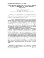
Novel dexamethasone-HPMA copolymer conjugate and its potential application in treatment of rheumatoid arthritis pdf
Bạn đang xem bản rút gọn của tài liệu. Xem và tải ngay bản đầy đủ của tài liệu tại đây (823.55 KB, 9 trang )
Open Access
Available online />Page 1 of 9
(page number not for citation purposes)
Vol 9 No 1
Research article
Novel dexamethasone-HPMA copolymer conjugate and its
potential application in treatment of rheumatoid arthritis
Dong Wang
1,2
, Scott C Miller
3
, Xin-Ming Liu
1
, Brian Anderson
3
, Xu Sherry Wang
1,4
and
Steven R Goldring
5,6
1
Department of Pharmaceutical Sciences, College of Pharmacy, University of Nebraska Medical Center, 986025 Nebraska Medical Center, COP
3026, Omaha, NE 68198-6025, USA
2
Department of Pharmaceutics and Pharmaceutical Chemistry, University of Utah, 30 South 2000 East, Salt Lake City, UT 84112, USA
3
Department of Radiology/Radiobiology Division, University of Utah, 729 Arapeen Dr., Salt Lake City, UT 84108, USA
4
Washington University in St. Louis, 6515 Wydown Blvd., Campus Box 3519, St. Louis, MO 63105, USA
5
Hospital for Special Surgery, 535 East 70th Street, New York, NY 10021, USA
6
New England Baptist Bone and Joint Institute, Harvard Institutes of Medicine, 4 Blackfan Circle, Boston, MA 02115, USA
Corresponding author: Dong Wang,
Received: 13 Oct 2006 Revisions requested: 17 Nov 2006 Revisions received: 4 Dec 2006 Accepted: 18 Jan 2007 Published: 18 Jan 2007
Arthritis Research & Therapy 2007, 9:R2 (doi:10.1186/ar2106)
This article is online at: />© 2007 Wang et al.; licensee BioMed Central Ltd.
This is an open access article distributed under the terms of the Creative Commons Attribution License ( />),
which permits unrestricted use, distribution, and reproduction in any medium, provided the original work is properly cited.
Abstract
Rheumatoid arthritis (RA) is a chronic autoimmune disease of
unknown etiology. Effective treatment of this disorder has been
hampered by the lack of availability of agents that selectively
target affected joint tissue. We developed a novel pH-sensitive
drug delivery system of dexamethasone (Dex) based on an N-(2-
hydroxypropyl)methacrylamide copolymer (P-Dex) and have
shown that the delivery system specifically accumulates in
inflamed joints in an animal model of arthritis. We hypothesize
that the arthrotropism of the delivery system and the local
acidosis-mediated drug release provide superior therapeutic
efficacy and potentially reduced side effects in RA treatment.
The initial in vitro drug-release study confirmed that the Dex
release is indeed dependent upon the environmental pH. At pH
5, 37°C, the conjugate shows the highest level of drug release.
When administered systemically in an adjuvant-induced arthritis
rat model, P-Dex offers superior and longer-lasting anti-
inflammatory effects compared with systemically administered
free Dex. In addition, greater bone and cartilage preservation
was observed with the P-Dex treatment compared with free Dex
treatment. Our data indicate that the differential effect of the
conjugate is related to its selective accumulation, potential
macrophage-mediated retention, and pH-sensitive drug release
(extracellular and intracellular) in arthritic joints. This newly
developed drug delivery system provides a unique method for
selective targeting of glucocorticoids to inflamed joints which
may potentially reduce systemic side effects.
Introduction
Rheumatoid arthritis (RA) is a chronic systemic inflammatory
disease that leads to the destruction of diarthrodial joints.
Many consider it to be an autoimmune disorder, although the
exact cause is unknown. The primary target of the inflammatory
process is synovial tissue. The inflamed synovium invades and
destroys articular bone and cartilage, leading to significant
pain and disability [1-3].
Currently, there is no cure for RA. The most commonly used
medications for treatment and management of the disease
include nonsteroidal anti-inflammatory drugs, glucocorticoids
(GCs), and disease-modifying anti-rheumatic drugs, including
AIA = adjuvant-induced arthritis; BMD = bone mineral density; Boc = tert-butoxycarbonyl; Boc-NHNH
2
= carbazic acid tert-butyl ester; DCC = N,N'-
dicyclohexylcarbodiimide; Dex = dexamethasone; EPR = enhanced permeability and retention; FPLC = fast protein liquid chromatography; GC =
glucocorticoid; HPLC = high-performance liquid chromatography; HPMA = N-(2-hydroxypropyl)methacrylamide; LA = N, N-dioctadecyl-N', N'-bis(2-
hydroxyethyl)propanediamine; MA-Gly-Gly-OH = N-methacryloylglycylglycine; MRI = magnetic resonance imaging; M
w
= weight average molecular
weight; NMR = nuclear magnetic resonance; P-Dex = N-(2-hydroxypropyl)methacrylamide copolymer-dexamethasone conjugate; pDXA = peripheral
dual x-ray absorptiometry; PVP = polyvinylpyrrolidone; RA = rheumatoid arthritis; SEC = size exclusion chromatography; SF = synovial fluid; TNBS
= 2,4,6-trinitrobenzenesulfonic acid.
Arthritis Research & Therapy Vol 9 No 1 Wang et al.
Page 2 of 9
(page number not for citation purposes)
the so-called biologic agents that target tumour necrosis fac-
tor-alpha and interleukin-1 [1,4]. There is also current empha-
sis on the early diagnosis and treatment of RA.
Although progress has been made in understanding the
molecular mechanisms and identification of novel therapeutic
targets for RA, challenges still remain. Most of the available
therapies for RA do not have tissue specificity. Therefore, to
reach effective drug concentrations in affected joint tissues,
high systemic doses of the therapeutic agent must often be
administered, which may lead to significant adverse systemic
and extra-articular side effects. Reductions in drug doses may
attenuate toxicity but may lead to reduced therapeutic efficacy.
To overcome this limitation, approaches that specifically target
agents to affected joints offer unique promise.
Arthrotropic drug delivery systems may be achieved based on
the unique pathophysiological features of RA. Severe synovial
membrane inflammation (synovitis) with significant angiogen-
esis and influx of inflammatory leukocytes is the hallmark of RA
[4]. The inflammatory synovial lining, especially the pannus tis-
sue, resembles solid tumors in many ways, including the leaky
nature of the associated blood capillaries. This leads to abnor-
mal serum protein infiltration into the synovium and higher pro-
tein content in synovial fluid (SF) from patients with RA
compared with healthy individuals [5,6]. In solid tumors, similar
pathophysiological characteristics lead to the so-called
'enhanced permeability and retention' (EPR) effect for macro-
molecules [7]. Based on this principle, many colloidal drug
delivery systems have been developed for improved cancer
chemotherapy [8-11]. There have been relatively few trials
using liposome [12], albumin [13], and polyvinylpyrrolidone
(PVP) [14] as carriers to deliver anti-rheumatic drugs. More
recently, magnetic resonance imaging (MRI) and histological
analysis have been used to demonstrate the arthrotropism of
N-(2-hydroxypropyl)methacrylamide (HPMA) copolymers in an
adjuvant-induced arthritis (AIA) rat model [15]. In addition to
the arthritic ankle joints, the copolymer showed minor deposi-
tion to other inflammatory tissues such as the knee joints and
the base of the tail where the adjuvant was given.
Another pathological feature of the rheumatic joint is the pres-
ence of low pH. pH values as low as 6.0 have been detected
in the SF from RA joints [16-21]. There also appears to be a
direct correlation between the acidity of the joint tissues and
indices of disease severity [22-24]. The imbalance between
increased metabolic activity and insufficient vascular supply,
which induces a shift toward anaerobic glycolysis and lactate
formation, has been suggested as the cause of the acidosis in
RA [17,20]. Similar pathophysiological conditions have been
found in solid tumors and exploited to provide a specific drug-
releasing mechanism for prodrugs [25,26].
Recently, we designed a novel dexamethasone-HPMA copol-
ymer conjugate (P-Dex) with a pH-sensitive drug-releasing
mechanism. Here, we report its synthesis, in vitro drug release,
and in vivo use to treat animals in a rat model of inflammatory
arthritis. Our results provide evidence that the therapeutic effi-
cacy of the conjugate is related to its selective accumulation
and pH-sensitive drug release (extracellular and intracellular)
in arthritic joints. This newly developed drug delivery system
provides a unique method for selective targeting of GCs to
inflamed joints which may potentially reduce adverse extra-
articular side effects.
Materials and methods
Materials
HPMA [27], MA-FITC (N-methacryloylaminopropyl fluorescein
thiourea) [28], N-methacryloylglycylglycine (MA-Gly-Gly-OH)
[29], and N, N-dioctadecyl-N', N'-bis(2-hydroxyethyl)propane-
diamine (LA) [30] were prepared as described previously.
Sephadex LH-20 resin was obtained from GE Healthcare (Pis-
cataway, NJ, USA). N-(3-Aminopropyl)diethanol amine and
carbazic acid tert-butyl ester (Boc-NHNH
2
) were obtained
from TCI America (Portland, OR, USA). Mycobacterium tuber-
culosis H37Ra (heat-killed, desiccated) was obtained from
VWR International (West Chester, PA, USA). Paraffin oil (low
viscosity, Bayol F) was obtained from Crescent Chemical
Company, Inc. (Islandia, NY, USA). Dexamethasone (Dex) was
purchased from Hawkins, Inc. (Minneapolis, MN, USA). If not
specified, all other reagents and solvents were purchased
from Sigma-Aldrich (St. Louis, MO, USA) and used without
further purification.
Characterization of the synthetic products
The weight average molecular weight (M
w
) and number aver-
age molecular weight of copolymers were determined by size
exclusion chromatography (SEC) using the ÄKTA fast protein
liquid chromatography (FPLC) system (GE Healthcare)
equipped with UV and refractive index detectors (KNAUER,
Berlin, Germany). SEC measurements were carried out on
Superose 12 columns (HR [high-resolution] 10/30) (GE
Healthcare) with phosphate-buffered saline (pH 7.3) as the
eluent. The average molecular weights of the polymers were
calculated using calibrations with poly(HPMA). UV spectra of
all tested compounds were obtained on a Cary 400 Bio UV-
Vis spectrometer (Varian, Inc., Palo Alto, CA, USA).
1
H NMR
spectra of all synthesized compounds were recorded on a Var-
ian Unity 500-MHz NMR spectrometer (Varian, Inc.). The sol-
vent peak was used for reference (d
6
-dimethyl sulfoxide, 2.49
ppm). Mass spectra of all synthesized compounds were
obtained using a Finnigan LCQ DECA mass spectrometer
(Thermo Electron Corporation, Waltham, MA, USA) interfaced
to an electrospray ionization (ESI) source.
Synthesis of P-Dex
P-Dex was synthesized with the following exemplary proce-
dure (Figure 1). HPMA (1 g, 7 mmol) and MA-Gly-Gly-OH
(0.156 g, 0.78 mmol) were copolymerized using AIBN (2,2'-
azobisisobutyronitrile) (0.007 g, 0.043 mmol) as initiator. The
Available online />Page 3 of 9
(page number not for citation purposes)
copolymer (1 g [-COOH] = 0.6 mmol) was then reacted with
Boc-NHNH
2
(1.68 g, 12.6 mmol) using N,N'-dicyclohexylcar-
bodiimide (DCC) as the coupling agent. After removal of the
resulting dicyclohexylurea and the extra DCC, the Boc (tert-
butoxycarbonyl) protection in the resulting conjugate was
removed by trifluroacetic acid treatment for 2 hours. The
resulting polymer was precipitated, dialyzed, and lyophilized to
obtain the HPMA copolymer-hydrazide conjugate ([-NHNH
2
]
= 4 × 10
-4
mol/g). This copolymer (0.75 g) was mixed with an
excess of Dex (0.36 g, 9.2 × 10
-4
mol) in N,N-dimethylforma-
mide (1 ml), and one drop of acetic acid was added to catalyze
the reaction. It was stirred overnight at room temperature and
then purified on an LH-20 column (×2) to remove the unre-
acted low molecular weight compounds.
In vitro Dex release
P-Dex (1.8 mg/ml) was dissolved in acetate buffer (0.01 M
with 0.15 M NaCl, pH 5.0) or phosphate buffer (0.01 M with
0.15 M NaCl, pH 6.0 and pH 7.4) and incubated at three dif-
ferent temperatures (4°C, 25°C, and 37°C). At selected time
intervals, the conjugate solution (300 μl) was withdrawn and
extracted with ethyl acetate (3 × 400 μl). After Speed Vac
®
(SC100, Savant Instruments Inc., Holbrook, NY, USA)
removal of the solvent, the isolated samples were dissolved in
acetonitrile/water (1:1, vol/vol, 600 μl) for high-performance
liquid chromatography (HPLC) analysis. An Agilent 1100
HPLC system (Agilent Technologies, Inc., Santa Clara, CA,
USA) with a quaternary pump (with degasser), an autosam-
pler, a fluorescence detector, and a diode-array-based UV
detector was used for the Dex-release study.
A reverse phase C
18
(Agilent, 4.6 × 150 mm, 5 μm) was used
for the analysis with acetonitrile/water = 1/1 as the mobile
phase. Its flow rate was set constant at 0.5 mL/min. The UV
detection wavelength was at 240 nm. The sample injection
volume was 10 μL for all evaluation. A linear external Dex cali-
bration curve was established in the range of 1 to 150 μg. The
calibration was performed with the analysis of each batch of
Dex sample. The analysis of each sample was repeated three
times. The resulting mean value was converted to the percent-
age of Dex released.
Treatment of AIA rats with P-Dex
Male Lewis rats (175 to 200 g) were obtained from Charles
River Laboratories, Inc. (Wilmington, MA, USA) and allowed to
acclimate for at least 1 week. To induce arthritis, M.
Figure 1
The synthesis of N-(2-hydroxypropyl)methacrylamide (HPMA) copolymer-dexamethasone conjugateThe synthesis of N-(2-hydroxypropyl)methacrylamide (HPMA) copolymer-dexamethasone conjugate. Conjugation to dexamethasone may occur at
either the 3 or the 20 carbonyl group (an example of the latter is shown). AIBN, 2,2'-azobisisobutyronitrile; Boc-NHNH
2
, carbazic acid tert-butyl
ester; DCC, N, N'-dicyclohexylcarbodiimide; TFA, trifluroacetic acid.
Arthritis Research & Therapy Vol 9 No 1 Wang et al.
Page 4 of 9
(page number not for citation purposes)
tuberculosis H37Ra (1 mg) and LA (5 mg) were mixed in par-
affin oil (100 μl), sonicated, and injected subcutaneously into
the base of the tail [20]. The progression of the joint inflamma-
tion was monitored daily. Special care was given to the rats as
the inflammation developed to ensure access to water and
food. All animal experiments were performed according to a
protocol approved by the University of Utah Institutional Ani-
mal Care and Use Committee and adhered to Principles of
Laboratory Animal Care (National Institutes of Health publica-
tion 85–23, revised in 1985).
Rats with established arthritis were selected and randomly
assigned into three groups (six or seven rats per group). A
fourth healthy, untreated group (seven rats) was included as
control. On the 13th day after arthritis induction, P-Dex (10
mg/kg, containing 2 mg of Dex/10 mg P-Dex) was injected
intravenously into one group of the RA rats. An equivalent total
dose of water-soluble free Dex (sodium phosphate) was
divided into four aliquots and administered in separate intrave-
nous injections to the second group of rats on days 13, 14, 15,
and 16. Saline was similarly given to a third group of rats (con-
trols). The changes in ankle size and body weight during the
treatment were monitored.
On day 22, all animals were euthanized and joint tissues were
collected. The bone mineral densities (BMDs) of the ankle
region (distal tibia to the phalanges of the foot) and the whole
femur and lumbar vertebral bodies (fourth and fifth) were
measured by peripheral dual x-ray absorptiometry (pDXA). The
tissues were fixed in phosphate-buffered formalin for 2 days,
and the intact ankle regions were then dehydrated in ascend-
ing concentrations of ethanol and embedded undecalcified in
methyl methacrylate. Sections of the entire joint were cut with
a low-speed saw using diamond-wafering blades. The sec-
tions were mounted on plastic slides and ground to approxi-
mately 50 μm in thickness, and the surface was stained using
a Giemsa stain modified for plastic sections [31]. The ankle
joints were assessed for the presence of inflammation and tis-
sue damage. The extent of osteoclastic eroded cancellous
bone surface was measured in the calcaneus using an image
analysis system (BIOQUANT Image Analysis Corporation,
Nashville, TN, USA). The percentage of the cancellous surface
undergoing osteoclastic bone resorption as determined by the
presence of osteoclasts and resorption pits (eroded surfaced)
was calculated.
Statistical methods
The differences between the groups were first tested by a one-
way analysis of variance followed by a Fisher's predicted least-
square difference test to determine the significance of individ-
ual group comparisons. Differences were considered to be
significant at a p value of less than 0.05.
Results
Synthesis and in vitro Dex release from P-Dex
After overnight polymer-analogue reaction between the pen-
dent hydrazide and Dex (acid catalyzed), the conjugate was
purified twice using an LH-20 column. A subsequent FPLC
analysis of the conjugate indicated that there was no detecta-
ble free Dex in the purified copolymer conjugate. The remain-
ing hydrazide in the conjugate was determined using the
TNBS (2,4,6-trinitrobenzenesulfonic acid or picrylsulfonic
acid) assay [32] and compared with the hydrazide content in
the polymer precursor. The reduction of hydrazide content was
due to Dex conjugation and the amount of Dex conjugated was
determined as 50 mg/g of polymer conjugate. The M
w
of the
conjugate was 73 kDa with a polydispersity index of 1.4 and
this material was selected for use in the treatment study of the
rats. Given that no difference in M
w
was observed for P-Dex
and its precursor, the possibility that both carbonyl groups in
Dex would react with hydrazide and cause cross-linking of the
polymer was minimal.
For the in vitro release study, another batch of P-Dex was syn-
thesized with the Dex content determined to be 106 mg/g pol-
ymer conjugate by the TNBS assay and by HPLC after full
hydrolysis. As shown in Figure 2, the Dex release rate depends
on temperature. When the incubation temperature increased
from 4°C to 37°C, the Dex releasing rate was greatly
increased. We also confirmed that the release of Dex from P-
Dex was indeed pH-dependent. As can be seen in Figure 2,
the conjugate demonstrated close to a zero-order release pro-
file during the 14 days at all pH levels tested. The highest drug
release occurred at 37°C in the most acidic buffer (pH 5.0),
with approximately 14% of the loaded Dex released at the end
of 14 days. This amounted to a release rate of approximately
1% of the loaded drug per day. However, all other drug-
release pH levels did not yield significant drug release (<5%
after 14 days).
Observation during the treatment of AIA rats
At 13 days after arthritis induction, the ankle size of most of the
AIA rats reached a plateau (data not shown). At this time, the
hind legs of the affected animals were less mobile and the ani-
mals had reduced body weights. Some inflammation was also
observed in the front limbs, but it was not as severe as the
inflammation observed in the hind limbs. The intravenous
administration of free Dex or P-Dex on day 13 led to a rapid
suppression of the inflammation. The ankle sizes of both treat-
ment groups were greatly reduced by day 14. Though not
quantified, the animals were more mobile and active after the
Dex or P-Dex treatments. The effects of P-Dex on the suppres-
sion of observable inflammation and mobility and activity lasted
for the duration of the entire study (euthanasia on day 22). This
was unlike the animals given four daily injections of Dex, in
which the inflammation and decreased mobility rapidly
returned after the cessation of the Dex injections. By the end
of the study on day 22, the animals that had been treated with
Available online />Page 5 of 9
(page number not for citation purposes)
Dex were indistinguishable from those in the untreated control
group.
BMD assessment
The measurements of BMD of the ankle region, whole femur,
and lumbar vertebral bodies (fourth and fifth) of all animals at
the end of the study, as determined by pDXA, are presented in
Figure 3. For the ankle region, the AIA+saline group had the
lowest BMDs and the values were significantly different from
all other groups. The AIA+Dex treatment group had signifi-
cantly greater BMDs than the untreated AIA+saline group, but
the values were significantly less than the healthy controls. The
AIA+P-Dex treatment group had BMDs that were significantly
greater than those in the AIA+saline and the AIA+Dex-treated
groups, and the values were not significantly different from the
healthy controls. In the femur, the BMDs of the healthy control
group were significantly greater than those of all the other
groups. Both the AIA+Dex and AIA+P-Dex treatment groups
had significantly greater BMDs than the AIA+saline group.
However, the difference between the AIA+Dex and AIA+P-
Dex treatment groups did not achieve statistical significance.
The BMDs of lumbar vertebral bodies in both the AIA+Dex and
AIA+P-Dex treatment groups were significantly greater than
those in the AIA+saline group but were not significantly differ-
ent from each other or from the healthy control. The BMDs of
lumbar vertebral bodies in AIA+saline group were significantly
less than those in the healthy control group.
Cancellous bone osteoclastic resorption (eroded)
surface measurement
The percentages of endosteal cancellous bone surfaces
undergoing active bone resorption (resorption surface) with
the different treatment groups are presented in Figure 4.
Almost 50% of the total endosteal surface in the calcaneus of
AIA+saline group was undergoing active bone resorption,
whereas only 3% of these surfaces were resorbing in the
healthy controls. The group treated with Dex (AIA+Dex) had
approximately 48% less osteoclast surface than the
AIA+saline group, whereas the AIA+P-Dex group had approx-
imately 82% less osteoclast surface. The osteoclast surface in
the AIA+P-Dex group was significantly different from both the
AIA+saline and AIA+Dex groups, but not from the healthy con-
trol group.
Histological evaluation of ankle joints
Representative histological sections of AIA rats' ankle joints
from all treatment groups are shown in Figure 5. Extensive
bone loss, inflammation, subchondral bone erosion, and
cartilage erosion were evident at the tibia-astrogalus junction
in the untreated rats (Figure 5a) compared with the same
region in the AIA+P-Dex group (Figure 5b). Cancellous bone
surfaces in the untreated controls and the AIA-Dex rats (Figure
5c) were populated with large osteoclasts resorbing bone as
indicated by the presence of cells in resorption pits on the
bone surfaces. Fewer osteoclasts and less active resorption
Figure 2
In vitro dexamethasone (Dex) release from N-(2-hydroxypropyl)methacrylamide (HPMA) copolymer-Dex conjugate at different temperatures and pH levelsIn vitro dexamethasone (Dex) release from N-(2-hydroxypropyl)methacrylamide (HPMA) copolymer-Dex conjugate at different temperatures and pH
levels. n = 3, standard deviation is less than 5% of mean value.
Figure 3
Bone mineral density (BMD) measurement of healthy and adjuvant-induced arthritis (AIA) ratsBone mineral density (BMD) measurement of healthy and adjuvant-
induced arthritis (AIA) rats. BMD was evaluated with peripheral dual x-
ray absorptiometry at the ankle, femur, and the fourth and fifth lumbar
vertebral bodies. *Significantly different from the healthy control group,
p < 0.05. **Significantly different from the AIA+saline group, p < 0.05.
***Significantly different from the AIA+Dex group, p < 0.05. Dex, dex-
amethasone; P-Dex, N-(2-hydroxypropyl)methacrylamide (HPMA)
copolymer-dexamethasone conjugate.
Arthritis Research & Therapy Vol 9 No 1 Wang et al.
Page 6 of 9
(page number not for citation purposes)
surfaces were observed in similar cancellous bone regions in
the AIA+P-Dex group (Figure 5d).
Discussion
Colloidal drug delivery systems, including water-soluble poly-
mers, have been used extensively to improve the safety and
efficacy of chemotherapeutic treatment for solid tumors
[10,11]. The pathophysiological 'EPR' effect is considered as
the driving force for their tumor tropic distribution patterns.
Drug-releasing mechanisms based on low tissue pH, hypoxia,
and unique expression patterns of certain enzymes have been
used to enhance the tissue specificity of these delivery sys-
tems [11,25,33]. The application of these drug delivery sys-
tems to improve the current treatment of RA has not been
extensively studied.
PEGylated liposome systems have been used with some suc-
cess to deliver GCs for the treatment of inflammatory arthritis
[12]. As a natural extension, a macromolecular chemothera-
peutic agent, albumin-methotrexate conjugate, has also been
tested in an arthritic rodent model [13]. Using MRI techniques,
we have previously demonstrated that the HPMA copolymer
can specifically accumulate and be retained (for 1 to 2 days)
in inflamed ankle joints in rats with AIA [15]. Based on these
observations, we hypothesized that, due to its preferential
deposition to arthritic tissues, HPMA copolymer could selec-
tively deliver a conjugated drug to the inflamed joint tissue
while minimizing exposure of extra-articular tissues to the
active agent. The benefits of this approach include the ability
to increase the therapeutic efficacy by increasing local drug
concentration in arthritic joints and the capacity to reduce sys-
temic side effects. Anti-rheumatic drugs with the potential to
produce systemic or organ-specific adverse side effects
would benefit the most from this approach.
In this proof-of-principle study, we selected Dex as the model
compound to be conjugated to the delivery system. Dex, a syn-
thetic GC, is a very potent anti-inflammatory drug that exhibits
a rapid therapeutic response. Dex and other GCs are often
used in the early phases of RA treatment to relieve symptoms.
It has also been reported that these agents can modify the dis-
ease progression in patients with RA [34]. However, when
used for long-term treatment, GCs are also well known for
their adverse side effects, including secondary osteoporosis,
muscle weakness and atrophy, suppression of the adrenal
gland, increased risk of infection, peptic ulcer disease, and
growth retardation [35]. Therefore, a delivery system that
could selectively direct GCs to arthritic joints but spare the
skeletal and soft tissues would have a significant therapeutic
advantage. If the delivery system could enhance the
therapeutic index and also reduce the side effects of GCs, it
Figure 4
Cancellous bone osteoclast surface measurement in the calcaneus of healthy and adjuvant-induced arthritis (AIA) ratsCancellous bone osteoclast surface measurement in the calcaneus of
healthy and adjuvant-induced arthritis (AIA) rats. Significant differences
were observed between the following groups: AIA+P-Dex versus
AIA+Dex, AIA+P-Dex versus AIA+saline, AIA+Dex versus AIA+saline,
AIA+Dex versus healthy, and AIA+saline versus healthy. Differences
observed between the AIA+P-Dex versus the healthy group were not
significant. Dex, dexamethasone; P-Dex, N-(2-hydroxypropyl)methacry-
lamide (HPMA) copolymer-dexamethasone conjugate.
Figure 5
Histological features of the ankle joint from adjuvant-induced arthritis (AIA) ratsHistological features of the ankle joint from adjuvant-induced arthritis
(AIA) rats. (a) Tibia (Tib) astrogalus (Ast) joint from rat with saline injec-
tion illustrating extensive bone loss, inflammation, and cartilage erosion
(arrows). (b) Same region from a P-Dex-treated animal showing intact
articular cartilage with less subchondral bone erosion. (c) Higher-
power photomicrograph of cancellous bone from a Dex-treated rat
showing extensive osteoclastic (arrows) bone resorption. (d) Same
region from a P-Dex-treated rat showing much less eroded bone com-
pared to the Dex-treated rat (c). Dex, dexamethasone; P-Dex, N-(2-
hydroxypropyl)methacrylamide (HPMA) copolymer-dexamethasone
conjugate.
Available online />Page 7 of 9
(page number not for citation purposes)
could also be used in conjugation with other anti-rheumatic
drugs.
The first challenge for this study was how to conjugate Dex to
HPMA copolymer. In a previous report, Dex was conjugated to
PVP via its hydroxyl group [14]. However, the ester bond con-
jugation proved to be too stable to adequately release the con-
jugated Dex. In the present study, we used a pH-sensitive
hydrazone bond to conjugate Dex to the HPMA copolymer
side chain. This chemical linkage has been used successfully
in conjugating doxorubicin to polymeric drug carriers for
improved treatment of solid tumors [25]. As discussed above,
acidosis is often associated with inflammatory arthritis. Thus, it
was anticipated that the hydrazone bond would be cleaved in
the low pH environment of the inflamed joint to release the
conjugated Dex, thus enhancing the specificity of the delivery
system.
Polymer-analogue reactions were used to conjugate Dex to
the HPMA copolymer (Figure 1). As the first step, hydrazide
was coupled to the side chain -COOH of HPMA copolymer.
After deprotection of the Boc group, Dex was conjugated to
the copolymer via a hydrazone bond with acetic acid as the
catalyst. The advantage of this synthetic route is its simplicity
and ease of purification. However, it is not known which of the
two carbonyl groups in the Dex structure was involved in the
conjugation. NMR analysis of P-Dex was inconclusive
because of the broad peak of the polymeric drug conjugate
(data not shown). As evident in this study, batch-to-batch var-
iation of the drug content in the conjugate was significant for
the polymer-analogue reaction approach. Synthesis of Dex-
containing monomer and its copolymerization with HPMA may
resolve the issue. However, the synthesis and especially the
purification of the Dex-containing monomer may continue to
be difficult.
To validate the pH sensitivity of P-Dex, the conjugate was incu-
bated at three different pH levels in isotonic buffers. As can be
seen in Figure 2, the release of Dex from the conjugate was
indeed pH-sensitive. At 37°C, Dex release under acidic pH
(5.0) is 10 times faster than that released under neutral pH
(7.4). The drug-releasing kinetics may be considered as zero-
order within the tested time frame, which indicates that the
Dex releasing rate is independent of the drug content in P-Dex.
Nevertheless, the overall Dex release from the conjugate was
only 14% of the original loading after 14 days at 37°C (pH
5.0), or approximately 1% per day. Compared to HPMA copol-
ymer-doxorubicin conjugates, this is rather low [36]. Poten-
tially, the in vivo Dex release may be accelerated due to the
presence of various proteins that bind hydrophobic drugs.
It is of interest to see that both the free Dex and the P-Dex
groups showed immediate relief of inflammation after adminis-
tration. Because drug-polymer conjugates are not easily rec-
ognized by their receptors, their therapeutic activity depends
mainly on the amount of free drug released from the conjugate.
It usually takes longer for non-targeted polymer-drug conju-
gates, such as P-Dex, to be endocytosed and incorporated
into the lysosomes where the acidic environment would act to
release the Dex [37]. The free Dex must then escape from the
lysosomal compartment to deliver its anti-inflammatory effect.
Therefore, the rapid anti-inflammatory response from the P-
Dex observed in this study is best explained by rapid
extracellular drug release in the arthritic joint mediated by low
extracellular pH. Such a response indirectly confirms the aci-
dosis condition in the arthritic joints of AIA rats. In addition to
making general observations of the animals, we measured the
change in ankle size with a digital caliper during the treatment.
The ankle size data generally agree with the observations dis-
cussed above. It is difficult to obtain more specific quantitative
measurements for comparison between different groups
because of the inconsistency of joint alignment and measure-
ment position.
A previous MRI study suggested that polymeric drug carriers
such as HPMA copolymers might be retained in the arthritic
joint for at least 1 to 2 days. They were gradually cleared
through the urinary tract. However, this cannot explain the
observed long-lasting (>9 days) therapeutic effect of P-Dex.
One potential explanation is that during its residence in the
synovial tissue, colloidal drug carriers (for example, liposome)
may be endocytosed by cells such as macrophages [38]. Sim-
ilarly, if some of the P-Dex was endocytosed, it would remain
in the acidic lysosomal compartments and act as a drug depot
to gradually release Dex for a prolonged period of time. Con-
sidering the relatively slow release of Dex from P-Dex (Figure
2), one may speculate that the amount of free Dex actually
needed at the arthritic joint to sustain the suppression of
inflammation may be very small.
The ankle joints, compared with other joints, were most
affected by the induced arthritis as determined in a previous
MRI study and in the present study by histopathology. The
advantages of using P-Dex compared with free Dex were also
most evident in the ankle joints. The BMD was significantly
greater, whereas the relative perimeter of cancellous bone sur-
faces undergoing active osteoclastic bone resorption was sig-
nificantly less in the joints from animals treated with one
injection of P-Dex compared with four injections of free Dex. In
the bones of the ankle joint, the BMD of the P-Dex group was
not significantly different from that in healthy controls, indicat-
ing that the P-Dex was more effective than free Dex in slowing
the bone loss during the disease progression. These differ-
ences were not as apparent, however, in the lumbar vertebra
and the femur, where both the P-Dex and Dex groups had a
greater BMD than the untreated group, but the values were
not significantly different from each other. As noted above, the
joints in the femur (knee joint) and the lumbar vertebra
(intervertebral disks) did not have the same level of inflamma-
tion observed in the ankle joints.
Arthritis Research & Therapy Vol 9 No 1 Wang et al.
Page 8 of 9
(page number not for citation purposes)
Osteoclasts are the cells that resorb bone and are responsible
for the bony destruction that accompanies the inflammatory
process in RA. The daily treatments with Dex suppressed
osteoclastic bone resorption in the ankle bones of the AIA rats
compared with the untreated AIA rats, consistent with previ-
ous observation. However, osteoclastic bone resorption was
further suppressed and was significantly less in the animals
given P-Dex compared with free Dex treatment. The fact that
the P-Dex was given 9 days prior to the end of the study and
that the BMD was preserved in this region indicates that the
P-Dex treatment had a sustained effect on limiting osteoclastic
bone resorption. There was also less joint destruction in the P-
Dex-treated animals compared with those treated with free
Dex as confirmed by histological analyses. Further studies will
be required to establish time- and dose-related effects of the
polymer delivery system on bone and joint metabolism.
In this study, the histology was used to substantiate the effi-
cacy of the treatment with respect to preservation of articular
bone structure. pDXA was also used to offer a sensitive meas-
urement of ankle BMD change during the treatment. In future
investigations, we will consider using additional imaging
modalities such as microcomputed tomography to provide
more useful information regarding the preservation of articular
bone morphology with this new treatment strategy.
The experiments presented here were not designed for rigor-
ous evaluation of systemic side effects of GC therapy. It would
be anticipated that, due to its unique design, this delivery sys-
tem (which may be viewed as a macromolecular prodrug)
would have less systemic toxicity. To activate the prodrug, two
conditions must be met: (a) a pathological condition (for exam-
ple, neovascularization in RA joint) that would allow local
enrichment of the prodrug and (b) an acidic environment (for
example, RA joint acidosis and lysosomal compartments) that
would trigger the release of active Dex from the polymer car-
rier. Because healthy tissues and organs would lack at least
one of these conditions, it would be predicted that this therapy
might also reduce the systemic side effects of GC therapy.
Exploration of proper animal models is under way to confirm
this hypothesis.
Conclusion
A novel HPMA copolymer-Dex conjugate was designed, syn-
thesized, and tested in an animal model of inflammatory arthri-
tis. The hydrazone bond linking Dex to HPMA copolymer side
chains was demonstrated to be cleavable under an acidic pH.
When administered systemically, P-Dex proved to offer supe-
rior and longer-lasting anti-inflammatory effects compared with
free Dex, consistent with its selective accumulation, retention,
and pH-sensitive drug release (extracellular and intracellular)
in arthritic joints. Greater bone and cartilage preservation was
observed with the P-Dex treatment compared with free Dex
treatment. This initial study demonstrates that this novel copol-
ymer system may offer therapeutic advantage for the delivery,
retention, and release of drugs in the treatment of RA and
related forms of inflammatory arthritis.
Competing interests
DW and SCM are inventors named in a patent application par-
tially related to the content of this manuscript. University of
Utah holds the full rights to this patent application. DW and
SCM have not received any financial benefit related to this pat-
ent application. All other authors declare that they have no
competing interests.
Authors' contributions
DW conceptualized the treatment strategy, designed and syn-
thesized the P-Dex conjugate, and prepared the manuscript.
SCM participated in the design of the treatment study, carried
out the histology study and data analysis, and participated in
the preparation of the manuscript. X-ML participated in the
synthesis, characterization, and in vitro evaluation of P-Dex.
BA participated in the histology study. SXW participated in the
synthesis and in vitro evaluation of P-Dex. SRG participated in
the design of the treatment study, data evaluation, and the
preparation of the manuscript. All authors read and approved
the final manuscript.
Acknowledgements
The authors are indebted to Drs. Jindřich Kopeček and Pavla Kopečková
for their constant support and helpful discussion during the early devel-
opment of this project. DW is grateful for the financial support he
received from the College of Pharmacy, University of Nebraska Medical
Center as a new member of the faculty. SXW acknowledges the
research fellowship she received from the College of Pharmacy, Univer-
sity of Nebraska Medical Center as a summer student.
References
1. O'Dell JR: Therapeutic strategies for rheumatoid arthritis. N
Engl J Med 2004, 350:2591-2602.
2. Firestein GS: Etiology and pathogenesis of rheumatoid arthri-
tis. In Kelley's Textbook of Rheumatology 7th edition. Edited by:
Harris ED Jr., Budd RC, Genovese MC, Firestein GS, Sargent JS,
Sledge CB. Philadelphia: Elsevier Saunders; 2005:996-1042.
3. McDuffie FC: Morbidity impact of rheumatoid arthritis on
society. Am J Med 1985, 78:1-5.
4. Smolen JS, Steiner G: Therapeutic strategies for rheumatoid
arthritis. Nat Rev Drug Discov 2003, 2:473-488.
5. Wallis WJ, Simkin PA, Nelp WB: Protein traffic in human syno-
vial effusions. Arthritis Rheum 1987, 30:57-63.
6. Levick JR: Permeability of rheumatoid and normal human syn-
ovium to specific plasma proteins. Arthritis Rheum 1981,
24:1550-1560.
7. Matsumura Y, Maeda H: A new concept for macromolecular
therapeutics in cancer chemotherapy: mechanism of tumori-
tropic accumulation of proteins and the antitumor agent
smancs. Cancer Res 1986, 46:6387-6392.
8. Seymour LW: Passive tumor targeting of soluble macromole-
cules and drug conjugates. Crit Rev Ther Drug Carrier Syst
1992, 9:135-187.
9. Tsukioka Y, Matsumura Y, Hamaguchi T, Koike H, Moriyasu F, Kak-
izoe T: Pharmaceutical and biomedical differences between
micellar doxorubicin (NK911) and liposomal doxorubicin
(Doxil). Jpn J Cancer Res 2002, 93:1145-1153.
10. Duncan R: The dawning era of polymer therapeutics. Nat Rev
Drug Discov 2003, 2:347-360.
11. Kopeček J, Kopečková P, Minko T, Lu Z: HPMA copolymer-anti-
cancer drug conjugates: design, activity, and mechanism of
action. Eur J Pharm Biopharm 2000, 50:61-81.
Available online />Page 9 of 9
(page number not for citation purposes)
12. Metselaar JM, Wauben MH, Wagenaar-Hilbers JP, Boerman OC,
Storm G: Complete remission of experimental arthritis by joint
targeting of glucocorticoids with long-circulating liposomes.
Arthritis Rheum 2003, 48:2059-2066.
13. Fiehn C, Muller-Ladner U, Gay S, Krienke S, Freudenberg-Konrad
S, Funk J, Ho AD, Sinn H, Wunder A: Albumin-coupled meth-
otrexate (MTX-HSA) is a new anti-arthritic drug which acts
synergistically to MTX. Rheumatology (Oxford) 2004,
43:1097-1105.
14. Timofeevski SL, Panarin EF, Vinogradov OL, Nezhentsev MV: Anti-
inflammatory and antishock water-soluble polyesters of glu-
cocorticoids with low level systemic toxicity. Pharm Res 1996,
13:476-480.
15. Wang D, Miller SC, Sima M, Parker D, Buswell H, Goodrich KC,
Kopečková P, Kopeček J: The arthrotropism of macromolecules
in adjuvant-induced arthritis rat model: a preliminary study.
Pharm Res 2004, 21:1741-1749.
16. Goldie I, Nachemson A: Synovial pH in rheumatoid knee-joints.
I. The effect of synovectomy. Acta Orthop Scand 1969,
40:634-641.
17. Falchuk KH, Goetzl EJ, Kulka JP: Respiratory gases of synovial
fluids. An approach to synovial tissue circulatory-metabolic
imbalance in rheumatoid arthritis. Am J Med 1970,
49:223-231.
18. Treuhaft PS, McCarty DJ: Synovial fluid pH, lactate, oxygen and
carbon dioxide partial pressure in various joint diseases.
Arthritis Rheum 1971, 14:475-484.
19. Goetzi EJ, Rynes RI, Stillman JS: Abnormalities of respiratory
gases in synovial fluid of patients with juvenile rheumatoid
arthritis. Arthritis Rheum 1974, 17:450-454.
20. Levick JR: Hypoxia and acidosis in chronic inflammatory arthri-
tis; relation to vascular supply and dynamic effusion pressure.
J Rheumatol 1990, 17:579-582.
21. Andersson SE, Lexmuller K, Johansson A, Ekstrom GM: Tissue
and intracellular pH in normal periarticular soft tissue and dur-
ing different phases of antigen induced arthritis in the rat. J
Rheumatol 1999, 26:2018-2024.
22. Farr M, Garvey K, Bold AM, Kendall MJ, Bacon PA: Significance
of the hydrogen ion concentration in synovial fluid in rheuma-
toid arthritis. Clin Exp Rheumatol 1985, 3:99-104.
23. Geborek P, Saxne T, Pettersson H, Wollheim FA: Synovial fluid
acidosis correlates with radiological joint destruction in rheu-
matoid arthritis knee joints. J Rheumatol 1989, 16:468-472.
24. Nordstrom T, Shrode LD, Rotstein OD, Romanek R, Goto T, Heer-
sche JN, Manolson MF, Brisseau GF, Grinstein S: Chronic extra-
cellular acidosis induces plasmalemmal vacuolar type H+
ATPase activity in osteoclasts. J Biol Chem 1997,
272:6354-6360.
25. Kratz F, Beyer U, Schutte MT: Drug-polymer conjugates con-
taining acid-cleavable bonds. Crit Rev Ther Drug Carrier Syst
1999, 16:245-288.
26. Kaneko T, Willner D, Monkovic I, Knipe JO, Braslawsky GR, Green-
field RS, Vyas DM: New hydrazone derivatives of adriamycin
and their immunoconjugates – a correlation between acid sta-
bility and cytotoxicity. Bioconjug Chem 1991, 2:133-141.
27. Kopeček J, Bažilová H: Poly[N-(2-hydroxypropyl)methacryla-
mide]. 1. Radical polymerization and copolymerization. Eur
Polym J 1973, 9:7-14.
28. Omelyanenko V, Kopečková P, Gentry C, Kopeček J: Targetable
HPMA copolymer-adriamycin conjugates. Recognition, inter-
nalization, and subcellular fate. J Control Release 1998,
53:25-37.
29. Rejmanová P, Labský J, Kopeček J: Aminolyses of monomeric
and polymeric p-nitrophenyl esters of methacryloylated amino
acids. Makromol Chem 1977, 178:2159-2168.
30. Cronin TH, Faubl H, Hoffman WW, Korst JJ, inventors: Xylene-
diamines as antiviral agents. US patent 4,034,040 . 5 July 1977
31. Wang D, Miller S, Sima M, Kopečková P, Kopeček J: Synthesis
and evaluation of water-soluble polymeric bone-targeted drug
delivery systems. Bioconjug Chem 2003, 14:853-859.
32. Hancock WS, Battersby JE: A new micro-test for the detection
of incomplete coupling reactions in solid-phase peptide syn-
thesis using 2,4,6-trinitrobenzenesulphonic acid.
Anal
Biochem 1976, 71:260-264.
33. Denny WA: Prospects for hypoxia-activated anticancer drugs.
Curr Med Chem Anticancer Agents 2004, 4:395-399.
34. Kirwan JR: The effect of glucocorticoids on joint destruction in
rheumatoid arthritis. The Arthritis and Rheumatism Council
Low-Dose Glucocorticoid Study Group. N Engl J Med 1995,
333:142-146.
35. Jacobs JWG, Bijlsma JWJ: Glucocorticoid therapy. In Kelley's
Textbook of Rheumatology 7th edition. Edited by: Harris ED Jr.,
Budd RC, Genovese MC, Firestein GS, Sargent JS, Sledge CB.
Philadelphia: Elsevier Saunders; 2005:870-874.
36. Etrych T, Jelinkova M, Rihova B, Ulbrich K: New HPMA copoly-
mers containing doxorubicin bound via pH-sensitive linkage:
synthesis and preliminary in vitro and in vivo biological
properties. J Control Release 2001, 73:89-102.
37. Jensen KD, Kopečková P, Bridge JH, Kopeček J: The cytoplasmic
escape and nuclear accumulation of endocytosed and micro-
injected HPMA copolymers and a basic kinetic study in Hep G2
cells. AAPS PharmSci 2001, 3:E32.
38. Metselaar JM, van den Berg WB, Holthuysen AE, Wauben MH,
Storm G, van Lent PL: Liposomal targeting of glucocorticoids to
synovial lining cells strongly increases therapeutic benefit in
collagen type II arthritis. Ann Rheum Dis 2004, 63:348-353.