Fibroblast-like synovial cells from normal and inflamed knee joints differently affect the expression of pain-related receptors in sensory neurones: a co-culture study ppsx
Bạn đang xem bản rút gọn của tài liệu. Xem và tải ngay bản đầy đủ của tài liệu tại đây (771.64 KB, 12 trang )
Open Access
Available online />Page 1 of 12
(page number not for citation purposes)
Vol 9 No 1
Research article
Fibroblast-like synovial cells from normal and inflamed knee
joints differently affect the expression of pain-related receptors in
sensory neurones: a co-culture study
Gisela Segond von Banchet
1
, Jonny Richter
1
, Marion Hückel
2,3
, Christina Rose
3
, Rolf Bräuer
3
and
Hans-Georg Schaible
1
1
Institute of Physiology, University of Jena, Teichgraben 8, D-07740 Jena, Germany
2
Current address: Roche Diagnostics GmbH, D-82377 Penzberg, Germany
3
Institute of Pathology, University of Jena, Ziegelmühlenweg, D-07740 Jena, Germany
Corresponding author: Hans-Georg Schaible,
Received: 31 Aug 2006 Revisions requested: 3 Oct 2006 Revisions received: 20 Dec 2006 Accepted: 25 Jan 2007 Published: 25 Jan 2007
Arthritis Research & Therapy 2007, 9:R6 (doi:10.1186/ar2112)
This article is online at: />© 2007 Segond von Banchet et al.; licensee BioMed Central Ltd.
This is an open access article distributed under the terms of the Creative Commons Attribution License ( />),
which permits unrestricted use, distribution, and reproduction in any medium, provided the original work is properly cited.
Abstract
Innervation of the joint with thinly myelinated and unmyelinated
sensory nerve fibres is crucial for the occurrence of joint pain.
During inflammation in the joint, sensory fibres show changes in
the expression of receptors that are important for the activation
and sensitization of the neurones and the generation of joint
pain. We recently reported that both neurokinin 1 receptors and
bradykinin 2 receptors are upregulated in dorsal root ganglion
(DRG) neurones (the cell bodies of sensory fibres) in the course
of acute and chronic antigen-induced arthritis in the rat. In this
study, we begin to address mechanisms of the interaction
between fibroblast-like synovial (FLS) cells and sensory
neurones by establishing a co-culture system of FLS cells and
DRG neurones. The proportion of DRG neurones expressing
neurokinin 1 receptor-like immunoreactivity was not altered in
the co-culture with FLS cells from normal joints but was
significantly upregulated using FLS cells from knee joints of rats
with antigen-induced arthritis. The proportion of DRG neurones
expressing bradykinin 2 receptors was slightly upregulated in
the presence of FLS cells from normal joints but upregulation
was more pronounced in DRG neurones co-cultured with FLS
cells from acutely inflamed joints. In addition, the expression of
the transient receptor potential V1 (TRPV1) receptor, which is
involved in inflammation-evoked thermal hyperalgesia, was
mainly upregulated by co-culturing DRG neurones with FLS
cells from chronically inflamed joints. Upregulation of neurokinin
1 receptors but not of bradykinin 2 and TRPV1 receptors was
also observed when only the supernatant of FLS cells from
acutely inflamed joint was added to DRG neurones. Addition of
indomethacin to co-cultures inhibited the effect of FLS cells
from acutely inflamed joints on neurokinin 1 receptor expression,
suggesting an important role for prostaglandins. Collectively,
these data show that FLS cells are able to induce an
upregulation of pain-related receptors in sensory neurones and,
thus, they could contribute to the generation of joint pain.
Importantly, the influence of FLS cells on DRG neurones is
dependent on their state of activity, and soluble factors as well
as direct cellular contacts are crucial for their interaction with
neurones.
Introduction
The inflammatory response in organs is produced by numer-
ous inflammatory cell types. These cell types communicate
with each other in order to develop an appropriate inflamma-
tory reaction. A large amount of information on the mecha-
nisms of interaction of different inflammatory cells has been
obtained from co-culture systems of different cell types, such
as T cells and monocytes [1-3], T cells and endothelial cells
[4], T cells and fibroblasts [5-7], monocytes and fibroblasts
[8,9], and macrophages and fibroblasts [10-12]. These data
have established the importance of both cell-cell contacts and
mediators for the production of the inflammatory activity.
AIA = antigen-induced arthritis; B2 = bradykinin 2; BK = bradykinin; BSA = bovine serum albumin; BSA-C = acetylated bovine serum albumin; COX
= cyclooxygenase; DMEM = Dulbecco's modified Eagle's medium; DRG = dorsal root ganglion; FCS = fetal calf serum; FLS = fibroblast-like synovial;
Ig = immunoglobulin; IL = interleukin; IR = immunoreactivity; NGF = nerve growth factor; NK1 = neurokinin 1; NSE = neurone-specific enolase; PBS
= phosphate-buffered saline; PGE
2
= prostaglandin E
2
; TRPV1 = transient receptor potential V1; TX-100 = Triton X-100.
Arthritis Research & Therapy Vol 9 No 1 von Banchet et al.
Page 2 of 12
(page number not for citation purposes)
Most tissues are innervated, and nerve fibres play an important
role in inflammatory diseases. The activation of nociceptive
sensory afferent fibres ('pain fibres') evokes pain, a major
symptom of inflammatory diseases [13]. Furthermore, there is
growing evidence that primary afferent neurones as well as
sympathetic nerve fibres influence the inflammatory process
through efferent processes [14-16]. Despite the functional
cross-talk between the inflammatory process and neurones,
the mechanistic analysis of interactions between non-neuronal
inflammatory cells and neurones has not been carried out in
great detail. Recently, a first report appeared on the influence
of neurones in the central nervous system on T cells and the
potential role of neurone-T cell interactions on experimental
autoimmune encephalomyelitis [17].
The somata of primary afferent neurones are located in the
dorsal root ganglia (DRG). Similar to the sensory endings, the
somata of these neurones express ion channels and receptors
that are important for the activation and/or sensitization of
these neurones, and they thus seem to represent the total pri-
mary afferent neuron in this respect [18]. In addition, the
expression of ion channels and receptors in the somata is
altered during peripheral inflammation. We recently took
DRGs from normal rats and from rats with unilateral acute and
chronic antigen-induced arthritis (AIA) in the knee, cultured
them for one day and determined which proportion of DRG
neurones express receptors for bradykinin (BK) and sub-
stance P (neurokinin 1 (NK1) receptors). In lumbar DRGs but
not in cervical DRGs from AIA rats we found a pronounced
increase in the proportion of neurones exhibiting BK receptors
and NK1 receptors [19]. The upregulation of these receptors
found in this study and in other studies on inflammation
[20,21] is thought to be involved in the inflammatory pain
response because both BK and substance P activate and/or
sensitize proportions of primary afferent neurones for mechan-
ical stimuli, which is a mechanism of mechanical hyperalgesia
[22-26]. Indeed AIA rats show limping of the inflamed knee
and a lowered pain threshold when pressure is applied to the
knee [19]. In addition, the transient receptor potential V1
(TRPV1) receptor is an ion channel that is involved in thermal
inflammatory hyperalgesia [27-29]. Some studies [30-33], but
not others [34,35], have identified an upregulation of TRPV1
receptors in DRGs in inflammatory models.
We have begun to identify mechanisms that cause the upreg-
ulation of BK, NK1 and TRPV1 receptors in DRG neurones. In
the present study we co-cultured DRG neurones with fibrob-
last-like synovial (FLS) cells from either normal knee joints or
from acutely or chronically inflamed knee joints from AIA rats.
FLS cells are key players in the propagation of joint inflamma-
tion and joint destruction during rheumatoid arthritis [36-39]
whereas DRG neurones are key players in the development of
chronic pain [18]. We addressed three questions. First, is the
expression of these receptors in DRG neurones influenced by
the presence of FLS cells? Second, do FLS cells from normal
and inflamed knee joints exert different effects on receptor
expression in DRG neurones? Third, are effects of FLS cells
on DRG neurones mediated by soluble mediators (is the
supernatant of FLS cells sufficient?) or is the presence of the
FLS cells with cellular contacts important? Preliminary results
have been reported [40,41].
Materials and methods
Induction of joint inflammation
In 17 10-week-old female Lewis rats (Charles River, Sulzfeld,
Germany), an inflammation was induced in the right knee joint.
In the first step the rats received a subcutaneous injection of
500 μg antigen (methylated BSA; Sigma, Deisenhofen, Ger-
many) in 500 μl saline emulsified with 500 μl complete Fre-
und's adjuvant (Sigma; supplemented to 2 mg/ml heat-killed
Mycobacterium tuberculosis strain H37RA, Difco, Detroit, MI,
USA). In addition, an intraperitoneal injection of 2 × 10
9
heat-
inactivated Bordetella pertussis (Chiron Behring, Marburg,
Germany) was performed on the same day. The same immuni-
sation procedure was repeated 7 days later. After a further 14
days, a sterile solution of antigen (methylated BSA), 500 μg in
50 μl saline, was injected into the right knee joint cavity (day
0). Either 3 days (acute AIA) or 20 to 28 days (chronic AIA)
after induction of inflammation in the knee joint, the rats were
killed by cervical dislocation during ether anaesthesia. In total,
17 untreated rats of the same age and sex were used as nor-
mal control animals. All rats were used for the preparation of
FLS cells. All procedures complied with the regulations of the
Thuringian Commission for Animal Protection.
Preparation of fibroblast-like synovial cells
Explant cultures of FLS cells were prepared from the knee
joints of normal rats or from rats 3 days (acute phase) or 20 to
28 days (chronic phase of inflammation) after induction of AIA.
The patella and the menisci of the joints with adjacent synovial
tissue were separated and cultured in 24-well plates in DMEM
(Gibco, BRL, Eggenstein-Leopoldshafen, Germany) contain-
ing 20% fetal calf serum (FCS, [Gibco]), 0.1 mg/ml streptomy-
cin (Grünenthal, Aachen, Germany), 100 U/ml penicillin
(Jenapharm, Jena, Germany), 2 mmol/l glutamine (Gibco), 10
mM Hepes (Gibco), and 1 mmol/l sodium pyruvate (Gibco) for
7 days at 37°C in a humidified incubator gassed with 5% CO
2
in air. During this time, out-growing FLS cells emerged from
the tissue. In the first 7 days the medium was replaced daily.
After 7 days the residual tissue was removed and 2 days later
the cells were transferred to new plates. For this purpose the
cells were washed with PBS and incubated for 2 to 4 minutes
in PBS containing 0.25% trypsin and 0.02% EDTA (Gibco).
Thereafter, the cells were collected, washed with DMEM con-
taining 20% FCS and disseminated. After another 3 to 6 days
the cells were transferred into new plates. For the co-culture,
cells were used after the third to fifth passage. FLS cells were
slowly cooled down with isopropanol to -70°C in DMEM con-
taining 10% dimethylsulfoxide and stored at -192°C over liq-
uid nitrogen until co-culturing.
Available online />Page 3 of 12
(page number not for citation purposes)
Primary culture of dorsal root ganglion neurones
Normal male Wistar rats, 60 days old, were sacrificed with a
lethal dose of ether. DRGs from all segments of the spinal cord
were dissected. Ganglia were incubated at 37°C with 215 U/
ml collagenase type II (Paesel and Lorei, Hanau, Germany) dis-
solved in Ham's F12 medium (Gibco) for 100 minutes. After
washing with Ca
2+
- and Mg
2+
-free PBS, the ganglia were
placed in DMEM (Gibco) containing 10,000 U/ml trypsin
(Sigma) for 11 minutes at 37°C. The cells were dispersed by
gentle agitation and aspiration with a fire polished Pasteur-
pipette. The dispersed cells were collected by centrifugation
(500 × g, 5 minutes), washed 3 times in DMEM and centri-
fuged. The cell pellets were suspended in Ham's F-12 medium
containing 10% heat-inactivated horse serum (Gibco), 100 U/
ml penicillin (Gibco), 100 μg/ml streptomycin (Gibco), and
100 ng/ml nerve growth factor (NGF; Paesel and Lorei). On
average, 200 to 300 DRG neurones were plated on poly-L-
lysine- (200 μg/ml) coated glass cover slips (diameter 13 mm)
situated in 35/10 mm dishes and maintained for 1 night at
37°C in a humidified incubator gassed with 3.5% CO
2
and air.
After this overnight setting period, the co-cultures of DRG
neurones and the FLS cells were prepared.
Co-culture of FLS cells and DRG neurones
Two days before co-culturing, the FLS cells (from normal rats
or from rats with acute or chronic AIA) were thawed and cul-
tured in DMEM containing 20% FCS in a concentration of 2 ×
100 cells/well in 24-well dishes. After 2 days, the FLS cells
were incubated with 0.25% trypsin (Gibco) and 0.02% EDTA
(Sigma) in DMEM for 2 to 4 minutes at 37°C. Thereafter, the
cells were washed 3 times with DMEM (containing 100 ng/ml
NGF) and added to the glass cover slips with the cultured
DRG neurones (see above) in a concentration of 10
5
/ml. The
two cell types were co-cultured for 24 hours in DMEM contain-
ing 100 ng/ml NGF maintained at 37°C in a humidified incu-
bator gassed with 5% CO
2
in air. As a control, either only DRG
neurones or only FLS cells were cultured and handled in the
same way as the co-cultures. After culturing for 24 hours, all
cells on the glass cover slips were fixed and used for immuno-
cytochemical labelling of the NK1, the BK 2 (B2) and the
TRPV1 receptors.
In addition, in some experiments, DRG neurones were cul-
tured in medium containing only the supernatant of FLS cells.
These FLS cells were isolated from either normal knee joints
or knee joints at the acute (day 3) or chonic state (20 to 28
days) of AIA and then cultured for 2 days. The supernatants of
these FLS cells were added to the DRG neurones, which were
kept for 24 hours at 37°C in a humidified incubator gassed
with 5% CO
2
in air, and, in addition, 100 ng/ml NGF were
administered.
Furthermore, co-cultures of DRG neurones and FLS cells from
normal or acutely and chronically inflamed knee joints were
made in a medium containing either the cyclooxygenase
(COX) inhibitor indomethacin (1 μmol/l; Calbiochem, Bad
Soden/Ts, Germany), an antibody against IL-6 (1 μmol/l;
BioTrend, Köln, Germany) or IgG from normal rat (1 μmol/l;
BioTrend). These co-cultures were kept at 37°C in a humidi-
fied incubator gassed with 5% CO
2
in air for 24 hours in
DMEM containing 100 ng/ml NGF.
Detection of bradykinin 2 receptors
Because a reliable B2 receptor antibody was not available, we
used BK-gold conjugates for labelling of B2 receptors. The
BK-gold conjugates were prepared as described earlier [42].
In brief, 1 μmol BK (Bachem, Heidelberg, Germany) was dis-
solved in 500 μl HEPES (20 mmol/l, pH 7.5). This solution
was added to 6 nmol sulfo-N-hydroxy-succinimido Nanogold
reagent (BioTrend), dissolved in 500 μl ddH
2
O, and incubated
for 1 hour at room temperature. To separate BK-gold conju-
gates from unbound BK, a membrane centrifugation (Amicon
microcon-10 system) was used. The BK-gold conjugate was
dissolved in PBS containing 0.1% BSA (Sigma), 0.2 mol/l
sucrose (Sigma), 4 μg/ml leupeptin (Sigma) and 10 mmol/l
sodium azide (Sigma). This solution was aliquoted and stored
at -20°C for a maximum of three months.
The cells were fixed with 2% paraformaldehyde in 0.1 mol
phosphate buffer (pH 7.2) for 30 minutes. After washing with
PBS (20 mmol/l, pH 7.4), the cells were pre-treated with 50
mmol/l glycine in PBS and, thereafter, with 5% BSA and 0.1%
gelatine in PBS for 30 minutes. Then the cells were washed
with 0.1% acetylated BSA (BSA-C; BioTrend) and incubated
overnight with 0.3 nmol/ml BK-gold in PBS containing 0.1%
BSA-C, bacitracin (40 μg/ml; Sigma), leupeptin (4 μg/ml;
Sigma) and chymostatin (2 μg/ml; Sigma) at 4°C in a moist
chamber. Following washing with PBS plus 0.1% BSA-C and,
thereafter, with PBS to remove unbound BK-gold, cells were
postfixed with 2% glutaraldehyde in PBS for 10 minutes. After
extensive washing with PBS and ddH
2
O, the gold particles
were intensified with silver enhancer (R-Gent, pH 5.5;
BioTrend) for 15 minutes at 22°C. The reaction was stopped
by washing in ddH
2
O. To examine whether the binding was
related to B2 receptors, 3 nmol/ml BK-gold was incubated in
parallel control dishes in the presence of 1 μmol/ml D-Arg
[Hyp
3
-Thi
5,8
-D-Phe
7
]-BK (Sigma), a BK analogue that specifi-
cally binds to the B2 receptor. This analogue produces a com-
plete displacement of BK-gold.
Immunocytochemical labelling of NK1 and TRPV1
receptors
The cover slips were transferred to 2% paraformaldehyde in
0.1 mol/l phosphate buffer (pH 7.2) plus 0.3% Triton X-100
(TX-100) for 30 minutes. After washing with PBS plus 0.3%
Triton X-100 (PBS TX-100), cells were incubated with 50
mmol/l glycine in PBS TX-100 and, thereafter, with 5% BSA
and 0.1% gelatine in PBS TX-100 for 30 minutes. Then the
cells were washed with PBS TX-100 and incubated for 30
minutes in PBS TX-100 containing 2% normal goat serum
Arthritis Research & Therapy Vol 9 No 1 von Banchet et al.
Page 4 of 12
(page number not for citation purposes)
(BioTrend). Thereafter, the cells were washed with PBS TX-
100 containing 0.1% BSA-C and incubated overnight with an
anti-NK1 antibody diluted 1:100 in PBS TX-100 plus 1% nor-
mal goat serum (the antibody was raised in rabbit against
amino acids 393 to 407 of the rat NK1 receptor; Sigma) or
with an antibody to the TRPV1 receptor (VR11-A, 1:75; Alpha
Diagnostics, San Antonio, USA) at 4°C in a moist chamber.
The cover slips were extensively rinsed in PBS TX-100 plus
0.1% BSA-C and, thereafter, in PBS TX-100. After washing,
the cells were incubated for 4 hours at 20°C with a gold-
labelled (10 nm) anti-rabbit antibody developed in goat
(BioTrend), diluted 1:100 in PBS TX-100 plus 1% normal goat
serum. After washing with PBS TX-100, PBS and ddH
2
O, the
gold particles were intensified with silver enhancer (R-Gent,
pH 5.5) for 20 minutes at 21°C. The reaction was stopped by
washing in ddH
2
O. To test for unspecificity of the detection
system, cells were incubated only with the secondary anti-
body. In these cultures no cells were labelled.
Double-labelling with an antibody against neurone-
specific enolase
To identify the neurones in the cultures, double-staining with
an anti-neurone-specific enolase (NSE) antibody was used in
all experiments in which B2, NK1, and TRPV1 receptor label-
ling was performed. After washing with PBS, the cover-slips
were incubated overnight at 4°C with the anti-NSE-antibody
(Sigma) developed in rabbit (diluted 1:100). Then the cover-
slips were incubated with a goat anti-rabbit antibody labelled
with Cy3 (diluted 1:200; Jackson ImmunoResearch, Cam-
bridgeshire, UK) for 2 hours. After washing with PBS, all cover
slips were embedded in Vectashield (Vector, Burlingame,
England). In control experiments in which the primary antibody
(anti-NSE) was omitted, no fluorescence signal was
detectable.
Prostaglandin E
2
and IL-6 measurement in the
supernatant of the cultures
The supernatants of cultured DRG neurones, cultured FLS
cells and co-cultures of DRG neurones and FLS cells (from
normal and acutely or chronically inflamed joints) were ana-
lysed for the production of prostaglandin E
2
(PGE
2
) and IL-6.
The samples were stored at -20°C until analysis. For each sub-
stance, four independent cultures were used. All samples
were measured twice. The supernatants were analysed with
commercial ELISA kits for PGE
2
(Cayman Chemicals, Ann
Arbor, Michigan, USA) and for rat IL-6 (OptEIA; BD Bio-
sciences, Heidelberg, Germany).
Data analysis
From each cover slip, 100 structurally intact and NSE-labelled
neurones were examined with a light microscope (Axioplan 2,
Zeiss, Germany) coupled to a CCD video camera and an
image analysing system (KS 300, Zeiss, Germany). The mean
area and mean grey value were determined for each neuronal
soma. To take into account differences in the basal grey values
on each coated cover slip, a relative grey value of each neu-
rone was calculated by dividing the mean grey value of the
neurone by the grey value of the cover slip background. For an
unbiased discrimination of cells with or without positive label-
ling with the antibodies against NK1 and TRPV1 or BK-gold,
neurones were considered positive if their relative grey value
was above that of neurones from the control incubations,
which were not treated with the antibodies against NK1 or
TRPV1 receptors. In experiments with BK-gold labelling, neu-
rones were considered positive if their relative grey value was
above that of neurones from the displacement control incuba-
tions with a BK analogue that specifically binds to the B2
receptor. This value was 0.16; thus, all neurones with grey
density >0.16 were considered as positive for the antibodies
or B2 receptor BK-gold binding. Proportions of labelled neu-
rones are expressed as the mean ± standard deviation. For
statistical analysis we used χ
2
-tests taking into account multi-
ple comparisons. Significant differences were acknowledged
if p < 0.05.
Results
Co-culture system
As a first step we characterized the morphology of FLS cells
and DRG neurones in the co-culture system. The FLS cells
formed a flat layer composed mainly of triangular NSE-nega-
tive cells (Figure 1a). After the setting period, DRG neurones
that showed strong NSE-like immunoreactivity (IR) were dis-
persed as single cells or small cell clusters on the FLS cell
layer (Figure 1b, neurones are labelled with stars). Only very
few FLS cells (maximum of 1%) showed some NSE-like IR
and, therefore, the antibody against NSE is a good tool to dif-
ferentiate between neurones and FLS cells. The neurones had
round perikarya of varying sizes and thin neurites spanning
over and along FLS cells (Figure 1b–d).
DRG neurones are characterized by the size of their cell body
and exhibit a typical size distribution with most cells in the
small- and medium-sized range (which give rise to unmyeli-
nated and thin myelinated axons) and fewer cells in the large-
sized range (which give rise to thick myelinated axons). To
compare the size distribution of DRG neurones in different cul-
tures, we determined the diameter of neurones in the DRG
mono-culture and in the co-cultures with FLS cells from
acutely and chronically inflamed knee joints. The size distribu-
tion of neurones was similar under all culture conditions, indi-
cating that the DRG cell samples were comparable (Figure 2).
The morphology of FLS cells was also similar in mono-cultures
and in co-cultures with DRG neurones.
Expression of NK1, B2, and TRPV1 receptor-like IR in
DRG neurones after co-culture with FLS cells
As a first approach we tested whether the presence of FLS
cells from normal, acutely or chronically inflamed knee joints
influences receptor expression in DRG neurones taken from
normal rats. DRG neurones were fixed 24 hours after co-cul-
Available online />Page 5 of 12
(page number not for citation purposes)
turing and receptor expression was determined in NSE-
labelled DRG neurones.
Neurokinin 1 receptor-like immunoreactivity
In the standard mono-culture of DRG neurones from adult nor-
mal rats, only a small proportion of neurones showed NK1
receptor-like IR, similar to previously reported studies [19,43].
On average, 8.8 ± 2.0% of the DRG neurones (4 cultures)
were labelled with the anti-NK1 receptor antibody (Figure 3c,
first bar). When DRG neurones were co-cultured with FLS
cells isolated from the knee joint of normal adult rats, a similar
proportion of DRG neurones (8.4 ± 2.1%, 5 cultures) showed
NK1 receptor-like IR (Figure 3c, second bar). However, in co-
cultures of DRG neurones from normal rats and FLS cells from
the inflamed knee of AIA rats, the proportion of neurones
expressing NK1 receptor-like IR was significantly higher (Fig-
ure 3c, third and fourth bar). Figure 3a,b displays a cover slip
with a co-culture of DRG neurones and FLS cells isolated from
acutely inflamed knee joints (3 days of AIA). The dark cell in
Figure 3a (see arrow) is labelled for NK1 receptor-like IR, and
Figure 3b shows that this cell is also NSE-positive. Counting
of double-labelled neurones showed that 31.3 ± 6.7% of the
DRG neurones expressed NK1 receptor-like IR after co-cul-
ture with FLS cells isolated from knee joints of rats at 3 days
of AIA (4 cultures), whereas 27.0 ± 2.9% of the DRG neu-
rones were labelled with the anti-NK1 receptor antibody after
co-culture with FLS cells isolated from the knee joint of rats at
21 to 28 days of AIA (chronic AIA, 4 cultures). At both time
points, NK1 receptor expression was increased significantly
compared to the mono-culture of DRG neurones and the DRG
co-culture with FLS cells from normal knee joints. Black col-
umns in Figure 2a show the proportions of neurones exhibiting
NK1 receptor-like IR. Thus, FLS cells isolated from both
acutely and chronically inflamed knee joints induced an upreg-
ulation of the NK1 receptor in DRG neurones from normal rats
whereas a co-culture with FLS cells from normal knee joints
did not. FLS cells themselves did not show NK1 receptor-like
IR.
Bradykinin 2 receptor-like labelling
Because a BK analogue that specifically binds to the B2
receptor completely displaced the BK-gold particle (see Mate-
rials and methods), we conclude that BK-gold binds only to B2
receptors in this experimental approach. In the standard mono-
culture of DRG neurones from normal adult rats, 36.8 ± 3.4%
of all DRG neurones (5 cultures) showed B2 receptor-like
labelling (Figure 4c, first bar), which is in the same range as in
previous studies [19,42,44,45]. When DRG neurones and
FLS cells isolated from knee joints of normal adult rats were
co-cultured, on average, 53.5 ± 7.1% of the DRG neurones (4
cultures) were labelled for B2 receptors. Thus, in contrast with
NK1 receptor expression, FLS cells from normal knee joints
caused a significant up-regulation of the proportion of DRG
neurones exhibiting B2 receptor-like labelling (Figure 4c, sec-
ond bar). Compared to co-cultures of DRG neurones and FLS
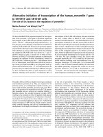
Figure 1
Morphology of cultured fibroblast-like synovial (FLS) cells and co-cul-tured dorsal root ganglion (DRG) neuronesMorphology of cultured fibroblast-like synovial (FLS) cells and co-cul-
tured dorsal root ganglion (DRG) neurones. (a) Mono-culture of FLS
cells. (b) Co-culture of FLS cells and DRG neurones. The DRG neu-
rones show neurone-specific enolase-like immunoreactivity (white stain-
ing indicated by stars). (c,d) Co-culture of FLS cells and DRG
neurones. The processes of DRG neurones are indicated with arrows.
Note the contact between the DRG neurones and the FLS cells (d).
Scale bars: 10 μm (a); 30 μm (b,c); 10 μm (d).
Arthritis Research & Therapy Vol 9 No 1 von Banchet et al.
Page 6 of 12
(page number not for citation purposes)
cells from normal joints, a significantly higher proportion of
DRG neurones (on average, 69.5 ± 9.9% of the DRG neu-
rones, 4 cultures) showed B2 receptor expression when FLS
cells from acutely inflamed knee joints were used for co-cul-
ture (Figure 4c, third bar). In co-cultures of DRG neurones and
FLS cells from chronically inflamed joints, 56.5 ± 16% of the
DRG neurones were labelled for B2 receptors (8 cultures, not
significantly different from co-cultures of DRG neurones and
FLS cells from normal knee joints). Figure 4a,b displays a
cover slip with a co-culture of DRG neurones and FLS cells
isolated from knee joint from rats at the acute AIA stage. The
size distribution of labelled neurones is shown in Figure 2b.
FLS cells did not exhibit B2 receptor labelling.
TRPV1 receptor-like immunoreactivity
In 13 DRG mono-cultures from normal adult rats, 21.0 ± 5.1%
of the neurones showed TRPV1 receptor-like IR (Figure 5c,
first bar). In 7 co-cultures of DRG neurones and FLS cells iso-
lated from normal knee joints, on average, 28.1 ± 11.3% of the
DRG neurones showed TRPV1 receptor-like IR (Figure 5c,
second bar), and this proportion was not different from that in
DRG neurones in mono-cultures. Co-cultures of DRG neu-
rones with FLS cells from knee joints of AIA rats yielded higher
proportions of neurones exhibiting TRPV1 receptor-like IR.
After co-culture with FLS cells from acutely inflamed knee
joints, 34.5 ± 5.5% of DRG neurones were immunopositive for
the TRPV1 receptor (4 cultures), and after co-culture with FLS
cells from chronically inflamed knee joints, 50.1 ± 6.8% of the
DRG neurones were immunopositive (7 cultures; Figure 5c,
third and fourth bar). These proportions of immunopositive
neurones were significantly higher than the proportion of those
in the DRG mono-culture but only the latter value (co-cultures
with FLS cells from chronically inflamed joints) was signifi-
cantly higher than the proportion of immunopositive neurones
in the co-culture with FLS cells from normal joints. Figure 5a,b
displays a cover slip with a co-culture of DRG neurones and
FLS cells isolated from acutely inflamed knee joints. Figure 2c
shows the size distribution of labelled neurones. FLS cells did
not show TRPV1 receptor-like IR.
Influence of soluble FLS cell mediators on the
expression of NK1, B2, and TRPV1 receptors
In a second approach, we tested whether soluble mediators
are responsible for the up-regulation of NK1, B2, and TRPV1
receptors that is observed in co-culture with FLS cells. We
cultured DRG neurones and added only the supernatant from
Figure 2
Size distribution of dorsal root ganglion (DRG) neurones under different culture conditionsSize distribution of dorsal root ganglion (DRG) neurones under different culture conditions. The open bars in the graphs show the proportions of
neurones (%) in the indicated size ranges; the black insets show the proportions of neurones that express neurokinin 1 (NK1) receptor-like immuno-
reactivity (a), bradykinin 2 (B2) receptor labelling (b), and transient receptor potential V1 (TRPV1) receptor-like immunoreactivity (c). Cells were
grown in DRG mono-culture (top) or together with fibroblast-like synovial (FLS) cells from normal, acutely inflamed and chronically inflamed knee
joints.
Available online />Page 7 of 12
(page number not for citation purposes)
cultured FLS cells sampled from normal and inflamed knee
joints. The results are displayed in Figure 6.
Only NK1 receptor-like IR was changed by addition of super-
natant and this effect was dependent on the source of the
supernatant (Figure 6a). The supernatant from FLS cells from
normal knee joints did not change the proportion of neurones
showing NK1 receptor-like IR. In 4 cultures, 15.4 ± 2.6% of
the neurones were immunopositive, versus 15.3 ± 2.6% of the
neurones in 5 control cultures with normal medium. After appli-
cation of the supernatant of FLS cells from acute AIA joints,
the proportion of DRG neurones with NK1 receptor-like IR sig-
nificantly increased to 48.2 ± 4.7% (4 cultures). Such an
effect was not seen when supernatant from FLS cells from
chronically inflamed knee joints was added to the neurones. In
this case, only 12.0 ± 4.0% (3 cultures) of the neurones
showed NK1 receptor-like IR. By contrast, none of the super-
natants influenced the proportion of DRG neurones showing
B2 receptor labelling (Figure 6b, each value is from three to
five cultures) and TRPV1 receptor labelling (Figure 6c, each
value is from three cultures).
Because we previously found that the expression of NK1
receptor-like IR in DRG neurones is upregulated by long-term
addition of either PGE
2
[43] or IL-6 [46] to the culture medium,
we measured the concentration of PGE
2
and IL-6 in the
Figure 3
Influence of fibroblast-like synovial (FLS) cells on the expression of neu-rokinin 1 (NK1) receptor-like immunoreactivity (IR) in dorsal root gan-glion (DRG) neuronesInfluence of fibroblast-like synovial (FLS) cells on the expression of neu-
rokinin 1 (NK1) receptor-like immunoreactivity (IR) in dorsal root gan-
glion (DRG) neurones. (a) Co-culture of DRG neurones from normal
rats and FLS cells isolated from inflamed knee joints three days after
induction of antigen-induced arthritis (AIA). The dark cell (see arrow)
shows labelling for NK1 receptor-like immunoreactivity. (b) Same cells
as in (a) double-labelled with an anti-neurone-specific enolase antibody.
Scale bar, 10 μm. (c) Influence of FLS cells from normal and acutely
and chronically inflamed knee joints on the expression of NK1 receptor-
like immunoreactivity in DRG neurones from normal rats.
#
Significant
difference to DRG mono-cultures.
$
Significant difference to co-culture
with FLS cells from normal knee joints. AIA, antigen-induced arthritis.
Figure 4
Influence of fibroblast-like synovial (FLS) cells on bradykinin 2 (B2) receptor-like labelling in dorsal root ganglion (DRG) neuronesInfluence of fibroblast-like synovial (FLS) cells on bradykinin 2 (B2)
receptor-like labelling in dorsal root ganglion (DRG) neurones. (a) Co-
culture of DRG neurones and FLS cells isolated after induction of acute
antigen-induced arthritis (AIA). Dark DRG neurones (arrows) show
bradykinin-gold labelling. (b) Same cells as in (a) double-labelled with
an anti-neurone-specific enolase antibody. Scale bar, 10 μm. (c) Influ-
ence of FLS cells isolated from normal, acutely or chronically inflamed
knees on B2 receptor-like labelling in DRG neurones.
#
Significant dif-
ference to DRG mono-cultures.
$
Significant difference to co-culture
with FLS cells from normal knee joints. AIA, antigen-induced arthritis.
Arthritis Research & Therapy Vol 9 No 1 von Banchet et al.
Page 8 of 12
(page number not for citation purposes)
supernatants from different cultures, and we tested whether
interference of PGE
2
and IL-6 would reduce the effect of FLS
cells on NK1 receptor expression in DRG neurones. While
PGE
2
and IL-6 were below the detection level in the superna-
tants from DRG mono-cultures, the supernatants of FLS cells
from normal joints contained both PGE
2
and IL-6 (Table 1).
The concentrations of both mediators were higher in superna-
tants of FLS cells from acutely inflamed joints. The concentra-
tion of PGE
2
tended to be even higher in supernatants of FLS
cells plus DRG neurones. The PGE
2
concentration was also
measured in the supernatant from chronically inflamed joints.
It was even more elevated than in the supernatant of FLS cells
from acutely inflamed joints but was not further enhanced by
addition of DRG neurones (Table 1). IL-6 was not found to be
elevated in the chronic stage of AIA [47] and was, therefore,
not determined in the present study.
The results of different treatments to interfere with PGE
2
and
IL-6 are shown in Figure 7. The expression of NK1 receptor-
like IR was analysed 24 hours after co-culturing. In co-cultures
of DRG neurones and FLS cells from acutely and chronically
inflamed knee joints without additional treatment, we found the
Figure 5
Influence of fibroblast-like synovial (FLS) cells on the expression of tran-sient receptor potential V1 (TRPV1) receptor-like immunoreactivity in dorsal root ganglion (DRG) neuronesInfluence of fibroblast-like synovial (FLS) cells on the expression of tran-
sient receptor potential V1 (TRPV1) receptor-like immunoreactivity in
dorsal root ganglion (DRG) neurones. (a) Labelled DRG neurones (see
arrows) expressing TRPV1 receptor-like immunoreactivity after co-cul-
ture with FLS cells isolated from acutely inflamed knee joints. (b) Same
cells as in (a) double-labelled with an anti-neurone-specific enolase
antibody. Scale bar, 10 μm. (c) Influence of FLS cells isolated from nor-
mal, acutely and chronically inflamed knee joints on the expression of
TRPV1 receptor-like immunoreactivity in co-cultured DRG neurones.
#
Significant difference to DRG mono-cultures.
$
Significant difference
to co-culture with FLS cells from normal knee joints. AIA, antigen-
induced arthritis.
Figure 6
Influence of soluble mediators on the expression of neurokinin 1 (NK1), bradykinin 2 (B2), and transient receptor potential V1 (TRPV1) recep-tors in dorsal root ganglion (DRG) neuronesInfluence of soluble mediators on the expression of neurokinin 1 (NK1),
bradykinin 2 (B2), and transient receptor potential V1 (TRPV1) recep-
tors in dorsal root ganglion (DRG) neurones. Proportion of DRG neu-
rones with (a) NK1 receptor-like immunoreactivity (IR), (b) B2 receptor-
like labelling, and (c) TRPV1 receptor-like immunoreactivity after cultur-
ing either in normal medium, or after administration of the supernatant
of fibroblast-like synovial (FLS) cells from normal, acutely or chronically
inflamed knee joints.
#
Significant difference to DRG mono-cultures.
$
Significant difference to co-culture with FLS cells from normal knee
joints. AIA, antigen-induced arthritis.
Available online />Page 9 of 12
(page number not for citation purposes)
typical upregulation of NK1 receptor-like IR (first three col-
umns, 4 cultures each). This was different when the COX
inhibitor indomethacin (1 μmol/l) was added to the medium
(columns 4 to 6). After addition of indomethacin, only few
DRG neurones co-cultured with FLS cells from normal joints
showed NK1 receptor-like IR, and the upregulation in the pres-
ence of FLS cells from acutely inflamed knees was prevented
because, under these conditions, only 8.9 ± 1.7% of the DRG
neurones showed NK1 receptor-like IR (black column). This
proportion is significantly lower than the proportion of NK1
receptor-immunopositive neurones from co-cultures with FLS
cells from acutely inflamed knee joints but without indometh-
acin. However, indomethacin did not prevent upregulation of
NK1 receptor-like IR in co-cultures with FLS cells from chron-
ically inflamed knee joints, consistent with the finding that the
supernatant from FLS cells from chronically inflamed knee
joints did not induce upregulation of NK1 receptor-like IR. By
contrast, neither the addition of an antibody to IL-6 (1 μmol/l)
nor addition of normal rat IgG (1 μmol/l) prevented the upreg-
ulation of NK1 receptor-like IR in the presence of FLS cells
from acutely and chronically inflamed knee joints (Figure 7,
each column shows the data from 4 co-cultures). These data
suggest that a COX product plays an important role in the
upregulation of NK1 receptor-like IR in the acute stage of
inflammation but not in the chronic one.
Discussion
The present study shows that FLS cells from the knee joint
influence the expression of pain-related receptors in DRG neu-
rones. The expression of NK1 receptors was affected only by
co-culture with FLS cells from inflamed knee joints. By con-
trast, B2 receptors were upregulated by FLS cells from normal
knee joints, and this effect was more pronounced in the co-cul-
ture with FLS cells from acutely inflamed joints. The expression
of TRPV1 receptors was slightly upregulated by FLS cells from
normal joints, but a significant upregulation was found only in
the presence of FLS cells from inflamed joints, with the strong-
est effects after co-culture with FLS cells from chronically
inflamed joints. The upregulation of NK1 receptor-like IR by
FLS cells from acutely inflamed joints was mimicked by the
supernatant of FLS cells from acutely inflamed joints, but the
expression of neither B2 nor TRPV1 receptors was influenced
by supernatants from FLS cells from normal, acutely or chron-
ically inflamed knee joints. Although FLS cells from both
acutely and chronically inflamed joints produced elevated lev-
Table 1
Concentration of IL-6 and PGE
2
in supernatants from cultured FLS cells and co-cultures from FLS cells and DRG neurones
PGE
2
(ng/ml) IL-6 (ng/ml)
DRG neurones ND ND
FLS cells, normal knee joints 4.8 ± 2.1 10.6 ± 2.2
FLS cells, acutely inflamed knee joints 28.6 ± 5.2 22.0 ± 4.2
FLS cells, chronically inflamed knee joints 92.9 ± 23.4 NM
DRG neurones + FLS cells, normal knee joints 11.7 ± 3.1 12.8 ± 2.0
DRG neurones + FLS cells, acutely inflamed joints 39.0 ± 4.1 26.0 ± 3.9
DRG neurones + FLS cells, chronically inflamed joints 75.7 ± 18.5 NM
DRG, dorsal root ganglion; FLS, fibroblast-like synovial; IL, interleukin; ND, not detected; NM, not measured; PGE
2
, prostaglandin E
2
.
Figure 7
Influence of cyclooxygenase (COX) inhibition on the expression of the neurokinin 1 (NK1) receptors in dorsal root ganglion (DRG) neurones in different co-cultures with fibroblast-like synovial (FLS) cellsInfluence of cyclooxygenase (COX) inhibition on the expression of the
neurokinin 1 (NK1) receptors in dorsal root ganglion (DRG) neurones
in different co-cultures with fibroblast-like synovial (FLS) cells. Propor-
tions of DRG neurones with NK1 receptor-like immunoreactivity (IR)
after culturing in normal medium, in medium with the COX inhibitor
indomethacin (1 μmol/l), in medium containing an anti-IL6 antibody (1
μmol/l) or in medium containing normal rat IgG (1 μmol/l). White bars
show co-cultures of DRG neurones with FLS cells from normal knee
joints, black bars show co-cultures of DRG neurones with FLS cells
from acutely inflamed knee joints (day 3), and grey bars show co-cul-
tures of DRG neurones with FLS cells from chronically inflamed joints.
#
Significant difference between untreated and indomethacin-treated
co-cultures of DRG neurones and FLS cells from acutely inflamed knee
joints.
Arthritis Research & Therapy Vol 9 No 1 von Banchet et al.
Page 10 of 12
(page number not for citation purposes)
els of PGE
2
, only the upregulation of NK1 receptor-like IR by
FLS cells from acutely inflamed joints was prevented by
indomethacin, suggesting that prostaglandins and/or other
COX products are relevant mediators for receptor regulation
in acute arthritis but not in chronic arthritis.
Initially, it was important to establish optimal conditions for co-
culturing FLS cells and DRG neurones. We chose a DMEM
medium that is routinely used for FLS cells. However, we did
omit FCS in order to avoid uncontrollable concentrations of
additives, such as growth factors, and added nerve growth
factor to support the development of neurites. Although the
major FLS cell type was morphologically different from DRG
neurones, some cells could not be identified. We therefore
used an antibody to NSE in the mono-cultures and the co-cul-
tures and found that this antibody reliably labels neurones but
does not label FLS cells. According to the literature, the anti-
NSE antibody labels about 95% of the neurones [48]. We
therefore used this antibody for double-labelling in all
experiments.
Several parameters indicate that the co-culture conditions are
suitable for the survival of neurones. First, neuronal perikarya
in the co-culture had a similar size distribution as perikarya in
DRG mono-cultures. Second, labelling for neuronal receptors
yielded similar values as for neurones in DRG mono-cultures.
In the absence of an inflammatory stimulus, about 10% of
DRG neurones show NK1 receptor-like IR in the co-culture
system as well as DRG mono-cultures, and about 40% of the
DRG neurones exhibit BK-gold binding in the co-culture sys-
tem and DRG mono-cultures. Only the expression of the
TRPV1 receptor was less widespread than expected. From
immunohistological staining of DRG sections one would
expect between 30% and 40% of the neurones to be immuno-
positive [49,50] but only 21% of the DRG neurones were
immunopositive in mono-culture, and 28% after co-culture
with FLS cells from normal knee joints. This may be due to a
decrease in receptor expression over time. In fact, in DRG
mono-cultures of 1 day, between 30% and 40% of the neu-
rones show an increase in [Ca
2+
]
i
after bath application of cap-
saicin, an agonist of the TRPV1 receptor, indicating a higher
proportion of TRPV1 receptor-positive neurones (unpublished
observations), and this is in line with the literature [51,52].
A major finding of this study is that receptor expression in
DRG neurones in the co-culture system was influenced simi-
larly to that under conditions of in vivo inflammation. In lumbar
DRGs from rats with acute AIA in the knee joint, up to 50% of
the DRG neurones showed NK1 receptor expression, and
about 80% of the DRG neurones showed BK-gold binding
[19]. Thus, receptor upregulation in the co-culture system
reached almost the same level as in the course of AIA in vivo.
In the case of the TRPV1 receptor, the proportion of positive
neurones increased by about 10% in the positive studies (see
Introduction), and TRPV1 receptor upregulation in the present
co-culture system was in the same range. These data show
that the co-culture system of FLS cells and DRG neurones is
a powerful tool for the study of interactions between inflamma-
tory processes and primary afferent neurones.
The present data provide the important novel finding that most
of the changes in the receptor expression occur only when
FLS cells are present, whereas the supernatant from FLS cells
was not sufficient to induce changes in receptor expression,
except for the upregulation of NK1 receptor expression (see
below). This finding matches our previous observation that
only the expression of the NK1 receptor could be manipulated
by adding inflammatory compounds to the culture medium
[46] whereas the expression of BK receptors was never
changed by adding inflammatory compounds (unpublished
observations). The data showing receptor regulation only in
the presence of non-neuronal cells such as FLS cells could be
a milestone in the study of mechanisms that induce changes
in neurones in the course of inflammation. The most likely
explanation for the positive influence of FLS cells on neurones
is that FLS cells provide signals to neurones that are absent in
the supernatant. Either direct cellular contacts are required to
mediate the effect of FLS cells on neurones, or FLS cells cre-
ate a local milieu that is not maintained in the supernatant, or
both mechanisms may be at work.
Notably, FLS cells from normal, acutely and chronically
inflamed joints had different effects on receptor expression in
DRG neurones. FLS cells from normal knee joints induced a
small upregulation of BK and TRPV1 receptors, suggesting
that the basal expression of these receptors is partly depend-
ent on extraneuronal factors. It is possible that explant cultures
of FLS cells from normal knee joints do not provide an entirely
physiological milieu and, therefore, it is difficult to make firm
conclusions about whether FLS cells exert trophic influences
on neurones under non-inflammatory conditions. However,
comparison of the effects of FLS cells from normal and
inflamed joints shows the potential of FLS cells to influence
neurones under inflammatory conditions. The finding that BK
receptors were mainly upregulated by FLS cells from acutely
inflamed joints, whereas TRPV1 receptors were mainly
increased by FLS cells from chronically inflamed joints, shows
the potential of this approach to define mechanisms involved
in neuronal receptor regulation at different stages of
inflammation.
While the expression of BK and TRPV1 receptors was entirely
dependent on the presence of FLS cells, the expression of
NK1 receptors was also influenced by the supernatant of FLS
cells from acutely inflamed knee joints, suggesting that soluble
factors play an important role. There is some evidence that
prostaglandins are involved. First, PGE
2
production is
increased in FLS cells from inflamed knee joints (see also
[38,39]). Second, addition of PGE
2
to the culture medium of
DRG neurones enhances the proportion of neurones with
Available online />Page 11 of 12
(page number not for citation purposes)
NK1 receptor-like IR through activation of adenylate cyclase
and protein kinase A [43]. Third, the effect of FLS cells from
acutely inflamed joints on NK1 receptor expression in DRG
neurones was blocked by application of the COX inhibitor
indomethacin to the co-culture. Together, these data indicate
a crucial role of prostaglandins in NK1 receptor upregulation
at the acute stage of inflammation.
However, this explanation is not applicable to the effect of FLS
cells from chronically inflamed knee joints on NK1 receptor
expression. Unlike the presence of FLS cells from chronically
inflamed joints, the supernatant of FLS cells from chronically
inflamed joints did not cause NK1 receptor upregulation, nor
was upregulation reduced by indomethacin. These data sug-
gest that cellular interactions are important for this effect. It is
unclear why the supernatant of FLS cells from chronically
inflamed joints did not induce NK1 receptor upregulation. One
possibility could be that FLS cells from chronically inflamed
joints secrete, in addition to PGE
2
, other mediators that coun-
teract the PGE
2
effect. For example, the PGE
2
effect on NK1
receptor expression can be blocked by somatostatin, which
inhibits adenylate cyclase [43].
Conclusion
The present study addresses the regulation of receptors in
DRG neurones that are involved in the generation of inflamma-
tory pain and hyperalgesia. Substance P sensitises joint affer-
ents for mechanical stimuli, thus inducing mechanical
hyperalgesia [24,25]. BK activates and sensitizes primary
afferents for mechanical and chemical stimuli [13,22,23,26]
and is, therefore, an important pain mediator. The TRPV1
receptor is an ion channel that is involved in thermal inflamma-
tory hyperalgesia [27-29]. All of these receptors are upregu-
lated at some stage of inflammation. In the present study, we
have established for the first time a co-culture system of FLS
cells and DRG neurones that enables the investigation of
mechanisms of interaction between cells contributing to joint
pathology and neurones involved in pain and neurogenic
mechanisms of inflammation. We provide evidence that three
different pain-related receptors in DRG neurones are
differently regulated by FLS cells and mediators from FLS
cells. While extracellular soluble mediators from FLS cells from
acutely inflamed knee joints are sufficient for the upregulation
of NK1 receptors, the presence and most likely direct cellular
contacts between FLS cells and sensory neurones are
required for the upregulation of B2 and TRPV1 receptors.
Importantly, the state of activity of FLS cells is crucial for their
impact on neurones and, therefore, they are likely to play a piv-
otal role in the generation of inflammatory pain.
Competing interests
The authors declare that they have no competing interests.
Authors' contributions
GSvB and JR produced cultures of DRG neurones and estab-
lished the co-culture system. They also carried out the labelling
of neurones. MH, CR and RB induced the antigen-induced
arthritis and produced the cultures of FLS cells, carried out the
ELISA study and collaborated in establishing the co-culture
system. HGS was involved in planning the study and the prep-
aration of the manuscript.
Acknowledgements
The authors thank Antje Wallner and Renate Stöckigt for excellent tech-
nical assistance. The study was supported by the Interdisziplinäres Zen-
trum für Klinische Forschung (IZKF) at the University of Jena.
References
1. Burger D, Dayer JM: The role of human T-lymphocyte-mono-
cyte contact in inflammation and tissue destuction. Arthritis
Res 2002, 4(Suppl 3):S169-S176.
2. Lacraz S, Isler P, Vey E, Welgus HG, Dayer JM: Direct contact
between T lymphocytes and monocytes is a major pathway for
induction of metalloproteinase expression. J Biol Chem 1994,
269:22027-22033.
3. Li JM, Isler P, Dayer JM, Burger D: Contact-dependent stimula-
tion of monocytic cells and neutrophils by stimulated human
T-cell clones. Immunology 1995, 84:571-576.
4. Lou J, Dayer JM, Grau GE, Burger D: Direct cell/cell contact with
stimulated T lymphocytes induces the expression of cell adhe-
sion molecules and cytokines by human brain microvascular
endothelial cells. Eur J Immunol 1996, 26:3107-3113.
5. Bombara MP, Webb DL, Conrad P, Marlor CW, Sarr T, Ranges
GE, Aun TM, Greve JM, Blue ML: Cell contact between T cells
and synovial fibroblasts causes induction of adhesion mole-
cules and cytokines. J Leukoc Biol 1993, 54:399-406.
6. Burger D, Rezzonico R, Li JM, Modoux X, Pierce RA, Welgus HG,
Dayer JM: Imbalance between interstitial collagenase and tis-
sue inhibitor of metalloproteinases 1 in synoviocytes and
fibroblasts upon direct contact with stimulated T lymphocytes:
involvement of membrane-associated cytokines. Arthritis
Rheum 1998, 41:1748-1759.
7. Yamamura Y, Gupta R, Morita Y, He X, Pai R, Endres J, Freiberg A,
Chung K, Fox DA: Effector function of resting T cells: activation
of synovial fibroblasts. J Immunol 2001, 166:2270-2275.
8. Blue ML, Conrad P, Webb DL, Sarr T, Macaro M: Interacting
monocytes and synoviocytes induce adhesion molecules by a
cytokine-regulated process. Lymphokine Cytokine Res 1993,
12:213-218.
9. Chomarat P, Rissoan MC, Pin JJ, Banchereau J, Miossec P: Con-
tribution of IL-1, CD14, and CD13 in the increased IL-6 produc-
tion induced by in vitro monocyte-synoviocyte interactions. J
Immunol 1995, 155:3645-3652.
10. Lind M, Trindade MC, Yaszay B, Goodman SB, Smith RL: Effects
of particulate debris on macrophage-dependent fibroblast
stimulation in coculture. J Bone Joint Surg Br 1998,
80:924-930.
11. Song E, Ouyang N, Horbelt M, Antus B, Wang M, Exton MS: Influ-
ence of alternatively and classically activated macrophages on
fibrogenic activities of human fibroblasts. Cell Immunol 2000,
204:19-28.
12. Steinhauser ML, Kunkel SL, Hogaboam CM, Evanoff H, Strieter
RM, Lukacs NW: Macrophage/fibroblast coculture induces
macrophage inflammatory protein-1 alpha production medi-
ated by intercellular adhesion molecule-1 and oxygen radicals.
J Leukoc Biol 1998, 64:636-641.
13. Schaible HG, Richter F: Pathophysiology of pain. Langenbecks
Arch Surg 2004, 389:237-243.
14. Jänig W, Levine JD: Autonomic-endocrine-immune interactions
in acute and chronic pain. In Wall and Melzacks's Textbook of
Pain Edited by: McMahon SB, Koltzenburg M. London: Elsevier;
2005:205-218.
Arthritis Research & Therapy Vol 9 No 1 von Banchet et al.
Page 12 of 12
(page number not for citation purposes)
15. Schaible HG, Del Rosso A, Matucci-Cerinic M: Neurogenic
aspects of inflammation. Rheum Dis Clin North Am 2005,
31:77-101.
16. Straub RH, Cutolo M: Involvement of the hypothalamic-pitui-
tary-adrenal/gonadal axis and the peripheral nervous system
in rheumatoid arthritis: viewpoint based on a systemic patho-
genetic role. Arthritis Rheum 2001, 44:493-507.
17. Liu Y, Teige I, Birnir B, Issazadeh-Navikas S: Neuron-mediated
generation of regulatory T cells from encephalitogenic T cells
suppresses EAE. Nature Med 2006, 12:518-525.
18. Marchand F, Perretti M, McMahon SB: Role of the immune sys-
tem in chronic pain. Nat Rev Neurosci 2005, 6:521-532.
19. von Banchet SG, Petrow PK, Bräuer R, Schaible HG: Monoartic-
ular antigen-induced arthritis leads to pronounced bilateral
upregulation of the expression of neurokinin
1
and bradykinin
2 receptors in dorsal root ganglion neurones of rats. Arthritis
Res 2000, 2:424-427.
20. Banik RK, Kozaki Y, Sato J, Gera L, Mizumura K: B2 receptor-
mediated enhanced bradykinin sensitivity of rat cutaneous C-
fiber nociceptors during persistent inflammation. J
Neurophysiol 2001, 86:2727-2735.
21. Carlton SM, Coggeshall RE: Inflammation-induced up-regula-
tion of neurokinin
1
-receptors in rat glabrous skin. Neurosci
Lett 2002, 326:29-36.
22. Dray A: Tasting the inflammatory soup: the role of peripheral
neurones. Pain Rev 1994, 1:153-171.
23. Dray A, Perkins M: Bradykinin and inflammatory pain. Trends
Neurosci 1993, 16:99-104.
24. Heppelmann B, Pawlak M: Sensitisation of articular afferents in
normal and inflamed knee joints by substance P in the rat.
Neurosci Lett 1997, 223:97-100.
25. Herbert MK, Schmidt RF: Sensitisation of group III articular
afferents to mechanical stimuli by substance P. Inflamm Res
2001, 50:275-282.
26. Liang YF, Haake B, Reeh PW: Sustained sensitization and
recruitment of cutaneous nociceptors by bradykinin and a
novel theory of its excitatory action. J Physiol 2001,
532:229-239.
27. Caterina MJ, Leffler A, Malmberg AB, Martin WJ, Trafton J,
Petersen-Zeitz KR, Koltzenburg M, Basbaum AI, Julius D:
Impaired nociception and pain sensation in mice lacking the
capsaicin receptor. Science 2000, 288:306-313.
28. Davis JB, Gray J, Gunthorpe MJ, Hatcher JP, Davey PT, Overend
P, Harries MH, Latcham J, Clapham C, Atkinson K, et al.: Vanilloid
receptor-1 is essential for inflammatory thermal hyperalgesia.
Nature 2000, 405:183-187.
29. Papapoutian A, Peier AM, Story GM, Viswanath V: ThermoTRP
channels and beyond: mechanisms of temperature sensation.
Nat Rev Neurosci 2003, 4:529-539.
30. Amaya F, Oh-Hashi K, Naruse Y, Iijima N, Ueda M, Shimosato G,
Tominaga Y, Tanaka Y, Tanaka M: Local inflammation increases
vanilloid receptor 1 expression within distinct subgroups of
DRG neurons. Brain Res 2003, 963:190-196.
31. Bron R, Klesse LJ, Shah K, Parada LF, Winter J: Activation of Ras
is necessary and sufficient for upregulation of vanilloid recep-
tor type 1 in sensory neurons by neurotrophic factors. Mol Cell
Neurosci 2003, 22:118-132.
32. Carlton SM, Coggeshall RE: Peripheral capsaicin receptors
increase in the inflamed rat hindpaw: a possible mechanism
for peripheral sensitization. Neurosci Lett 2001, 310:53-56.
33. Ji RR, Samad TA, Jin SX, Schmoll R, Woolf CJ: p38MAPK activa-
tion by NGF in primary sensory neurons after inflammation
increases TRPV1 levels and maintains heat hyperalgesia.
Neuron 2002, 36:57-68.
34. Bär KJ, Schaible HG, Bräuer R, Halbhuber KJ, von Banchet SG:
The proportion of TRPV1 protein-positive lumbar DRG neu-
rones does not increase in the course of acute and chronic
antigen-induced arthritis in the knee joint of the rat. Neurosci
Lett 2004, 361:172-175.
35. Zhou Y, Li GD, Zhao ZQ: State-dependent phosphorylation of
ε-isozyme of protein kinase C in adult rat dorsal root ganglia
after inflammation and nerve injury. J Neurochem 2003,
85:571-580.
36. Edwards JC: Fibroblast biology. Development and differentia-
tion of synovial fibroblasts in arthritis. Arthritis Res 2000,
2:344-347.
37. Konttinnen YT, Li TF, Hukkanen M, Ma J, Xu JW, Virtanen I: Fibrob-
last biology. Signals targeting the synovial fibroblast in
arthritis. Arthritis Res 2000, 2:348-355.
38. Mor A, Abramson SB, Pillinger MH: The fibroblast-like synovial
cell in rheumatoid arthritis: a key player in inflammation and
joint destruction. Clin Immunol 2005, 115:118-128.
39. Ritchlin C: Fibroblast biology. Effector signals released by the
synovial fibroblast in arthritis. Arthritis Res 2000, 2:356-360.
40. Segond von Banchet G, Pilecki J, Hückel M, Bräuer R, Schaible H-
G: Studies on receptor expression in DRG neurones in a co-
culture of DRG neurones and synoviocytes. Eur J Physiol
2003, 445:P16-1.
41. Segond von Banchet G, Pilecki J, Hückel M, Bräuer R, Schaible H-
G: Synoviocytes from the rat knee joint influences receptor
expression in dorsal root ganglion neurones in a co-culture
system. Brain Behav Immunity 2004:18.
42. Segond von Banchet G, Petersen M, Heppelmann B: Bradykinin
receptors at cultured rat dorsal root ganglion cells: influence
of length of time in culture. Neuroscience 1996, 75:1211-1218.
43. Segond von Banchet G, Scholze A, Schaible HG: Prostaglandin
E2 increases the expression of the neurokinin1 receptor in
adult sensory neurones in culture – a novel role of
prostaglandins.
Br J Pharmacol 2003, 139:672-680.
44. Petersen M, Eckert AS, Segond von Banchet G, Heppelmann B,
Klusch A, Kniffki KD: Plasticity in the expression of bradykinin
binding sites in sensory neurons after mechanical nerve injury.
Neuroscience 1998, 83:949-959.
45. Eckert A, Segond von Banchet G, Sopper S, Petersen M: Spatio-
temporal pattern of induction of bradykinin receptors and
inflammation in rat dorsal root ganglia after unilateral nerve
ligation. Pain 1999, 83:487-497.
46. von Banchet SG, Kiehl M, Schaible HG: Acute and long-term
effects of IL-6 on cultured dorsal root ganglion neurones from
adult rat. J Neurochem 2005, 94:238-248.
47. Mentzel K, Bräuer R: Matrix metalloproteinases, IL-6, and nitric
oxide in rat antigen-induced arthritis. Clin Exp Rheumatol
1998, 16:269-276.
48. Vega JA, Rodriguez C, Medina M, del Valle ME: Neuron-specific
enolase (NSE)-like and neurofilament protein (NFP)-like
immunoreactivities in the rat dorsal root ganglia and sciatic
nerve. Cell Mol Biol 1990, 36:537-546.
49. Greffrath W, Binzen U, Schwarz ST, Saaler-Reinhardt S, Treede
RD: Co-expression of heat sensitive vanilloid receptor subu-
nits in rat dorsal root ganglion neurones. Neuroreport 2003,
14:2251-2255.
50. Ahluwalia J, Rang H, Nagy I: The putative role of vanilloid recep-
tor-like protein-1 in mediating high threshold noxious heat-
sensitivity in rat cultured primary sensory neurons. Eur J
Neurosci 2002, 16:1483-1489.
51. Anand U, Otto WR, Casula MA, Day NC, Davis JB, Bountra R,
Anand P: The effect of neurotrophic factors on morphology,
TRPV1 expression and capsaicin responses of cultured
human DRG sensory neurones. Neurosci Lett 2006, 399:51-56.
52. Senba E, Katanosaka K, Yajima H, Mizumura K: The immunosup-
pressant FK506 activates capsaicin- and bradykinin-sensitive
DRG neurons and cutaneous C-fibers. Neurosci Res 2004,
50:257-262.