Intra-articular injection of a nutritive mixture solution protects articular cartilage from osteoarthritic progression induced by anterior cruciate ligament transection in mature rabbits: a randomized controlled trial ppsx
Bạn đang xem bản rút gọn của tài liệu. Xem và tải ngay bản đầy đủ của tài liệu tại đây (482.54 KB, 9 trang )
Open Access
Available online />Page 1 of 9
(page number not for citation purposes)
Vol 9 No 1
Research article
Intra-articular injection of a nutritive mixture solution protects
articular cartilage from osteoarthritic progression induced by
anterior cruciate ligament transection in mature rabbits: a
randomized controlled trial
Yoo-Sin Park
1
, Si-Woong Lim
2,3
, Il-Hoon Lee
2,4
, Tae-Jin Lee
5
, Jong-Sung Kim
6
and Jin Soo Han
7
1
Institute of Biomedical Science, College of Medicine 1F, Hanyang University, Haengdang-dong 17, Seongdong-gu, Seoul, 133-791, South Korea
2
Department of Physical Medicine and Rehabilitation, School of Medicine, Inje University, Gaekeum-dong 633-165, Pusanjin-gu, Pusan, 614-735,
South Korea
3
Chamsarang PM&R Clinic, Chonho-dong 455, Gangdong-gu, Seoul, 134-020, South Korea
4
Kwangmyung PM&R Clinic, Kwangmyung-dong 340-5, Kwangmyung, Gyunggi-do, 423-016, South Korea
5
Department of Pathology, School of Medicine, Chungang University, Heukseok-dong, Dongjak-gu, Seoul, 155-756, South Korea
6
Laboratory Animal Research Center, Samsung Biomedical Research Institute, Samsung Medical Center, Ilwon-dong 50, Gangnam-gu, Seoul, 135-
710, South Korea
7
Department of Laboratory Animal Medicine, College of Veterinary Medicine, Konkuk University, Hwayang-dong 1, Gwangjin-gu, Seoul, 143-701,
South Korea
Corresponding author: Yoo-Sin Park,
Received: 30 Jun 2006 Revisions requested: 10 Aug 2006 Revisions received: 15 Dec 2006 Accepted: 26 Jan 2007 Published: 26 Jan 2007
Arthritis Research & Therapy 2007, 9:R8 (doi:10.1186/ar2114)
This article is online at: />© 2007 Park et al.; licensee BioMed Central Ltd.
This is an open access article distributed under the terms of the Creative Commons Attribution License ( />),
which permits unrestricted use, distribution, and reproduction in any medium, provided the original work is properly cited.
Abstract
Osteoarthritis (OA) is a degenerative disease that disrupts the
collagenous matrix of articular cartilage and is difficult to cure
because articular cartilage is a nonvascular tissue. Treatment of
OA has targeted macromolecular substitutes for cartilage
components, such as hyaluronic acid or genetically engineered
materials. However, the goal of the present study was to
examine whether intra-articular injection of the elementary
nutrients restores the matrix of arthritic knee joints in mature
animals. A nutritive mixture solution (NMS) was composed of
elementary nutrients such as glucose or dextrose, amino acids
and ascorbic acid. It was administered five times (at weeks 6, 8,
10, 13 and 16) into the unilateral anterior cruciate ligament
transected knee joints of mature New Zealand White rabbits,
and the effect of NMS injection was compared with that of
normal saline. OA progression was histopathologically
evaluated by haematoxylin and eosin staining, by the Mankin
grading method and by scanning electron microscopy at week
19. NMS injection decreased progressive erosion of articular
cartilage overall compared with injection of normal saline (P <
0.01), and nms joints exhibited no differences relative to normal
cartilage that had not undergone transection of the anterior
cruciate ligament, as assessed using the mankin grading
method. Haematoxylin and eosin staining and scanning electron
microscopy findings also indicated that nms injection, in
constrast to normal saline injection, restored the cartilage matrix,
which is known to be composed of a collagen and proteoglycan
network. thus, nms injection is a potent treatment that
significantly retards oa progression, which in turn prevents
progressive destruction of joints and functional loss in mature
animals.
Introduction
Osteoarthritis (OA) is induced by complex mechanisms such
as progressive erosion of articular cartilage, proteoglycan
(PG) degradation and disruption of the collagen network, all of
which lead to progressive destruction of joints and functional
loss [1]. Until recently the only therapies available to patients
with OA were short-term relief agents [1-3], oral nutrient sup-
plements [4-6], proliferative or regenerative therapies [7-10]
and total surgical replacement of articular cartilage [11]. In
Korea, intra-articular injection is becoming increasingly
ACLT = anterior cruciate ligament transection; H&E = haematoxylin and eosin; NMS = nutritive mixture solution; Normal = normal articular cartilage;
NS = normal saline; NSAIDs = nonsteroidal anti-inflammatory drugs; OA = osteoarthritis; PG = proteoglycan; SEM = scanning electron microscopy.
Arthritis Research & Therapy Vol 9 No 1 Park et al.
Page 2 of 9
(page number not for citation purposes)
popular because of its convenience and rapid effects. Agents
that are commonly administered by injection include analge-
sics, nonsteroidal anti-inflammatory drugs (NSAIDs), steroids,
hyaluronic acid and glucose [12]. Analgesics, NSAIDs and
steroids have anti-inflammatory effects. However, analgesics
and NSAIDs provide only temporary pain relief [3,13], and
steroids are of limited use because of the resulting sympto-
matic 'dry' knees [12,14]. Hyaluronic acid improves only
molecular-weight-related short-term viscoelasticity of the joint
synovial fluid [15,16]. Injection of glucose or dextrose is used
to manage chronic musculoskeletal pain, soft tissue injuries,
and ligament and joint laxity [17]. Although the therapeutic
effects of dextrose or glucose are stronger with increased con-
centration [18], severe pain caused by inflammatory reactions
at the injection site can also occur as a result of increased con-
centration [19].
Most patients would prefer treatments that are inexpensive
and have long-term efficacy, and are less painful, less invasive
and more easily accessible, and with fewer side effects than
with existing treatments [17]. Therefore, given the needs of
OA patients and the limitations of existing OA treatments, we
designed an intra-articular injection material that might confer
greater therapeutic benefit in OA and fulfill patients' needs.
This material is a nutritive mixture solution (NMS), and it is for-
mulated to supply nutrients to chondrocytes, which in turn syn-
thesize collagen or proteoglycan (PG) to maintain the matrix
network [20,21]. Collagen fibres, especially type II collagen
and PG, hold water to give tensile and compressive stiffness,
and cartilage integrity depends on a successful symbiotic rela-
tionship between chondrocytes and interstitial matrix [22,23].
NMS is composed of glucose or dextrose, several amino acids
and ascorbic acid. Among the NMS components, glucose or
dextrose plays a role in elevating levels of certain growth fac-
tors in ligaments after injury [24] and in serving as an energy
substrate for chondrocytes and promoting matrix metabolism
[20]. The amino acids that we selected are the substrates for
fibril forming collagen or PG in articular cartilage. They include
glycine, proline, hydroxyproline, glutamate, alanine, aspartate,
serine, glutamine, arginine, lysine and methionine [25,26].
Cysteine is not a substrate of fibril forming collagen, but it pro-
tects cartilage from oxidative damage by acting as a thiol anti-
oxidant [27]. The anti-OA roles that these amino acids play in
articular cartilage have been examined in in vitro or in vivo
studies [27-38]. Finally, ascorbic acid is required for synthesis
of type II collagen and PG as a cofactor in articular cartilage
[39,40].
In light of the physiological roles played by each of these ele-
mentary nutrients, we investigated the therapeutic effects of
intra-articular injection of NMS on osteoarthritic knee joints, as
compared with the effect of injection of normal saline (NS).
The study was conducted using an experimental model in
which OA develops as a result of anterior cruciate ligament
transection (ACLT) in New Zealand White Rabbits with closed
growth plates.
Materials and methods
Experimental materials
The NMS was composed of 20% dextrose or glucose solu-
tion, 20% amino acid solution and 5% ascorbic acid solution,
and they were mixed at a ratio of 50:40:10 (Table 1). For injec-
tion, dextrose or glucose was finally diluted to a 10% solution,
which has been shown to be the effective and most tolerable
concentration and so the most acceptable to the patients [41].
The amino acids were selected based on their frequency of
occurrence in fibril forming collagen, especially type II colla-
gen; thus proline, hydroxyproline and glycine – the major
amino acids required for the triple helical structure of collagen
– were selected [25,42]. All of the remaining amino acids with
a frequency of occurrence of about 10% or greater in the tri-
ple-helical structure of collagen were also selected. These
minor amino acids were glutamate, alanine, aspartate, serine,
glutamine, arginine, lysine and methionine. Cysteine and
ascorbic acid were added as cofactors that promote the syn-
thesis of type II collagen in articular cartilage [27,40]. Ascorbic
solution should be made just before injection, because it is
very unstable and highly reactive. The effect of injection of NS
(0.9% sodium chloride solution) into osteoarthritic joints was
Table 1
Compositions of the NMS
Compositions Contents of 100 ml
solution
Contents of 0.5 ml
NMS
Dextrose solution
a
20.0 g (20%) 0.25 ml
Amino acids
solution
b
20.0 g (20%) 0.20 ml
Glycine 4.0 g 0.008 g
Proline 4.0 g 0.008 g
Hydroxyproline 2.0 g 0.004 g
Glutamate 2.0 g 0.004 g
Alanine 1.0 g 0.002 g
Aspartate 1.0 g 0.002 g
Serine 1.0 g 0.002 g
Glutamine 0.5 g 0.001 g
Arginine 1.0 g 0.002 g
Lysine 1.0 g 0.002 g
Methionine 1.5 g 0.003 g
Cysteine 1.0 g 0.002 g
Ascorbic acid
solution
b
5.0 g (5%) 0.05 ml
a
Dextrose solution (20%) was purchased from Daehan
Pharmaceutical Co. Ltd.
b
Amino acids and ascorbic acid were
purchased from Sigma Co. Ltd., and they were dissolved in sterilized
distilled water. NMS, nutritive mixture solution.
Available online />Page 3 of 9
(page number not for citation purposes)
also evaluated as a control intervention. Dextrose or glucose
solution (20%) and sodium chloride solution (0.9%) were pur-
chased from Daehan Pharmaceutical Co., Ltd (Seoul, South
Korea) and all of the amino acids and ascorbic acid were pur-
chased from Sigma Co., Ltd (St. Louis, MO, USA).
Experimental animals
Twenty-four mature New Zealand White rabbits (female, age 9
± 2 months, body weight 3.6 ± 0.2 kg) were examined in this
study. Ten rabbits were from the Laboratory Animal Research
Center, Samsung Biomedical Research Institute (Samsung
Medical Center, Seoul, South Korea) and 14 were from the
Laboratory Animal Research Center, ChemOn Institute
(Yongin, Gyunggi-do, South Korea). The rabbits were housed
individually and had free access to tap water and commercial
rabbit diet. The animal experiments were performed in accord-
ance with internationally accredited guidelines, and were
approved by each laboratory's Institutional Animal Care and
Use Committee.
ACLT surgery for induction of osteoarthritis
Experimental OA in rabbits was induced by ACLT surgery in
both groups. The rabbits were anaesthetized with intramuscu-
lar injection of ketamine (5 mg/kg) and butorphanol (0.1 mg/
kg). After shaving and sterilizing the surgical site, ACLT was
performed using a para-medial approach with the skin incision
in the left knee medial para-patellar area. To achieve optimal
visualization of the anterior cruciate ligament, the patellar bone
was displaced laterally and the knee was placed in full flexion.
The anterior stability was confirmed by an anterior drawer test
[43]. The synovium and the incised skin were sutured, and
sterile dressing was applied. Following the surgical procedure,
gentamycin (5 mg/kg) was injected intramuscularly into each
rabbit once daily for a week.
Experimental protocol for treatment
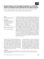
A schematic diagram of the experimental protocol is pre-
sented in Figure 1. All the rabbits in both laboratories were
evenly divided into two groups by weights a week after the
ACLT procedure. Generally, articular cartilage exhibits degen-
erate changes approximately 3–8 weeks after ACLT in experi-
mental rabbits [44,45]. Thus, 0.5 ml of each reagent was
injected intra-articularly to the left knee with ACLT at week 6
after surgery. Four more injections were administered at 2-
week or 3-week intervals at weeks 8, 10, 13 and 16 after sur-
gery. One group was injected with NMS and the other was
injected with NS. In both groups the intact right cartilage
served as the normal articular cartilage (Normal) group, and
did not undergo any treatment. We lost two rabbits because
of infection in the NMS group, and the remaining rabbits were
killed by infusion of potassium chloride 3 weeks after the last
injection, at week 19 after ACLT. For the Normal group, sam-
ples were randomly taken from the untreated right knees of the
rabbits in both groups: three from the NMS group and four
from the NS group. Haematoxylin and eosin (H&E) staining
was applied to tissue samples from each of the three groups.
Scanning electron microscopy (SEM) images of the articular
cartilage were also examined to confirm the histopathological
findings in the three groups.
Figure 1
Schematic diagram of the experimental protocolSchematic diagram of the experimental protocol. The Normal group includes right knee joints from the mature rabbits, which had not undergone any
surgery or treatment (n = 7). In the ACLT+NMS group, joints had undergone anterior cruciate ligament transection (ACLT) surgery followed by injec-
tions of nutritive mixture solution (NMS; n = 7). In the ACLT+NS group, joints had undergone ATLC surgery followed by normal saline (NS) injec-
tions (n = 11). The intra-articular injection volume was 0.5 ml (NMS or NS), and injections were given for 13 consecutive weeks starting on the week
6 after surgery and with 2-week or 3-week intervals. All of the rabbits were dissected at week 19, and histopathological examinations (such as hae-
matoxylin and eosin staining, Mankin grading method and scanning electron microscopy) were performed in all three groups.
Arthritis Research & Therapy Vol 9 No 1 Park et al.
Page 4 of 9
(page number not for citation purposes)
Histopathological examinations: H&E staining
After the rabbits had been killed, the knee joints of the rabbits
were dissected. The medial tibial plateaus and medial femoral
condyles of the rabbits were fixed in 10% phosphate-buffered
formalin (pH 7.4) with 1% cetylpyridinium (CPC) for 24 hours
and decalcified with 20% EDTA. The decalcified specimens
were embedded in paraffin and 1 μm thin sections were
stained with H&E for light microscopic examination (×100)
[46]. The severity of articular cartilage lesions was graded
through double-blind observations, using the histological
grading method proposed by Mankin and coworkers [47]. The
Mankin grading method is a well known and proven method for
the histological evaluation of OA cartilage. This method evalu-
ates the severity of erosion and/or fissures of cartilage, disor-
ganization or loss of chondrocytes, and pannus formation.
Thus, the method adequately satisfies the criteria for measur-
ing osteoarthritic changes in human and experimental animals
[16,48]. Other parenchymal organs were also examined to
investigate possible deleterious effects of the treatment
material.
Histopathological examinations: scanning electron
microscopy
The extent of fibrillation and abrasion on the cartilage surface
was observed in the photographs obtained by SEM (Joel
35CF; Tokyo, Japan; ×6,000). The microsections of cartilage
of the medial tibial plateau, which is part of a weight-bearing
joint, were washed with normal saline and pre-fixed in 2.5%
glutaraldehyde-1/15 M phosphate buffer solution. After serial
dehydration with ethanol, the ethanol was replaced with iso-
amyl acetate, and the samples were completely dried in a
dryer. An ionic coater was used for gold deposition, and the
coated samples were imaged by SEM [16].
Statistical analysis
The histopathological evaluation gradings obtained using the
Mankin grading method [47] were pooled for the normal group
(n = 7), the NMS group (n = 7), and the NS group (n = 11).
The mean values of the grades were compared among the
three groups by one-way analysis of variance and the Tukey
HSD test or Kruskal-Wallis test, depending on normality of
data (P < 0.05).
Results
Histopathological results: H&E staining
The cartilage surfaces of the weight-bearing parts, such as the
medial tibial plateau and medial femoral condyle, were evalu-
ated and graded for the extent of degradation. Table 2
presents the histopathological results of H&E staining using
the Mankin grading method [47]. A set of photomicrographs
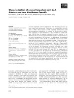
by light microscopy is also presented in Figure 2, which shows
the representative medial tibial plateaus in the three groups.
The least changes were noted in the Normal group: slight sur-
face irregularities, and slight to moderate hypercellularities in
the transitional and radial zones (Table 2 and Figure 2a). In the
NMS group the few changes noted were moderate surface
irregularities, swelling of chondrocytes in the tangential zones,
and moderate to severe hypercellularities in the transitional
Table 2
Histopathologic evaluation of H&E stained cartilage by the Mankin grading method
Items Normal (n = 7) NMS (n = 7) NS (n = 11) P*
Medial tibial plateau
Structure 0.57 (0.30)
a
2.14 (0.46)
a
5.27 (0.60)
b
0.000
Cell in tangential zone 0.29 (0.18)
a
0.71 (0.18)
a
1.50 (0.15)
b
0.000
Cell in transitional and radial zone 1.00 (0.69)
a
2.57 (0.43)
a
6.05 (0.75)
b
0.000
Pannus formation 0.00 (0.00)
a
0.29 (0.18)
a
1.18 (0.30)
b
0.005
Sum of scores 1.86 (1.12)
a
5.71 (0.84)
b
14.00 (0.75)
c
0.000
Medial femoral condyle
Structure 0.57 (0.30)
a
1.29 (0.47)
ab
2.50 (0.54)
b
0.031
Cell in tangential zone 0.29 (0.18)
a
0.43 (0.20)
ab
0.95 (0.11)
b
0.010
Cell in transitional and radial zone 1.00 (0.69)
a
2.29 (0.68)
a
5.36 (0.78)
b
0.001
Pannus formation 0.00 (0.00) 0.43 (0.20) 0.73 (0.24) 0.062
Sum of scores 1.86 (1.12)
a
4.43 (1.17)
a
9.55 (1.30)
b
0.001
Data represent the means (standard error) for each group. As the severity of cartilage lesions increases, so does the score obtained using the
Mankin grading method. The Normal group includes normal articular cartilage group that did not undergo any surgery or treatment. The nutritive
mixture solution (NMS) group received NMS after surgery. The normal saline (NS) group received NS after surgery. *Differences among the three
groups were considered to be statistically significant when the P value was under 0.05 by one-way analysis of variance and Tukey HSD test, or
Kruskal-Wallis test, depending on normality of data.
a,b,c
Data with different letters in the three groups are statistically significantly different.
Available online />Page 5 of 9
(page number not for citation purposes)
and radial zones (Figure 2b). However, in terms of the degen-
erative changes observed, there were no significant differ-
ences between the NMS group and the Normal group (Table
2). In addition, almost all of the histological changes in articular
cartilage, especially the degenerative changes in the medial
tibial plateau, were significantly less severe in the NMS group
than in the NS group (P ≤ 0.001). On the other hand, signifi-
cant degenerative findings were noted in the NS group (Table
2 and Figure 2c), such as severe surface irregularities, a cleft
in the radial zone, swelling or disappearance of chondrocytes
in the tangential zone, moderate to severe cloning in the tran-
sitional and radial zones and slight pannus formation, as com-
pared with the Normal and NMS groups (P ≤ 0.01). In
particular, in the NS group there was disappearance of sur-
face layer cells, and loss of the cartilage matrix extended to the
calcified zone (Figure 2c). These findings indicate that NMS
injection reduced loss of the superficial layer and erosion of
cartilage as compared with NS injection, and conferred pro-
tection effects against the OA-like degenerative changes in
the articular cartilage. Other parenchymal organs taken from
the treatment group did not exhibit any remarkable deleterious
changes (data not shown).
Histopathological results: scanning electron microscopy
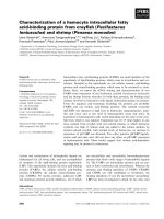
A set of samples evaluated by SEM shows the surface of the
medial tibial plateau in the three groups (Figure 3). The Normal
group exhibited the smoothest and the most intact superficial
surface of the articular cartilage (Figure 3a). In the NMS group
(Figure 3b), the medial tibial plateau was partially exposed
where the cartilage matrix was missing. However, the medial
tibial plateau of the NMS group was less damaged than that of
the NS group, particularly in the full thickness and the superfi-
cial zone of the articular cartilage. The loss of cartilage matrix
in the NMS group was obviously less severe than that in the
NS group, and the configuration of the articular cartilage in the
NMS group was much closer to that in the Normal group. As
opposed to the NMS and Normal groups, in the NS group no
cartilage matrix remained, and some of calcified layer of carti-
lage was exposed (Figure 3c).
Discussion
Our experimental material, NMS, is a nutritive mixture solution
that is designed to upregulate chondrocytes' regenerative
potential to synthesize a collagen and PG network. The com-
ponents of NMS are solutions of dextrose or glucose, amino
acids and ascorbic acid. These elementary nutrients are sub-
strates that can be delivered into the articular cartilage via the
synovial route, which is a major nutrient transport pathway for
ligaments and menisci of the articular joint [49]. Articular car-
tilage has extremely small pores (estimated at 50 Å) in the
superficial zone, and so only low-molecular-weight com-
pounds (<20 kDa) in synovial fluid may diffuse into the tissue
[26]. All of the components of NMS can move freely through
the tissue because they are not heavy molecular compounds.
Moreover, articular chondrocytes have special transporter sys-
tems for glucose and ascorbic acid [50]. Glucose is delivered
to the chondrocytes via synovial microcirculation and taken up
by glucose uptake (GLUT) proteins. The intracellular glucose
pool is used for glycolysis and extracellular matrix macromole-
cules [51]. The supply of glucose for anaerobic metabolism is
essential to the survival and proliferation of chondrocytes and
for the maintenance of matrix integrity. Therefore, impaired glu-
cose uptake would compromise chondrocyte function, and
potentially result in an imbalance in cartilage matrix synthesis
and degradation, leading to OA [20]. Ascorbic acid is trans-
ported into chondrocytes by the sodium-dependent vitamin C
transporter (SVCT2), and has been shown to upregulate the
expression of type II collagen and aggrecan [52]. Ascorbic
acid also plays an important role in chondrocyte proliferation
and protection from oxidative stress [32].
In the case of amino acids, transporter systems in cartilage
chondrocytes have not yet been identified, but glycine, proline,
glutamine and glutamate transporters in chondrocytes have
Figure 2
Representative photomicrographs of articular cartilage of the medial tibial plateauRepresentative photomicrographs of articular cartilage of the medial tibial plateau. These photomicrographs were taken at 19 weeks after anterior
cruciate ligament transection surgery, the stain used is haematoxylin and eosin, and the magnificantion is ×100. (a) Normal group. (b) Nutritive mix-
ture solution (NMS) group. (c) Normal saline (NS) group.
Arthritis Research & Therapy Vol 9 No 1 Park et al.
Page 6 of 9
(page number not for citation purposes)
recently been investigated [53,54]. Amino acids are expected
not only to control chondrocyte gene expression [55] but also
to synthesize collagen by chondrocytes [56]. Therefore, amino
acids in NMS were selected to provide substrates for fibril
forming collagen and PG, based on their prevalence in the tri-
ple-helical structure of collagen, and depending on their spe-
cific biochemical and physiologic characteristics [21,25].
Some amino acids' abilities to maintain cartilage integrity have
already been revealed. For example, hydroxyproline stabilizes
the collagen fibres to hold water [33] and glutamate prevents
cartilage calcification [37,38]. Glutamine protects articular
chondrocytes from heat stress and nitric oxide induced apop-
tosis [28], it regulates collagen gene expression in cultured
human fibroblasts [36], and it also increases collagen gene
transcription [31]. Arginine and lysine increase insulin-like
growth factor-1 production and collagen synthesis [29].
Lysine also slows the loss of collagen and PG from disrupted
articular cartilage surfaces [32]. Methionine stimulates
synthesis and deposits of PG in articular cartilage [30,34], and
cysteine activates a signalling pathway in articular chondro-
cytes [27] and protects chondrocytes and cartilage from oxi-
dative damage and degenerative processes such as OA [35].
Therefore, sufficient nutrients from the metabolically active
synovium reach the chondrocytes, presumably by diffusion
through the cartilage matrix via the synovial fluid and various
transporter systems. Finally, all of the components of NMS
cooperate with each other in promoting chondrocyte activities
to regenerate a collagen and PG network, and in preventing
degenerative changes in articular cartilage. This is the great
benefit of intra-articular injection of NMS, and one that existing
OA treatments can not provide.
Existing OA treatments, such as intra-articular injections of
either glucose or dextrose solution (5–25%) alone, are
expected to yield osmotic changes and production of precur-
sors for extracellular matrix macromolecules in the articular
cartilage [24]. Hypertonic solution is known to generate a bet-
ter therapeutic effect, but it causes more discomfort from an
inflammatory reaction [19,57,58]. The osmotic change in the
knee joint cavity induced by 10% hypertonic dextrose, which
we used in this study, is as follows: [296 (synovial fluid) + 505
(10% dextrose)]/2 = 400.5 mOsm. This osmolarity of 400
mOsm is of excellent therapeutic value, because it exerts a
strong influence on proliferation of cells such as chondrocytes,
osteocytes and fibroblasts [19]. It also influences protein syn-
thesis and amino acid (proline) transport without any cellular
toxicity [59], and produces less pain than 20% dextrose solu-
tion does [41]. Oral administration of glucosamine or chon-
droitin sulphate, which is a component of PG, plays a role as
a symptomatic slow-acting drug in degenerative OA, but its
effect is slow, small, or temporary [5,60]. Intra-articular injec-
tion of hyaluronic acid plays a role in cartilage as a lubricant
that lessens the frictional resistance of the cartilage [15], but
it only generates temporary or placebo effects [61]. Repeated
injections of hyaluronic acid may deteriorate chondrocytes'
PG biosynthetic ability [62], because hyaluronic acid is not a
substrate for PG but a terminal material. Chondrocyte prolifer-
ation therapies, such as arthroscopic abrasion of the articular
surface [7], osteotomy [63], transplantation of chondrocytes
[8] or soft-tissue grafts [10,64,65], injections of growth fac-
tors [9,66] and autologous blood [67], are also administered
into the articular cartilage to stimulate proliferation of
chondrocytes and repair cartilage matrix. However, these pro-
cedures are too expensive for general use, or they require
long-term follow up because of the potential risk for cancer
[68] or haemorrhagic arthritis [67].
Generally, cartilage in mature rabbits does not readily regener-
ate [69], and so histological changes after ACLT in rabbit
knees, including cartilage hypertrophy, reduced cell density
and matrix alterations preceding cartilage fibrillation, lead to
progressive degeneration of cartilage [70]. With ageing, the
nutritional supply of cartilage diminishes because of degener-
ative changes of the joint cavity and decreased metabolism.
However, if joint cavities were supplied with sufficient nutri-
ents, they might recover from the nutritional deficiency caused
Figure 3
Scanning electron micrographs of articular cartilage surface of the medial tibial plateauScanning electron micrographs of articular cartilage surface of the medial tibial plateau. These micrographs were taken at 19 weeks after anterior
cruciate ligament transection surgery and the magnification is ×6,000 (scale bar is 10 μm). (a) Normal group. (b) Nutritive mixture solution (NMS)
group. (c) Normal saline (NS) group.
Available online />Page 7 of 9
(page number not for citation purposes)
by ageing, and OA progression might be inhibited. In this
regard, intra-articular injection of NMS has the potential to
induce chondrocytes to synthesize a collagen and PG net-
work, which in turn maintains the cartilage matrix and protects
against OA progression in the mature rabbit model, whereas
NS injection has no such effect.
In summary, 0.5 ml of NMS or NS was intra-articularly admin-
istered into the knee joint cavity of mature rabbits for 13 con-
secutive weeks starting on week 6 after ACLT at 2-week or 3-
week intervals, when arthritic changes had begun. It was found
that only NMS injection significantly restored the extracellular
matrix and inhibited the progression of OA-like changes in
articular cartilage that had undergone ACLT. We suggest fur-
ther comparative studies with other existing OA treatments,
because in this study we only examined the effects of NMS on
OA progression in comparison with a control (NS) treatment.
Conclusion
This study is the first trial to administer intra-articularly injecta-
ble material, not in the form of a macromolecular compound
but in the form of a mixture of elementary nutrients, into the
osteoarthritic articular cartilage. Each composition of the mix-
ture, NMS, is likely to promote upregulated energy production
in chondrocytes and extracellular matrix metabolism in articular
cartilage, and to exert antioxidative effects in ageing chondro-
cytes. Based on the results of this study, NMS injection may
be applied to osteoarthritic articular cartilage of adult animals
as a very simple and effective treatment without significant
adverse effects.
Competing interests
SWL and IHL have a patent on NMS in South Korea.
Authors' contributions
YSP participated in the design of the study, performed animal
surgery, analyzed and interpreted the data, and drafted and
revised the manuscript. SWL and IHL conceived the study,
participated in its design and coordination, performed animal
surgery, and helped to draft the manuscript. TJL participated
in the study design, performed histopathological examinations,
interpreted the data, and reviewed the manuscript. JSH and
JSK participated in the study design, animal handling and care,
and reviewed the manuscript. All authors read and approved
the final manuscript.
Acknowledgements
The authors would like to thank Daehan Pharmaceutical Co. Ltd.(Seoul,
South Korea) for supporting experimental materials, and Eugene Kim,
the Director of Laboratory Animal Center, ChemOn Institute (Yongin,
Gynggi-do, South Korea) for supporting animal handling and care.
References
1. Woessner JF, Howell DS: Joint Cartilage Degradation: Basic and
Clinical Aspects New York: Marcel Dekker; 1993.
2. Brandt KD: Should nonsteroidal anti-inflammatory drugs be
used to treat osteoarthritis? Rheum Dis Clin North Am 1993,
19:29-44.
3. Creamer P, Hunt M, Dieppe P: Pain mechanisms in osteoarthri-
tis of the knee: effect of intraarticular anesthetic. J Rheumatol
1996, 23:1031-1036.
4. Shikhman AR, Amiel D, D'Lima D, Hwang SB, Hu C, Xu A, Hashi-
moto S, Kobayashi K, Sasho T, Lotz MK: Chondroprotective
activity of N-acetylglucosamine in rabbits with experimental
osteoarthritis. Ann Rheum Dis 2005, 64:89-94.
5. Bucsi L, Poor G: Efficacy and tolerability of oral chondroitin sul-
fate as a symptomatic slow-acting drug for osteoarthritis
(SYSADOA) in the treatment of knee osteoarthritis. Osteoar-
thritis Cartilage 1998:31-36.
6. Najm WI, Reinsch S, Hoehler F, Tobis JS, Harvey PW: S-adenosyl
methionine (SAMe) versus celecoxib for the treatment of oste-
oarthritis symptoms: a double-blind cross-over trial.
[ISRCTN36233495]. BMC Musculoskelet Disord 2004, 5:6.
7. Levy AS, Lohnes J, Sculley S, LeCroy M, Garrett W: Chondral
delamination of the knee in soccer players. Am J Sports Med
1996, 24:634-639.
8. Noguchi T, Oka M, Fujino M, Neo M, Yamamuro T: Repair of oste-
ochondral defects with grafts of cultured chondrocytes. Com-
parison of allografts and isografts. Clin Orthop Relat Res 1994,
302:251-258.
9. Buckwalter JA, Lohmander S: Operative treatment of osteoar-
throsis. Current practice and future development. J Bone Joint
Surg Am 1994, 76:1405-1418.
10. Bobic V: Arthroscopic osteochondral autograft transplantation
in anterior cruciate ligament reconstruction: a preliminary clin-
ical study. Knee Surg Sports Traumatol Arthrosc 1996,
3:262-264.
11. Waikakul S, Vanadurongwan V, Bintachitt P: The effects of patel-
lar resurfacing in total knee arthroplasty on position sense: a
prospective randomized study. J Med Assoc Thai 2000,
83:
975-982.
12. Uthman I, Raynauld JP, Haraoui B: Intra-articular therapy in
osteoarthritis. Postgrad Med J 2003, 79:449-453.
13. Papathanassiou NP: Intra-articular use of tenoxicam in degen-
erative osteoarthritis of the knee joint. J Int Med Res 1994,
22:332-337.
14. Raynauld JP: Clinical trials: impact of intraarticular steroid
injections on the progression of knee osteoarthritis. Osteoar-
thritis Cartilage 1999, 7:348-349.
15. Mandell BF, Lipani J: Refractory osteoarthritis. Differential diag-
nosis and therapy. Rheum Dis Clin North Am 1995,
21:163-178.
16. Yoshimi T, Kikuchi T, Obara T, Yamaguchi T, Sakakibara Y, Itoh H,
Iwata H, Miura T: Effects of high-molecular-weight sodium
hyaluronate on experimental osteoarthrosis induced by the
resection of rabbit anterior cruciate ligament. Clin Orthop Relat
Res 1994, 298:296-304.
17. Hauser GS: Answers to common questions about prolother-
apy. In Prolo Your Pain Away 1st edition. Edited by: Hauser RA,
Hauser MA, Pottinger K. Oak Park, IL: Beulah Land Press;
1998:47-66.
18. Peyron JG: Intraarticular hyaluronan injections in the treatment
of osteoarthritis: state-of-the-art review. J Rheumatol Suppl
1993, 39:10-15.
19. Reeves KD, Hassanein K: Randomized prospective double-
blind placebo-controlled study of dextrose prolotherapy for
knee osteoarthritis with or without ACL laxity. Altern Ther
Health Med 2000, 6:68-74.
20. Windhaber RA, Wilkins RJ, Meredith D: Functional characterisa-
tion of glucose transport in bovine articular chondrocytes.
Pflugers Arch 2003, 446:572-577.
21. Bhattacharjee A, Bansal M: Collagen structure: the Madras tri-
ple helix and the current scenario. IUBMB Life 2005,
57:161-172.
22. Buckwalter JA, Mankin HJ: Articular cartilage: tissue design and
chondrocyte-matrix interactions. Instr Course Lect 1998,
47:477-486.
23. Quinn TM, Morel V:
Microstructural modeling of collagen net-
work mechanics and interactions with the proteoglycan gel in
articular cartilage. Biomech Model Mechanobiol 2007, 6:73-82.
24. Liu YK, Tipton CM, Matthes RD, Bedford TG, Maynard JA, Walmer
HC: An in situ study of the influence of a sclerosing solution in
Arthritis Research & Therapy Vol 9 No 1 Park et al.
Page 8 of 9
(page number not for citation purposes)
rabbit medial collateral ligaments and its junction strength.
Connect Tissue Res 1983, 11:95-102.
25. Persikov AV, Ramshaw JA, Kirkpatrick A, Brodsky B: Amino acid
propensities for the collagen triple-helix. Biochemistry 2000,
39:14960-14967.
26. Mankin HJ, Mow VC, Buckwalter JA, Iannotti JP, Ratclffe A: Artic-
ular cartilage structure, composition, and function. In Ortho-
paedic Basic Science: Biology and Biomechanics of the
Musculoskeletal System 2nd edition. Edited by: Buckwalter JA,
Eidhorn TA, Simon SR. Rosemont, IL: American Academy of
Orthopaedic Surgeons; 2000:443-470.
27. Li WQ, Dehnade F, Zafarullah M: Thiol antioxidant, N-acetyl-
cysteine, activates extracellular signal-regulated kinase sign-
aling pathway in articular chondrocytes. Biochem Biophys Res
Commun 2000, 275:789-794.
28. Tonomura H, Takahashi KA, Mazda O, Arai Y, Inoue A, Terauchi R,
Shin-Ya M, Kishida T, Imanishi J, Kubo T: Glutamine protects
articular chondrocytes from heat stress and NO-induced
apoptosis with HSP70 expression. Osteoarthritis Cartilage
2006, 14:545-553.
29. Chevalley T, Rizzoli R, Manen D, Caverzasio J, Bonjour JP:
Arginine increases insulin-like growth factor-I production and
collagen synthesis in osteoblast-like cells. Bone 1998,
23:103-109.
30. Harmand MF, Vilamitjana J, Maloche E, Duphil R, Ducassou D:
Effects of S-adenosylmethionine on human articular chondro-
cyte differentiation. An in vitro study. Am J Med 1987,
83:48-54.
31. Bellon G, Chaqour B, Wegrowski Y, Monboisse JC, Borel JP:
Glutamine increases collagen gene transcription in cultured
human fibroblasts. Biochim Biophys Acta 1995, 1268:311-323.
32. Hering TM, Kollar J, Huynh TD, Varelas JB, Sandell LJ: Modulation
of extracellular matrix gene expression in bovine high-density
chondrocyte cultures by ascorbic acid and enzymatic
resuspension. Arch Biochem Biophys 1994, 314:90-98.
33. Kawahara K, Nishi Y, Nakamura S, Uchiyama S, Nishiuchi Y, Naka-
zawa T, Ohkubo T, Kobayashi Y: Effect of hydration on the sta-
bility of the collagen-like triple-helical structure of [4(R)-
hydroxyprolyl-4(R)-hydroxyprolylglycine]10. Biochemistry
2005, 44:15812-15822.
34. Malemud CJ, Papay RS: Stimulation of cyclic AMP in chondro-
cyte cultures: effects on sulfated-proteoglycan synthesis.
FEBS Lett 1984, 167:343-351.
35. Beckman KB, Ames BN: Oxidative decay of DNA.
J Biol Chem
1997, 272:19633-19636.
36. Kahn A, Pottenger LA, Albertini JG, Taitz AD, Thonar EJ: Chemical
stabilization of cartilage matrix. J Surg Res 1994, 56:302-308.
37. Yagami K, Suh JY, Enomoto-Iwamoto M, Koyama E, Abrams WR,
Shapiro IM, Pacifici M, Iwamoto M: Matrix GLA protein is a devel-
opmental regulator of chondrocyte mineralization and, when
constitutively expressed, blocks endochondral and intramem-
branous ossification in the limb. J Cell Biol 1999,
147:1097-1108.
38. Wang L, Hinoi E, Takemori A, Takarada T, Yoneda Y: Abolition of
chondral mineralization by group III metabotropic glutamate
receptors expressed in rodent cartilage. Br J Pharmacol 2005,
146:732-743.
39. Schwartz ER, Adamy L: Effect of ascorbic acid on arylsulfatase
activities and sulfated proteoglycan metabolism in chondro-
cyte cultures. J Clin Invest 1977, 60:96-106.
40. Sandell LJ, Daniel JC: Effects of ascorbic acid on collagen
mRNA levels in short term chondrocyte cultures. Connect Tis-
sue Res 1988, 17:11-22.
41. Reeves KD, Hassanein KM: Long-term effects of dextrose prol-
otherapy for anterior cruciate ligament laxity. Altern Ther
Health Med 2003, 9:58-62.
42. Downs JT, Lane CL, Nestor NB, McLellan TJ, Kelly MA, Karam GA,
Mezes PS, Pelletier JP, Otterness IG: Analysis of collagenase-
cleavage of type II collagen using a neoepitope ELISA. J Immu-
nol Methods 2001, 247:25-34.
43. Calliet R: Ligamentous injuries. In Knee Pain and Disability 3rd
edition. Edited by: Calliet R. Philadelphia, Seoul: FA. Davis and
Yeong Mun; 1992:112-142.
44. Hellio Le Graverand MP, Vignon E, Otterness IG, Hart DA: Early
changes in lapine menisci during osteoarthritis development:
Part I: cellular and matrix alterations. Osteoarthritis Cartilage
2001, 9:56-64.
45. Batiste DL, Kirkley A, Laverty S, Thain LM, Spouge AR, Holdsworth
DW: Ex vivo characterization of articular cartilage and bone
lesions in a rabbit ACL transection model of osteoarthritis
using MRI and micro-CT. Osteoarthritis Cartilage 2004,
12:
986-996.
46. Kikuchi T, Yamada H, Shimmei M: Effect of high molecular
weight hyaluronan on cartilage degeneration in a rabbit model
of osteoarthritis. Osteoarthritis Cartilage 1996, 4:99-110.
47. Mankin HJ, Dorfman H, Lippiello L, Zarins A: Biochemical and
metabolic abnormalities in articular cartilage from osteo-
arthritic human hips. II. Correlation of morphology with bio-
chemical and metabolic data. J Bone Joint Surg Am 1971,
53:523-537.
48. Jean YH, Wen ZH, Chang YC, Lee HS, Hsieh SP, Wu CT, Yeh
CC, Wong CS: Hyaluronic acid attenuates osteoarthritis
development in the anterior cruciate ligament-transected
knee: Association with excitatory amino acid release in the
joint dialysate. J Orthop Res 2006, 24:1052-1061.
49. Amiel D, Abel MF, Kleiner JB, Lieber RL, Akeson WH: Synovial
fluid nutrient delivery in the diathrial joint: an analysis of rabbit
knee ligaments. J Orthop Res 1986, 4:90-95.
50. Goggs R, Vaughan-Thomas A, Clegg PD, Carter SD, Innes JF,
Mobasheri A, Shakibaei M, Schwab W, Bondy CA: Nutraceutical
therapies for degenerative joint diseases: a critical review. Crit
Rev Food Sci Nutr 2005, 45:145-164.
51. Mobasheri A, Vannucci SJ, Bondy CA, Carter SD, Innes JF,
Arteaga MF, Trujillo E, Ferraz I, Shakibaei M, Martin-Vasallo P: Glu-
cose transport and metabolism in chondrocytes: a key to
understanding chondrogenesis, skeletal development and
cartilage degradation in osteoarthritis. Histol Histopathol 2002,
17:1239-1267.
52. Clark AG, Rohrbaugh AL, Otterness I, Kraus VB: The effects of
ascorbic acid on cartilage metabolism in guinea pig articular
cartilage explants. Matrix Biol 2002, 21:175-184.
53. Barker GA, Wilkins RJ, Golding S, Ellory JC: Neutral amino acid
transport in bovine articular chondrocytes. J Physiol 1999,
514:795-808.
54. Hinoi E, Wang L, Takemori A, Yoneda Y: Functional expression
of particular isoforms of excitatory amino acid transporters by
rodent cartilage. Biochem Pharmacol 2005, 70:70-81.
55. Fafournoux P, Bruhat A, Jousse C: Amino acid regulation of gene
expression. Biochem J 2000, 351:
1-12.
56. Haussinger D: The role of cellular hydration in the regulation of
cell function. Biochem J 1996, 313:697-710.
57. Reeves KD: Prolotherapy: Basic science, clinical studies, and
technique. In Pain Procedures in Clinical Practice 2nd edition.
Edited by: Lennard TA. Philadelphia: Hanley & Belfus;
2000:172-190.
58. Reeves KD: Prolotherapy: present and future applications in
soft tissue pain and disability. Phys Med Rehab Clin North Am
1995, 6:917-926.
59. Borghetti P, Della Salda L, De Angelis E, Maltarello MC, Petronini
PG, Cabassi E, Marcato PS, Maraldi NM, Borghetti AF: Adaptive
cellular response to osmotic stress in pig articular
chondrocytes. Tissue Cell 1995, 27:173-183.
60. James CB, Uhl TL: A review of articular cartilage pathology and
the use of glucosamine sulfate. J Athl Train 2001, 36:413-419.
61. Lo GH, LaValley M, McAlindon T, Felson DT: Intra-articular
hyaluronic acid in treatment of knee osteoarthritis: a meta-
analysis. Jama 2003, 290:3115-3121.
62. Handley CJ, Lowther DA: Inhibition of proteoglycan biosynthe-
sis by hyaluronic acid in chondrocytes in cell culture. Biochim
Biophys Acta 1976, 444:69-74.
63. Weisl H: Intertrochanteric osteotomy for osteoarthritis. A long-
term follow-up. J Bone Joint Surg Br 1980, 62-B:37-42.
64. Beaver RJ, Mahomed M, Backstein D, Davis A, Zukor DJ, Gross
AE: Fresh osteochondral allografts for post-traumatic defects
in the knee. A survivorship analysis. J Bone Joint Surg Br 1992,
74:105-110.
65. Paletta GA, Arnoczky SP, Warren RF: The repair of osteochon-
dral defects using an exogenous fibrin clot. An experimental
study in dogs. Am J Sports Med 1992, 20:725-731.
66. Isogai N, Morotomi T, Hayakawa S, Munakata H, Tabata Y, Ikada
Y, Kamiishi H: Combined chondrocyte-copolymer implantation
with slow release of basic fibroblast growth factor for tissue
engineering an auricular cartilage construct. J Biomed Mater
Res A 2005, 74:408-418.
Available online />Page 9 of 9
(page number not for citation purposes)
67. Gilbert MS, Aledort LM, Seremetis S, Needleman B, Oloumi G,
Forster A: Long term evaluation of septic arthritis in hemophilic
patients. Clin Orthop Relat Res 1996, 328:54-59.
68. Chodorowska G, Tomczyk M, Glowacka A: Basic-fibroblast
growth factor (b-FGF): its biological role in physiologic and
pathologic conditions. Ann Univ Mariae Curie Sklodowska
[Med] 2004, 59:286-291.
69. Caplan AI, Elyaderani M, Mochizuki Y, Wakitani S, Goldberg VM:
Principles of cartilage repair and regeneration. Clin Orthop
Relat Res 1997, 342:254-269.
70. Buckwalter JA: Evaluating methods of restoring cartilaginous
articular surfaces. Clin Orthop Relat Res 1999:S224-S238.