Báo cáo y học: " Developments in the synovial biology field 2006" potx
Bạn đang xem bản rút gọn của tài liệu. Xem và tải ngay bản đầy đủ của tài liệu tại đây (126.1 KB, 8 trang )
Page 1 of 8
(page number not for citation purposes)
Available online />Abstract
Synovial pathophysiology is a complex and synergistic interplay of
different cell populations with tissue components, mediated by a
variety of signaling mechanisms. All of these mechanisms drive the
affected joint into inflammation and drive the subsequent
destruction of cartilage and bone. Each cell type contributes
significantly to the initiation and perpetuation of this deleterious
concert, especially in rheumatoid arthritis. Rheumatoid arthritis
synovial fibroblasts and macrophages, both cell types with pivotal
roles in inflammation and destruction, but also T cells and B cells
are crucial for complex network in the inflamed synovium. An even
more complex cellular crosstalk between these key players
maintains a process of chronic inflammation. As outlined in the
present review, in the past year substantial progress has been
made to elucidate further details of the rich pathophysiology of
rheumatoid arthritis, which may also facilitate the identification of
novel targets for future therapeutic strategies.
Introduction
The shift from physiology to pathophysiology – driving
factors
In a healthy joint the synovium covers the joint cavity and
regulates the transport of nutrients and other molecules
between the joint cavity and the adjacent tissue. The
synovium consists of few cell layers of fibroblast-like synovio-
cytes and macrophage-like synoviocytes [1]. Of these, the
fibroblast-like synoviocytes, also termed synovial fibroblasts
(SF), synthesize and secrete a rich but balanced variety of
products, including cytokines, matrix metalloproteinases
(MMPs), hyaluronan and proteoglycans into the synovial fluid.
In joints affected by rheumatoid arthritis (RA), the synovial
membrane becomes hyperplastic due to the proliferation of
genuine synovial cells such as SF [2] and a massive
infiltration of inflammatory cells [3]. As a consequence, the
inner layer of the synovium, the lining layer, increases in size
up to 10 cell layers and more. Similarly, in the normal state
the lining layer contains only a small number of blood vessels,
and oxygenation and nutrition is facilitated by the blood
vessels from the sublining. In diseased synovium, the
proliferation of cells (for example, SF) and the infiltration of
blood-borne cells (for example, macrophages, B cells, T cells,
plasma cells) subsequently result in hypoxic conditions in the
tissue because of the increasing distance to a blood vessel
and the increased demand for oxygen in the hyperplastic
tissue. Neovascularization is thus a prerequisite in the
formation and maintenance for the pannus, and intensive
neovascularization with blood vessels close to the ultimate
lining layer can be observed [4,5]. In addition, there is a close
association between rheumatoid synovitis and the formation
of complex lymphoid microstructures [6].
At the site of invasion into the adjacent cartilage and bone,
the pannus consists mainly of activated fibroblasts. SF
mediate also the perichondrocytic cartilage degradation and
promote bone destruction by influencing osteoclastogenesis
in cooperation with macrophages [7].
As summarized in the present review, numerous researchers
have addressed the details of this transition of healthy tissue
to diseased synovial tissue. One of the key examples is the
work by Steenvorden and colleagues, who have shown that
the original epithelial-like phenotype of SF is replaced by a
cell showing mesenchymal/fibrotic characteristics, which
includes the expression of collagen type I and α-smooth
muscle actin [8].
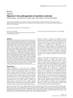
As outlined below and shown in Figure 1, the stimulating
factors for this development, which have been examined
intensively and have revealed numerous novel aspects in the
past year, are cell-derived microparticles, hitherto unknown
cytokines and chemoattractive molecules.
Review
Developments in the synovial biology field 2006
Anette Knedla, Elena Neumann and Ulf Müller-Ladner
Department for Internal Medicine and Rheumatology, Justus-Liebig-University Giessen, Kerckhoff-Clinic, Bad Nauheim, Benekestr. 2-8,
D-61231 Bad Nauheim, Germany
Corresponding author: Anette Knedla,
Published: 10 April 2007 Arthritis Research & Therapy 2007, 9:209 (doi:10.1186/ar2140)
This article is online at />© 2007 BioMed Central Ltd
IFN = interferon; IKK = Iκβ kinase; IL = interleukin; MAPK = mitogen-activated protein kinase; MKK = mitogen-activated protein kinase kinase; MMP =
matrix metalloproteinase; NF = nuclear factor; RA = rheumatoid arthritis; RANK = receptor activator of nuclear factor κB; RANKL = receptor activa-
tor of nuclear factor κB ligand; RASF = rheumatoid arthritis synovial fibroblasts; SF = synovial fibroblasts; TNF = tumor necrosis factor.
Page 2 of 8
(page number not for citation purposes)
Arthritis Research & Therapy Vol 9 No 2 Knedla et al.
Microparticles
Microparticles are a heterogeneous population of small
membrane-coated vesicles that can be released from all cell
types, including macrophages, monocytes, epithelial cells as
well as B cells and T cells. Microparticles in synovial fluid were
first described by Berckmans and coworkers, who showed that
these particles originate mainly from monocytes and
granulocytes [9]. The potential function of microparticles in
inflammation and as part of mechanisms of the innate immunity
was recently reviewed by Distler and colleagues [10].
Microparticles emerge by budding from their parental cells
upon apoptosis or activation. The composition of the
membrane of the microparticles therefore depends on the cell
type of origin. Microparticles inherit all characteristics of the
parental cell, including the respective cell surface molecules
and receptors, and can therefore act as mediators of cellular
interactions. Whether intracellular contents such as cytosolic
or nuclear proteins are present within the microparticles or
even contribute to their biologic activity remains largely unclear.
In this context, recent findings shed light on these
phenomena by indicating that microparticles derived from
leukocytes can play a role in inflammatory arthritis by inducing
the synthesis of MMPs, chemokines and cytokines in SF
[11,12]. It could also be shown that, in particular, the
synthesis of MMP-1, MMP-3, MMP-9 and MMP-13 was
strongly induced by microparticles but the expression of
MMP-2, MMP-14 and the tissue inhibitors TIMP-1, TIMP-2
and TIMP-3 was unaffected [13]. Moreover, in the same
study it could be demonstrated that microparticles increased
the synthesis of IL-6, IL-8 and the monocyte chemoattractant
proteins MCP-1 and MCP-2. As was demonstrated recently,
bound complement components and activator molecules are
present on microparticles ex vivo [14]. In RA synovial fluid,
therefore, microparticles might modulate the increased
complement activation.
Cytokines
Increasing evidence was provided in 2006 that the more
recently discovered ‘novel’ cytokines are also involved in
promoting joint inflammation in RA. For example, IL-32, which
is intensively expressed in RA synovial tissue, resulted in joint
inflammation and a mild cartilage damage when injected
intraarticularly in murine knee joints [15]. IL-32 was first
described by Kim and colleagues [16]. They demonstrated
that IL-32 was able to induce the expression of TNFα, IL-1β,
IL-6 and several other chemokines in a human acute
monocytic leukemia cell line (THP-1), for example, through
the cytokine signaling pathways of NF-κB and p38 mitogen-
activated protein kinase (MAPK). IL-32 can therefore be
considered a proinflammatory mediator in RA.
IL-1F8, a new member of the IL-1 family that is known to play
a pivotal role in immune and inflammatory reactions, also
exerts proinflammatory effects in primary human joint cells
[17]. Another ‘novel’ cytokine, IL-17, which is synthesized
primarily by T cells and exhibits proinflammatory activities, has
also been associated with RA. Interestingly, it could be
shown that IL-17, which is a potent inducer of TNFα and
IL-1β, acts independently of TNFα in RA and was able to
enhance inflammation and cartilage damage in a TNF-
deficient mouse model [18].
A new member of the IL-10 family, IL-20, is known to play a
role in skin inflammation and the development of hemato-
poetic cells. The potential role of IL-20 in RA and arterio-
sclerosis was recently analyzed by Wei and coworkers [19].
In this context, IL-20 was shown to be upregulated in the
synovial fluid of RA patients. Furthermore, in a collagen-
induced arthritis model in rats it could be shown that both
IL-20 and its receptor IL-20RI are present, which confirmed
the involvement of IL-20 in the pathogenesis of RA [20]. IL-21
is a CD4
+
T-cell-derived cytokine being involved in innate and
adaptive immune response. Overexpression of IL-21 and its
receptor IL-21R could also be identified in the inflamed
synovial membrane and in synovial fluid leukocytes of RA
patients [21]. Stimulation of peripheral-blood T cells or
synovial-fluid T cells isolated from RA patients with IL-21
resulted in enhanced T-cell activation, proliferation and
secretion of proinflammatory cytokines, including TNFα and
IFNγ. Furthermore, it is known that activated macrophages
produce central inflammatory cytokines such as TNFα and
IL-1. As the anti-inflammatory cytokine IL-10 suppresses the
macrophage-dependent synthesis of both TNFα and IL-1 in
nonmalignant conditions, it was most interesting to see that
Figure 1
Interactions in the pathophysiology of joint destruction in rheumatoid
arthritis. SF, synovial fibroblasts; MMPs, matrix metalloproteinases.
Page 3 of 8
(page number not for citation purposes)
the responses to IL-10 are dysregulated in RA macrophages,
resulting in an inefficient suppression of inflammation [22].
Besides their role in energy metabolism, cytokines derived
from adipocytes (for example, adiponectin and resistin)
appear to play a pivotal role in the pathogenesis of RA. A
strong stimulatory effect of adiponectin on rheumatoid
arthritis synovial fibroblasts (RASF) could be detected.
Hereby, adiponectin induced IL-6 and MMP-1 in a p38 MAPK
pathway-dependent manner [23]. Similarly, it could be shown
that an increased concentration of adiponectin in the synovial
fluid of RA patients is negatively correlated with the local
inflammatory process [24]. Resistin, another so-called
adipokine, may also influence proinflammation in RA. For
example, an upregulated concentration of resistin was found
at local sites of inflammation in arthritis, and the serum
resistin levels correlated with inflammation and the activity of
the disease in RA [25]. In contrast, circulating levels of the
prototype adipocytokine leptin appear not to have a
correlation with RA activity [26,27].
Chemokines
Chemokines are small chemotactic proteins that play a role in
the migration of circulating cells into tissue and migration of
cells within the tissue. As recently reviewed by Vergunst and
colleagues and by Tarrant and Patel, chemokines play a
substantial role in the inflammatory process of RA by
promoting leukocyte trafficking into the synovium [28,29].
The regulation of chemokine ligand CCL18, a T-cell-attracting
chemokine, was described by van Lieshout and coworkers
[30]. These authors showed that IL-10 in combination with IL-
4 and IL-13 induced synergistically the secretion of CCL18 in
monocytes and monocyte-derived cells. This finding
supported the idea that CCL18 is involved in the regulation of
the immune system in health and disease.
In several diseases including RA or osteoarthritis, however,
chemokines and their receptors are considered potential
future therapeutic targets. Based on this idea, a recent study
by Haringman and colleagues investigated the expression of
the ligands of chemokine receptors CCR1 and CCR5 in the
inflamed synovium [31]. They found an abundant expression
of both receptors CCR1 and CCR5 in the synovial tissue of
RA patients, whereas the percentages of CCR1-positive and
CCR5-positive monocytes in the peripheral blood of RA
patients were found to be decreased. The blockade of CCR1
and CCR5 could therefore be part of an effective future
therapy for RA.
Synovial fibroblasts
Pathways to proliferation
‘Receptor cells’ of the disease-promoting factors outlined
above are mainly synovial fibroblasts and macrophages,
which are also the predominant cell types in the inflamed
synovium [8,32]. With regard to SF it is not known what
initiates the initial proliferation of these cells in the early
stages of RA, but this pivotal event can occur prior to the
onset of inflammation [33]. In this regard, the investigation of
the mode of proliferation revealed an upregulation of the
metastatic lymph node MLN51 gene in hyperactive RASF
[34]. Even growth-retarded SF showed a significant up-
regulation of MLN51 when treated with granulocyte–
macrophage colony-stimulating factor or with synovial fluid.
As MLN51 was originally identified in breast cancer, this
observation once more emphasizes distinct similarities of the
mechanisms of cellular activation in RA and in malignant
diseases.
Cell survival and resistance to apoptosis
Besides unrestricted proliferation, the increasing number of
RASF in the synovial lining layer may be also due to an
altered apoptosis. It is known that the deficiency or the lack of
tumor suppressor genes such as p53, the ‘phosphatase and
tensin homolog deleted on chromosome 10’ PTEN, small
ubiquitin-like modifier and p21 leads to long-term cell growth,
to extended survival and potentially to tumor formation.
Woods and colleagues demonstrated that the cell-cycle
inhibitor p21 is significantly reduced in RA synovial lining,
particularly in RASF. In addition, p21 is able to repress
migration of SF – and, vice versa, loss of p21, which occurs
also in RASF, may contribute to the excessive invasion and
extended survival of these cells [35]. Moreover, although
overexpression of p53 is found in RA synovial tissue, only few
synoviocytes undergo apoptosis [36]. This effect could be
explained in part by a low expression of proapoptotic genes.
In a study from Cha and coworkers using synovial tissue and
SF, it could be shown that a deficient p53-upregulated
modulator of apoptosis can inhibit apoptosis of SF [37].
Furthermore, the lack of the ‘phosphatase and tensin
homolog’ PTEN in the RA synovial lining was able to
contribute to the survival of RASF at sites of destruction [38].
Connor and coworkers showed also that this phenomenon
could be due to the PTEN-dependent effect on IκB/NF-κB
interactions and other nuclear factors (for example,
akt/protein kinase B).
As recent data indicated a role of protein geranylgeranylation
and RhoA/RhoA kinase blocking in regulation of apoptosis,
Nagashima and colleagues suggested lipophilic statins as
therapeutic agents for RA, since they are able to induce
apoptosis in RASF; for example, through mitochondrial-
dependent and caspase-3-dependent pathways and the
inhibition of mevalonate pathways [39]. Moreover, the
antiapoptotic molecule myeloid cell leukemia Mcl-1, which is
known to be critical for the survival of T lymphocytes and
B lymphocytes and of macrophages [40], appears also to be
relevant in the survival of RASF [41].
TNF, being one of the key molecules in driving the
inflammatory process in RA synovium, is also linked directly to
SF apoptosis. For example, a study conducted by Wang and
colleagues [42] revealed that the antiapoptotic effect of
Available online />TNFα in RASF is regulated by the Jun activating binding
protein JAB1, because specific knockdown of JAB1 with an
antisense RNA construct resulted in TNFα-induced apoptosis
response in RASF. Moreover, Wang and coworkers showed
that this antiapoptotic signaling might be due to a JAB1-
mediated ubiquitination of TNF-receptor-associated-factor 2.
The potential role of the TNF ligand receptor superfamily in
the antiapoptotic pathways in RA was recently reviewed by
Hsu and colleagues [43].
Degradation of cartilage and bone
In 2006 numerous groups supported the idea of RASF being
key players in the pathogenesis of RA [8,32,44]. For example,
a recent study showed that the expression of the extracellular
matrix metalloproteinase inducer CD147 was more intensively
expressed on RASF than on osteoarthritic SF [45]. The
authors concluded that the increased expression of CD147
might be responsible for both the elevated secretion of
MMPs and the invasive potential of SF. Of the subsequently
activated family members, the collagenases MMP-1 and
MMP-13, the gelatinases MMP-2 and MMP-9, the stromelysin
MMP-3 and the membrane-type MMPs can be found in active
RA synovium. Of these, the expression of MMP-3 mRNA is
higher in diseased RA pannus tissue compared with adjacent
nondiseased RA synovium [46]. Most strikingly, although
MMP-1 appears to have a function in degrading cartilage
collagen type II, it does not appear to derive from pannus
tissue but to be secreted by chondrocytes [46]. In addition, in
vitro inhibition of the membrane type I MT1-MMP with an
antisense RNA construct resulted in a significant reduction of
cartilage degradation by RASF [47]. Also, a study by Bauer
and colleagues addressing the expression of fibroblast
activation protein by RASF revealed that the expression of
FAP is colocalized with MMP-1 and MMP-13, indicating that
fibroblast activation protein might be an additional factor in
cartilage and bone destruction in RA joints [48].
An effective future therapy for RA could be the selective
inhibition of MAPK kinases [49]. MKK3 and MKK6 play key
roles in the activation of p38 MAPK, which in turn
upregulates the expression of cytokines and MMPs in SF.
Inoue and colleagues investigated the potential of MKK3 as a
therapeutic target. They could show that MKK3 deficiency
significantly decreases synovial inflammation and cytokine
production in a mouse model of arthritis.
As recently reviewed by Ruocco and Karin, the Iκβ kinase
IKKβ is essential for the inflammatory cytokine-induced
activation of NF-κB [50]. Blocking of IKKβ could therefore be
part of a therapeutic strategy for the treatment of inflam-
mation. In this context, it was demonstrated by Wen and
coworkers that the inhibition of IKKβ with the β-carboline
derivative ML120B inhibits NF-κB signaling in human SF,
chondrocytes and mast cells [51]. Moreover, it could be
shown that ML120B administration reduces NF-κB activity in
rats with induced polyarthritis [52].
RASF play an important role in osteoclast formation [53]. The
molecular basis for this property is the synthesis of the ligand
for the receptor activator of nuclear factor β (RANKL) [54].
Binding of receptor activator of nuclear factor (RANK) with its
ligand RANKL regulates the differentiation of bone-resorbing
osteoclasts from monocytes/macrophages progenitor cells.
In addition, Lee and coworkers revealed that RASF produce
actively RANKL, and thus are part of the RANK/RANKL
interaction system [55]. Interestingly, a study by Pettit and
colleagues demonstrated a focal RANKL, RANK and
osteoprotegerin expression in the RA bone microenvironment
[56]. Taking these results together, RASF most probably
perpetuate actively osteoclastogenesis and bone destruction
in RA.
Macrophages
Physiological function
In synovial homeostasis, the physiologic function of
macrophages is the induction and regulation of inflammation
after infection. Similar to RASF, macrophages are key players
in promoting inflammation and joint destruction in RA by
secreting proinflammatory cytokines such as IL-1 and TNFα
and by the induction and perpetuation of osteoclastogenesis
[57]. Macrophages migrate out of the bloodstream as
monocytes and accumulate in the synovial membrane. As
demonstrated recently, this migratory capacity is dependent
on distinct enzymes. Miyata and colleagues showed that, in
contrast to osteoarthritis, patients with RA have a significant
increase in cathepsin G activity [58]. Interestingly, cathepsin G
was able to induce the migration of monocytes in a
microchemotaxis chamber and thus cathepsin G appears to
promote synovial inflammation in addition to the hydrolytic
function of cathepsins in matrix degradation.
With regard to cellular accumulation, Gregory and coworkers
showed that macrophage migration inhibitory factor induces
the release of CC chemokine ligand 2 from primary micro-
vascular cells [59]. This function of macrophage migration
inhibitory factor might therefore further promote the patho-
genesis of RA by inducing monocyte migration into the
synovium.
Activation
Activation of macrophages in the RA synovium can take place
by several mechanisms [57]; for example, activation by T cells
that secrete stimulatory cytokines such as IFNγ and IL-2. Direct
cell–cell contact between macrophages and T cells can also
result in macrophage activation. In a recent study from Beech
and coworkers it was shown that RA synovial T cells are able to
induce the chemokine production by monocytes in a cell-
contact-dependent manner [60]. Moreover, there appears also
to be a correlation between B cells and macrophage activation.
Treatment of RA patients with rituximab, a chimeric antibody
against CD20-expressing B cells, resulted in a significant
decrease of TNFα in the supernatant of isolated human
monocyte-derived macrophages [61].
Arthritis Research & Therapy Vol 9 No 2 Knedla et al.
Page 4 of 8
(page number not for citation purposes)
Resistance to apoptosis
Not only fibroblasts but also macrophages appear to be
resistant to apoptosis, thereby increasing further the number
of macrophages in the synovium. In this context, the
antiapoptotic B-cell leukemia Bcl-2 family member Mcl-1 may
contribute to the survival of these cells. This was supported
by a recent study from Liu and coworkers who revealed an
increased expression of Mcl-1 in CD14
+
macrophages
derived from the synovial fluid of RA patients. Furthermore,
the same group was able to show the induction of apoptosis
in synovial macrophages by blocking the PI 3-kinase/Akt-1 or
STAT-3 pathways [40].
Osteoclastogenesis
Macrophages/monocytes are not only involved in inflam-
matory reactions, but also in the remodeling processes of the
bone. Two studies published in the past year provided new
insights in the role of monocytes in osteoclast formation.
CD14
+
synovial macrophages isolated from patients with
osteoarthritis, RA and pyrophosphate arthropathy have been
shown to differentiate into osteoclasts when treated with
RANKL [62]. Stimulation with TNFα and IL-1α resulted in
osteoclast formation of macrophages from RA and
pyrophosphate arthropathy patients. Another study confirmed
the involvement of monocytes as potential precursors of
macrophages and osteoclasts. Komano and colleagues
revealed that CD16 monocytes, a subset of human peripheral
blood monocytes, bear the potential to differentiate into
osteoclasts when stimulated with RANKL and macrophage
colony-stimulating factor [63].
In 2006, different groups investigated the potential role of the
tyrosine kinase inhibitor imatinib in the treatment of RA
[64-66]. For example, Ando and colleagues showed that
imatinib inhibits the proliferation of macrophage colony-
stimulating factor-dependent osteoclast precursor cells and
the formation of osteoclasts in vitro [64]. Moreover, they
showed that the administration of imatinib suppressed joint
destruction in a collagen-induced arthritis model in rats.
Similarly, it was demonstrated that imatinib enhances osteo-
clast apoptosis in a cell culture model using rabbit
osteoclasts [66]. A recent study by Paniagua and coworkers
demonstrated that, in a collagen-induced arthritis model in
mice, imatinib affects the proliferation of B cells and
monocytes/macrophages, and inhibits several tyrosine
kinases that are directly implicated in the pathogenesis of RA
[65]. Taken together, the selective inhibition of tyrosine
kinases by imatinib could be a promising future therapy for
RA.
B cells
There is increasing evidence that B cells play an important
role in the pathogenesis of RA. The production of auto-
antibodies directed against self-antigens is an important
characteristic of RA that can be found prior to the onset of
the clinical onset of the disease [67]. Samuels and coworkers
were able to show that part of the B-cell-dependent
pathophysiology appears to be a failure of the efficient
removement of polyreactive B cells in RA and that there are
defects at the early B-cell tolerance checkpoint in the bone
marrow [68]. Thus, in RA patients the peripheral mature naïve
B cells are able to accumulate which then contribute actively
to the development of the disease. The importance of B cells
in the perpetuation of RA is underlined by the successful
treatment of RA patients with biologic agents and drugs
selectively affecting B cells.
As recently reviewed by Keystone and by Looney, the
targeting and depletion of B cells with a mouse–human
chimeric monoclonal antibody against the B-cell-specific
antigen CD20 resulted in a significant beneficial effect in RA
patients [69,70]. Moreover, treatment of RA patients with a
fully human monoclonal antibody against the B-lymphocyte
stimulator, which is a growth and survival factor for B cells,
appears to be a promising therapy for the future [71].
T cells
As recently reviewed by Leipe and coworkers and by
Skapenko and colleagues, T cells play an important role in
the pathogenesis of RA [72,73]. An important subset of
regulatory T cells is CD4
+
CD25
+
T cells, which are known to
control the development of autoimmune diseases. This cell
population is enriched in synovial fluid of RA patients but
appears to be reduced in peripheral blood. Moreover, the lack
of CD4
+
CD25
+
T cells in peripheral blood can be observed
in early active RA [74].
Several studies addressed the paradox that although the
number of inhibitory regulatory T cells is increased in synovial
fluid, inflammation still occurs in the rheumatoid joint. In this
regard, Sakaguchi and colleagues reported that complete
depletion of the regulatory T-cell transcription factor FOXP3
was able to activate even weak or rare self-reactive T-cell
clones and to induce severe autoimmune diseases [75].
Another review published in 2006 discusses the function of
cytokines in the generation and maintenance of regulatory
T cells [76]. In this context, a recent study from Zorn and
coworkers underlined the potential role of IL-2 in the
maintenance of FOXP3
+
CD4
+
CD25
+
regulatory T cells [77].
It could be shown that IL-2 upregulated selectively the
expression of FOXP3 in an in vitro culture of CD4
+
CD25
+
T cells. With regard to a potential therapeutic approach,
Gonzalez-Rey and colleagues determined the ability of
vasoactive intestinal peptide to induce functional regulatory
T cells in the collagen-induced arthritis mouse model [78].
They were able to show that the administration of vasoactive
intestinal peptide resulted in the expansion of
FOXP3
+
CD4
+
CD25
+
regulatory T cells, including the joints.
The vasoactive intestinal peptide-triggered transfer of the
regulatory T cells suppressed the progression of the disease,
and might therefore bear the potential to suit as a therapeutic
tool in the future.
Available online />Page 5 of 8
(page number not for citation purposes)
The important role of cytokines in the development and
chronic progression of CD4
+
T-cell-mediated chronic auto-
immune disease was also demonstrated in the novel animal
model for RA, the Sakaguchi SKG mice [79]. Hata and
coworkers showed that the synovial fluid of arthritic SKG
mice contain high concentrations of IL-6, TNFα and IL-1.
Furthermore, their study revealed that the deficiency in either
IL-6, IL-1 or TNFα can inhibit the development and the
progression of arthritis in this mouse model, whereas IL-10
deficiency leads to an exacerbation of the disease. A recently
published study by Hirota and coworkers provided evidence
that IL-6 is a key factor in the differentiation process of self-
reactive T cells [80]. These authors could demonstrate in a
mouse model that self-reactive T cells stimulate antigen-
presenting cells to secrete IL-6. Together with T cells,
antigen-presenting cells form an IL-6 cytokine milieu, which
drives naïve self-reactive T cells to differentiate into IL-17-
secreting CD4
+
helper T cells (Th17 cells). Moreover, it was
shown that IL-17 or IL-6 deficiency leads to a complete
inhibition of arthritis.
Conclusion
The past year has contributed significantly to the deeper
understanding of synovial biology. Of the various aspects that
have been addressed, predominantly extracellular pathways
including novel cytokines, adipokines and chemokines as well
as stimulating microparticles have been introduced in this
fascinating field. Among the various cellular players, fibroblasts,
macrophages, T cells and B cells especially have been in the
scope of interest of worldwide rheumatology research – which
has identified numerous hitherto unknown mechanisms
involved in the activation and proliferation of these cells and
their interaction with other articular components.
Competing interests
The authors declare that they have no competing interests.
References
1. Smith M, Barg E, Weedon H, Papangelis V, Smeets T, Tak PP,
Kraan M, Coleman M, Ahern M: Microarchitecture and protec-
tive mechanisms in synovial tissue from clinically and arthro-
scopically normal knee joints. Ann Rheum Dis 2003, 62:
303-307.
2. Qu Z, Garcia C, O’Rourke L, Planck S, Kohli M, Rosenbaum J:
Local proliferation of fibroblast-like synoviocytes contributes
to synovial hyperplasia. Results of proliferating cell nuclear
antigen/cyclin, c-myc, and nucleolar organizer region staining.
Arthritis Rheum 1994, 37:212-220.
3. Kraan M, Versendaal H, Jonker M, Bresnihan B, Post W, t Hart B,
Breedveld F, Tak P: Asymptomatic synovitis precedes clinically
manifest arthritis. Arthritis Rheum 1998, 41:1481-1488.
4. Paleolog EM: Angiogenesis in rheumatoid arthritis. Arthritis
Res Ther 2002, 4:S81-S90.
5. Roccaro A, Russo F, Cirulli T, Di Pietro G, Vacca A, Dammacco F:
Antiangiogenesis for rheumatoid arthritis. Curr Drug Targets
Inflamm Allergy 2005, 4:27-30.
6. Takemura S, Braun A, Crowson C, Kurtin PJ, Cofield RH, O’Fallon
WOM, Goronzy JJ, Weyand CM: Lymphoid neogenesis in
rheumatoid synovitis. J Immunol 2001, 167:1072-1080.
7. Karouzakis E, Neidhart M, Gay R, Gay S: Molecular and cellular
basis of rheumatoid joint destruction. Immunol Lett 2006, 106:
8-13.
8. Steenvoorden M, Tolboom T, van der Pluijm G, Lowik C, Visser C,
Degroo tJ, Gittenberger-Degroot A, Deruiter M, Wisse B, Huizinga
T, et al.: Transition of healthy to diseased synovial tissue in
rheumatoid arthritis is associated with gain of mesenchy-
mal/fibrotic characteristics. Arthritis Res Ther 2006, 8:R165.
9. Berckmans RJ, Nieuwland R, Tak PP, Boing AN, Romijn FP, Kraan
MC, Breedveld FC, Hack CE, Sturk A: Cell-derived microparti-
cles in synovial fluid from inflamed arthritic joints support
coagulation exclusively via a factor VII-dependent mecha-
nism. Arthritis Rheum 2002, 46:2857-2866.
10. Distler J, Pisetsky D, Huber L, Kalden J, Gay S, Distler O:
Microparticles as regulators of inflammation: novel players of
cellular crosstalk in the rheumatic diseases. Arthritis Rheum
2005, 52:3337-3348.
11. Distler J, Huber L, Gay S, Distler O, Pisetsky D: Microparticles as
mediators of cellular cross-talk in inflammatory disease.
Autoimmunity 2006, 39:683-690.
12. Berckmans RJ, Nieuwland R, Kraan MC, Schaap MC, Pots D,
Smeets TJ, Sturk A, Tak PP: Synovial microparticles from
arthritic patients modulate chemokine and cytokine release
by synoviocytes. Arthritis Res Ther 2005, 7:R536-R544.
13. Distler J, Jüngel A, Huber L, Seemayer C, Reich C, Gay R, Michel
B, Fontana A, Gay S, Pisetsky D, et al.: The induction of matrix
metalloproteinase and cytokine expression in synovial fibrob-
lasts stimulated with immune cell microparticles. Proc Natl
Acad Sci U S A 2005, 102:2892-2897.
14. Biro E, Nieuwland R, Tak PP, Pronk LM, Schaap MC, Sturk A,
Hack CE, Activated complement components and comple-
ment activator molecules on the surface of cell-derived
microparticles in patients with rheumatoid arthritis and
healthy individuals. Ann Rheum Dis 2007 [Epub ahead of print].
15. Joosten L, Netea M, Kim S, Yoon D, Oppers-Walgreen B, Rad-
stake T, Barrera P, van de Loo F, Dinarello C, van den Berg W: IL-
32, a proinflammatory cytokine in rheumatoid arthritis. Proc
Natl Acad Sci U S A 2006, 103:3298-3303.
16. Kim S, Han S, Azam T, Yoon D, Dinarello C: Interleukin-32: a
cytokine and inducer of TNF
αα
. Immunity 2005, 22:131-142.
17. Magne D, Palmer G, Barton J, Mezin F, Talabot-Ayer D, Bas S,
Duffy T, Noger M, Guerne P, Nicklin M, et al.: The new IL-1 family
member IL-1F8 stimulates production of inflammatory media-
tors by synovial fibroblasts and articular chondrocytes. Arthri-
tis Res Ther 2006, 8:R80.
18. Koenders M, Lubberts E, van de Loo F, Oppers-Walgreen B, van
den Bersselaar L, Helsen M, Kolls J, Di Padova F, Joosten L, van
den Berg W: Interleukin-17 acts independently of TNF-
αα
under
arthritic conditions. J Immunol 2006, 176:6262-6269.
19. Wei C, Hsu Y, Li H, Wang Y, Hsieh M, Chen W, Hsing C, Chang
M: IL-20: biological functions and clinical implications.
J Biomed Sci 2006, 13:601-612.
20. Hsu Y, Li H, Hsieh M, Liu M, Huang K, Chin L, Chen P, Cheng H,
Chang M: Function of interleukin-20 as a proinflammatory
molecule in rheumatoid and experimental arthritis. Arthritis
Rheum 2006, 54:2722-2733.
21. Li J, Shen W, Kong K, Liu Z: Interleukin-21 induces T-cell acti-
vation and proinflammatory cytokine secretion in rheumatoid
arthritis. Scand J Immunol 2006, 64:515-522.
22. Antoniv T, Ivashkiv L: Dysregulation of interleukin-10-depen-
dent gene expression in rheumatoid arthritis synovial macro-
phages. Arthritis Rheum 2006, 54:2711-2721.
23. Ehling A, Schaffler A, Herfarth H, Tarner I, Anders S, Distler O,
Paul G, Distler J, Gay S, Scholmerich J, et al.: The potential of
adiponectin in driving arthritis. J Immunol 2006, 176:4468-
4478.
24. Senolt L, Pavelka K, Housa D, Haluzik M: Increased adiponectin
is negatively linked to the local inflammatory process in
patients with rheumatoid arthritis. Cytokine 2006, 35:247-
252.
25. Senolt L, Housa D, Vernerova Z, Jirasek T, Svobodova R, Veigl D,
Anderlova K, Muller-Ladner U, Pavelka K, Haluzik M: Resistin in
rheumatoid arthritis synovial tissue, synovial fluid and serum.
Ann Rheum Dis 2007, 66:458-463.
26. Gunaydin R, Kaya T, Atay A, Olmez N, Hur A, Koseoglu M: Serum
leptin levels in rheumatoid arthritis and relationship with
disease activity. South Med J 2006, 99:1078-1083.
27. Hizmetli S, Kisa M, Gokalp N, Bakici M: Are plasma and synovial
fluid leptin levels correlated with disease activity in rheuma-
toid arthritis? Rheumatol Int 2007, 27:335-338.
Arthritis Research & Therapy Vol 9 No 2 Knedla et al.
Page 6 of 8
(page number not for citation purposes)
28. Vergunst CE, van de Sande MGH, Lebre MC, Tak PP: The role of
chemokines in rheumatoid arthritis and osteoarthritis. Scand
J Rheumatol 2005, 34:415-425.
29. Tarrant T, Patel D: Chemokines and leukocyte trafficking in
rheumatoid arthritis. Pathophysiology 2006, 13:1-14.
30. van Lieshout A, van der Voort R, le Blanc L, Roelofs M, Schreurs
B, van Riel P, Adema G, Radstake T: Novel insights in the regu-
lation of CCL18 secretion by monocytes and dendritic cells via
cytokines, toll-like receptors and rheumatoid synovial fluid.
BMC Immunol 2006, 7:23.
31. Haringman J, Smeets T, Reinders-Blankert P, Tak P: Chemokine
and chemokine receptor expression in paired peripheral
blood mononuclear cells and synovial tissue of patients with
rheumatoid arthritis, osteoarthritis, and reactive arthritis. Ann
Rheum Dis 2006, 65:294-300.
32. Meinecke I, Rutkauskaite E, Gay S, Pap T: The role of synovial
fibroblasts in mediating joint destruction in rheumatoid arthri-
tis. Curr Pharm Des 2005, 11:563-568.
33. Ospelt C, Neidhart M, Gay RE, Gay S: Synovial activation in
rheumatoid arthritis. Front Biosci 2004, 9:2323-2334.
34. Jang J, Lim D-S, Choi Y-E, Jeong Y, Yoo S-A, Kim W-U, Bae Y-S:
MLN51 and GM-CSF involvement in the proliferation of fibrob-
last-like synoviocytes in the pathogenesis of rheumatoid
arthritis. Arthritis Res Ther 2006, 8:R170.
35. Woods J, Klosowska K, Spoden D, Stumbo N, Paige D, Scatizzi J,
Volin M, Rao M, Perlman H: A cell-cycle independent role for
p21 in regulating synovial fibroblast migration in rheumatoid
arthritis. Arthritis Res Ther 2006, 8:R113.
36. Seemayer CA, Kuchen S, Neidhart M, Kuenzler P, Rihosková V,
Neumann E, Pruschy M, Aicher WK, Müller-Ladner U, Gay RE, et
al.: p53 in rheumatoid arthritis synovial fibroblasts at sites of
invasion. Ann Rheum Dis 2003, 62:1139-1144.
37. Cha H, Rosengren S, Boyle D, Firestein GS: PUMA regulation
and proapoptotic effects in fibroblast-like synoviocytes. Arthri-
tis Rheum 2006, 54:587-592.
38. Connor A, Berger S, Narendam A, Keystone E: Inhibition of
protein geranylgeranylation induces apoptosis in synovial
fibroblasts. Arthritis Res Ther 2006, 8:R94.
39. Nagashima T, Okazaki H, Yudoh K, Matsuno H, Minota S: Apop-
tosis of rheumatoid synovial cells by statins through the
blocking of protein geranylgeranylation: a potential therapeu-
tic approach to rheumatoid arthritis. Arthritis Rheum 2006, 54:
579-586.
40. Liu H, Huang Q, Shi B, Eksarko P, Temkin V, Pope RM: Regula-
tion of Mcl-1 expression in rheumatoid arthritis synovial
macrophages. Arthritis Rheum 2006, 54:3174-3181.
41. Liu H, Eksarko P, Temkin V, Haines GR, Perlman H, Koch A,
Thimmapaya B, Pope R: Mcl-1 is essential for the survival of
synovial fibroblasts in rheumatoid arthritis. J Immunol 2005,
175:8337-8345.
42. Wang J, Li C, Liu Y, Mei W, Yu S, Zhang L, Cao X, Kimberly R,
Grizzle W, Zhang H: JAB1 determines the response of
rheumatoid arthritis synovial fibroblasts to tumor-necrosis
factor-
αα
. Am J Pathol 2006, 169:889-902.
43. Hsu H, Wu Y, Mountz J: Tumor necrosis factor ligand-receptor
superfamily and arthritis. Curr Dir Autoimmun 2006, 9:37-54.
44. Huber L, Distler O, Tarner I, Gay RE, Gay S, Pap T: Synovial
fibroblasts: key players in rheumatoid arthritis. Rheumatology
2006, 45:669-675.
45. Zhu P, Lu N, Shi Z, Zhou J, Wu Z, Yang Y, Ding J, Chen Z: CD147
overexpression on synoviocytes in rheumatoid arthritis
enhances matrix metalloproteinase production and invasive-
ness of synoviocytes. Arthritis Res Ther 2006, 8:R44.
46. Ainola MM, Mandelin JA, Liljeström MP, Li T-F, Hukkanen MVJ,
Konttinen YT: Pannus invasion and cartilage degradation in
rheumatoid arthritis: involvement of MMP-3 and interleukin-
1
ββ
. Clin Exp Rheumatol 2005, 23:644-650.
47. Rutkauskaite E, Volkmer D, Shigeyama Y, Schedel J, Pap G,
Muller-Ladner U, Meinecke I, Alexander D, Gay R, Drynda S, et al.:
Retroviral gene transfer of an antisense construct against
membrane type 1 matrix metalloproteinase reduces the inva-
siveness of rheumatoid arthritis synovial fibroblasts. Arthritis
Rheum 2005, 52:2010-2014.
48. Bauer S, Jendro M, Wadle A, Kleber S, Stenner F, Dinser R, Reich
A, Faccin E, Godde S, Dinges H, et al.: Fibroblast activation
protein is expressed by rheumatoid myofibroblast-like syn-
oviocytes. Arthritis Res Ther 2006, 8:R171.
49. Inoue T, Boyle DL, Corr M, Hammaker D, Davis RJ, Flavell RA,
Firestein GS: Mitogen-activated protein kinase kinase 3 is a
pivotal pathway regulating p38 activation in inflammatory
arthritis. Proc Natl Acad Sci U S A 2006, 103:5484-5489.
50. Ruocco MG, Karin M: IKK
ββ
as a target for treatment of inflam-
mation induced bone loss. Ann Rheum Dis, 2005 64 (Suppl 4):
iv81-iv85.
51. Wen D, Nong Y, Morgan JG, Gangurde P, Bielecki A, Dasilva J,
Keaveney M, Cheng H, Fraser C, Schopf L, et al.: A selective
small molecule I
κκ
B kinase
ββ
inhibitor blocks nuclear factor
κκ
B-mediated inflammatory responses in human fibroblast-
like synoviocytes, chondrocytes, and mast cells. J Pharmacol
Exp Ther 2006, 317:989-1001
52. Schopf L, Savinainen A, Anderson K, Kujawa J, DuPont M, Silva
M, Siebert E, Chandra S, Morgan J, Gangurde P, et al.: IKK
ββ
inhi-
bition protects against bone and cartilage destruction in a rat
model of rheumatoid arthritis. Arthritis Rheum 2006, 54:3163-
3173.
53. Neumann E, Gay S, Muller-Ladner U: The RANK/RANKL/osteo-
protegerin system in rheumatoid arthritis: new insights from
animal models. Arthritis Rheum 2005, 52:2960-2967.
54. Shigeyama Y, Pap T, Kunzler P, Simmen B, Gay RE, Gay S:
Expression of osteoclast differentiation factor in rheumatoid
arthritis. Arthritis Rheum 2000, 43:2523-2530.
55. Lee H, Jeon H, Song E, Han M, Park S, Lee S, Yun H, Kim J, Kim
J, Lee Y, et al.: CD40 ligation of rheumatoid synovial fibrob-
lasts regulates RANKL-mediated osteoclastogenesis: evi-
dence of NF-
κκ
B-dependent, CD40-mediated bone destruction
in rheumatoid arthritis. Arthritis Rheum 2006, 54:1747-1758.
56. Pettit A, Walsh N, Manning C, Goldring S, Gravallese E: RANKL
protein is expressed at the pannus–bone interface at sites of
articular bone erosion in rheumatoid arthritis. Rheumatology
2006, 45:1068-1076.
57. Ma Y, Pope RM: The role of macrophages in rheumatoid
arthritis. Curr Pharm Des 2005, 11:569-580.
58. Miyata J, Tani K, Sato K, Otsuka S, Urata T, Lkhagvaa B, Furukawa
C, Sano N, Sone S: Cathepsin G: the significance in rheuma-
toid arthritis as a monocyte chemoattractant. Rheumatol Int
2007, 27:375-382.
59. Gregory J, Morand E, McKeown S, Ralph J, Hall P, Yang Y,
McColl S, Hickey M: Macrophage migration inhibitory factor
induces macrophage recruitment via CC chemokine ligand 2.
J Immunol 2006, 177:8072-8079.
60. Beech J, Andreakos E, Ciesielski C, Green P, Foxwell B, Brennan
F: T-cell contact-dependent regulation of CC and CXC
chemokine production in monocytes through differential
involvement of NF
κκ
B: implications for rheumatoid arthritis.
Arthritis Res Ther 2006, 8:R168.
61. Toubi E, Kessel A, Slobodin G, Boulman N, Pavlotzky E, Zisman
D, Rozenbaum M, Rosner I: Macrophage function changes fol-
lowing rituximab treatment in patients with rheumatoid arthri-
tis. Ann Rheum Dis 2006 [Epub ahead of print].
62. Adamopoulos I, Sabokbar A, Wordsworth B, Carr A, Ferguson D,
Athanasou N: Synovial fluid macrophages are capable of
osteoclast formation and resorption. Am J Pathol 2006, 208:
35-43.
63. Komano Y, Nanki T, Hayashida K, Taniguch K, Miyasaka N: Identi-
fication of a human peripheral blood monocyte subset that
differentiates into osteoclasts. Arthritis Res Ther 2006, 8:R152.
64. Ando W, Hashimoto J, Nampei A, Tsuboi H, Tateishi K, Ono T,
Nakamura N, Ochi T, Yoshikawa H: Imatinib mesylate inhibits
osteoclastogenesis and joint destruction in rats with collagen-
induced arthritis (CIA). J Bone Miner Metab 2006, 24:274-282.
65. Paniagua RT, Sharpe O, Ho PP, Chan SM, Chang A, Higgins JP,
Tomooka BH, Thomas FM, Song JJ, Goodman SB, et al.: Selec-
tive tyrosine kinase inhibition by imatinib mesylate for the
treatment of autoimmune arthritis. J Clin Invest 2006, 116:
2633-2642.
66. El Hajj Dib I, Gallet M, Mentaverri R, Sevenet N, Brazier M, Kamel
S: Imatinib mesylate (Gleevec) enhances mature osteoclast
apoptosis and suppresses osteoclast bone resorbing activity.
Eur J Pharmacol 2006, 551:27-33.
67. Leslie D, Lipsky PE, Notkins AL: Autoantibodies as predictors of
disease. J Clin Invest 2001, 108:1417-1422.
68. Samuels J, Ng YS, Coupillaud C, Paget D, Meffre E: Impaired
early B cell tolerance in patients with rheumatoid arthritis.
J Exp Med 2005, 201:1659-1667.
Available online />Page 7 of 8
(page number not for citation purposes)
69. Keystone E: B cell targeted therapies. Arthritis Res Ther 2005,
7(Suppl 3):S13-S18.
70. Looney RJ: B cell-targeted therapy for rheumatoid arthritis: an
update on the evidence. Drugs 2006, 66:625-639.
71. Stohl W: Therapeutic targeting of B lymphocyte stimulator
(BLyS) in the rheumatic diseases. Endocr Metab Immune
Disord Drug Targets 2006, 6:351-358.
72. Leipe J, Skapenko A, Lipsky PE, Schulze-Koops H: Regulatory T
cells in rheumatoid arthritis. Arthritis Res Ther 2005, 7:93-99.
73. Skapenko A, Leipe J, Lipsky PE, Schulze-Koops H: The role of
the T cell in autoimmune inflammation. Arthritis Res Ther
2005, 7(Suppl 2):S4-S14.
74. Lawson C, Brown A, Bejarano V, Douglas S, Burgoyne C, Green-
stein A, Boylston A, Emery P, Ponchel F, Isaacs J: Early rheuma-
toid arthritis is associated with a deficit in the CD4
+
CD25
high
regulatory T cell population in peripheral blood. Rheumatology
2006, 45:1210-1217.
75. Sakaguchi S, Ono M, Setoguchi R, Yagi H, Hori S, Fehervari Z,
Shimizu J, Takahashi T, Nomura T: Foxp3
+
CD25
+
CD4
+
natural
regulatory T cells in dominant self-tolerance and autoimmune
disease. Immunol Rev 2006, 212:8-27.
76. Wan YY, Flavell RA: The roles for cytokines in the generation
and maintenance of regulatory T cells. Immunol Rev 2006,
212:114-130.
77. Zorn E, Nelson EA, Mohseni M, Porcheray F, Kim H, Litsa D, Bel-
lucci R, Raderschall E, Canning C, Soiffer RJ, et al.: IL-2 regu-
lates FOXP3 expression in human CD4
+
CD25
+
regulatory T
cells through a STAT-dependent mechanism and induces the
expansion of these cells in vivo. Blood 2006, 108:1571-1579.
78. Gonzalez-Rey E, Fernandez-Martin A, Chorny A, Delgado M:
Vasoactive intestinal peptide induces CD4
+
,CD25
+
T regula-
tory cells with therapeutic effect in collagen-induced arthritis.
Arthritis Rheum 2006, 54:864-876.
79. Hata H, Sakaguchi N, Yoshitomi H, Iwakura Y, Sekikawa K, Azuma
Y, Kanai C, Moriizumi E, Nomura T, Nakamura T, et al. Distinct
contribution of IL-6, TNF-
αα
, IL-1, and IL-10 to T cell-mediated
spontaneous autoimmune arthritis in mice. J Clin Invest 2004,
114:582-588.
80. Hirota K, Hashimoto M, Yoshitomi H, Tanaka S, Nomura T, Yam-
aguchi T, Iwakura Y, Sakaguchi N, Sakaguchi S: T cell self-reac-
tivity forms a cytokine milieu for spontaneous development of
IL-17
+
Th cells that cause autoimmune arthritis. J Exp Med
2007, 204:41-47.
Arthritis Research & Therapy Vol 9 No 2 Knedla et al.
Page 8 of 8
(page number not for citation purposes)