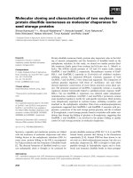
Báo cáo y học: "The identification and characterization of a novel protein, c19orf10, in the synovium" docx
Bạn đang xem bản rút gọn của tài liệu. Xem và tải ngay bản đầy đủ của tài liệu tại đây (1.35 MB, 9 trang )
Open Access
Available online />Page 1 of 9
(page number not for citation purposes)
Vol 9 No 2
Research article
The identification and characterization of a novel protein,
c19orf10, in the synovium
Tracey Weiler
1
, Qiujiang Du
1
, Oleg Krokhin
1
, Werner Ens
2
, Ken Standing
2
, Hani El-Gabalawy
1,3
and John A Wilkins
1,3
1
Department of Internal Medicine and Manitoba Centre for Proteomics and Systems Biology, University of Manitoba, Room 799, John Buhler
Research Centre, 715 McDermot Avenue, Winnipeg, Manitoba, R3E 3P4, Canada
2
Department of Physics and Astronomy, University of Manitoba, 510 Allen Building, Winnipeg, Manitoba, R3T 2N2, Canada
3
Section of Rheumatology, Department of Internal Medicine, Faculty of Medicine, University of Manitoba, RR149 Rehab Hospital, 800 Sherbrook
Street, Winnipeg, Manitoba, R3A 1M4, Canada
Corresponding author: John A Wilkins,
Received: 16 Jul 2006 Revisions requested: 24 Jul 2006 Revisions received: 14 Feb 2007 Accepted: 15 Mar 2007 Published: 15 Mar 2007
Arthritis Research & Therapy 2007, 9:R30 (doi:10.1186/ar2145)
This article is online at: />© 2007 Weiler et al.; licensee BioMed Central Ltd.
This is an open access article distributed under the terms of the Creative Commons Attribution License ( />),
which permits unrestricted use, distribution, and reproduction in any medium, provided the original work is properly cited.
Abstract
Joint inflammation and destruction have been linked to the
deregulation of the highly synthetic fibroblast-like synoviocytes
(FLSs), and much of our current understanding of the
mechanisms that underlie synovitis has been collected from
studies of FLSs. During a proteomic analysis of FLS cells, we
identified a novel protein, c19orf10 (chromosome 19 open
reading frame 10), that was produced in significant amounts by
these cells. The present study provides a partial characterization
of c19orf10 in FLSs, synovial fluid, and the synovium. Murine
monoclonal and chicken polyclonal antibodies were produced
against recombinant human c19orf10 protein and used to
examine the distribution of c19orf10 in cultured FLSs and in
synovial tissue sections from patients with rheumatoid arthritis
or osteoarthritis. The intracellular staining pattern of c19orf10 is
consistent with localization in the endoplasmic reticulum/Golgi
distribution. Sections of rheumatoid arthritis and osteoarthritis
synovia expressed similar patterns of c19orf10 distribution with
perivascular and synovial lining staining. Double-staining in situ
analysis suggests that fibroblast-like synovial cells produced
c19orf10, whereas macrophages, B cells, or T cells produced
little or none of this protein. There is evidence of secretion into
the vascular space and the extracellular matrix surrounding the
synovial lining. A competitive enzyme-linked immunosorbent
assay confirmed the presence of microgram levels of c19orf10
in the synovial fluids of patients with one of various
arthropathies. Collectively, these results suggest that c19orf10
is an FLS-derived protein that is secreted into the synovial fluid.
However, the significance of this protein in synovial biology
remains to be determined. The absence of known structural
motifs or domains and its relatively late evolutionary appearance
raise interesting questions about its function.
Introduction
The healthy synovial membrane consists of a thin layer of
fibroblast-like synoviocytes (FLSs) and macrophages. These
cells produce glycosaminoglycans such as hyaluronic acid
and lubricating glycoproteins for secretion into the synovial
fluid [1]. Healthy homeostasis within the joint can be disturbed
by development of inflammatory diseases such as rheumatoid
arthritis (RA). In this situation, the synovium becomes enlarged
and the cellular composition changes. The intimal layer exhib-
its an increased number of FLSs, there is an increase in the
number of blood vessels, and the sub-intima becomes infil-
trated with lymphocytes and plasma cells forming ectopic lym-
phoid follicles [2]. The phenotype of the synovial cells also
changes. In vitro, RA FLSs exhibit a transformed phenotype
reminiscent of that seen in tumors (increased proliferative
potential and resistance to apoptosis), whereas the vascular
2DE = two-dimensional electrophoresis; bp = base pair; BSA = bovine serum albumin; c19orf10 = chromosome 19 open reading frame 10; ELISA
= enzyme-linked immunosorbent assay; FLS = fibroblast-like synoviocyte; GPM = Global Proteome Machine; GST = glutathione S transferase; HRP
= horseradish peroxidase; Ig = immunoglobulin; IL = interleukin; MS = mass spectrometry; MS/MS = tandem mass spectrometry; NCBI = National
Center for Biotechnology Information; OA = osteoarthritis; PBS = phosphate-buffered saline; RA = rheumatoid arthritis; rhc19orf10 = recombinant
chromosome 19 open reading frame 10.
Arthritis Research & Therapy Vol 9 No 2 Weiler et al.
Page 2 of 9
(page number not for citation purposes)
endothelium displays an increased ratio of apoptotic to prolif-
erative cells, which is indicative of vascular remodeling [3].
These changes may contribute to the maintenance of synovial
inflammation, aggravating the destruction of cartilage and
bone and stimulating the development of the pannus. Because
FLSs can exhibit significant phenotypic changes under differ-
ent pathological conditions, studies were initiated to examine
the repertoire of proteins produced by these cells. Ultimately,
these studies could provide information about differences in
protein synthesis by FLSs in health and disease.
Previously, proteomic studies were initiated to determine the
protein composition and expression patterns of FLSs [4]. One
of the proteins identified was a major FLS protein encoded by
the novel gene, c19orf10 (chromosome 19 open reading
frame 10). c19orf10 has been identified in two other reports
in the literature. Tulin and colleagues [5] performed a genetic
complementation screening approach looking for stromal cell-
derived factors involved in cell proliferation and identified
c19orf10 as a murine bone marrow stroma-derived growth
factor (SF20/interleukin [IL]-25). Subsequent work revealed
that the proliferation data described in the initial report could
not be replicated and the report was withdrawn [6]. In another
report, Wang and colleagues [7] profiled the proteins
secreted from pre-adipocytes (3T3-L1) by means of two-
dimensional electrophoresis (2DE) mass spectrometry (MS).
The authors identified a number of secreted proteins, including
c19orf10 (SF20/IL-25), which was upregulated during differ-
entiation of adipocytes, leading to the suggestion that it is
involved in adipogenesis. Although these studies have postu-
lated that c19orf10 is involved in cell proliferation and differen-
tiation, none of these studies has revealed any functional
information about the molecule. We undertook the characteri-
zation of c19orf10 in the synovium because it is a quantita-
tively significant product of FLSs [4] and little is known about
the processing and function of the c19orf10 molecule.
Materials and methods
Cells, tissues, and synovial fluid
Synovial tissue was obtained with informed consent from
patients with RA or osteoarthritis (OA) at the time of knee or
hip arthroplasty. All patients with RA met American College of
Rheumatology criteria [8]. FLSs were isolated and cultured as
previously described [9]. Synovial fluid was obtained with
informed consent from five patients diagnosed with RA, reac-
tive arthritis, or gout. All samples were obtained according to
the guidelines approved by the Ethics Committee of the Uni-
versity of Manitoba (Winnipeg, MB, Canada).
Sample preparation, two-dimensional electrophoresis
analysis, in-gel digestion, and mass spectrometry
Cell lysates were prepared and isolated as previously
described [4]. The preparative 2DE and MS were performed
as described by Dasuri and colleagues [4]. Digests were ana-
lyzed using a matrix-assisted laser desorption/ionization quad-
rupole time-of-flight mass spectrometer [10], and proteins
were identified by single MS (peptide mass fingerprinting)
using ProFound [11] and by tandem mass spectrometry (MS/
MS) using Tandem search engine [12,13]. The National
Center for Biotechnology Information (NCBI) (Bethesda, MD,
USA) non-redundant human database was used in both
cases.
cDNA cloning and expression constructs
A full-length c19orf10 IMAGE (Integrated Molecular Analysis
of Genomes and their Expression) clone (4562455) corre-
sponding to GenBank accession number NM_019107
was
obtained from Open Biosystems (Huntsville, AL, USA). A set
of oligonucleotide primers was designed to amplify the coding
region of the c19orf10 sequence (amino acids 32V to 173L),
excluding a putative N-terminal signal sequence. Primers
Orf10F (GGTGTCCGAGCCCACGACGGT) and Orf10R1
(catggctcgaGTCACAGCTCAGTGCG) were used to amplify
a 431-base pair (bp) sequence of c19orf10 encompassing
nucleotides 162 to 592, excluding the region coding for the
putative N-terminal signal peptide but including the 3' stop
codon. This sequence was inserted into the PshA1 and XhoI
sites of the pET41b vector (Novagen, part of EMD Bio-
sciences, Inc., San Diego, CA, USA), resulting in a glutathione
S transferase (GST)-His-c19orf10 fusion gene. Primers
Orf10NdeI (gaattccatatGGTGTCCGAGCCCACGA) and
Orf10R2 (catggctcgagcAGCTCAGTGCGCGAT) were used
to amplify a 426-bp sequence of c19orf10 encompassing
nucleotides 162 to 587, excluding both the region coding for
the putative N-terminal signal peptide and the 3' stop codon.
This sequence was inserted into the NdeI and XhoI sites of the
pET41b vector (Novagen), resulting in a c19orf10-His fusion
gene. Primers Orf10BamHI (catgcggatccCGGTGTC-
CGAGCCCA) and Orf10R1 (catggctcgaGTCACAGCT-
CAGTGCG) were used to amplify a 431-bp sequence of
c19orf10 encompassing amino acids 32V to 173L, including
the C-terminal stop codon. This sequence was inserted into
the BamHI and XhoI sites of the pGEX-5X-2 vector (Amer-
sham Biosciences, now part of GE Healthcare, Little Chalfont,
Buckinghamshire, UK), resulting in a GST-c19orf10 fusion
gene. Restriction enzyme analysis and DNA sequencing were
used to confirm the fidelities of the plasmids. The expression
constructs were then transformed into Escherichia coli strain
BL21 or the Rosetta2 (DE3) strain enhanced with seven
human tRNA genes (Novagen).
Recombinant protein expression was induced with 1 mM iso-
propyl-β-D-thiogalactopyranoside (Novagen) for 3 hours at
37°C. The cells were subsequently collected and frozen over-
night. The frozen cell pellet was resuspended in BugBuster
reagent (Novagen) in 1X phosphate-buffered saline (PBS),
and the cells were lysed by treatment with lysozyme (Sigma-
Aldrich, St. Louis, MO, USA) and benzonase (Novagen) for 30
minutes at room temperature and then centrifuged at 15,000g
for 20 minutes. The supernatant was collected and applied to
Available online />Page 3 of 9
(page number not for citation purposes)
a Sepharose nickel affinity column (Novagen) or a Sepharose
GST affinity column (Novagen) according to the manufac-
turer's instructions. Rhc19orf10-His tag was eluted from the
Sepharose nickel affinity column with 300 mM imidazole.
Recombinant GST-tagged c19orf10 was eluted from the glu-
tathione column with 10 mM reduced glutathione. Unlabelled
c19orf10 (amino acids 32 to 173) was produced by digestion
of GST-c19orf10 with Factor Xa for 4 hours at room tempera-
ture and then purified through another GST affinity column.
The released recombinant protein was collected in the efflu-
ent. Protein quality and purity were assessed by 12% SDS-
PAGE and visualized with Imperial purple protein stain (Pierce,
Rockford, IL, USA).
Antibody production
Monoclonal antibodies were generated in mice as previously
described [14]. Female BALB/c mice were immunized with
recombinant c19orf10 (rhc19orf10) fusion protein containing
both GST and s (GST-His-c19orf10) or c19orf10-His. Ten or
twenty-five micrograms of c19orf10 fusion protein was mixed
with Titer-Max Gold adjuvant (Cedarlane Laboratories Ltd.,
Burlington, ON, Canada) and administered subcutaneously.
The mice were boosted 1 month later. Three months later, one
mouse was boosted without adjuvant intraperitoneally 4 days
prior to the fusion. After the fusion, hybridomas that were
enzyme-linked immunosorbent assay (ELISA)-positive to
c19orf10 were selected for further analysis and cloning.
Cloned hybridomas were grown to death in RPMI-1640 con-
taining 10% fetal bovine serum, and supernatants were col-
lected and used as a source of antibodies. The antibodies
were characterized using an Isotyping Monoclonal Antibodies
Kit from GE Healthcare. One of the anti-rhc19orf10 clones,
1B6, was grown in serum-free media, and antibody was puri-
fied using a KaptivM column (BioCan Scientific, now part of
MediCorp Inc., Montreal, QC, Canada).
The ELISA studies were performed using Nunc Maxisorp
multi-well plates (Nalge Nunc, Naperville, IL, USA) coated
overnight at 4°C with 1.0 μg/well of c19orf10-His fusion pro-
tein. Plates were washed and then blocked with 1% bovine
serum albumin (BSA) in PBS. The plates were incubated
either with primary antibodies in culture supernatants contain-
ing 10% serum or with purified antibody. The plates were incu-
bated with goat anti-mouse immunoglobulin (Ig) G (whole
molecule) alkaline phosphatase conjugate (Sigma-Aldrich),
and the reaction was developed using p-nitrophenyl phos-
phate substrate (Sigma-Aldrich). The response was quantified
at 405 nm on an ELISA plate reader.
A chicken polyclonal antibody was produced by immunization
with recombinant His-tagged c19orf10 (Gallus Immunotech,
Inc., Fergus, ON, Canada). A hen was immunized twice with
100 μg of orf10-His and then twice with 50 μg of orf10-His.
Immune eggs were collected and egg yolk IgY was purified by
Gallus Immunotech, Inc. A competitive ELISA was developed
using this antibody. Nunc Maxisorp multi-well plates were
coated overnight at 4°C with GST-c19orf10 fusion protein
(0.8 μg/well). Plates were washed and then blocked with 1%
BSA in PBS. The plates were incubated with anti-c19orf10
IgY or anti-c19orf10 IgY pre-incubated with GST-c19orf10 or
synovial fluid. The plates were washed and incubated with
donkey anti-chicken IgY horseradish peroxidase (HRP) conju-
gate (1:15,000) (Gallus Immunotech, Inc.). The reaction was
monitored with tetramethylbenzidine ELISA substrate solution
(Sigma-Aldrich) and read at 450 nm.
Immunofluorescence
FLSs were stained with monoclonal anti-rhc19orf10 antibod-
ies. Twenty-one-spot microscope slides (Erie Scientific Com-
pany, Portsmouth, NH, USA) coated with 5 or 10 μg/ml of
fibronectin (Sigma-Aldrich) were seeded with 5 × 10
4
cells
per spot and incubated overnight in 37°C with 10% CO
2
.
After incubation, the cells were washed in PBS and then fixed
in 4% paraformaldehyde (Polysciences, Inc., Warrington, PA,
USA) in PBS for 15 minutes. The cells were then washed and,
in some cases, permeabilized using 0.2% Triton X-100
(Sigma-Aldrich) in PBS for 5 minutes and washed again. The
slides were treated with supernatants containing anti-
rhc19orf10 antibodies or purified antibody for 1 hour at room
temperature. Excess primary antibody was removed with three
washes of PBS, and the cells were then treated with Oregon
Green 488-conjugated phalloidin (Invitrogen Corporation,
Carlsbad, CA, USA) in combination with cyanine 3-conjugated
goat anti-mouse Ig (Jackson ImmunoResearch Laboratories,
Inc., West Grove, PA, USA). Fluorescence was visualized
using an epifluorescence microscope (Olympus BX60; Olym-
pus, Tokyo, Japan) equipped with a xenon arc lamp, light pipe
(Lambda LS; Sutter Instrument Company, Novato, CA, USA),
and a Sensicam digital camera (The Cooke Corporation, Rom-
ulus, MI, USA). Images were processed with Image-Pro soft-
ware (Media Cybernetics, Inc., Silver Spring, MD, USA).
Immmunohistochemistry
Fresh frozen synovial biopsies were sectioned at 5 μm using a
cryostat. Two sequential sections were placed side-by-side on
a charged microscope slide (ProbOn; Fisher Scientific Co.,
Pittsburgh, PA, USA). Tissue staining was carried out using
Dako's ABC system (Dako North America, Inc., Carpinteria,
CA, USA). Tissue sections were fixed in chilled acetone, air-
dried, and rehydrated in PBS. Endogenous tissue peroxidase
was blocked by incubating the sections with hydrogen perox-
ide solution (Dako North America, Inc.). Sections were also
blocked by incubation with normal serum from the animal spe-
cies used for secondary antibody generation. Primary antibod-
ies were then added to each tissue section and incubated
overnight at 4°C in a humidified slide chamber. The appropri-
ate biotinylated secondary antibody, streptavidin-HRP, and
diaminobenzidine substrate (all from Dako North America, Inc.)
were used to detect the binding of the primary antibodies.
Murine Ig of irrelevant specificity was added to a tissue section
Arthritis Research & Therapy Vol 9 No 2 Weiler et al.
Page 4 of 9
(page number not for citation purposes)
adjacent to the primary antibody and used as a negative
control.
Paraffin blocks were prepared by immersing tissue samples in
neutral buffered 10% formalin for 8 hours for fixation. They
were dehydrated in ascending graded ethanol and infiltrated
and embedded in low-melting paraffin at 56°C in a heated
oven. The tissue-paraffin mold was solidified on a cold plate to
form a block. Four-micrometer sections were cut by a micro-
tome. The slides were then rehydrated in descending graded
ethanol and processed through the same immunostaining
steps as for frozen sections.
Results
Mass spectrometric analysis of two-dimensional SDS-PAGE-
separated synovial fibroblast lysates identified a 16.5-kDa
spot as the product of c19orf10 [4]. Total peptide coverage
corresponded to 47% of the predicted peptide sequence with
a ProFound expectation score of 2.5 × 10
-6
[11] (Figure 1a).
Subsequent MS/MS analysis of three of the peptides derived
from this spot confirmed that the sequence corresponded to
that of c19orf10 with a GPM log (e) score of -16.8 (Figure 1b–
d) [12]. Furthermore, manual MS/MS analysis of a 2,992.5-Da
parent ion indicated that it was a non-tryptic peptide corre-
sponding to amino acids 32 to 60.
The c19orf10 gene is located on chromosome 19p13.3 and
spans approximately 30 kbp [15] (Figure 2). A survey of the
available cDNA clones suggests the possibility of three splice
variants for c19orf10 [16] (Figure 2b). The most common var-
iant, c19orf10.b, has six exons and is supported by sequence
data from 468 clones. The other two predicted variants (a and
c) have been identified with sequence data support from one
clone each. It is likely that variant c is incomplete at the 5' end.
Predicted protein products of the three splice variants are
illustrated in Figure 2c. All of the c19orf10 peptides identified
by our analysis are present in the c19orf10.b sequence (high-
lighted peptides, Figure 2c). However, based on the predicted
molecular weight of variant c, it is not expected to be in the
same molecular weight regions on the gels as the variants a
and b. Hence, it is not possible to comment on its presence in
the FLSs.
The c19orf10 gene product variant c is predicted to be a 173-
amino acid protein with a theoretical molecular weight of 18.8
kDa and a theoretical isoelectric point of 6.2. Two cysteine
residues, at positions 63 and 92 (Figure 2c, outlined with a
rectangle), are predicted to be disulfide-bonded [17]. A 31-
amino acid signal peptide is predicted using the SignalP [18]
(and PSORT II [19] algorithms (Figure 2c, underlined),
suggesting that this is a secreted protein. The presence of a
Figure 1
Proteomic identification of c19orf10Proteomic identification of c19orf10. (a) Mass spectra of in-gel digest of c19orf10 with six peptides mapping to the c19orf10 protein labeled. (b)
Tandem mass spectrum of c19orf10 parent ion 1,154 Da, 146-TAVAHRPGAFK-156. (c) Tandem mass spectrum of c19orf10 parent ion 1,196 Da,
101-SYLYFTQFK-109. (d) Tandem mass spectrum of c19orf10 parent ion 1,750 Da, 131-ESDVPLKTEEFEVTK-145. Peaks contributing to the
score are labeled. The mass and sequence of each parent ion are indicated on the appropriate spectrum. c19orf10, chromosome 19 open reading
frame 10.
Available online />Page 5 of 9
(page number not for citation purposes)
cleavable signal peptide is consistent with the results of the
MS/MS analysis, which found a non-tryptic cleavage site at
position 32. This corresponds to the exact site at which the
signal peptide is predicted to be cut to generate the mature
protein.
The c19orf10 sequence was also examined for other
sequence patterns that might predict molecular features or
properties. Domain and pattern searches using InterProScan
to query ProDom, PFAM, SMART, and PRINTS [20] suggest
that c19orf10 does not possess any known domains or motifs.
Two potential O-glycosylation sites are predicted at thre-
onines 36 and 37 using NetOGlyc3.1[21]. The fact that the
2,992.5-Da peak corresponding to the N-terminal peptide
containing unmodified threonines 36 and 37 was observed
suggests that not all of the protein is necessarily glycosylated.
Eight potential phosphorylation sites at residues S33, S84,
S132, S169, T36, Y61, Y67, and Y119 are predicted by the
NetPhos 2.0 server [22]. These results suggest that c19orf10
is a secreted protein with unique structural features.
A panel of murine monoclonal antibodies was produced
against rhc19orf10 for immunohistochemistry and immu-
noassays in an effort to define the distribution of c19orf10 in
synovium. Three hybridomas were selected based on reactiv-
ity with rhc19orf10. The antibodies are very effective in immun-
ofluorescence as well as immunohistochemistry on both
frozen and paraffin sections, but they do not work on Western
blot. All of the antibodies give similar patterns of reactivity in a
comparative staining analysis. These antibodies were all of the
IgM class and this may be attributable to the fact that there is
91% identity between the predicted mature human and murine
proteins. Thus, the immunogenicity of c19orf10 would be
expected to be low and an isotype switch to IgG would be
more difficult to achieve.
The staining of permeabilized low-passage FLSs with the mon-
oclonal antibody, 1B6, revealed a perinuclear punctate distri-
bution, which was consistent with an endoplasmic reticulum/
Golgi distribution (Figure 3a,c). This staining pattern was
detected with the other antibodies to c19orf10, whereas there
was no evidence of staining with control antibodies (Figure
3b,d). In many (but not all) cases, there appeared to be a faint
staining on the substrate surrounding the cells (Figure 3c,
arrow), suggesting that the protein was secreted.
Figure 2
Genomic organization, alternative splicing, and protein sequence of c19orf10Genomic organization, alternative splicing, and protein sequence of c19orf10. (a) The chromosomal localization of the region containing the
c19orf10 gene is indicated on the ideogram. The area containing the c19orf10 gene is expanded and the base-pair positions are indicated. The
location of a microsatellite marker linked to juvenile rheumatoid arthritis, D19S216, is also indicated. (b) The expanded region of chromosome 19
containing the c19orf10 gene. Three putative splicing variants are indicated (c19orf10.a, c19orf10.b, and c19orf10.c). Thick blocks indicate trans-
lated exons, open blocks indicate untranslated exons, horizontal lines indicate introns, and the arrows indicate the direction of transcription. (c) Align-
ment of protein products of c19orf10 splicing variants. Variants a and b seem to be complete sequences starting with an N-terminal methionine.
Variant c does not start with an N-terminal methionine and is probably incomplete at the N-terminus. Lines above the sequence map the exons to the
protein sequence. Shaded sequences indicate peptides observed by mass spectrometry. The putative N-terminal signal peptide is underlined with a
solid black line. C63 and 92 are indicated by rectangles. c19orf10, chromosome 19 open reading frame 10.
Arthritis Research & Therapy Vol 9 No 2 Weiler et al.
Page 6 of 9
(page number not for citation purposes)
The distributions of c19orf10 in the synovial tissues from
patients with RA or OA are very similar (Figure 4). The majority
of staining is in the synovial lining with some cells interspersed
in the deeper tissues (Figure 4a,b). Staining in the perivascular
regions of the small vessels is also noted (Figure 4c). In these
cases, the staining appears to be largely extracellular, sug-
gesting that the secreted proteins are bound at these sites. In
regions of high mononuclear cellularity (Figure 4d), there is
limited intermittent staining of a subset of cells. In addition,
areas of synovial hyperplasia show variable staining patterns.
In some cases, areas of hyperplasia are positive for c19orf10
staining (Figure 4e,g), whereas in other cases, there is an
apparent absence of c19orf10 staining (Figure 4f,h). These
observations raise the possibility that there may be either func-
tional or compositional changes in the synovial cellular content
which result in altered synthesis of this protein. Clearly, the sig-
nificance and relationship to disease will require detailed com-
parative analysis.
The cellular origins of the synovial tissues were examined
using double-staining for c19orf10 and either CD68 as a
marker for macrophage-like cells or CD59 as a marker for
fibroblasts. There is clear colocalization of CD59 and
c19orf10 staining throughout the synovial lining and in the
underlying tissues (Figure 5). This contrasts with the situation
with CD68, in which there was no obvious association
between the bulk of c19orf10 distribution and the presence of
CD68, suggesting that these cells are not the major producers
of c19orf10 in the tissues examined. Similarly, staining for B
cells (CD20) and T cells (CD25) also failed to show any evi-
dence of codistribution with c19orf10 (data not shown).
Figure 3
c19orf10 immunofluorescence staining of fibroblast-like synoviocytes (FLSs)c19orf10 immunofluorescence staining of fibroblast-like synoviocytes
(FLSs). (a) FLSs were labeled with anti-c19orf10 monoclonal antibody,
1B6, and visualized using red fluorescent cyanine 3 (Cy3) goat anti-
mouse immunoglobulin G (IgG) (heavy and light chain reactive [H&L])
antibody. The cells were counterstained with green fluorescent Oregon
Green phalloidin to visualize the F-actin. (b) Negative control with no
primary antibody, stained as above. (c) FLSs were labeled with anti-
c19orf10 monoclonal antibody, 1B6, and visualized using red fluores-
cent Cy3 goat anti-mouse IgG (H&L) antibody. The cells were counter-
stained with green fluorescent Oregon Green phalloidin to visualize the
F-actin (arrow). (d) Negative control with no primary antibody, stained
as above. c19orf10, chromosome 19 open reading frame 10.
Figure 4
Expression of c19orf10 in rheumatoid arthritis (RA) and osteoarthritis (OA) synoviumExpression of c19orf10 in rheumatoid arthritis (RA) and osteoarthritis
(OA) synovium. (a,d,f,h) Expression of c19orf10 in RA synovium.
(b,c,e,g) Expression of c19orf10 in OA synovium. (a) Intense staining
of the synovial lining layer and perivascular regions of RA (OCT sec-
tion) tissue.(b) Intense staining of the synovial lining layer and perivas-
cular regions of OA (paraffin section) tissue. (c) An area demonstrating
a thin lining layer and perivascular region populated with c19orf10-pos-
itive cells. Note that the sublining stroma in this area is virtually devoid
of c19orf10 staining. This pattern of staining is typical of that seen in
both RA and OA sections. (d) In most lymphocytic aggregates, there
was minimal staining of the lymphocytes although some mononuclear
cells in the aggregates stained positively (arrow). (e) Intense staining of
individual cells in the lining layer of a typical OA synovium. (f) An area of
an OA synovium demonstrates a lining layer completely devoid of
c19orf10 staining. (g) Intense staining of a hyperplastic RA synovial lin-
ing cell layer. This staining was typical of most areas of RA synovium
where the lining was hyperplastic. (h) An area of an RA synovial lining
layer that is not positive for c19orf10 staining. c19orf10, chromosome
19 open reading frame 10.
Available online />Page 7 of 9
(page number not for citation purposes)
In some tissue sections, c19orf10 deposition was observed
on the luminal surface of the synovium, suggesting that it was
being secreted into the joint space. We developed a compet-
itive inhibition ELISA assay as a direct test of this possibility to
measure the c19orf10 levels in synovial fluid. Initial assays
using the murine IgM hybridomas were unsatisfactory for this
purpose, so chicken polyclonal antibodies were produced.
Chicken is phylogenetically more distant from humans than
mouse is, and as such there is greater divergence in the
c19orf10 sequence (see below). This was expected to
increase the immunogenicity of the human c19orf10. A hen
was immunized with c19orf10-His fusion protein and the reac-
tivity and specificity of the purified IgY were confirmed using
GST-c19orf10 and a panel of unrelated His and GST fusion
proteins. The antibody was then used to establish a competi-
tive ELISA for the detection of c19orf10 in synovial fluids (Fig-
ure 6). The synovial fluids from five patients with different
arthropathies were examined for the presence of c19orf10.
The c19orf10 levels ranged from 7 to 184 μg/ml. This corre-
sponds to millimolar concentrations of the protein. Specificity
controls confirmed that only c19orf10 was detected in this
assay, lending further support to the contention that this pro-
tein represents a major component of synovial fluid. At this
time, it is unwarranted to try to draw any links between the rel-
ative fluid levels of c19orf10 and the various diseases. The
only clear conclusion that can be drawn is that c19orf10 is
present in synovial fluids.
The tissue distribution of c19orf10 is unknown at this time;
however, based on the reverse transcription-polymerase chain
reaction studies of Tulin and colleagues [5], it appears that the
protein may be broadly expressed. High levels of gene expres-
sion were observed in testis, spleen, and heart, and moderate
levels were observed in the lung and liver. In contrast, brain,
kidney, and skeletal muscle did not show any expression.
Significant expression levels were found in carcinomas of the
lung, breast, and colon, whereas respective healthy tissues
were negative. Resting CD8
+
cells, CD19
+
cells, and mononu-
clear cells were shown to have significant c19orf10 expres-
sion. Conversely, resting CD4
+
and CD14
+
cells were
negative for c19orf10 as were activated CD19
+
cells, CD4+
cells, and mononuclear cells. The proteomic studies of Wang
and colleagues [7] identified c19orf10 as a secreted product
of 3T3 fibroblasts as they differentiate into adipocytes. Collec-
tively, these observations suggest that c19orf10 may not be
unique to the synovial compartment; however, the results of
the present study clearly indicate that it is a significant synovial
fluid component, at least in some disease states.
Extensive database searches identified c19orf10 homologues
in 18 species of vertebrates, including mammals, amphibians,
birds, and fishes. The c19orf10 aliases identified in the
databases include R33729 (EMBL|CAB96948.1) and lym-
phocyte antigen 6 complex, locus E ligand (ly6e,
GB|NP_543027.1). Several of the sequences are not full-
length and were omitted from further analyses. A multiple
Figure 5
The cellular origins of c19orf10 in the synoviumThe cellular origins of c19orf10 in the synovium. Sections of rheumatoid arthritis synovial tissue stained with anti-c19orf10 stained red. (a) Sections
stained with anti-CD68 to detect macrophage-like cells stained brown (arrow). (b) Sections stained with anti-CD55 to detect fibroblasts stained
brown (arrows). There was a clear colocalization with CD55-staining cells, whereas the association with CD63-positive cells was much less appar-
ent. c19orf10, chromosome 19 open reading frame 10.
Figure 6
Demonstration of c19orf10 in synovial fluidsDemonstration of c19orf10 in synovial fluids. Synovial fluid c19orf10
levels were determined by competitive enzyme-linked immunosorbent
assay for five patients with the indicated arthropathies. Each fluid was
measured at two different dilutions. The concentrations for each are
indicated in the table. A representative standard curve is presented in
the graph. c19orf10, chromosome 19 open reading frame 10; GST-
orf10, glutathione S transferase-open reading frame 10; SF, synovial
fluid.
Arthritis Research & Therapy Vol 9 No 2 Weiler et al.
Page 8 of 9
(page number not for citation purposes)
alignment of the remaining sequences was performed using
the T-Coffee server [23] (Figure 7). The sequences were sim-
ilar in size, ranging from 161 to 174 amino acids. The N-termi-
nal 40 residues in the area of the putative signal peptide were
poorly conserved although the majority of the residues were
non-polar. The remainder of the protein showed a high degree
of conservation between the human sequence and all other
sequences (Figure 7). The two cysteine residues at positions
63 and 92 of the human sequence were completely con-
served, consistent with the prediction that they participate in a
disulfide bond. Twenty-eight percent of the residues are iden-
tical across the species listed, and an additional 19% of the
residues are highly conserved.
We have chosen not to assign a name to the c19orf10 prod-
uct at this time because there is no clear understanding of the
function of this protein. The previous name, IL-25, was based
on an apparent growth-factor activity [6]. The claim of this
activity was subsequently retracted and this has led to
significant confusion in the literature and the databases
regarding the designation IL-25. c19orf10 does not show any
similarity to either IL-25 or IL-27. The IL-25 annotation has
been changed in the NCBI databases, and the HUGO
(Human Genome Organisation) Gene Nomenclature Commit-
tee (London, UK) [24] has indicated that the designations IL-
25 and IL-27 would not be used. We suggest that c19orf10
not be named at this time.
Conclusion
The protein encoded for by c19orf10 displays novel structural
features. The protein is produced by cultured fibroblast-like
synovial cells and cells with fibroblast markers in synovial tis-
sues. The c19orf10 protein is found in significant concentra-
tions in the synovial fluids of patients with one of a variety of
arthropathies. The patterns of synthesis in the synovium may
change with synovial tissue cellularity, possibly indicating that
alterations in production rates or biosynthetic capabilities may
occur in subsets of synovial fibroblasts in RA and OA. How-
ever, the role of this protein in synovial biology in both health
and disease remains to be determined.
Competing interests
JAW, HE-G, and OK hold a provisional patent,
'CHROMOSOME19 ORF10: Molecular properties and distri-
bution' (US Patent and Trademark Office serial number 60/
746,828). The remaining authors declare that they have no
competing interests.
Authors' contributions
TW participated in the design and analysis of the study,
drafted the manuscript, performed c19orf10 in silico analysis
and the multiple sequence alignment, purified recombinant
protein, and carried out the immunofluorescence, immunohis-
tochemistry, and ELISA assays. QD cloned c19orf10
constructs and generated recombinant protein. OK analyzed
Figure 7
The presence of c19orf10 homologues is predicted in multiple vertebrate speciesThe presence of c19orf10 homologues is predicted in multiple vertebrate species. All sequences were obtained from GenBank [25]. Homo sapiens,
NP_061980
; Mus musculus, NP_543027; Rattus norvegicus, XM_347126; Sus scrofa, SSC.4092; Bos taurus, NP_01001164; Canis familiaris,
ENSCAFT30192
; Gallus gallus, NP_001006342; Danio rerio, NP_001002480; Tetraodon nigroviridis, CAG08012; and Leucoraja erinacea,
CV067465
(translated in reading frame +3); Squalus acanthias, CX789984 (translated in reading frame +2). c19orf10, chromosome 19 open read-
ing frame 10.
Available online />Page 9 of 9
(page number not for citation purposes)
the MS data. WE and KS provided instrumentation. HE-G per-
formed immunohistology and reviewed the manuscript. JAW
initiated the study, planned the studies, and revised the manu-
script. All authors read and approved the final manuscript.
Acknowledgements
We thank Kumar Dasuri for the initial 2DE gel that resulted in the discov-
ery of c19orf10. We acknowledge the technical assistance of Patty
Sauder for production of the murine monoclonal antibodies, Miranda Ma
for assistance in immunohistochemistry, and Keng Wong for samples of
cells and synovial fluids. We thank Tim Reid for his assistance with the
multiple sequence alignment and John Cortens for manuscript editing.
This work was supported by the Canadian Institutes for Health Research
(operating grant to JAW) and Health Sciences Centre Foundation of
Winnipeg (postdoctoral fellowship to TW).
References
1. Firestein GS: Invasive fibroblast-like synoviocytes in rheuma-
toid arthritis. Passive responders or transformed aggressors?
Arthritis Rheum 1996, 39:1781-1790.
2. Middleton J, Americh L, Gayon R, Julien D, Aguilar L, Amalric F, Gir-
ard JP: Endothelial cell phenotypes in the rheumatoid syn-
ovium: activated, angiogenic, apoptotic and leaky. Arthritis Res
Ther 2004, 6:60-72.
3. Funk JL, Wei H, Downey KJ, Yocum D, Benjamin JB, Carley W:
Expression of PTHrP and its cognate receptor in the rheuma-
toid synovial microcirculation. Biochem Biophys Res Commun
2002, 297:890-897.
4. Dasuri K, Antonovici M, Chen K, Wong K, Standing K, Ens W, El-
Gabalawy H, Wilkins JA: The synovial proteome: analysis of
fibroblast-like synoviocytes. Arthritis Res Ther 2004,
6:R161-R168.
5. Tulin EE, Onoda N, Nakata Y, Maeda M, Hasegawa M, Nomura H,
Kitamura T: SF20/IL-25, a novel bone marrow stroma-derived
growth factor that binds to mouse thymic shared antigen-1
and supports lymphoid cell proliferation. J Immunol 2001,
167:6338-6347.
6. Tulin EE, Onoda N, Nakata Y, Maeda M, Hasegawa M, Nomura H,
Kitamura T: SF20/IL-25, a novel bone marrow stroma-derived
growth factor that binds to mouse thymic shared antigen-1
and supports lymphoid cell proliferation. J Immunol 2003,
170:1593.
7. Wang P, Mariman E, Keijer J, Bouwman F, Noben JP, Robben J,
Renes J: Profiling of the secreted proteins during 3T3-L1 adi-
pocyte differentiation leads to the identification of novel
adipokines. Cell Mol Life Sci 2004, 61:2405-2417.
8. Arnett FC, Edworthy SM, Bloch DA, McShane DJ, Fries JF, Cooper
NS, Healey LA, Kaplan SR, Liang MH, Luthra HS: The American
Rheumatism Association 1987 revised criteria for the classifi-
cation of rheumatoid arthritis. Arthritis Rheum 1988,
31:315-324.
9. Hitchon C, Wong K, Ma G, Reed J, Lyttle D, El-Gabalawy H:
Hypoxia-induced production of stromal cell-derived factor 1
(CXCL12) and vascular endothelial growth factor by synovial
fibroblasts. Arthritis Rheum 2002, 46:2587-2597.
10. Loboda AV, Krutchinsky AN, Bromirski M, Ens W, Standing KG: A
tandem quadrupole/time-of-flight mass spectrometer with a
matrix-assisted laser desorption/ionization source: design
and performance. Rapid Commun Mass Spectrom 2000,
14:1047-1057.
11. Zhang W, Chait BT:
ProFound: an expert system for protein
identification using mass spectrometric peptide mapping
information. Anal Chem 2000, 72:2482-2489.
12. Craig R, Beavis RC: TANDEM: matching proteins with tandem
mass spectra. Bioinformatics 2004, 20:1466-1467.
13. The Global Proteome Machine Organization [
gpm.org]
14. Weiler T, Sauder P, Cheng K, Ens W, Standing K, Wilkins JA: A
proteomics-based approach for monoclonal antibody
characterization. Anal Biochem 2003, 321:217-225.
15. AceView [ />]
16. AceView Human August 2005 Assembly, UCSC
GenomeBrowser [ />]
17. PredictProtein [
]
18. SignalP 3.0 Server [ />]
19. PSORT II Prediction [ />]
20. InterProScan Sequence Search [ />Scan/]
21. NetOGlyc 3.1 Server [ />NetOGlyc/]
22. NetPhos 2.0 Server [ />]
23. T-COFFEE Server [ />fee.html]
24. HGNC Gene Family Nomenclature [ />nomenclature/genefamily/interleukins.html]
25. National Center for Biotechnology Information(Bethesda, MD,
USA) [ />]