Báo cáo y học: "Peroxisome proliferator-activated receptor γ1 expression is diminished in human osteoarthritic cartilage and is downregulated by interleukin-1β in articular chondrocytes" doc
Bạn đang xem bản rút gọn của tài liệu. Xem và tải ngay bản đầy đủ của tài liệu tại đây (709.41 KB, 11 trang )
Open Access
Available online />Page 1 of 11
(page number not for citation purposes)
Vol 9 No 2
Research article
Peroxisome proliferator-activated receptor γ1 expression is
diminished in human osteoarthritic cartilage and is
downregulated by interleukin-1β in articular chondrocytes
Hassan Afif
1
, Mohamed Benderdour
2
, Leandra Mfuna-Endam
1
, Johanne Martel-Pelletier
1
, Jean-
Pierre Pelletier
1
, Nicholas Duval
3
and Hassan Fahmi
1
1
Osteoarthritis Research Unit, Centre Hospitalier de l'Université de Montréal (CHUM), Notre-Dame Hospital, Department of Medicine, University of
Montreal, Montreal, 1560 Sherbrooke East, Pavillon J.A DeSève, Y-2628, Montreal, QC, H2L 4M1, Canada
2
Centre de Recherche, Sacré-Coeur Hospital, 5400 Boulevard Gouin Ouest, Montréal, QC, H4J 1C5, Canada
3
Centre de Convalescence, Pavillon de Charmilles, 1487 Boulevard des Laurentides, Montréal, QC, H7M 2Y3, Canada
Corresponding author: Hassan Fahmi,
Received: 30 Oct 2006 Revisions requested: 11 Jan 2007 Revisions received: 26 Feb 2007 Accepted: 26 Mar 2007 Published: 26 Mar 2007
Arthritis Research & Therapy 2007, 9:R31 (doi:10.1186/ar2151)
This article is online at: />© 2007 Afif et al.; licensee BioMed Central Ltd.
This is an open access article distributed under the terms of the Creative Commons Attribution License ( />),
which permits unrestricted use, distribution, and reproduction in any medium, provided the original work is properly cited.
Abstract
Peroxisome proliferator-activated receptor γ (PPARγ) is a
nuclear receptor involved in the regulation of many cellular
processes. We and others have previously shown that PPARγ
activators display anti-inflammatory and chondroprotective
properties in vitro and improve the clinical course and
histopathological features in an experimental animal model of
osteoarthritis (OA). However, the expression and regulation of
PPARγ expression in cartilage are poorly defined. This study
was undertaken to investigate the quantitative expression and
distribution of PPARγ in normal and OA cartilage and to evaluate
the effect of IL-1β, a prominent cytokine in OA, on PPARγ
expression in cultured chondrocytes. Immunohistochemical
analysis revealed that the levels of PPARγ protein expression
were significantly lower in OA cartilage than in normal cartilage.
Using real-time RT-PCR, we demonstrated that PPARγ1 mRNA
levels were about 10-fold higher than PPARγ2 mRNA levels,
and that only PPARγ1 was differentially expressed: its levels in
OA cartilage was 2.4-fold lower than in normal cartilage (p <
0.001). IL-1 treatment of OA chondrocytes downregulated
PPARγ1 expression in a dose- and time-dependent manner. This
effect probably occurred at the transcriptional level, because IL-
1 decreases both PPARγ1 mRNA expression and PPARγ1
promoter activity. TNF-α, IL-17, and prostaglandin E
2
(PGE
2
),
which are involved in the pathogenesis of OA, also
downregulated PPARγ1 expression. Specific inhibitors of the
mitogen-activated protein kinases (MAPKs) p38 (SB203580)
and c-Jun N-terminal kinase (SP600125), but not of extracellular
signal-regulated kinase (PD98059), prevented IL-1-induced
downregulation of PPARγ1 expression. Similarly, inhibitors of
NF-κB signaling (pyrrolidine dithiocarbamate, MG-132, and SN-
50) abolished the suppressive effect of IL-1. Thus, our study
demonstrated that PPARγ1 is downregulated in OA cartilage.
The pro-inflammatory cytokine IL-1 may be responsible for this
downregulation via a mechanism involving activation of the
MAPKs (p38 and JNK) and NF-κB signaling pathways. The IL-1-
induced downregulation of PPARγ expression might be a new
and additional important process by which IL-1 promotes
articular inflammation and cartilage degradation.
Introduction
Osteoarthritis (OA) is the most common joint disorder,
accounting for a large proportion of disability in adults. It is
characterized by the progressive destruction of articular carti-
lage, and excessive production of several pro-inflammatory
mediators [1-3]. Among these mediators, IL-1β has been
shown to be predominantly involved in the initiation and pro-
gression of the disease [1-3]. Exposure of chondrocytes to IL-
AP-1 = activator protein 1; COX = cyclooxygenase; DMEM = Dulbecco's modified Eagle's medium; ERK – extracellular signal-regulated kinase; FCS
= fetal calf serum; GAPDH = glyceraldehyde-3-phosphate dehydrogenase; IL = interleukin; JNK = c-Jun N-terminal kinase; MAPK = mitogen-activated
protein kinase; MMP = metalloproteinase; mPGES = membrane-associated prostaglandin E synthase; NF-κB = nuclear factor-κB; OA = osteoarthri-
tis; PDTC = pyrrolidine dithiocarbamate; PG = prostaglandin; PGE
2
= prostaglandin E
2
; PPAR = peroxisome proliferator-activated receptor; RT-PCR
= reverse-transcriptase-mediated polymerase chain reaction; TNF = tumor necrosis factor.
Arthritis Research & Therapy Vol 9 No 2 Afif et al.
Page 2 of 11
(page number not for citation purposes)
1 induces a cascade of inflammatory and catabolic events
including the upregulation of genes encoding matrix metallo-
proteinases (MMPs), aggrecanases, inducible nitric oxide syn-
thase, cyclooxygenase-2 (COX-2), and microsomal
prostaglandin E synthase-1 (mPGES-1) [1-4], leading to artic-
ular inflammation and destruction. Although the role of
increased inflammatory and catabolic responses in OA is well
documented, little is known about the endogenous signals and
pathways that negatively regulate these events. Thus, identifi-
cation and characterization of these pathways is of major
importance in improving our understanding of the pathogene-
sis of OA and may be helpful in the development of new effi-
cacious therapeutic strategies.
Peroxisome proliferator-activated receptors (PPARs) are a
family of ligand-activated transcription factors belonging to the
nuclear receptor superfamily [5]. So far, three PPAR subtypes
have been identified: PPARα, PPARβ/δ, and PPARγ. PPARα
is present mostly in the liver, heart, and muscle, where it is the
target of the fibrate class of drugs and is believed to function
in the catabolism of fatty acid [6]. PPARβ/δ is fairly ubiquitous
and seems to be important in lipid and energy homeostasis
[7]. PPARγ is the most studied form of PPAR. At least two
PPARγ isoforms have been identified that are derived from the
same gene by the use of alternative promoters and differential
mRNA splicing [8,9]. PPARγ1 is found in a broad range of tis-
sues, whereas PPARγ2 is expressed mainly in adipose tissue
[10].
Several lines of evidence suggest that PPARγ activation may
have therapeutic benefits in OA and possibly other chronic
articular diseases. We and others have shown that PPARγ is
expressed and functionally active in chondrocytes and that
PPARγ activators modulate the expression of several genes
considered essential in the pathogenesis of OA. PPARγ acti-
vation inhibits the IL-1-induced expression of inducible nitric
oxide synthase, MMP-13, COX-2, and mPGES-1 in chondro-
cytes [4,11,12]. Moreover, pretreatment with PPARγ activa-
tors prevents IL-1-induced proteoglycan degradation [13].
Additionally, PPARγ activation in synovial fibroblasts prevents
the expression of IL-1, TNF-α, MMP-1, COX-2, and mPGES-1
[14-16]. The inhibitory effect of PPARγ is partly due to antag-
onizing the transcriptional activity of the transcription factors
NF-κB, activator protein 1 (AP-1), signal transducers and acti-
vators of transcription (STATs), and Egr-1 [16,17]. The protec-
tive effect of PPARγ activators has also been demonstrated in
several animal models of arthritis, including a guinea-pig model
of OA [18]. In that study, pioglitazone, a PPARγ activator,
reduced cartilage degradation as well as IL-1 and MMP-13
expression [18]. Together, these data indicate that PPARγ
may constitute a new therapeutic target in treating OA.
Although a considerable amount is known on the effects of
PPARγ activation on inflammatory and catabolic responses in
articular tissues, little is known about PPARγ expression and
regulation in these tissues. To improve our understanding of
the biology of PPARγ in OA, we compared the expression of
PPARγ in normal and OA cartilage. In addition, we investi-
gated the effect of IL-1 on PPARγ expression in human OA
chondrocytes.
Materials and methods
Reagents
Recombinant human IL-1β was obtained from Genzyme (Cam-
bridge, MA, USA), and recombinant human TNF-α and recom-
binant human IL-17 were from R&D Systems (Minneapolis,
MN, USA). Prostaglandin E
2
(PGE
2
) was from Cayman Chem-
ical Co. (Ann Arbor, MI, USA). SB203580, SP600125,
PD98059, pyrrolidine dithiocarbamate (PDTC), MG-132 and
SN-50 were from Calbiochem (La Jolla, CA, USA). DMEM,
penicillin and streptomycin, FCS, and TRIzol
®
reagent were
from Invitrogen (Burlington, ON, Canada). All other chemicals
were purchased from either Bio-Rad (Mississauga, ON, Can-
ada) or Sigma-Aldrich Canada (Oakville, ON, Canada).
Specimen selection and chondrocyte culture
Human normal cartilage (from femoral chondyles) was
obtained at necropsy, within 12 hours of death, from donors
with no history of arthritic diseases (n = 18, age 61 ± 15 years
(mean ± SD)). To ensure that only normal tissue was used,
cartilage specimens were thoroughly examined both macro-
scopically and microscopically. Only those with neither altera-
tion were processed further. Human OA cartilage was
obtained from patients undergoing total knee replacement (n
= 41, age 64 ± 14 years (mean ± SD)). All patients with OA
were diagnosed on criteria developed by the American Col-
lege of Rheumatology Diagnostic Subcommittee for OA [19].
At the time of surgery, the patients had symptomatic disease
requiring medical treatment in the form of non-steroidal anti-
inflammatory drugs or selective COX-2 inhibitors. Patients
who had received intra-articular injections of steroids were
excluded. The Clinical Research Ethics Committee of Notre-
Dame Hospital approved the study protocol and the use of
human tissues.
Chondrocytes were released from cartilage by sequential
enzymatic digestion as described previously [11]. In brief, this
consisted of 2 mg/ml pronase for 1 hour followed by 1 mg/ml
collagenase for 6 hours (type IV; Sigma-Aldrich) at 37°C in
DMEM and antibiotics (100 U/ml penicillin, 100 μg/ml strep-
tomycin). The digested tissue was briefly centrifuged and the
pellet was washed. The isolated chondrocytes were seeded at
high density in tissue culture flasks and cultured in DMEM sup-
plemented with 10% heat-inactivated FCS. At confluence, the
chondrocytes were detached, seeded at high density, and
allowed to grow in DMEM supplemented as above. The cul-
ture medium was changed every second day, and 24 hours
before the experiment the cells were incubated in fresh
medium containing 0.5% FCS. Only first-passaged chondro-
cytes were used.
Available online />Page 3 of 11
(page number not for citation purposes)
Immunohistochemistry
Cartilage specimens were processed for immunohistochemis-
try as described previously [4]. The specimens were fixed in
4% paraformaldehyde and embedded in paraffin. Sections (5
μm thick) of paraffin-embedded specimens were deparaffin-
ized in toluene, then dehydrated in a graded ethanol series.
The specimens were then preincubated with chondroitinase
ABC (0.25 U/ml in PBS, pH 8.0) for 60 minutes at 37°C, fol-
lowed by incubation with Triton X-100 (0.3%) for 30 minutes
at 25°C. Slides were then washed in PBS followed by 2%
hydrogen peroxide/methanol for 15 minutes. They were further
incubated for 60 minutes with 2% normal serum (Vector Lab-
oratories, Burlingame, CA, USA) and overlaid with primary
antibody for 18 hours at 4°C in a humidified chamber. The anti-
body was a rabbit polyclonal anti-human PPARγ (Santa Cruz
Biotechnology, Santa Cruz, CA, USA), used at 10 μg/ml. This
antibody recognizes the epitope of the sequence mapping of
amino acids 8 to 106 at the N terminus of PPARγ. Each slide
was washed three times in PBS, pH 7.4, and stained with the
use of the avidin-biotin complex method (Vectastain ABC kit;
Vector Laboratories). The color was developed with 3,3'-
diaminobenzidine (DAB) (Vector Laboratories) containing
hydrogen peroxide. The slides were counterstained with eosin.
The specificity of staining was evaluated by using antibody
that had been preadsorbed (1 hour at 37°C) with a 20-fold
molar excess of the protein fragment corresponding to amino
acids 6 to 105 of human PPARγ (Santa Cruz), and by replac-
ing the primary antibody with non-immune rabbit IgG (Chemi-
con, Temecula, CA, USA; used at the same concentration as
the primary antibody). The evaluation of positive-staining
chondrocytes was performed with our previously published
method [4]. For each specimen, six microscopic fields were
examined under ×40 magnification. The total number of
chondrocytes and the number of positive-staining chondro-
cytes were evaluated and results were expressed as the per-
centage of chondrocytes that stained positive (cell score).
RNA extraction and reverse transcriptase-polymerase
chain reaction
Total RNA from homogenized cartilage or stimulated chondro-
cytes was isolated by using TRIzol
®
reagent (Invitrogen) in
accordance with the manufacturer's instructions. To remove
contaminating DNA, isolated RNA was treated with RNase-
free DNase I (Ambion, Austin, TX, USA). The RNA was quan-
tified with the RiboGreen RNA quantitation kit (Molecular
Probes, Eugene, OR, USA), dissolved in diethylpyrocar-
bonate-treated water and stored at -80°C until use. Total RNA
(1 μg) was reverse-transcribed with Moloney murine leukemia
virus reverse transcriptase (Fermentas, Burlington, ON, Can-
ada) as detailed in the manufacturer's guidelines. One-fiftieth
of the reverse transcriptase reaction was analyzed by real-time
quantitative PCR as described below. The following primers
were used: PPARγ1 sense, 5'-AAA-
GAAGCCAACACTAAACC-3'; PPARγ2 sense, 5'-GCGAT-
TCCTTCACTGATAC-3'; common PPARγ1 and PPARγ2
antisense, 5'-CTTCCATTACGGAGAGATCC-3'; glyceralde-
hyde-3-phosphate dehydrogenase (GAPDH) sense, 5'-
CAGAACATCATCCCTGCCTCT-3'; and GAPDH antisense,
5'-GCTTGACAAAGTGGTCGTTGAG-3'.
Real-time quantitative PCR
Quantitative PCR analysis was performed in a total volume of
50 μl containing template DNA, 200 nM sense and antisense
primers, 25 μl of SYBR
®
Green master mix (Qiagen, Missis-
sauga, ON, Canada) and uracil-N-glycosylase (UNG, 0.5 U;
Epicentre Technologies, Madison, WI, USA). After incubation
at 50°C for 2 minutes (UNG reaction), and at 95°C for 10 min-
utes (UNG inactivation and activation of the AmpliTaq Gold
enzyme), the mixtures were subjected to 40 amplification
cycles (15 s at 95°C for denaturation, and 1 minute for anneal-
ing and extension at 60°C). Incorporation of SYBR Green dye
into PCR products was monitored in real time with a Gene-
Amp 5700 Sequence detection system (Applied Biosystems,
Foster City, CA, USA) allowing determination of the threshold
cycle (C
t
) at which exponential amplification of PCR products
begins. After PCR, dissociation curves were generated with
one peak, indicating the specificity of the amplification. A
threshold cycle (C
t
value) was obtained from each amplifica-
tion curve with the software provided by the manufacturer
(Applied Biosystems).
Relative amounts of mRNA in normal and OA cartilage were
determined with the use of the standard curve method. Serial
dilutions of internal standards (plasmids containing cDNA of
target genes) were included in each PCR run, and standard
curves for the target gene and for GAPDH were generated by
linear regression with a plot of log(C
t
) against log(cDNA rela-
tive dilution). C
t
values were then converted to the number of
molecules. Relative mRNA expression in cultured chondro-
cytes was determined with the ΔΔC
t
method, as detailed in the
manufacturer's guidelines (Applied Biosystems). A ΔC
t
value
was first calculated by subtracting the C
t
value for the house-
keeping gene GAPDH from the C
t
value for each sample. A
ΔΔC
t
value was then calculated by subtracting the ΔC
t
value of
the control (unstimulated cells) from the ΔC
t
value of each
treatment. Fold changes compared with the control were then
determined by raising 2 to the -ΔΔC
t
power. Each PCR reac-
tion generated only the expected specific amplicon as shown
by the melting-temperature profiles of the final product and by
gel electrophoresis of test PCR reactions. Each PCR was per-
formed in triplicate on two separate occasions for each inde-
pendent experiment.
Plasmids and transient transfection
The luciferase reporter construct pGL3-PPARγ1p3000, con-
taining a 3,000-base-pair fragment of the human PPARγ1
gene promoter, was kindly provided by Dr Johan Auwerx (Insti-
tut de Génétique et de Biologie Moléculaire et Cellulaire,
Illkirch, France) [9]. β-Galactosidase reporter vector under the
control of SV40 promoter (pSV40-β-galactosidase) was from
Arthritis Research & Therapy Vol 9 No 2 Afif et al.
Page 4 of 11
(page number not for citation purposes)
Promega (Madison, WI, USA). Transient transfection experi-
ments were performed with FuGene-6 (1 μg of DNA to 4 μl of
FuGene 6; Roche Applied Science, Laval, QC, Canada) in
accordance with the manufacturer's recommended protocol.
In brief, chondrocytes were seeded and grown to 50 to 60%
confluence. The cells were transfected with 1 μg of the
reporter construct and 0.5 μg of the internal control pSV40-β-
galactosidase. Six hours later, the medium was replaced with
DMEM containing 1% FCS. The next day, the cells were
treated for 18 hours with or without IL-1. After harvesting, luci-
ferase activity was determined and normalized to β-galactosi-
dase activity [16].
Western blot analysis
Chondrocytes were lysed in ice-cold lysis buffer (50 mM Tris-
HCl, pH 7.4, 150 mM NaCl, 2 mM EDTA, 1 mM PMSF, 10 μg/
ml each of aprotinin, leupeptin, and pepstatin, 1% Nonidet
P40, 1 mM Na
3
VO
4
, 1 mM NaF). Lysates were sonicated on
ice and centrifuged at 12,000 r.p.m. for 15 minutes. The pro-
tein concentration of the supernatant was determined with the
bicinchoninic acid method (Pierce, Rockford, IL, USA). Total
cell lysate (20 μg) was subjected to SDS-PAGE and electro-
transferred to a nitrocellulose membrane (Bio-Rad). After
blocking in 20 mM Tris-HCl, pH 7.5, containing 150 mM NaCl,
0.1% Tween 20, and 5% (w/v) non-fat dry milk, blots were
incubated overnight at 4°C with the primary antibody and
washed with a Tris buffer (Tris-buffered saline, pH 7.5, con-
taining 0.1% Tween 20). The blots were then incubated with
horseradish peroxidase-conjugated secondary antibody
(Pierce), washed again, incubated with SuperSignal Ultra
Chemiluminescent reagent (Pierce), and, finally, exposed to
Kodak X-Omat film (Eastman Kodak Ltd, Rochester, NY,
USA).
Statistical analysis
Data are expressed as means ± SEM unless stated otherwise.
Statistical significance was assessed by the two-tailed Stu-
dent's t test; p < 0.05 was considered significant.
Results
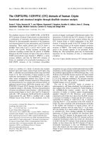
Decreased expression of PPARγ1 in OA cartilage
To examine the expression and localization of PPAR-γ protein
in cartilage, we performed an immunohistochemical analysis.
We found that chondrocytes in both normal and OA cartilage
express PPARγ protein. The immunostaining for PPARγ was
essentially located in the superficial zones, and was lower in
OA cartilage than in normal cartilage. Statistical evaluation of
the cell score for PPARγ indicated significant differences
between normal cartilage (22 ± 2.5% (mean ± SEM)) and car-
tilage from mild to moderate OA (11 ± 3%; Figure 1a,b). Sim-
ilarly, PPARγ expression was significantly reduced in severe
OA cartilage (10 ± 2%, data not shown). By contrast, in intact
OA cartilage, the positive staining seemed lower, but the dif-
ferences were not significant (data not shown). The specificity
of the staining was confirmed by using antibodies that had
been preadsorbed (1 hour, 37°C) with a 20-fold molar excess
of the protein fragment corresponding to amino acids 6 to 105
of human PPARγ (Figure 1c) or non-immune serum (Figure
1c). PPARα and PPARβ were also expressed in normal, mild
to moderate, and severe OA cartilage, but no significant differ-
ences were observed between the cartilage groups (Addi-
tional file 1).
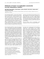
PPARγ has two isoforms, PPARγ1 and PPARγ2, which are
generated by alternative promoters and differential splicing
[9]. To examine which PPARγ transcripts were expressed in
cartilage, we determined absolute mRNA concentrations of
PPARγ1 and PPARγ2 by quantitative real-time PCR. As shown
in Figure 2, PPARγ1 abundance represents about 90% of the
total PPARγ mRNA. Thus, human cartilage expresses high lev-
els of γ1 mRNA, the isoform that is generally expressed in var-
ious tissues, and low levels of the γ2 isoform, which is more
selectively expressed in adipose tissue [10]. The level of
PPARγ1 expression in OA cartilage was 2.4-fold lower than in
normal cartilage (p < 0.005). However, no significant differ-
ences in mRNA levels of PPARγ2 were seen between normal
and OA cartilage (Figure 2). These observations demonstrate
a selective downregulation of PPARγ1 in OA cartilage. In pre-
liminary experiments we showed that the amplification effi-
ciency of PPARγ1, PPARγ2, and GAPDH were approximately
equal, ranging between 1.95 and 2.
Time-course and dose-dependent effect of IL-1 on
PPARγ1 expression in chondrocytes
The reduced expression of PPARγ1 in OA cartilage suggests
that humoral factors produced in the OA joint downregulate
PPARγ1 expression. We therefore evaluated the effect of IL-1,
one of the most prominent mediators in OA, on PPARγ1
expression in cultured chondrocytes. OA chondrocytes were
treated with 100 pg/ml IL-1 for 0, 3, 6, 12, and 24 hours; the
levels of PPARγ1 protein were then analyzed by Western blot-
ting. In preliminary experiments we found that, as in cartilage,
cultured chondrocytes express predominantly the PPARγ1
isoform but not the adipocyte-specific PPARγ2 isoform. As
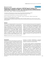
shown in Figure 3a, PPARγ1 protein expression was not sig-
nificantly affected after 3 hours of stimulation with IL-1. The
level of PPARγ1 protein then started to decline gradually at 6
hours and remained low until at least 24 hours. Subsequently,
we examined the effect of various concentrations of IL-1 on
PPARγ1 protein expression. As shown in Figure 3b, the
expression of PPARγ1 was downregulated by IL-1 in a con-
centration-dependent manner; significant decreases were
observed at a concentration as low as 10 pg/ml. Maximal
decreases were obtained at an IL-1 concentration of 100 pg/
ml (Figure 3b). No modulation of PPARα and PPARβ expres-
sion was seen (Additional file 2).
In addition to IL-1, the pro-inflammatory mediators TNF-α, IL-
17, and PGE
2
also contribute to the pathogenesis of OA [1-3].
We therefore examined their effects on PPARγ1 protein
Available online />Page 5 of 11
(page number not for citation purposes)
expression. Cultured OA chondrocytes were incubated for 24
hours with IL-1 (100 pg/ml), TNF-α (1 and 10 ng/ml), IL-17
(10 and 100 ng/ml), and PGE
2
(0.1 and 1 μM), and the expres-
sion levels of PPARγ1 were determined by Western blotting.
As shown in Figure 4, and like IL-1, TNF-α, IL-17, and PGE
2
also downregulated PPARγ1 protein expression. Similar
results were obtained with normal chondrocytes (n = 3; data
not shown).
Downregulation by IL-1 of PPARγ1 expression at the
transcriptional level
To elucidate the mechanism responsible for the changes in
amounts of PPARγ1 protein, we measured the steady-state
level of PPARγ1 mRNA by quantitative real-time PCR. Expres-
sion of the gene encoding GAPDH was used for normaliza-
tion. The relative expression level of PPARγ1 mRNA was
plotted as a percentage decrease compared with untreated
control cells (Figure 5a). Consistent with its effects on protein
expression (Figure 3b), IL-1 downregulates PPARγ1 mRNA
expression in a dose-dependent manner in OA chondrocytes.
The effect of IL-1 on PPARγ1 mRNA expression was maximal
(about 85% decrease) at 100 pg/ml. A dose-dependent effect
of IL-1 on PPARγ1 mRNA expression was also observed in
normal chondrocytes (n = 3; data not shown).
To characterize the effect of IL-1 on PPARγ1 expression fur-
ther, we performed transient transfection experiments with the
reporter construct pGL3-PPARγ1p3000, containing about
3,000 base pairs of regulatory sequence of the gene encoding
human PPARγ1 [9]. As shown in Figure 5b, IL-1 suppressed
PPARγ1 promoter activity in a dose-dependent manner. The
effect of IL-1 on PPARγ1 promoter activity was optimal at 100
pg/ml (about 65% decrease). Taken together, these data
strongly suggest that IL-1 suppressed PPARγ1 expression at
the transcriptional level.
The MAPKs JNK and p38, but not ERK, are involved in IL-
1-induced downregulation of PPARγ1
IL-1 is known to induce its effects in chondrocytes through
activation of a plethora of signaling pathways, including the
mitogen-activated protein kinases (MAPKs) c-Jun N-terminal
kinase (JNK), p38, and extracellular signal-regulated kinase
(ERK) [20]. To assess the contribution of these pathways in
the IL-1-mediated downregulation of PPARγ1, OA
Figure 1
Expression of PPARγ protein in normal and osteoarthritis cartilageExpression of PPARγ protein in normal and osteoarthritis cartilage. Representative immunostaining of human normal cartilage (a) and cartilage from
mild to moderate osteoarthritis (OA) (b) for peroxisome proliferator-activated receptor γ (PPARγ). (c) Normal specimens treated with anti-PPARγ
antibody that was preadsorbed with a 20-fold molar excess of the protein fragment corresponding to amino acids 8 to 106 of human PPARγ (control
for staining specificity). (d) Percentage of chondrocytes expressing PPARγ in normal and OA cartilage. The results are means ± SEM for 10 normal
and 11 OA specimens. *p < 0.05 compared with normal cartilage.
Arthritis Research & Therapy Vol 9 No 2 Afif et al.
Page 6 of 11
(page number not for citation purposes)
chondrocytes were pretreated for 30 minutes with selective
inhibitors for the above pathways, and then stimulated or not
with IL-1 for 18 hours. Total cell lysates were analyzed for
PPARγ1 protein expression by Western blotting. As shown in
Figure 6a, IL-1 reduced PPARγ1 expression remarkably, con-
firming the results seen previously (Figure 3). Pretreatment
with SB203580, a specific p38 MAPK inhibitor, as well as
pretreatment with SP600125, a selective inhibitor of JNK,
dose-dependently abolished IL-1-induced downregulation of
PPARγ1 expression. Conversely, PD98059, a selective inhib-
itor of ERK, had no effect on IL-1-induced downregulation of
PPARγ expression, even at a high concentration (20 μM).
None of the MAPK inhibitors had an effect on PPARγ expres-
sion in the absence of IL-1. These results suggest that the
MAPKs JNK and p38, but not ERK, are involved in the sup-
pression of PPARγ1 expression by IL-1.
Mediation of IL-1-induced downregulation of PPARγ1 by
NF-κB
Because NF-κB mediates many of the effects of IL-1 in a vari-
ety of cell types including chondrocytes, we examined the role
of this transcription factor in the repression of PPARγ1. We
used three different pharmacological inhibitors of the NF-κB
pathway: the antioxidant PDTC, a proteasome inhibitor MG-
132, and an inhibitor of NF-κB translocation (SN-50). Cells
were pretreated with increasing concentrations of each
inhibitor for 30 minutes and then subsequently treated with
100 pg of IL-1 for 18 hours.
As shown in Figure 6b, treatment with IL-1 decreased PPARγ1
expression, but this IL-1 effect was dose-dependently abol-
ished in the presence of each of the three NF-κB inhibitors
(PDTC, MG-132, and SN-50). None of the NF-κB inhibitors
had an effect on basal PPARγ1 expression. These results
imply that NF-κB activation participates in the IL-1-mediated
downregulation of PPARγ1 expression.
Discussion
There is considerable evidence for the importance of PPARγ
in OA because of its potential beneficial effects. It is expressed
by all major cells in joints, including chondrocytes [11,13].
Natural and synthetic ligands of PPARγ were shown to inhibit
the expression of several inflammatory and catabolic genes in
cultured chondrocytes [4,11,12] and to exhibit anti-inflamma-
tory and chondroprotective effects in an experimental animal
model of OA [18]. However, little is known about the expres-
sion and regulation of PPARγ expression in cartilage. Here, we
analyzed the expression of PPARγ in OA and normal cartilage,
and studied the effect of IL-1, a prominent cytokine in OA, on
PPARγ expression in cultured chondrocytes.
This is the first study to demonstrate that human cartilage
expresses predominantly PPARγ1 mRNA and that the levels of
PPARγ1 are decreased in OA in comparison with normal car-
tilage. Our immunohistochemistry analysis showed that
PPARγ was located essentially in the superficial zone of carti-
Figure 2
PPARγ1 and PPARγ2 mRNA levels in normal and osteoarthritis human cartilagePPARγ1 and PPARγ2 mRNA levels in normal and osteoarthritis human
cartilage. RNA was extracted from normal (n = 7) and osteoarthritis (n
= 8) cartilage, reverse transcribed into cDNA, and processed for real-
time PCR. The threshold cycle values were converted to the number of
molecules, as described in the Materials and methods section. Data
were expressed as copies of the gene's mRNA detected per 1,000
glyceraldehyde-3-phosphate dehydrogenase copies. *p < 0.05 com-
pared with normal samples. PPAR, peroxisome proliferator-activated
receptor.
Figure 3
Effect of IL-1 on PPARγ1 protein expression in osteoarthritis chondrocytesEffect of IL-1 on PPARγ1 protein expression in osteoarthritis chondro-
cytes. (a) Osteoarthritis (OA) chondrocytes were treated with 100 pg/
ml IL-1 for the indicated periods. (b) OA chondrocytes were treated
with increasing concentrations of IL-1 for 24 hours. Cell lysates were
prepared and analyzed for peroxisome proliferator-activated receptor
γ1 (PPARγ1) protein by Western blotting (upper panels). The blots
were stripped and reprobed with a specific anti-β-actin antibody (lower
panels). The blots are representative of similar results obtained from
four independent experiments.
Available online />Page 7 of 11
(page number not for citation purposes)
lage and that the levels of PPARγ expression in OA cartilage
were lower than in normal cartilage.
Altered expression of PPARγ was observed in several other
inflammatory disorders. For instance, PPARγ expression was
shown to be reduced in atherosclerotic tissues [21], in epithe-
lial cells from patients with ulcerative colitis [22], in peripheral
blood mononuclear cells from patients with multiple sclerosis
[23], in alveolar macrophages from patients with allergic
asthma [24], and in nasal polyposis from patients with allergic
rhinitis [25]. In contrast, PPARγ expression was shown to be
elevated in brains of patients with Alzheimer's disease [26], in
bronchial epithelium and airway smooth muscle cells of asth-
matic patients [27], and in T cells isolated from patients with
sepsis [28]. Taken together, these results suggest that tissue-
specific regulation of PPARγ expression is extremely complex.
To determine which factors might downregulate PPARγ
expression in cartilage, we tested the impact of IL-1, which
accumulates in chondrocytes in the superficial zone of OA car-
tilage [29,30] and has a pivotal role in the initiation and pro-
gression of OA [1-3]. Our results revealed that exposure to IL-
1 downregulates PPARγ protein expression in chondrocytes in
a time- and dose-dependent manner. It should be noted that
TNF-α, IL-17, and PGE
2
, which are known to contribute to the
pathogenesis of OA, also downregulate PPARγ gene expres-
sion. We therefore cannot exclude the possibility of a role for
these inflammatory mediators in PPARγ downregulation in car-
tilage in vivo. Given the anti-inflammatory and anti-catabolic
functions of PPARγ, it is reasonable to speculate that the sup-
pression of PPARγ expression by inflammatory mediators in
chondrocytes presents a new and additional mechanism by
which these mediators contribute to the pathogenesis of OA.
Our findings are consistent with other studies showing that
pro-inflammatory stimuli downregulate PPARγ expression in
chondrocytes [31-33] and synovial fibroblasts [34,35]. In con-
trast, Shan and colleagues [36] found that IL-1 upregulates
PPARγ expression in chondrocytes. The reasons for these dis-
crepancies are not clear and could be due to small differences
in chondrocyte preparation, culture conditions, and/or detec-
tion methods.
Suppression of PPARγ1 expression by IL-1 in chondrocytes
probably occurs at the transcriptional level, because reporter
gene assays revealed a decrease in PPARγ1 promoter activity
by IL-1. As an alternative to an effect on PPARγ1 promoter, we
could not exclude a specific effect of IL-1 on the stability of
PPARγ1 mRNA.
The MAPKs JNK, p38, and ERK are activated by IL-1 and
mediate many of the effects of IL-1 in chondrocytes [20]. To
determine whether these MAPKs are involved in the IL-1-medi-
ated downregulation of PPARγ1 expression, we employed
specific inhibitors of the three MAPKs. We found that
SB203580 and SP600125 – specific inhibitors of the MAPKs
p38 and JNK, respectively – almost completely abolished the
IL-1-mediated downregulation of PPARγ1 expression,
whereas PD98059 – an inhibitor of the MAPK ERK- was
without effect. These data suggest that the MAPKs JNK and
p38, but not ERK, mediate IL-1-induced downregulation of
PPARγ1 expression in chondrocytes. The NF-κB pathway also
mediates many effects of IL-1 in chondrocytes [37-41]. We
demonstrate here that three compounds that interfere with NF-
κB activation, the anti-oxidant PDTC, the proteasome inhibitor
MG-132, and an inhibitor of NF-κB translocation SN-50,
blocked the suppressive effect of IL-1, suggesting the involve-
ment of NF-κB in the IL-1-mediated downregulation of
PPARγ1 in chondrocytes. Thus, IL-1 engages both the MAPK
(JNK and p38) and the NF-κB pathways to suppress PPARγ1
expression, although it is not clear whether these pathways act
on the same axis or in parallel. Downstream nuclear events in
JNK, p38, and NF-κB signaling pathways leading to the regu-
lation of gene expression in chondrocytes include the
activation of the transcription factors AP-1 and NF-κB
Figure 4
Effect of TNF-α, IL-17 and prostaglandin E
2
on PPARγ1 protein expression in osteoarthritis chondrocytesEffect of TNF-α, IL-17 and prostaglandin E
2
on PPARγ1 protein expression in osteoarthritis chondrocytes. Cells were treated with IL-1 (100 pg/ml),
TNF-α (1 and 10 ng/ml), IL-17 (10 and 100 ng/ml), and prostaglandin E
2
(0.1 and 1 μM). After 24 hours, cell lysates were prepared and analyzed for
peroxisome proliferator-activated receptor γ1 (PPARγ1) protein expression by Western blotting. In the lower panel, the blots were stripped and rep-
robed with a specific anti-β-actin antibody. The blots are representative of similar results obtained from four independent experiments.
Arthritis Research & Therapy Vol 9 No 2 Afif et al.
Page 8 of 11
(page number not for citation purposes)
[20,37,38,40-43]. The human PPARγ1 promoter contains
binding sites for both AP-1 and NF-κB [9]. It is therefore pos-
sible that AP-1 and NF-κB mediate IL-1-induced downregula-
tion of PPARγ1 expression. Although they are historically
characterized as transcriptional activators, several reports
have recently defined AP-1 and NF-κB as transcriptional
Figure 5
IL-1 downregulates PPARγ1 expression at the transcriptional levelIL-1 downregulates PPARγ1 expression at the transcriptional level. (a)
Osteoarthritis (OA) chondrocytes were treated with increasing concen-
trations of IL-1 for 12 hours. Total RNA was isolated and reverse tran-
scribed into cDNA, and peroxisome proliferator-activated receptor γ1
(PPARγ1) and glyceraldehyde-3-phosphate dehydrogenase mRNAs
were quantified by real-time PCR. All experiments were performed in
triplicate, and negative controls without template RNA were included in
each experiment. (b) OA chondrocytes were co-transfected with 1 μg
per well of the PPARγ1 promoter (pGL3-PPARγ1p3000) and 0.5 μg
per well of the internal control pSV40-β-galactosidase, using FuGene 6
transfection reagent. The next day, transfected cells were treated with
increasing concentrations of IL-1 for 18 hours. Luciferase activity val-
ues were determined and normalized to β-galactosidase activity.
Results are expressed as percentage changes, taking the value of
untreated cells as 100%, and show means ± SEM for four independent
experiments. *p < 0.05 compared with untreated cells.
Figure 6
Effect of mitogen-activated protein kinase and NF-κB inhibitors on IL-1-induced downregulation of PPARγ1 expressionEffect of mitogen-activated protein kinase and NF-κB inhibitors on IL-1-
induced downregulation of PPARγ1 expression. (a) Osteoarthritis (OA)
chondrocytes were exposed to increasing concentrations of
SB203580 (p38 mitogen-activated protein kinase inhibitor),
SP600125 (c-Jun N-terminal kinase inhibitor) and PD98059 (extracel-
lular signal-regulated kinase inhibitor) for 30 minutes before treatment
with or without IL-1 (100 pg/ml). (b) OA chondrocytes were exposed
to increasing concentrations of various inhibitors of NF-κB (pyrrolidine
dithiocarbamate, MG-132, and SN-50) for 30 minutes before stimula-
tion with or without IL-1 (100 pg/ml). After 24 hours, cell lysates were
prepared and analyzed for peroxisome proliferator-activated receptor
γ1 (PPARγ1) protein expression by Western blotting. In the lower pan-
els, the blots were stripped and reprobed with a specific anti-β-actin
antibody. The blots are representative of similar results obtained from
four independent experiments.
Available online />Page 9 of 11
(page number not for citation purposes)
repressors [44-50]. Analysis of PPARγ1 promoter in a pro-
moter reporter construct, with mutation of the AP-1 and NF-κB
response elements and the use of small interfering RNA tech-
nology, will contribute to our understanding of the importance
of AP-1 and NF-κB in the IL-1-induced downregulation of
PPARγ1 expression.
The physiological significance of reduced expression of
PPARγ in OA cartilage is of considerable interest, given the
protective functions of PPARγ in cartilage. Indeed, we and
others have previously reported that PPARγ activators inhibit
several inflammatory and catabolic events involved in the
pathogenesis of OA [4,11,12,32-34]. PPARγ activation was
also shown to prevent the proteoglycan degradation induced
by pro-inflammatory cytokines [13]. Furthermore, PPARγ
ligands were shown to reduce the incidence and severity of
OA in an experimental model, preventing inflammatory and cat-
abolic responses as well as cartilage degradation [18]. All
these data suggest that PPARγ has a protective role in OA.
This is strengthened by the observation that PPARγ haploin-
sufficiency exacerbates experimentally induced arthritis [51]. It
is therefore tempting to speculate that diminished expression
of PPARγ in OA cartilage may, at least in part, be involved in
increased expression of inflammatory and catabolic genes,
promoting articular inflammation and cartilage degradation. In
addition, the observation that IL-1 and other pro-inflammatory
mediators downregulate PPARγ1 expression in chondrocytes
has important implications for our understanding of the patho-
physiology of OA.
Conclusion
The decreased expression of PPARγ in OA cartilage and the
literature supporting a protective role for PPARγ in OA raise
the possibility that upregulation of PPARγ may be beneficial in
the context of preventing and treating OA. Additional studies
to define the molecular mechanisms controlling the expression
of PPARγ are therefore urgently needed. Such research will no
doubt add to our understanding of the pathogenesis of OA,
and could lead to the development of new therapeutic strate-
gies in the prevention and treatment of OA and possibly other
arthritic diseases.
Competing interests
The authors declare that they have no competing interests.
Authors' contributions
HA conceived the study, designed and performed cell and
real-time RT-PCR experiments and some immunohistochemis-
try experiments. MB participated in the study design and data
analysis. LM-E performed some immunohistochemistry
experiments. JM-P, J-PP, and ND helped to obtain tissues, par-
ticipated in some immunohistochemistry studies and gave crit-
ical comments on the manuscripts. HF conceived, designed,
and coordinated the study, performed some cell experiments,
and drafted the manuscript. All authors read and approved the
final manuscript.
Additional files
Acknowledgements
The authors thank J Auwerx for the PPARg1 promoter, and M Boily for
help and critical comments. This work was supported by the Canadian
Institutes of Health Research (CIHR) Grant IMH-63168, and the Fonds
de la Recherche du Centre de Recherche du Centre Hospitalier de
l'Université de Montréal (CHUM). HF is a Research Scholar of the Fonds
de Recherche en Santé du Québec (FRSQ).
References
1. Goldring MB: The role of cytokines as inflammatory mediators
in osteoarthritis: lessons from animal models. Connect Tissue
Res 1999, 40:1-11.
2. Pelletier JP, Martel-Pelletier J, Abramson SB: Osteoarthritis, an
inflammatory disease: potential implication for the selection of
new therapeutic targets. Arthritis Rheum 2001, 44:1237-1247.
3. Goldring MB, Berenbaum F: The regulation of chondrocyte
function by proinflammatory mediators: prostaglandins and
nitric oxide. Clin Orthop Relat Res 2004:S37-S46.
4. Li X, Afif H, Cheng S, Martel-Pelletier J, Pelletier JP, Ranger P,
Fahmi H: Expression and regulation of microsomal prostaglan-
din E synthase-1 in human osteoarthritic cartilage and
chondrocytes. J Rheumatol 2005, 32:887-895.
5. Fahmi H, Pelletier JP, Martel-Pelletier J: PPARγ ligands as modu-
lators of inflammatory and catabolic responses on arthritis. An
overview. J Rheumatol 2002, 29:3-14.
6. Braissant O, Foufelle F, Scotto C, Dauca M, Wahli W: Differential
expression of peroxisome proliferator-activated receptors
(PPARs): tissue distribution of PPAR-α, -β, and -γ in the adult
rat. Endocrinology 1996, 137:354-366.
7. Barish GD, Narkar VA, Evans RM: PPAR δ: a dagger in the heart
of the metabolic syndrome. J Clin Invest 2006, 116:590-597.
8. Zhu Y, Qi C, Korenberg JR, Chen XN, Noya D, Rao MS, Reddy JK:
Structural organization of mouse peroxisome proliferator-
activated receptor γ (mPPAR γ) gene: alternative promoter use
and different splicing yield two mPPAR γ isoforms. Proc Natl
Acad Sci USA 1995, 92:7921-7925.
9. Fajas L, Auboeuf D, Raspe E, Schoonjans K, Lefebvre AM, Saladin
R, Najib J, Laville M, Fruchart JC, Deeb S, et al.: The organization,
promoter analysis, and expression of the human PPAR
γ gene.
J Biol Chem 1997, 272:18779-18789.
10. Vidal-Puig A, Jimenez-Linan M, Lowell BB, Hamann A, Hu E,
Spiegelman B, Flier JS, Moller DE: Regulation of PPAR γ gene
expression by nutrition and obesity in rodents. J Clin Invest
1996, 97:2553-2561.
The following Additional files are available online:
Additional file 1
A PDF file showing the expression of PPARα and PPARβ
proteins in normal and OA cartilage.
See />supplementary/ar2151-S1.pdf
Additional file 2
A PowerPoint file showing the effect of IL-1 on PPARα
and PPARβ protein expression in OA chondrocytes.
See />supplementary/ar2151-S2.ppt
Arthritis Research & Therapy Vol 9 No 2 Afif et al.
Page 10 of 11
(page number not for citation purposes)
11. Fahmi H, Di Battista JA, Pelletier JP, Mineau F, Ranger P, Martel-
Pelletier J: Peroxisome proliferator-activated receptor γ activa-
tors inhibit interleukin-1β-induced nitric oxide and matrix met-
alloproteinase 13 production in human chondrocytes. Arthritis
Rheum 2001, 44:595-607.
12. Fahmi H, Pelletier JP, Mineau F, Martel-Pelletier J: 15d-PGJ
2
is act-
ing as a 'dual agent' on the regulation of COX-2 expression in
human osteoarthritic chondrocytes. Osteoarthritis Cartilage
2002, 10:845-848.
13. Bordji K, Grillasca JP, Gouze JN, Magdalou J, Schohn H, Keller JM,
Bianchi A, Dauca M, Netter P, Terlain B: Evidence for the pres-
ence of peroxisome proliferator-activated receptor (PPAR) α
and γ and retinoid Z receptor in cartilage. PPARγ activation
modulates the effects of interleukin-1β on rat chondrocytes. J
Biol Chem 2000, 275:12243-12250.
14. Ji JD, Cheon H, Jun JB, Choi SJ, Kim YR, Lee YH, Kim TH, Chae IJ,
Song GG, Yoo DH, et al.: Effects of peroxisome proliferator-
activated receptor-γ (PPAR-γ) on the expression of inflamma-
tory cytokines and apoptosis induction in rheumatoid synovial
fibroblasts and monocytes. J Autoimmun 2001, 17:215-221.
15. Fahmi H, Pelletier JP, Di Battista JA, Cheung HS, Fernandes J, Mar-
tel-Pelletier J: Peroxisome proliferator-activated receptor γ acti-
vators inhibit MMP-1 production in human synovial fibroblasts
by reducing the activity of the activator protein 1. Osteoarthritis
Cartilage 2002, 10:100-108.
16. Cheng S, Afif H, Martel-Pelletier J, Pelletier JP, Li X, Farrajota K,
Lavigne M, Fahmi H: Activation of peroxisome proliferator-acti-
vated receptor γ inhibits interleukin-1β-induced membrane-
associated prostaglandin E2 synthase-1 expression in human
synovial fibroblasts by interfering with Egr-1. J Biol Chem
2004, 279:22057-22065.
17. Jiang C, Ting AT, Seed B: PPAR-γ agonists inhibit production of
monocyte inflammatory cytokines. Nature 1998, 391:82-86.
18. Kobayashi T, Notoya K, Naito T, Unno S, Nakamura A, Martel-Pel-
letier J, Pelletier JP: Pioglitazone, a peroxisome proliferator-
activated receptor γ agonist, reduces the progression of
experimental osteoarthritis in guinea pigs. Arthritis Rheum
2005, 52:479-487.
19. Altman R, Asch E, Bloch D, Bole G, Borenstein D, Brandt K,
Christy W, Cooke TD, Greenwald R, Hochberg M, et al.: Develop-
ment of criteria for the classification and reporting of osteoar-
thritis. Classification of osteoarthritis of the knee. Diagnostic
and Therapeutic Criteria Committee of the American Rheuma-
tism Association. Arthritis Rheum 1986, 29:1039-1049.
20. Geng Y, Valbracht J, Lotz M: Selective activation of the mitogen-
activated protein kinase subgroups c-Jun NH2 terminal kinase
and p38 by IL-1 and TNF in human articular chondrocytes. J
Clin Invest 1996, 98:2425-2430.
21. Soumian S, Gibbs R, Davies A, Albrecht C: mRNA expression of
genes involved in lipid efflux and matrix degradation in occlu-
sive and ectatic atherosclerotic disease. J Clin Pathol 2005,
58:1255-1260.
22. Dubuquoy L, Jansson EA, Deeb S, Rakotobe S, Karoui M,
Colombel JF, Auwerx J, Pettersson S, Desreumaux P: Impaired
expression of peroxisome proliferator-activated receptor
gamma in ulcerative colitis. Gastroenterology 2003,
124:1265-1276.
23. Klotz L, Schmidt M, Giese T, Sastre M, Knolle P, Klockgether T,
Heneka MT: Proinflammatory stimulation and pioglitazone
treatment regulate peroxisome proliferator-activated receptor
γ levels in peripheral blood mononuclear cells from healthy
controls and multiple sclerosis patients. J Immunol 2005,
175:4948-4955.
24. Kobayashi M, Thomassen MJ, Rambasek T, Bonfield TL, Ray-
chaudhuri B, Malur A, Winkler AR, Barna BP, Goldman SJ, Kavuru
MS: An inverse relationship between peroxisome proliferator-
activated receptor γ and allergic airway inflammation in an
allergen challenge model. Ann Allergy Asthma Immunol 2005,
95:468-473.
25. Cardell LO, Hagge M, Uddman R, Adner M: Downregulation of
peroxisome proliferator-activated receptors (PPARs) in nasal
polyposis. Respir Res 2005, 6:132.
26. Kitamura Y, Shimohama S, Koike H, Kakimura J, Matsuoka Y,
Nomura Y, Gebicke-Haerter PJ, Taniguchi T: Increased expres-
sion of cyclooxygenases and peroxisome proliferator-acti-
vated receptor-γ in Alzheimer's disease brains. Biochem
Biophys Res Commun 1999, 254:582-586.
27. Benayoun L, Letuve S, Druilhe A, Boczkowski J, Dombret MC,
Mechighel P, Megret J, Leseche G, Aubier M, Pretolani M: Regu-
lation of peroxisome proliferator-activated receptor
γ expres-
sion in human asthmatic airways: relationship with
proliferation, apoptosis, and airway remodeling. Am J Respir
Crit Care Med 2001, 164:1487-1494.
28. Soller M, Tautenhahn A, Brune B, Zacharowski K, John S, Link H,
von Knethen A: Peroxisome proliferator-activated receptor γ
contributes to T lymphocyte apoptosis during sepsis. J Leukoc
Biol 2006, 79:235-243.
29. Tetlow LC, Adlam DJ, Woolley DE: Matrix metalloproteinase and
proinflammatory cytokine production by chondrocytes of
human osteoarthritic cartilage: associations with degenera-
tive changes. Arthritis Rheum 2001, 44:585-594.
30. Towle CA, Hung HH, Bonassar LJ, Treadwell BV, Mangham DC:
Detection of interleukin-1 in the cartilage of patients with oste-
oarthritis: a possible autocrine/paracrine role in
pathogenesis. Osteoarthritis Cartilage 1997, 5:293-300.
31. Boyault S, Simonin MA, Bianchi A, Compe E, Liagre B, Mainard D,
Becuwe P, Dauca M, Netter P, Terlain B, Bordji K: 15-Deoxy-δ
12,14-PGJ
2
, but not troglitazone, modulates IL-1β effects in
human chondrocytes by inhibiting NF-κB and AP-1 activation
pathways. FEBS Lett 2001, 501:24-30.
32. Francois M, Richette P, Tsagris L, Raymondjean M, Fulchignoni-
Lataud MC, Forest C, Savouret JF, Corvol MT: Peroxisome pro-
liferator-activated receptor-γ down-regulates chondrocyte
matrix metalloproteinase-1 via a novel composite element. J
Biol Chem 2004, 279:28411-28418.
33. Poleni PE, Bianchi A, Etienne S, Koufany M, Sebillaud S, Netter P,
Terlain B, Jouzeau JY: Agonists of peroxisome proliferators-
activated receptors (PPAR) α, β/δ or γ reduce transforming
growth factor (TGF)-β-induced proteoglycans' production in
chondrocytes. Osteoarthritis Cartilage 2006 in press.
34. Simonin MA, Bordji K, Boyault S, Bianchi A, Gouze E, Becuwe P,
Dauca M, Netter P, Terlain B: PPAR-γ ligands modulate effects
of LPS in stimulated rat synovial fibroblasts. Am J Physiol Cell
Physiol 2002, 282:C125-C133.
35. Moulin D, Bianchi A, Boyault S, Sebillaud S, Koufany M, Francois
M, Netter P, Jouzeau JY, Terlain B: Rosiglitazone induces inter-
leukin-1 receptor antagonist in interleukin-1β-stimulated rat
synovial fibroblasts via a peroxisome proliferator-activated
receptor β/δ-dependent mechanism. Arthritis Rheum 2005,
52:759-769.
36. Shan ZZ, Masuko-Hongo K, Dai SM, Nakamura H, Kato T, Nishioka
K: A potential role of 15-deoxy-δ (12,14)-prostaglandin J
2
for
induction of human articular chondrocyte apoptosis in
arthritis. J Biol Chem 2004, 279:37939-37950.
37. Ding GJ, Fischer PA, Boltz RC, Schmidt JA, Colaianne JJ, Gough
A, Rubin RA, Miller DK: Characterization and quantitation of NF-
κB nuclear translocation induced by interleukin-1 and tumor
necrosis factor-α. Development and use of a high capacity flu-
orescence cytometric system. J Biol Chem 1998,
273:28897-28905.
38. Mengshol JA, Vincenti MP, Coon CI, Barchowsky A, Brinckerhoff
CE: Interleukin-1 induction of collagenase 3 (matrix metallo-
proteinase 13) gene expression in chondrocytes requires p38,
c-Jun N-terminal kinase, and nuclear factor κB: differential reg-
ulation of collagenase 1 and collagenase 3. Arthritis Rheum
2000, 43:801-811.
39. Mendes AF, Caramona MM, Carvalho AP, Lopes MC: Role of
mitogen-activated protein kinases and tyrosine kinases on IL-
1-induced NF-κB activation and iNOS expression in bovine
articular chondrocytes. Nitric Oxide 2002, 6:35-44.
40. Liacini A, Sylvester J, Li WQ, Huang W, Dehnade F, Ahmad M,
Zafarullah M: Induction of matrix metalloproteinase-13 gene
expression by TNF-α is mediated by MAP kinases, AP-1, and
NF-κB transcription factors in articular chondrocytes. Exp Cell
Res 2003, 288:208-217.
41. Fan Z, Bau B, Yang H, Aigner T: IL-1β induction of IL-6 and LIF
in normal articular human chondrocytes involves the ERK, p38
and NFκB signaling pathways. Cytokine 2004, 28:17-24.
42. Liacini A, Sylvester J, Li WQ, Zafarullah M: Inhibition of inter-
leukin-1-stimulated MAP kinases, activating protein-1 (AP-1)
and nuclear factor κB (NF-κB) transcription factors down-reg-
ulates matrix metalloproteinase gene expression in articular
chondrocytes. Matrix Biol 2002, 21:251-262.
Available online />Page 11 of 11
(page number not for citation purposes)
43. Miyazaki Y, Tsukazaki T, Hirota Y, Yonekura A, Osaki M, Shindo H,
Yamashita S: Dexamethasone inhibition of TGF β-induced cell
growth and type II collagen mRNA expression through ERK-
integrated AP-1 activity in cultured rat articular chondrocytes.
Osteoarthritis Cartilage 2000, 8:378-385.
44. Bushel P, Kim JH, Chang W, Catino JJ, Ruley HE, Kumar CC: Two
serum response elements mediate transcriptional repression
of human smooth muscle α-actin promoter in ras-transformed
cells. Oncogene 1995, 10:1361-1370.
45. Schreiber M, Kolbus A, Piu F, Szabowski A, Mohle-Steinlein U,
Tian J, Karin M, Angel P, Wagner EF: Control of cell cycle pro-
gression by c-Jun is p53 dependent. Genes Dev 1999,
13:607-619.
46. Takakura M, Kyo S, Inoue M, Wright WE, Shay JW: Function of
AP-1 in transcription of the telomerase reverse transcriptase
gene (TERT) in human and mouse cells. Mol Cell Biol 2005,
25:8037-8043.
47. Toliver-Kinsky T, Wood T, Perez-Polo JR: Nuclear factor κB/p49
is a negative regulatory factor in nerve growth factor-induced
choline acetyltransferase promoter activity in PC12 cells. J
Neurochem 2000, 75:2241-2251.
48. Wachtel M, Bolliger MF, Ishihara H, Frei K, Bluethmann H, Gloor
SM: Down-regulation of occludin expression in astrocytes by
tumour necrosis factor (TNF) is mediated via TNF type-1
receptor and nuclear factor-κB activation. J Neurochem 2001,
78:155-162.
49. Sohur US, Dixit MN, Chen CL, Byrom MW, Kerr LA: Rel/NF-κB
represses bcl-2 transcription in pro-B lymphocytes. Gene Expr
1999, 8:219-229.
50. Todorov VT, Volkl S, Muller M, Bohla A, Klar J, Kunz-Schughart LA,
Hehlgans T, Kurtz A: Tumor necrosis factor-α activates NFκB to
inhibit renin transcription by targeting cAMP-responsive
element. J Biol Chem 2004, 279:1458-1467.
51. Setoguchi K, Misaki Y, Terauchi Y, Yamauchi T, Kawahata K, Kad-
owaki T, Yamamoto K: Peroxisome proliferator-activated recep-
tor-
γ haploinsufficiency enhances B cell proliferative
responses and exacerbates experimentally induced arthritis. J
Clin Invest 2001, 108:1667-1675.