Báo cáo y học: "Accelerated cellular senescence in degenerate intervertebral discs: a possible role in the pathogenesis of intervertebral disc degeneration" ppt
Bạn đang xem bản rút gọn của tài liệu. Xem và tải ngay bản đầy đủ của tài liệu tại đây (922.68 KB, 12 trang )
Open Access
Available online />Page 1 of 12
(page number not for citation purposes)
Vol 9 No 3
Research article
Accelerated cellular senescence in degenerate intervertebral
discs: a possible role in the pathogenesis of intervertebral disc
degeneration
Christine Lyn Le Maitre, Anthony John Freemont and Judith Alison Hoyland
Tissue Injury and Repair Group, School of Medicine, Stopford Building, The University of Manchester, Oxford Road, Manchester, UK, M13 9PT
Corresponding author: Judith Alison Hoyland,
Received: 13 Mar 2007 Revisions requested: 16 Apr 2007 Revisions received: 26 Apr 2007 Accepted: 11 May 2007 Published: 11 May 2007
Arthritis Research & Therapy 2007, 9:R45 (doi:10.1186/ar2198)
This article is online at: />© 2007 Le Maitre et al.; licensee BioMed Central Ltd.
This is an open access article distributed under the terms of the Creative Commons Attribution License ( />),
which permits unrestricted use, distribution, and reproduction in any medium, provided the original work is properly cited.
Abstract
Current evidence implicates intervertebral disc degeneration as
a major cause of low back pain, although its pathogenesis is
poorly understood. Numerous characteristic features of disc
degeneration mimic those seen during ageing but appear to
occur at an accelerated rate. We hypothesised that this is due
to accelerated cellular senescence, which causes fundamental
changes in the ability of disc cells to maintain the intervertebral
disc (IVD) matrix, thus leading to IVD degeneration. Cells
isolated from non-degenerate and degenerate human tissue
were assessed for mean telomere length, senescence-
associated β-galactosidase (SA-β-gal), and replicative potential.
Expression of P16
INK4A
(increased in cellular senescence) was
also investigated in IVD tissue by means of
immunohistochemistry. RNA from tissue and cultured cells was
used for real-time polymerase chain reaction analysis for matrix
metalloproteinase-13, ADAMTS 5 (a disintegrin and
metalloprotease with thrombospondin motifs 5), and P16
INK4A
.
Mean telomere length decreased with age in cells from non-
degenerate tissue and also decreased with progressive stages
of degeneration. In non-degenerate discs, there was an age-
related increase in cellular expression of P16
INK4A
. Cells from
degenerate discs (even from young patients) exhibited
increased expression of P16
INK4A
, increased SA-β-gal staining,
and a decrease in replicative potential. Importantly, there was a
positive correlation between P16
INK4A
and matrix-degrading
enzyme gene expression. Our findings indicate that disc cell
senescence occurs in vivo and is accelerated in IVD
degeneration. Furthermore, the senescent phenotype is
associated with increased catabolism, implicating cellular
senescence in the pathogenesis of IVD degeneration.
Introduction
Approximately 11 million people in the UK experience low
back pain (LBP) for at least 1 week each month, leading to a
considerable loss of working days and impacting significantly
on the National Health Service. The cause of LBP is not
known, but it is the intervertebral disc (IVD) and the age-
related degenerative changes that occur within it that have
been most frequently associated with LBP [1]. The incidence
of disc degeneration increases with age, and the majority of
lumbar IVDs show some evidence of degeneration by the fifth
decade [2]. Although imaging studies indicate a link between
degeneration of the IVD and LBP [1], clearly not all degenerate
discs are symptomatic. Discs from symptomatic and asympto-
matic individuals show similar radiographic, structural, and
biochemical features. However, people who have LBP exhibit
more severe degeneration than those who are asymptomatic,
suggesting that IVDs of symptomatic individuals undergo
either an acceleration or exacerbation (possibly due to envi-
ronmental or genetic factors) of the ageing process. Thus, disc
degeneration can be viewed as a predictable natural part of
ageing, which in some people occurs at an accelerated rate
for reasons that are currently unknown.
ADAMTS 5 = a disintegrin and metalloprotease with thrombospondin motifs 5; AF = annulus fibrosus; BLAST = Basic Local Alignment Search Tool;
bp = base pairs; Ct = cycle at which threshold is reached; DMEM = Dulbecco's modified Eagle's medium; GAPDH = glyceraldehyde-3-phosphate
dehydrogenase; gDNA = genomic DNA; hTERT = human telomerase reverse transcriptase; IAF = inner annulus fibrosus; IgG = immunoglobulin G;
IHC = immunohistochemistry; IL-1 = interleukin-1; IVD = intervertebral disc; LBP = low back pain; MMP-13 = matrix metalloproteinase-13; MTL =
mean telomere length; NP = nucleus pulposus; OAF = outer annulus fibrosus; PBS = phosphate-buffered saline; PCR = polymerase chain reaction;
RS = replicative senescence; SA-β-gal = senescence-associated β-galactosidase; SIPS = stress-induced premature senescence.
Arthritis Research & Therapy Vol 9 No 3 Le Maitre et al.
Page 2 of 12
(page number not for citation purposes)
During ageing and degeneration, the matrix of the IVD under-
goes substantial structural, molecular, and mechanical
changes, including a loss in the demarcation between the
annulus fibrosus (AF) and the nucleus pulposus (NP), altera-
tions in collagen content, and a decrease in proteoglycan,
resulting in loss of structural integrity, decreased hydration,
and an inability to withstand load [3,4]. Because matrix
changes largely reflect alterations in the biology of the cells, it
is not surprising to find that during ageing and degeneration,
the cells of the NP exhibit altered patterns of gene and protein
expression for matrix molecules, degrading enzymes, and cat-
abolic cytokines [5-9]. Accompanying this is a deterioration in
the overall function of the disc cells, together with a decrease
in tissue cellularity and cell viability of remaining disc cells,
leading to an age-related impairment of IVD repair [6].
Cellular processes that lead to a reduction in fully functional
cells and altered cellular activity include apoptosis and cellular
senescence. Although apoptosis has been reported in age-
related IVD degeneration, with higher rates of apoptosis
present in older individuals [10], no studies, to date, have com-
prehensively investigated cellular senescence in ageing or
degenerate IVDs. The accumulation of senescent cells in vivo
with age, together with their changed pattern of gene expres-
sion [11], implicates cellular senescence in ageing and age-
related pathologies. Indeed, Roberts and colleagues [12] and
Gruber and colleagues [13] have shown increased staining for
senescence-associated β-galactosidase (SA-β-gal) in cells
from herniated discs and degenerate discs, respectively.
Based on this one biomarker of senescence, they postulate
that cellular senescence may be involved in the pathogenesis
of disc degeneration. Similarly, the involvement of cellular
senescence has been linked to osteoarthritis, and investiga-
tors have shown that chondrocytes in articular cartilage from
older individuals and osteoarthritic cartilage display a senes-
cent phenotype (as assessed by several markers) that corre-
lates with changes in matrix homeostasis, leading to matrix
destruction [14,15]. However, to date, no such studies corre-
lating senescence and altered cell function have been con-
ducted in cells from degenerate IVD tissue.
Here, we hypothesise that cellular senescence (assessed by
mean telomere length [MTL], SA-β-gal staining, p16
INK4A
expression, and cell growth kinetics) occurs at an accelerated
rate in IVD degeneration and that, importantly, the senescent
phenotype is related to altered disc cell function associated
with the characteristic features of IVD degeneration.
Materials and methods
Tissue samples
Human IVD tissue was obtained either at surgery, where
patients were selected on the basis of magnetic resonance
imaging-diagnosed degeneration and progression to anterior
resection for either spinal fusion or disc replacement surgery
for chronic LBP, or at post mortem examination. Whole discs
were removed (as detailed previously [9]) following local
research ethics committee approval and informed consent of
the patient or relatives. Herniated disc samples were excluded
from the study.
General procedure for tissue specimens
A block of tissue (incorporating AF and NP in continuity) was
fixed in 10% neutral buffered formalin and processed to paraf-
fin wax. Sections were taken for haematoxylin and eosin stain-
ing to score the degree of morphological degeneration
according to previously published criteria [8]. In brief, sections
were scored for the presence of cell clusters and fissures and
for loss of demarcation and haematoxophilia (indicating
reduced proteoglycan content). A score of 0 to 3 indicates a
histologically normal (non-degenerate) IVD, 4 to 7 indicates
evidence of intermediate degeneration, and 8 to 12 indicates
severe degeneration. Additional sections were taken for immu-
nohistochemistry (IHC).
Isolation of disc cells
Whole disc tissue was separated into NP and AF and finely
minced and digested with 2 U/ml protease (Sigma-Aldrich
Company Ltd., Poole, UK) in Dulbecco's modified Eagle's
medium (DMEM) + F-12 media for 30 minutes at 37°C and
washed twice in DMEM + F-12. NP and AF cells were isolated
in 2 mg/ml collagenase type 1 (Invitrogen Corporation, Pais-
ley, UK) for 4 hours at 37°C.
Evidence for senescence biomarkers in vivo
Telomere length assay
Following extraction of cells from IVD tissue, 31 disc cell sam-
ples (samples 1 to 31 inclusive in Table 1) were taken for DNA
extraction and analysis of MTL. Genomic DNA (gDNA) was
isolated from approximately 1 × 10
6
cells by means of a
DNeasy kit (Qiagen Ltd., Crawley, West Sussex, UK) accord-
ing to the manufacturer's instructions. Analysis of MTL was
performed using the Telo TTAGGG telomere length assay
according to the manufacturer's instructions (Roche Diagnos-
tics Ltd, Burgess Hill, UK). Briefly, 1 μg of gDNA was digested
with Hinf I and Rsa I for 2 hours and separated by electro-
phoresis. Southern transfer was performed and terminal
restriction fragments were detected by hybridization to a dig-
oxigenin-labeled telomeric oligonucleotide and chemilumines-
cence detection by alkaline phosphatase-conjugated anti-
digoxigenin antibodies according to the manufacturer's proto-
col. Membranes were exposed to x-ray film for 5 minutes, and
the MTL was determined using Gene Snap and Gene Tools
from Syngene (SLS, Manchester, UK). Regression analysis
and Spearman rank correlation were performed to analyse cor-
relations between age and MTL in non-degenerate and degen-
erate discs. Multivariate linear regression adjusted for age
(using Stata 9 statistical package; StataCorp LP, College Sta-
tion, TX, USA) was used to assess the association between
MTL and IVD degeneration. Mann-Whitney U tests were used
Available online />Page 3 of 12
(page number not for citation purposes)
to investigate statistical differences in MTL with degree of
degeneration.
Expression and localisation of P16
INK4A
IHC was used to localise the senescence marker P16
INK4a
in
22 paraffin-embedded disc samples (samples 32 to 53 in
Table 1). Tonsil tissue was used as a positive control. The IHC
protocol followed was as previously published [5]. Briefly, fol-
lowing blocking of endogenous peroxidase and antigen
retrieval with citrate buffer at 95°C for 20 minutes, sections
were incubated overnight at 4°C with mouse monoclonal pri-
mary antibody against human p16
INK4a
(Autogen Bioclear UK
Ltd., Calne, Wiltshire, UK) (1:300 dilution in 25% wt/vol rabbit
serum in 1% wt/vol bovine serum albumin [Sigma-Aldrich
Company Ltd.]). Negative controls in which mouse immu-
noglobulin G (IgG) (Dako UK Ltd., Ely, Cambridgeshire, UK)
replaced the primary antibody were used. After washing, sec-
tions were incubated with biotinylated rabbit anti-mouse
antiserum (1:400; Dako UK Ltd.) for 30 minutes at room tem-
perature. Disclosure of secondary antibody binding was by the
streptavidin-biotin complex (Dako UK Ltd.) technique with
3,3'-diaminobenzidine tetrahydrochloride solution (Sigma-
Aldrich Company Ltd.). Sections were counterstained with
Mayers Haematoxylin (Raymond A Lamb Limited, Eastbourne,
East Sussex, UK), dehydrated, and mounted in XAM (BDH,
Liverpool, UK).
For analysis, each disc section was divided morphologically
into three areas: the NP, inner AF (IAF), and outer AF (OAF).
Regions situated at the junction of IAF and OAF or of NP and
IAF were not included in the analysis. Within each area, five
fields of view were analysed and the percentage immunoposi-
tivity was calculated. Data were non-parametric, thus Mann-
Whitney U tests were used to compare the numbers of immu-
nopositive cells in degenerate groups (4 to 7 and 8 to 12) to
non-degenerate discs (scores 0 to 3) for each area of the disc.
Regression analysis and Spearman rank correlation were also
performed to analyse correlations between age and p16
INK4a
immunopositivity. In addition, multivariate linear regression
adjusting for age was performed to analyse correlations
between grade of degeneration and p16
INK4a
immunopositiv-
ity.
Senescence-associated
β
-galactosidase staining
Following extraction of cells from IVD tissue, six samples of NP
cells (Table 1) were taken for SA-β-gal staining. Directly
extracted cells were seeded onto 10-cm
2
flaskettes (SLS) at a
cell density of 0.2 × 10
6
cells per flaskette. Cells were cultured
in standard media [9] on flaskettes for 48 hours and then fixed
in 4% wt/vol paraformaldehdye/phosphate-buffered saline
(PBS) for 20 minutes. Following washing in PBS, cells were
stained overnight for SA-β-gal using the β-Gal Staining Set
(Roche Diagnostics Ltd), with buffer adjusted to pH 6. Sec-
tions were washed in PBS, counterstained with Mayers Hae-
matoxylin (Raymond A Lamb Ltd), dehydrated, and mounted in
XAM (BDH). Cells were visualised using a Leica RMDB
research microscope (Leica Camera Limited, Knowlhill, Milton
Keynes, UK), images were captured using a digital camera and
Bioquant Nova image analysis system (Bioquant Image Analy-
sis Corporation, Nashville, TN, USA), and the percentage of
SA-β-gal-positive cells was calculated.
Senescence biomarkers in human intervertebral disc
cells in vitro
Assessment of growth kinetics
Growth kinetics were examined in NP cells extracted from four
discs (two non-degenerate discs from one post mortem [L2/3:
grade 1, L4/5: grade 2; 37-year-old male] and two degenerate
discs from one patient undergoing surgery [L4/5: grade 4, L5/
S1: grade 8; 49-year-old male]). Following extraction, cells
were seeded into T75 flasks at a cell density of 0.25 × 10
6
,
cultured to 75% confluence, and serially passaged until cells
ceased dividing (failure of population doubling in 4 weeks).
Time in culture and cell number were recorded for each pas-
sage, and cumulative population doublings were calculated.
At each passage, an aliquot of approximately 1 × 10
6
cells was
taken for analysis of MTL, and regression analysis and Spear-
man rank correlation were performed to analyse MTL in cells
following prolonged culture. Aliquots of cells (0.5 × 10
6
cells)
were also taken in duplicate prior to culture (that is, directly
extracted cells) and at each passage for analysis of p16
INK4a
,
MMP-13 (matrix metalloproteinase-13), ADAMTS 5 (a disin-
tegrin and metalloprotease with thrombospondin motifs 5),
and hTERT (human telomerase reverse transcriptase) gene
expression.
Human telomerase reverse transcriptase polymerase chain
reaction
Reverse transcriptase-polymerase chain reaction (PCR) was
used to investigate the gene expression of hTERT in the sam-
ples detailed above to assess the ability of disc cells to repair
telomeres and prevent telomere shortening. RNA was
extracted with Trizol
®
reagent (Invitrogen Corporation) and
cDNA was synthesised using Bioscript RNase H minus
reverse transcriptase (Bioline Ltd., London, UK) and random
hexamers (Roche). PCR was performed with 5 μl of cDNA (50
ng/μl) from each test sample and positive control cDNA (gen-
erated from hTERT-infected cells (a kind gift from Basem
Abdallah and Moustapha Kassem, Odense University Hospi-
tal, Odense, Denmark)). Glyceraldehyde-3-phosphate dehy-
drogenase (GAPDH) primers were designed using Amplify
1.2 software (Professor B Engels, University of Wisconsin,
USA) and gene specificity was confirmed by Basic Local
Alignment Search Tool (BLAST) searches (GenBank data-
base sequences). hTERT primers were a kind gift from B.
Abdallah and M. Kassem (Table 2).
Arthritis Research & Therapy Vol 9 No 3 Le Maitre et al.
Page 4 of 12
(page number not for citation purposes)
Table 1
Intervertebral disc samples used for telomere length assay, senescence-associated β-galactosidase staining, and p16
INK4A
immunohistochemistry
Laboratory number Gender Age (years) Cell type Disc level Cell source Histological grade
1 M 37 AF L4/5 PM 1
2 M 37 AF L5/S1 PM 1
3
a
M37 NPL4/5PM 1
4
a
M37 NPL5/S1PM 1
5 M 47 AF L2/3 PM 1
6 M 47 AF L3/4 PM 1
7
a
M47 NPL2/3PM 1
8 M 47 NP L3/4 PM 1
9 M 47 NP L5/S1 PM 1
10 M 47 AF L4/5 PM 2
11 M 47 NP L4/5 PM 2
12 M 59 NP L4/5 PM 2
13 M 59 AF L4/5 PM 2
14 M 62 AF L3/4 PM 2
15 M 62 AF L4/5 PM 2
16
a
M62 NPL3/4PM 2
17 M 62 NP L4/5 PM 2
18 M 37 AF L1/2 PM 3
19 M 37 NP L1/2 PM 3
20 M 74 AF L3/4 PM 3
21 M 37 AF L2/3 PM 4
22 M 37 AF L3/4 PM 4
23 M 37 NP L2/3 PM 4
24 M 37 NP L3/4 PM 4
25
a
F 49 NP L4/5 Surgical 4
26 M 44 NP L4/5 Surgical 5
27 M 62 AF L5/S1 PM 5
Available online />Page 5 of 12
(page number not for citation purposes)
28 M 62 NP L5/S1 PM 5
29 M 74 AF L4/5 PM 5
30 M 74 AF L2/3 PM 6
31
a
F 49 NP L5/S1 Surgical 8
32 F 15 Tissue L4/5 Surgical 0
33 F 27 Tissue L5/S1 Surgical 0
34 M 39 Tissue L4/5 Surgical 0
35 F 44 Tissue L4/5 Surgical 0
36 F 20 Tissue L5/S1 Surgical 2
37 M 40 Tissue L4/5 Surgical 2
38 M 47 Tissue L4/5 Surgical 2
39 F 27 Tissue L4/5 Surgical 3
40 M 31 Tissue L4/5 Surgical 3
41 F 57 Tissue L4/5 Surgical 3
42 M 59 Tissue L5/S1 Surgical 3
43 M 28 Tissue L4/5 Surgical 5
44 F 34 Tissue L3/4 Surgical 5
45 M 39 Tissue L5/S1 Surgical 5
46 M 55 Tissue L3/4 Surgical 5
47 F 27 Tissue L4/5 Surgical 7
48 F 56 Tissue L5/S1 Surgical 7
49 M 33 Tissue L5/S1 Surgical 8
50 F 40 Tissue L4/5 Surgical 8
51 M 54 Tissue L4/5 Surgical 8
52 M 32 Tissue L4/5 Surgical 10
53 F 41 Tissue L5/S1 Surgical 12
Intervertebral disc samples 1 to 31 were used for telomere length assay, and samples 32 to 53 were used for p16
INK4a
immunohistochemistry.
a
Intervertebral disc samples used for senescence-associated β-galactosidase staining. AF, annulus fibrosus; F, female; M, male; NP, nucleus
pulposus; PM, postmortem tissue.
Table 1 (Continued)
Intervertebral disc samples used for telomere length assay, senescence-associated β-galactosidase staining, and p16
INK4A
immunohistochemistry
Arthritis Research & Therapy Vol 9 No 3 Le Maitre et al.
Page 6 of 12
(page number not for citation purposes)
Correlation of senescent phenotype with altered
expression of matrix-degrading enzyme genes
Real-time PCR was performed for 18s, p16
INK4a
, MMP-13,
and ADAMTS 5 using the standard curve method of analysis
on directly extracted cells and expanded cells.
Primers and probe design
Primers and probes were designed using the Primer Express
program (Applied Biosystems, Warrington, UK) within a single
exon to allow detection of target genes in gDNA and cDNA
samples. Total gene specificity was confirmed by BLAST
searches (GenBank database sequences). Primers and
probes were purchased from Applied Biosystems (Table 2).
Genomic standard curve
gDNA was used to generate standard curves for absolute
quantification of copy number per reaction. Briefly, gDNA
(Promega UK Ltd., Southampton, UK) was homogenised,
diluted to 25,000 pg/μl, and sonicated (Soniprep 150; MSE,
Wolf Laboratories Limited, Pocklington York, UK) on ice.
Serial dilutions of gDNA were prepared to generate standards
with gene copy numbers of 15,000, 3,000, 600, 120, 24, and
0 copies per 2 μl of gDNA.
Polymerase chain reaction amplification
PCRs were performed and monitored using the ABI Prism
7000 Sequence detection System (Applied Biosystems). The
PCR master mix was based on the AmpliTaq Gold DNA
polymerase (Applied Biosystems). On each real-time PCR
plate, a gDNA standard curve was included and cDNA sam-
ples (2 μl [50 ng cDNA/μl] in a total volume of 25 μl) were ana-
lysed in duplicate. Primers were used at a concentration of
900 nM, and probe at a concentration of 250 nM. After an ini-
tial denaturation step and Taq activation at 95°C for 10 min-
utes, the cDNA products were amplified with 40 PCR cycles
consisting of a denaturation step at 95°C for 15 seconds and
an annealing and extension step at 60°C for 1 minute.
Analysis of real-time polymerase chain reaction
Following real-time amplification, the ABI Prism 7000
expressed the data as an amplification plot, from which a base-
line was set from cycle number 3 up to a few cycles prior to
the first visible amplification. A threshold was set at a level
above background levels and within the exponential phase of
the PCR amplification. Vales of Ct (cycle at which the set
threshold is reached) were then exported into an Excel file
(Microsoft Corporation, Manchester, UK), and absolute quan-
tification analysis was performed using the gDNA standard
curve.
Absolute quantification
Standard curves were generated for the housekeeping gene
(18s) and each target gene by plotting log
10
copy number
against Ct value. Line of best fit was then drawn, and the equa-
tion of the line and R
2
was taken. Efficiency (E) was measured
as E = 10
[-1/slope]
[16], R
2
values were accepted if greater than
0.95, and all efficiencies were 97% or greater (Table 2). Ct val-
ues for test samples were converted into copy number per
100 ng of cDNA using the appropriate standard curve for each
gene. Copy numbers obtained for 18s were used to generate
a correction factor for normalization of target genes using the
equation: (maximum 18s copy number)/(18s copy number for
each individual sample), and the correction factor was then
multiplied by the copy number for each target gene for each
sample to give copy number of target gene normalized to 18s
per 100 ng of cDNA. Regression analysis and Spearman rank
correlation were performed to analyse correlations between
p16
INK4a
and matrix-degrading enzymes (MMP-13 and
Table 2
Polymerase chain reaction primer and probe sequences, amplicon sizes, and efficiencies
Standard polymerase chain reaction conditions
Target Forward primer Reverse primer Amplicon size
GAPDH 5' CCC ATC ACC ATC TTC CAG G 3' 5' GGC CAT CCA CAG TCT TCT G 3' 354 bp
hTERT 5' GCC TGA GCT GTA CTT TGT CAA 3' 5' AGG CTG CAG AGC AGC GTG GAG AGG 3' 422 bp
Real-time polymerase chain reaction primers and probes
Target Forward primer Probe Reverse primer Efficiency
18s PDAR PDAR PDAR 99.65%
P16
INK4a
5' GGC TCT ACA CAA GCT TCC TTT
CC 3'
5' 6 FAM – CCC CCA CCC TGG CTC
TGA CCA – TAMRA
5' TCA TGA CCT GCC AGA GAG AAC A
3'
99.22%
MMP-13 5' CCC CAG GCA TCA CCA TTC AAG
3'
5' 6 FAM – AGG GGT CCT GGC TGC
CTT CCT CTT C – TAMRA 3'
5' GAC AAA TCA TCT TCA TCA CCA
CCA C 3'
99.77%
ADAMTS 5 5' GGA CCT ACC ACG AAA GCA GAT
C 3'
5' 6 FAM – CCC AGG ACA GAC CTA
CGA TGC CAC C – TAMRA 3'
5' GCC GGG ACA CAC GGA GTA 3' 99.74%
ADAMTS 5, a disintegrin and metalloprotease with thrombospondin motifs 5; bp, base pairs; GAPDH, glyceraldehyde-3-phosphate
dehydrogenase; hTERT, human telomerase reverse transcriptase; MMP-13, matrix metalloproteinase-13; PDAR, pre-developed assay reagent.
Available online />Page 7 of 12
(page number not for citation purposes)
ADAMTS 5) gene expression.
Results
Evidence for senescence biomarkers in vivo
Mean telomere length in cells directly extracted from human
intervertebral disc tissue
MTL was investigated in cells directly extracted from 20 histo-
logically non-degenerate discs, 10 histologically graded inter-
mediate degenerate discs, and 1 histologically graded severe
degenerate disc. MTL decreased significantly with increasing
age in non-degenerate and degenerate discs (P < 0.05), with
an average decrease in MTL of 0.85 kbp per decade of life in
non-degenerate discs (Figure 1a). Interestingly, the MTL dif-
fered according to the degree of degeneration in two discs
from the same individual (grade 4 disc: MTL 8.56; grade 8
disc: MTL 7.7), and following the statistical correction of
results for age, a significant correlation was observed between
degeneration state (that is, non-degenerate versus degener-
ate) and MTL (P < 0.05). Degenerate discs (grades 4 to 7)
showed significantly shorter MTL compared to non-degener-
ate discs (P < 0.05), with a progressive shortening seen with
increasing grade of degeneration (Figure 1b).
p16
INK4A
Immunohistochemical localisation in human
intervertebral disc tissue
Immunopositive cells were found in all areas of the disc,
although less positivity was observed in the OAF (Figure 1c).
Degenerate discs showed significantly higher proportions of
p16
INK4a
immunopositive cells than non-degenerate discs in all
areas of the IVD (P < 0.05), except for the NP in severe grades
(8 to 12) of degeneration (Figure 1c), where there was a non-
significant increase compared to non-degenerate NP. Non-
degenerate disc samples showed a significant positive corre-
lation in p16
INK4a
immunopositive cells with increasing age (P
Figure 1
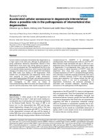
The expression of senescence biomarkers in vivoThe expression of senescence biomarkers in vivo. (a) Mean telomere length (MTL) in cells directly extracted from non-degenerate and degenerate
human intervertebral discs (IVDs): correlation with age. Samples are from 20 non-degenerate discs (6 aged 37 years, 7 aged 47 years, 2 aged 59
years, 4 aged 62 years, and 1 aged 74 years), 10 intermediate degenerate discs (4 aged 37 years, 1 aged 44 years, 1 aged 49 years, 2 aged 62
years, and 2 aged 74 years), and 1 severely degenerate disc (aged 49 years). Spearman rank correlation P < 0.05. (b) MTL in cells directly
extracted from non-degenerate and degenerate human IVDs: effect of degree of degeneration. *Intermediate degenerate samples are significantly
different from non-degenerate samples (P < 0.05). Disc samples are as described in (a). Data are shown as average MTL ± standard error of the
mean (SEM) for each disease state. (c) Quantification and localisation of p16
INK4a
immunopositivity in human IVDs correlated with degree of degen-
eration. *Samples are significantly different from non-degenerate samples (P < 0.05). Samples are from 11 non-degenerate discs, 6 intermediate
degenerate discs, and 5 severely degenerate discs. Averages ± SEM are presented. (d) p16
INK4a
immunopositive cells in human IVDs correlated
with age. Samples are as detailed in (c). Intermediate degenerate (grades 4 to 7) and severely degenerate (grades 8 to 12) samples are grouped for
correlation analysis. Spearman rank correlation for non-degenerate samples P < 0.05 and for degenerate samples P = 0.26. IAF, inner annulus fibro-
sus; kbp, kilobase pairs; NP, nucleus pulposus; OAF, outer annulus fibrosus.
Arthritis Research & Therapy Vol 9 No 3 Le Maitre et al.
Page 8 of 12
(page number not for citation purposes)
< 0.05), although in degenerate samples no such correlation
was observed (P = 0.26) (Figure 1d). A significant positive
correlation was observed between grade of degeneration and
number of p16
INK4a
immunopositive cells following correction
for age (P < 0.05). Immunoreactivity for p16
INK4a
was
restricted to the nucleus and cytoplasm of native disc cells in
all disc samples investigated, with no immunopositivity
observed in the matrix or blood vessels (Figure 2a, b). IgG con-
trols were all negative.
Senescence-associated
β
-galactosidase staining in cells
directly extracted from human intervertebral disc tissue
SA-β-gal staining was not observed in any of the NP cells iso-
lated from the four non-degenerate discs investigated. How-
ever, staining was observed in a number of NP cells extracted
from both grade 4 (12.25% SA-β-gal-positive) and grade 8
(10.25% SA-β-gal-positive) degenerate discs (Figure 2c, d).
Senescence biomarkers in human intervertebral disc
cells in vitro
Culture of NP cells derived from two non-degenerate discs
showed similar growth kinetics, achieving 34 and 38 cumula-
tive population doublings before reaching senescence (Figure
3a). NP cells derived from degenerate discs showed slower
growth kinetics with a reduced capacity to proliferate, achiev-
ing replicative senescence (RS) after 27 cumulative popula-
tion doublings (cells from a grade 4 disc) and 21 cumulative
population doublings (cells from a grade 8 disc) (Figure 3a).
Cells derived from degenerate NP completed 50% of their life
span within 50 days in culture, whereas cells derived from non-
degenerate NP were cultured for approximately 75 days prior
to 50% of their life span being completed (Figure 3b).
MTL in NP cells derived from non-degenerate discs showed a
negative correlation with increasing population doublings (P <
0.05) (Figure 3c), with telomere shortening of 180 to 210
base pairs (bp) per cell division (Figure 3c). A negative corre-
lation was also seen in the NP cells from the low-grade degen-
erate disc (P < 0.05) but not in the NP cells from the severe
degenerate disc (P = 0.25) (Figure 3c).
Expression of human telomerase reverse transcriptase
by intervertebral disc cells
GAPDH was expressed in all samples, but hTERT was
detected only in the positive control, with no expression seen
in any of the disc samples.
Correlation of senescence phenotype with features of
disc degeneration
Evidence from directly extracted cells
No gene expression for p16
INK4a
, MMP-13, or ADAMTS 5
was observed in directly extracted NP cells from non-degener-
ate discs, but expression for these genes was seen in NP cells
directly extracted from degenerate discs (average: p16
INK4a
,
1,893 copies/100 ng of cDNA; MMP-13, 9,386 copies/100
ng of cDNA; ADAMTS 5, 21,220 copies/100 ng of cDNA).
Correlation of p16
INK4A
and matrix-degrading enzyme
gene expression
The combination of all samples investigated demonstrated a
significant positive correlation between p16
INK4a
gene
expression and the gene expression for the matrix-degrading
enzymes MMP-13 and ADAMTS 5 (P values < 0.05) (Figure
4a, b).
Discussion
We hypothesised that, during ageing and degeneration of the
disc, the chondrocyte-like disc cells become senescent,
resulting in phenotypic changes that can lead to the altered
cell function and extracellular matrix characteristic of disc
degeneration. This study has shown for the first time that in
non-degenerate discs the incidence of senescent cells
increases with age. In particular, we have found that telomeric
erosion increases with age together with increased levels of
p16
INK4a
. Importantly, this study has shown that degenerate
discs exhibit accelerated senescence with decreased tel-
omere length, reduced cell replication potential, and elevated
levels of p16
INK4a
and SA-β-gal staining compared to non-
degenerate discs from age-matched individuals. Furthermore,
the senescent phenotype is associated with features charac-
teristic of disc degeneration, namely increased catabolic cell
function.
There are two known mechanisms for the induction of senes-
cence in a cell: RS and stress-induced premature senescence
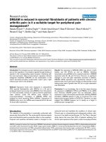
Figure 2
Senescence biomarker immunohistochemistrySenescence biomarker immunohistochemistry. (a) p16
INK4a
immunopo-
sitivity in the nucleus pulposus of human intervertebral discs. (b) Immu-
noglobulin G controls were negative. (c) Senescence-associated β-
galactosidase staining in directly extracted cells from non-degenerate
discs. (d) Senescence-associated β-galactosidase staining in directly
extracted cells from degenerate discs (positive cells indicated with
arrows). Scale bars = 190 μm (a, b) and 370 μm (c, d).
Available online />Page 9 of 12
(page number not for citation purposes)
(SIPS). RS is generally regarded as the result of telomere
shortening accumulated as cells undergo repeated cell divi-
sions [17]. The exact turnover rate of NP cells in the IVD is not
known but is thought to be low. However, Martin and Buckwal-
ter [14] examined cells in articular cartilage, which share many
characteristics with those of the NP, and suggested that
although turnover is slow the very long life of the chondrocyte
may mean that in older people chondrocytes may have gone
through sufficient replications to induce RS. SIPS is the alter-
native explanation for cellular senescence and has come from
the discovery that various insults, including mechanical load,
Figure 3
Senescence biomarkers in human intervertebral disc (IVD) cells in vitroSenescence biomarkers in human intervertebral disc (IVD) cells in vitro. (a) Cell growth kinetics: cumulative population doublings in nucleus pulpo-
sus (NP) cells extracted from non-degenerate and degenerate IVDs. (b) Percentage of life span completed over time in culture of NP cells extracted
from non-degenerate and degenerate IVDs. (c) Mean telomere length in NP cells extracted from non-degenerate and degenerate IVDs with increas-
ing population doubling. Samples used consisted of two non-degenerate discs from one post mortem (L2/3: grade 1, L4/5: grade 2; 37-year-old
male) and two degenerate discs from one patient undergoing surgery (L4/5: grade 4, L5/S1: grade 8; 49-year-old male).
Figure 4
Correlation of senescent phenotype with expression of matrix-degrading enzymesCorrelation of senescent phenotype with expression of matrix-degrading enzymes. (a) Correlation of MMP-13 and p16
INK4a
gene expression in
human intervertebral disc (IVD) cells. Spearman rank correlation P < 0.05. (b) Correlation of ADAMTS 5 and p16
INK4a
gene expression in human
IVD cells. Spearman rank correlation P < 0.05. ADAMTS 5, a disintegrin and metalloprotease with thrombospondin motifs 5; MMP-13, matrix
metalloproteinase-13.
Arthritis Research & Therapy Vol 9 No 3 Le Maitre et al.
Page 10 of 12
(page number not for citation purposes)
high levels of oxygen and cytokines such as interleukin-1 (IL-
1), can lead to cellular senescence [18,19]. This is an appeal-
ing explanation for the senescent biomarker expression seen
in the degenerate IVD of young people as degeneration can be
induced by mechanical load and cytokines such as IL-1, which
we have shown to be increased in IVD degeneration [9]. Fur-
thermore, the increased vascularisation also seen during disc
degeneration [20,21] could lead to increased oxygen tension
and hence induction of senescence.
One feature of senescent cells which appears as a universal
and predictable marker is telomere shortening [22]. Telomeres
are repetitive DNA sequences at the end of chromosomes
which are essential for chromosomal replication but also help
sustain normal chromosome function by sealing the chromo-
some ends and preventing enzymatic degradation. Upon each
cell division, telomeres degrade because replication of the
extreme ends of DNA is not possible. To counteract telomere
shortening, cells can express the enzyme telomerase (hTERT),
which synthesizes new telomeric repeats, thereby maintaining
or increasing telomere length. We have demonstrated that the
NP cells extracted from both the non-degenerate and degen-
erate IVD do not show expression of hTERT and thus are fully
susceptible to telomere erosion.
Telomere length is often considered a good indicator of the
cell's replicative history [17]. Telomeres, however, can also be
shortened during SIPS in a manner independent from replica-
tion [18,23,24]. Thus, MTL can be considered a marker of rep-
licative history and of the cumulative history of stress inducers
of senescence, as well as an indicator of the probability of cell
senescence [25]. In this study, we have investigated telomere
erosion in disc cells both in vitro and in vivo. Martin and Buck-
walter [14] demonstrated that in vitro telomeres in articular
chondrocytes shortened by 100 to 200 bp per cell division,
and Parsch and colleagues [26] showed telomere shortening
of approximately 300 bp per cumulative population doubling in
the same cell type. In the current study (albeit only in two
samples), we demonstrated that in NP cells derived from non-
degenerate discs, expansion in monolayer resulted in a pro-
gressive shortening of MTL, with a reduction of 180 to 210 bp
per cellular division, matching the attrition rate seen in vitro in
articular chondrocytes [14,26]. In NP cells isolated from the
non-degenerate discs, RS was induced when telomeres
reached a critical level of approximately 5 to 6.5 kbp, which
matched the critical level of approximately 5 to 7.6 kbp
observed previously in articular chondrocytes [14].
We have demonstrated that in vivo in 20 non-degenerate sam-
ples telomeres shortened at a rate of approximately 85 bp per
year, suggesting an in vivo replication rate of one cell division
every 2 years. The attrition rate seen in disc cells in vivo is
higher than the 30 bp/year attrition rate seen in articular
chondrocytes [27] but is similar to the attrition rate of 102 bp/
year seen in iliac artery cells [28]. This would suggest that disc
cells have a higher rate of cell turnover or are exposed to more
stress than articular chondrocytes in vivo. Indeed, the degen-
erative process in IVD begins as early as the second decade
of life, with associated increased occurrence of LBP [29].
However, articular cartilage does not show degenerative
changes until later in life, with the incidence of osteoarthritis
increasing dramatically after the age of 40 years [14]. Our data
suggest that senescent cells accumulate in different tissues at
different rates, with non-degenerate disc cells ageing faster
than cells from articular cartilage, which may be a result of
environmental factors such as mechanical stress, cytokine
exposure, or injury. Furthermore, our data suggest that cells
from degenerate discs exhibit accelerated senescence. (For
example, the MTL of 7.7 kbp in a severe degenerate sample
would have been predicted to be from an 80-year-old; how-
ever, this disc sample came from a donor who was only 49
years old.)
Hayflick [30] showed that normal cells could divide only a lim-
ited number of times in culture (the maximum number of divi-
sions is known as the Hayflick limit), after which cells remain
viable but are completely incapable of entering cell division
and are thus termed senescent. Since this time, the reduced
ability of cells to divide in culture has been used as an assess-
ment of premature senescence [31]. The Hayflick limit for
human fibroblasts has been estimated at approximately 60
population doublings, whereas the estimated limit for human
chondrocytes is approximately 35 doublings [14]. We have
shown that NP cells from non-degenerate discs were capable
of 35 to 40 population doublings prior to reaching the Hayflick
limit, which matches that seen previously for articular chondro-
cytes. However, in NP cells derived from degenerate discs, a
reduced capability to divide was seen with cells capable of
only 20 to 25 population doublings prior to senescence.
A number of studies have shown increased levels of p16
INK4a
with increased occurrence of senescence [32,33]. p16
INK4a
is
thought to be involved in the activation of the retinoblastoma
cell cycle inhibitory pathway, leading to permanent growth
arrest and cellular senescence [34]. We have demonstrated
that in non-degenerate discs p16
INK4a
increases with age but
that degenerate discs show overexpression of p16
INK4a
com-
pared to age-matched non-degenerate samples. This is similar
to the increased expression of p16
INK4a
seen in osteoarticular
cartilage [35] and suggests that p16
INK4a
may be physiologi-
cally involved in the senescence process, particularly as
p16
INK4a
may accumulate in response to specific forms of
stress, including oxidative damage [18].
Since the initial description of the pH-dependent staining of
senescent fibroblasts by β-galactosidase at pH 6 [36], this
simple histological stain has been used in a number of studies
to indicate the presence of senescent cells [14,27,37], includ-
ing in the IVD [12,13]. Like Roberts and colleagues [12] and
Gruber and colleagues [13], we have shown that NP cells
Available online />Page 11 of 12
(page number not for citation purposes)
stain for SA-β-gal, but our results differ in that we found no
staining in non-degenerate NP cells. However, as in these pre-
vious studies, with degeneration, there was increased SA-β-
gal staining. Because these discs (in our study) also showed
shorter MTLs, reduced ability to divide, and increased
numbers of p16
INK4a
immunopositive cells compared to cells
from non-degenerate discs, our data clearly illustrates an
increase in cellular senescence in degenerate discs compared
to non-degenerate discs, corroborating the recent data pro-
duced by Gruber and colleagues [13].
During cell senescence, cell function can deteriorate before
cell cycle arrest occurs, with cells showing abnormal protein
synthesis and an altered phenotype (including over expression
of p16
INK4a
[38]), and in chondrocytes, increased levels of
MMPs and aggrecanases have been observed [37]. Here, we
demonstrate for the first time that, in cells extracted from
human NP tissue, increased levels of p16
INK4a
were
associated with increased gene expression of the degradative
enzymes MMP-13 and ADAMTS 5, which is characteristic of
disc degeneration [5,39]. We have previously shown that cells
from degenerate discs respond differently to IL-1 compared to
cells from non-degenerate discs [9]. Cellular senescence may
be responsible for this as it has been shown that senescent
cells show altered responses to cytokines and growth factors
[15]. Our data indicate that the senescent phenotype is linked
to the increased production of degradation enzymes which
may be brought about by the catabolic cytokine IL-1 known to
be increased in disc degeneration [9].
Conclusion
We have shown tissue-specific cellular senescence and
accelerated senescence in the degenerate IVD and that this is
associated with increased catabolic cell function. Cellular
senescence can be prevented, bypassed, or reversed in other
settings and perhaps here too [35,40,41]. Our data suggest
that disc cell senescence has an important role in the develop-
ment and progression of IVD degeneration; thus, understand-
ing the nature of cellular senescence will be paramount in
devising new approaches for its prevention and treatment. Fur-
thermore, the cellular senescence we have identified could be
imperative in dictating the success of possible future biologic
therapies, which may require the insertion of new metabolically
active cells into the degenerate disc to achieve success.
Competing interests
The authors declare that they have no competing interests.
Authors' contributions
CLM participated in the design of the study, performed the
majority of the laboratory work and analysis, and drafted the
manuscript. AJF participated in the design of the study and
interpretation of data. JAH conceived the study, secured fund-
ing, contributed to the design and coordination of the study,
and participated in interpretation of data and extensive prepa-
ration of the final manuscript. All authors read and approved
the final manuscript.
Acknowledgements
The authors wish to thank Sara Rollinson (Department of Clinical Neu-
rosciences, The University of Manchester, Manchester, UK) for her inval-
uable statistical advice, Kulvir S Hundal for assistance with the p16
INK4a
immunohistochemistry, Stephen Richardson and Sian Parker for assist-
ance with long-term culturing of disc cells, Sarah Heathfield for assist-
ance with data analysis, and Basem Abdallah and Moustapha Kassem
for the kind gift of the hTERT primers and positive control. This work was
funded by a grant from DISCS (Diagnostic Investigation of Spinal Con-
ditions and Sciatica) and was undertaken in the Human Tissue Profiling
Laboratories of the Tissue Injury and Repair research group that receive
core support from the Arthritis Research Campaign (Integrated Clinical
Arthritis Centre grant F0551) and Medical Research Council (MRC)
(Co-operative Group Grant G9900933) and the joint Research Coun-
cils (MRC, Biotechnology and Biological Sciences Research Council,
and Engineering and Physical Sciences Research Council) of the UK
Centre for Tissue Engineering (34/TIE 13617).
References
1. Luoma K, Riihimaki H, Luukkonen R, Raininko R, Viikari-Juntura E,
Lamminen A: Low back pain in relation to lumbar disc
degeneration. Spine 2000, 25:487-492.
2. Miller JA, Schmatz C, Schultz AB: Lumbar disc degeneration:
correlation with age, sex, and spine level in 600 autopsy
specimens. Spine 1988, 13:173-178.
3. Roughley PJ, Alini M, Antoniou J: The role of proteoglycans in
aging, degeneration and repair of the intervertebral disc. Bio-
chem Soc Trans 2002, 30:869-874.
4. Roughley PJ: Biology of intervertebral disc aging and degener-
ation: involvement of the extracellular matrix. Spine 2004,
29:2691-2699.
5. Le Maitre CL, Freemont AJ, Hoyland JA: Localization of degrada-
tive enzymes and their inhibitors in the degenerate human
intervertebral disc. J Pathol 2004, 204:47-54.
6. Boos N, Weissbach S, Rohrbach H, Weiler C, Spratt KF, Nerlich
AG: Classification of age-related changes in lumbar interver-
tebral discs: 2002 Volvo Award in basic science. Spine 2002,
27:2631-2644.
7. Weiler C, Nerlich A, Zipperer J, Bachmeier BE, Boos N: 2002 SSE
Award Competition in Basic Science: expression of major
matrix metalloproteinases is associated with intervertebral
disc degradation and resorption. Eur Spine J 2002,
11:308-320.
8. Sive JI, Baird P, Jeziorsk M, Watkins A, Hoyland JA, Freemont AJ:
Expression of chondrocyte markers by cells of normal and
degenerate intervertebral discs. Mol Pathol 2002, 55:91-97.
9. Le Maitre CL, Freemont AJ, Hoyland JA: The role of interleukin-1
in the pathogenesis of human intervertebral disc
degeneration. Arthritis Res Ther 2005, 7:R732-R745.
10. Gruber HE, Hanley EN Jr: Analysis of aging and degeneration of
the human intervertebral disc. Comparison of surgical speci-
mens with normal controls [see comments]. Spine 1998,
23:751-757.
11. Campisi J, Kim SH, Lim CS, Rubio M: Cellular senescence, can-
cer and aging: the telomere connection. Exp Gerontol 2001,
36:
1619-1637.
12. Roberts S, Evans EH, Kletsas D, Jaffray DC, Eisenstein SM:
Senescence in human intervertebral discs. Eur Spine J 2006,
15(Suppl 3):S312-S316.
13. Gruber HE, Ingram JA, Norton HJ, Hanley EN Jr: Senescence in
cells of the aging and degenerating intervertebral disc: immu-
nolocalization of senescence-associated beta-galactosidase
in human and sand rat discs. Spine 2007, 32:321-327.
14. Martin JA, Buckwalter JA: Aging, articular cartilage chondrocyte
senescence and osteoarthritis. Biogerontology 2002,
3:257-264.
Arthritis Research & Therapy Vol 9 No 3 Le Maitre et al.
Page 12 of 12
(page number not for citation purposes)
15. Martin JA, Buckwalter JA: The role of chondrocyte senescence
in the pathogenesis of osteoarthritis and in limiting cartilage
repair. J Bone Joint Surg Am 2003, 85-A(Suppl 2):106-110.
16. Pfaffl MW: Quantification strategies in real time PCR. In A-Z of
Quantitative PCR Edited by: Bustin SA. La Jolla, CA: International
University Line; 2004:87-112.
17. Campisi J: Replicative senescence and immortalization. In The
Molecular Basis of Cell Cycle and Growth Control Edited by:
Stein GS. New York: Wiley-Liss; 1999:348-373.
18. Toussaint O, Medrano EE, von Zglinicki T: Cellular and molecular
mechanisms of stress-induced premature senescence (SIPS)
of human diploid fibroblasts and melanocytes. Exp Gerontol
2000, 35:927-945.
19. Yudoh K, Nguyen T, Nakamura H, Hongo-Masuko K, Kato T, Nish-
ioka K: Potential involvement of oxidative stress in cartilage
senescence and development of osteoarthritis: oxidative
stress induces chondrocyte telomere instability and downreg-
ulation of chondrocyte function. Arthritis Res Ther 2005,
7:R380-R391.
20. Hoyland JA, Freemont AJ, Jayson MI: Age related changes in the
structures within and bordering the intervertebral foramen:
associations with low back pain. In The Ageing Spine Edited by:
Hukins DW, Nelson MA. Manchester: Manchester University
Press; 1987:94-110.
21. Freemont AJ, Watkins A, Le Maitre C, Baird P, Jeziorska M, Knight
MT, Ross ER, O'Brien JP, Hoyland JA: Nerve growth factor
expression and innervation of the painful intervertebral disc. J
Pathol 2002, 197:286-292.
22. Coates PJ: Markers of senescence? J Pathol 2002,
196:371-373.
23. Duan J, Duan J, Zhang Z, Tong T: Irreversible cellular senes-
cence induced by prolonged exposure to H
2
O
2
involves DNA-
damage-and-repair genes and telomere shortening. Int J Bio-
chem Cell Biol 2005, 37:1407-1420.
24. Dumont P, Balbeur L, Remacle J, Toussaint O: Appearance of
biomarkers of in vitro ageing after successive stimulation of
WI-38 fibroblasts with IL-1alpha and TNF-alpha: senescence
associated beta-galactosidase activity and morphotype
transition. J Anat 2000, 197(Pt 4):529-537.
25. von Zglinicki T, Martin-Ruiz CM: Telomeres as biomarkers for
ageing and age-related diseases. Curr Mol Med 2005,
5:197-203.
26. Parsch D, Brummendorf TH, Richter W, Fellenberg J: Replicative
aging of human articular chondrocytes during ex vivo
expansion. Arthritis Rheum 2002, 46:2911-2916.
27. Martin JA, Buckwalter JA: Telomere erosion and senescence in
human articular cartilage chondrocytes. J Gerontol A Biol Sci
Med Sci 2001, 56:B172-B179.
28. Chang E, Harley CB: Telomere length and replicative aging in
human vascular tissues. Proc Natl Acad Sci USA 1995,
92:11190-11194.
29. Anderson DG, Tannoury C: Molecular pathogenic factors in
symptomatic disc degeneration. Spine J 2005, 5:260S-266S.
30. Hayflick L: The limited in vitro lifetime of human diploid cell
strains. Exp Cell Res 1965, 37:614-636.
31. Campisi J: Cancer, aging and cellular senescence. In Vivo 2000,
14:183-188.
32. Krishnamurthy J, Torrice C, Ramsey MR, Kovalev GI, Al-Regaiey K,
Su L, Sharpless NE: Ink4a/Arf expression is a biomarker of
aging. J Clin Invest 2004, 114:1299-1307.
33. Satyanarayana A, Rudolph KL: p16 and ARF: activation of teen-
age proteins in old age. J Clin Invest 2004, 114:1237-1240.
34. Sherr CJ, Roberts JM: CDK inhibitors: positive and negative
regulators of G1-phase progression.
Genes Dev 1999,
13:1501-1512.
35. Zhou HW, Lou SQ, Zhang K: Recovery of function in osteoar-
thritic chondrocytes induced by p16INK4a-specific siRNA in
vitro. Rheumatology (Oxford) 2004, 43:555-568.
36. Dimri GP, Lee X, Basile G, Acosta M, Scott G, Roskelley C,
Medrano EE, Linskens M, Rubelj I, Pereira-Smith O, et al.: A
biomarker that identifies senescent human cells in culture and
in aging skin in vivo. Proc Natl Acad Sci USA 1995,
92:9363-9367.
37. Price JS, Waters JG, Darrah C, Pennington C, Edwards DR, Donell
ST, Clark IM: The role of chondrocyte senescence in
osteoarthritis. Aging Cell 2002, 1:57-65.
38. Foreman KE, Tang J: Molecular mechanisms of replicative
senescence in endothelial cells. Exp Gerontol 2003,
38:1251-1257.
39. Le Maitre CL, Freemont A, Hoyland J: Human disc degeneration
is associated with increased MMP 7 expression. Biotech
Histochem 2006, 81:125-131.
40. Piera-Velazquez S, Jimenez SA, Stokes D: Increased life span of
human osteoarthritic chondrocytes by exogenous expression
of telomerase. Arthritis Rheum 2002, 46:683-693.
41. Shay JW, Wright WE: Use of telomerase to create bioengi-
neered tissues. Ann N Y Acad Sci 2005, 1057:479-491.