Báo cáo y học: "Regulation of the JNK pathway by TGF-beta activated kinase 1 in rheumatoid arthritis synoviocytes" pps
Bạn đang xem bản rút gọn của tài liệu. Xem và tải ngay bản đầy đủ của tài liệu tại đây (731.8 KB, 9 trang )
Open Access
Available online />Page 1 of 9
(page number not for citation purposes)
Vol 9 No 3
Research article
Regulation of the JNK pathway by TGF-beta activated kinase 1 in
rheumatoid arthritis synoviocytes
Deepa R Hammaker
1
, David L Boyle
1
, Tomoyuki Inoue
2
and Gary S Firestein
1
1
Division of Rheumatology, Allergy and Immunology, UCSD School of Medicine, Gilman Dr., La Jolla, CA 92093, USA
2
Medicinal Research Laboratories, Taisho Pharmaceutical Co. Ltd, Yoshino-Cho, Kita-Ku, Saitama 331-9530, Japan
Corresponding author: Deepa R Hammaker,
Received: 20 Apr 2007 Revisions requested: 22 May 2007 Revisions received: 25 May 2007 Accepted: 8 Jun 2007 Published: 8 Jun 2007
Arthritis Research & Therapy 2007, 9:R57 (doi:10.1186/ar2215)
This article is online at: />© 2007 Hammaker et al.; licensee BioMed Central Ltd.
This is an open access article distributed under the terms of the Creative Commons Attribution License ( />),
which permits unrestricted use, distribution, and reproduction in any medium, provided the original work is properly cited.
Abstract
c-Jun N-terminal kinase (JNK) contributes to metalloproteinase
(MMP) gene expression and joint destruction in inflammatory
arthritis. It is phosphorylated by at least two upstream kinases,
the mitogen-activated protein kinase kinases (MEK) MKK4 and
MKK7, which are, in turn, phosphorylated by MEK kinases
(MEKKs). However, the MEKKs that are most relevant to JNK
activation in synoviocytes have not been determined. These
studies were designed to assess the hierarchy of upstream
MEKKs, MEKK1, MEKK2, MEKK3, and transforming growth
factor-β activated kinase (TAK)1, in rheumatoid arthritis (RA).
Using either small interfering RNA (siRNA) knockdown or
knockout fibroblast-like synoviocytes (FLSs), MEKK1, MEKK2,
or MEKK3 deficiency (either alone or in combination) had no
effect on IL-1β-stimulated phospho-JNK (P-JNK) induction or
MMP expression. However, TAK1 deficiency significantly
decreased P-JNK, P-MKK4 and P-MKK7 induction compared
with scrambled control. TAK1 knockdown did not affect p38
activation. Kinase assays showed that TAK1 siRNA significantly
suppressed JNK kinase function. In addition, MKK4 and MKK7
kinase activity were significantly decreased in TAK1 deficient
FLSs. Electrophoretic mobility shift assays demonstrated a
significant decrease in IL-1β induced AP-1 activation due to
TAK1 knockdown. Quantitative PCR showed that TAK1
deficiency significantly decreased IL-1β-induced MMP3 gene
expression and IL-6 protein expression. These results show that
TAK1 is a critical pathway for IL-1β-induced activation of JNK
and JNK-regulated gene expression in FLSs. In contrast to other
cell lineages, MEKK1, MEKK2, and MEKK3 did not contribute to
JNK phosphorylation in FLSs. The data identify TAK1 as a pivotal
upstream kinase and potential therapeutic target to modulate
synoviocyte activation in RA.
Introduction
Rheumatoid arthritis (RA) is a chronic inflammatory disease
characterized by synovial lining hyperplasia and sublining infil-
tration of inflammatory cells [1]. Fibroblast-like synoviocytes
(FLSs) play a crucial role in joint damage as well as the prop-
agation of inflammation [2]. In response to potent pro-inflam-
matory cytokines such as IL-1β, FLSs produce large amounts
of matrix metalloproteinases (MMP), which are key drivers of
matrix destruction [3-5]. MMP production is, in turn, regulated
by several signal transduction pathways, including the
mitogen-activated protein kinases (MAPKs) [6,7].
All three MAPK families have been implicated in RA, including
extracellular signal-regulated kinase (ERK), c-Jun N-terminal
kinase (JNK), and p38 [8-10]. JNK plays an especially impor-
tant role in extracellular matrix turnover because it is activated
in RA synovium, regulates MMP gene expression in cultured
FLSs, and mediates joint destruction in rat adjuvant arthritis
[11-16]. JNK is phosphorylated by upstream MAPK kinases
(MAPKKs), which are dual specific enzymes that phosphor-
ylate threonine and tyrosine residues [17]. Two MAPKKs (or
mitogen-activated protein kinases [MEKs]), MKK4 and MKK7,
form a complex with JNK [18], although only the latter is
Ct = threshold cycle; DMEM = Dulbecco's modeified Eagle's medium; ELISA = enzyme-linked immunosorbent assay; ERK = extracellular signal-reg-
ulated kinase; FCS = fetal calf serum; FGF = fibroblast growth factor; FLS = fibroblast-like synoviocyte; GAPDH = glyceraldehyde-3-phosphate dehy-
drogenase; GST = glutathione S-transferase; IL = interleukin; IRAK = IL-1 receptor-associated kinase; JNK = c-Jun N-terminal kinase; MAP3K =
MAPKK kinase; MAPK = mitogen-activated protein kinase; MAPKK = MAPK kinase; MEF = murine embryonic fibroblast; MEK = mitogen-activated
protein kinase, MEKK = MEK kinase; MMP = matrix metalloproteinase; NF = nuclear factor; P = phospho; sc, scrambled; siRNA, small interfering
RNA; TAB = TAK1-binding protein, TAK = transforming growth factor-β activated kinase; TRAF6 = Tumor necrosis factor receptor-associated factor
6.
Arthritis Research & Therapy Vol 9 No 3 Hammaker et al.
Page 2 of 9
(page number not for citation purposes)
required for cytokine-mediated engagement of this pathway in
FLSs [19].
Multiple upstream MAPKK kinases (MAP3Ks) that activate the
MAPKKs and the JNK cascade have been identified in RA. For
instance, MEK kinase (MEKK)1, MEKK2, and transforming
growth factor-β activated kinase (TAK)1 are the most abun-
dant in inflamed synovium as well as cultured FLSs [20]. Of
these MAP3Ks, MEKK2 initially appeared to be the most
important in RA because it forms a functional complex with
JNK. In the present study, TAK1 functioned as the dominant
MAP3K for JNK activation in IL-1-stimulated FLSs. These
results were unexpected because several groups have shown
that MEKK1, MEKK2 and MEKK3 are indispensable for JNK
activation. For instance, MEKK1 is the predominant kinase
required for JNK activation in corneal epithelia [20] and murine
embryonic fibroblasts (MEFs) [20]. In other culture conditions,
JNK activation is inhibited in MEKK3-/- MEFs stimulated with
IL-1 [21]. Similarly, fibroblast growth factor (FGF)-2-induced
JNK activation and JNK phosphorylation-induced T cell recep-
tor ligation require MEKK2 [22]. Based on our studies using
MAP3K deficient cells, these MAP3Ks appear to be redundant
in JNK activation in cultured FLSs. Therefore, the diverse and
complex functions of MAP3Ks vary depending on the cell type
as well as the stimulus. It is precisely this signaling diversity
that offers an opportunity to target upstream kinases in the
JNK cascade that regulate pathogenic responses in arthritis
while potentially sparing other functions that are critical to host
responses. This study suggests that TAK1 is a crucial activator
of the JNK pathway in FLSs and is a potential target for arthritis
therapy.
Materials and methods
Fibroblast-like synoviocytes
FLSs were isolated from synovial tissues obtained from RA
patients at the time of joint replacement as described previ-
ously [3]. The diagnosis of RA conformed to the American Col-
lege of Rheumatology 1987 revised criteria [23]. The protocol
was approved by the UCSD Human Subjects Research Pro-
tection Program. Synovial tissues were minced and incubated
with 0.5 mg/ml collagenase VIII (Sigma, St. Louis, MO, USA)
in serum-free RPMI (Mediatech, Herndon, VA, USA) for 1.5 h
at 37°C, filtered through a 0.22 μm cell strainer, extensively
washed, and cultured in DMEM supplemented with 10% FCS
(endotoxin content <0.006 ng/ml; Gemini Biosciences, Cala-
basas, CA, USA), penicillin, streptomycin, gentamicin and L-
glutamine in a humidified 5% CO
2
incubator. After overnight
culture, nonadherent cells were removed, and adherent cells
were trypsinized, split at a 1:3 ratio, and cultured. Synovio-
cytes were used from passage 4 through 9, when FLSs were
a homogeneous population with <1% CD11b, <1% phago-
cytic, and <1% FcRγII positive cells.
Mice knee and ankle synovial tissues were isolated, minced
and incubated with 0.5 mg/ml collagenase VIII (Sigma) in
serum-free RPMI (Mediatech) for 1.5 h at 37°C, extensively
washed, and cultured in DMEM supplemented with 10% FCS
(endotoxin content <0.006 ng/ml; Gemini Biosciences), peni-
cillin, streptomycin, gentamicin and L-glutamine in a humidified
5% CO
2
incubator. After three days of culture, non-adherent
cells were removed, and adherent cells were trypsinized, split
at a 1:3 ratio, and cultured. Synoviocytes were then used from
passage 4 through 9.
Antibodies and reagents
Affinity purified rabbit polyclonal MEKK1, MEKK2, mouse
monoclonal TAK1, mouse monoclonal GAPDH, goat polyclo-
nal actin antibodies and secondary antibodies were pur-
chased from Santa Cruz Biotechnology (Santa Cruz, CA,
USA). Rabbit polyclonal phospho-JNK (P-JNK), P-p38, P-ERK,
P-MKK4, P-MKK7, JNK, and secondary horse-raddish peroxi-
dase (HRP)-conjugated antibodies and GST-c-Jun were pur-
chased from Cell Signaling Technology (Danvers, MA, USA).
Anti-MEKK3, MKK4, MKK7, and appropriate secondary anti-
bodies were purchased from Upstate Biotechnology (Lake
Placid, NY, USA). rhIL-1β was purchased from R&D Systems
(Minneapolis, MN, USA).
Fibroblast-like synoviocyte transfection
Using the Amaxa Human Dermal Fibroblast Nucleofector kit
(NHDF-adult) with program U-23, 2 to 5 × 10
5
cells (passages
4 to 6) were transfected with 1 to 5 μg of MEKK1, MEKK2,
MEKK3, TAK1, or scrambled (sc) negative control Smartpool
small interfering RNA (siRNA; Dharmacon, Lafayette, CO,
USA), according to the manufacturer's protocol (Amaxa,
Gaithersburg, MD, USA) [19].
Western blot analysis
After transfection, FLSs were cultured in DMEM with 10%
FCS in six-well plates for appropriate times and synchronized
in DMEM with 0.1% FCS. FLSs were then treated with
medium or rhIL-1β (2 ng/ml; R&D Systems) for 15 minutes.
Cell lysates were obtained as described previously [19].
Whole cell lysates (50 μg) were fractionated on Tris-glycine-
buffered 10% SDS-PAGE and transferred to nitrocellulose
membrane (Biorad, Hercules, CA, USA). The membranes
were blocked with 5% nonfat milk in 0.05% Tween 20/Tris-
buffered saline(TBS) for 1 h at room temperature, followed by
incubation with primary antibody (1:1000) overnight at 4°C.
The blots were then incubated in the secondary antibody for 2
h at room temperature. Immunoreactive protein was detected
with enhanced chemiluminescence (Perkin Elmer, Waltham,
MA, USA) and autoradiography, which was analyzed using
NIH Image (version 1.63) and normalized to actin or GAPDH
expression.
Immunoprecipitation and kinase assays
siRNA-transfected FLSs were stimulated with either medium
or IL-1 (2 ng/ml) and lysed at appropriate times, as previously
described [19]. The lysate (100 μg) was then incubated with
Available online />Page 3 of 9
(page number not for citation purposes)
anti-JNK, anti-MKK4, or anti-MKK7 antibodies (2 μg) for 4 h at
4°C on a rotator, followed by incubation with protein A-agar-
ose overnight. The immunoprecipitates were washed and
resuspended in 25 μl of kinase buffer (50 mM HEPES, pH 7.4,
1 mM MgCl
2
, 20 mM β-glycerophosphate, 1 mM Na
3
VO
4
, 0.2
mM dithiothreitol, 10 μg/ml aprotinin, 1 μM pepstatin A, and 1
mM phenylmethylsulfonyl fluoride) containing 5 mCi of [γ-
32
P]-
ATP, 25 μM ATP, and 8 μg of GST-c-Jun, and incubated at
37°C for 30 minutes. Reactions were stopped with 5 μl of 6×
SDS sample buffer (100 mM Tris, pH 6.8, 2% SDS, 10% glyc-
erol, 5% 2-ME, 0.25% bromophenol blue). After electrophore-
sis and autoradiography, the data were analyzed using NIH
Image (version 1.63).
Electrophoretic mobility shift assay
Following transfection, FLSs were seeded in 10 cm dishes
and cultured in DMEM with 10% FCS at 37°C for 24 h. The
cells were incubated in fresh media for 48 h and subsequently
serum starved (0.1% FCS/DMEM) for 48 h. FLSs were then
treated with either medium or IL-1β (2 ng/ml) for 60 minutes.
The cells were rinsed twice with phosphate-buffered saline
and nuclear extracts were isolated using a nuclear protein
extraction kit (Chemicon, Temecula, CA, USA) and protein
estimation was performed using the micro-BCA kit (Pierce,
Rockford, IL, USA). Nuclear extracts (10 μg) were incubated
with [γ-
32
P]-ATP labeled or unlabeled AP-1 (5'-CGCTTGAT-
GAGTCAGCCGGAA-3'), nuclear factor (NF)-κB (5'-AGTT-
GAGGGGACTTTCCCAGGC-3') oligonucleotides
(Promega, Madison, WI, USA) for 20 minutes at room temper-
ature and run on a 5% acrylamide/Tris-base EDTA (TBE) gel
for 25 minutes at 200 V. The gel was dried and exposed to
film. The autoradiograph was analyzed using NIH Image (ver-
sion 1.63).
IL-6 ELISA
After transfection, FLSs were seeded in 12-well plates and
cultured in DMEM with 10% FCS at 37°C for 24 h. The super-
natants were aspirated and replaced with fresh medium for 24
h. FLSs were then treated with medium or rhIL-1β (2 ng/ml) for
24 h and the supernatants were harvested. Samples were
assayed for IL-6 by ELISA (R&D Systems).
Quantification of MMP mRNA in FLS
mRNA from cultured FLSs was isolated using RNA-STAT (Tel-
Stat, Friendswood, TX, USA) and cDNA was prepared,
according to the manufacturer's instructions using GeneAmp
2400 (Applied Biosystems, Foster City, CA, USA). Quantita-
tive real-time PCR was performed using Assays On Demand
(Applied Biosystems) to determine relative mRNA levels using
the GeneAmp 5700 Sequence Detection System (Applied
Biosystems) as described previously [24]. Sample threshold
cycle (Ct) values were used to calculate the number of cell
equivalents in the test samples. The data were then normalized
to GAPDH expression to obtain relative cell equivalents.
Statistical analysis
Data are expressed as mean ± standard error of the mean.
Comparisons between two groups were performed using Stu-
dent's t-test. A comparison was considered statistically signif-
icant if p < 0.05.
Results
MAP3K knockdown by siRNA transfection in RA FLSs
To determine the optimal conditions for inhibiting MAP3K
expression, FLSs were transfected with 1 or 5 μg of MEKK1,
MEKK2, MEKK3, TAK1 or sc siRNA and lysates were pre-
pared 3 to 5 days later. Western blot analysis was then per-
formed using anti-MEKK1, -MEKK2, -MEKK3 and -TAK1
antibodies. As shown in Figure 1, each siRNA inhibited the
respective kinase expression. Optimal inhibition of MEKK1 (5
μg siRNA), MEKK2 (1 μg siRNA) and MEKK3 (1 μg siRNA)
expression was observed on day 3. TAK1 expression was
inhibited using 1 μg siRNA on day 5.
Figure 1
MAP3K knockdown by small interfering RNA (siRNA) in rheumatoid arthritis fibroblast-like synoviocytes (FLSs)MAP3K knockdown by small interfering RNA (siRNA) in rheumatoid
arthritis fibroblast-like synoviocytes (FLSs). Cultured FLSs were trans-
fected with 1 or 5 μg of MEKK1, MEKK2, MEKK3, TAK1 or scrambled
negative control (sc) siRNA as described in Materials and methods.
FLSs were then incubated for 3, 5 and 7 days and western blot analy-
sis was performed. siRNA specifically inhibited respective kinase
expression by >75%. Optimal knockdown of MEKK1 (5 μg siRNA),
MEKK2 (1 μg siRNA) and MEKK3 (1 μg siRNA) was observed on day
3 and on day 5 for TAK1 (1 μg siRNA).
Arthritis Research & Therapy Vol 9 No 3 Hammaker et al.
Page 4 of 9
(page number not for citation purposes)
MEKK1, MEKK2 and MEKK3 knockdown do not alter IL-
1-induced MAPK activation
To determine the relative contributions of MEKK1, MEKK2,
and MEKK3 to IL-1β-induced MAPK activation, FLSs were
transfected with the corresponding siRNA, individually or in
combination. On day 3, the serum starved FLSs were stimu-
lated with IL-1β (2 ng/ml) and the lysates were evaluated by
western blot analysis using anti-P-JNK, anti-P-p38, and anti-P-
ERK antibodies. Figure 2a shows that MEKK1, MEKK2, and
MEKK3 deficiency alone or in combination had no effect on IL-
1-stimulated JNK, p38 or ERK activation. We then repeated
the experiment using MEKK1-/- mouse FLS. Western blot
analysis of IL-1β or medium treated MEKK1-/- FLS lysates
confirmed data obtained from human FLSs transfected with
MEKK1 siRNA (Figure 2b). Next, we transfected MEKK1-/-
mouse FLS with MEKK2 and MEKK3 siRNA. IL-1β-induced
JNK, p38, and ERK activation was then determined (Figure
2c). The results indicate that MEKK1, MEKK2 and MEKK3
deficiency do not alter the activation of JNK, p38 or ERK in IL-
1β-stimulated FLSs.
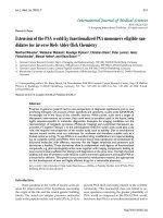
Effect of TAK1 knockdown on IL-1-induced JNK
activation
To determine the effect of TAK1 knockdown on JNK and p38
activation, FLSs were transfected with TAK1 siRNA and then
stimulated on day 5 with 2 ng/ml of rhIL-1β. As shown in Fig-
ure 3, TAK1 deficiency in FLSs significantly inhibited IL-1β-
induced JNK, MKK4, and MKK7 phosphorylation compared
with sc control (mean inhibition: JNK, 58 ± 1% (p = 0.01);
MKK4, 49 ± 3% (p = 0.01); MKK7, 49 ± 7% (p = 0.04); n =
3 each). However, p38 activation was not affected by TAK1
deficiency. To determine the effect of TAK1 deficiency on JNK
function, kinase assays were performed using anti-JNK, anti-
MKK4, or anti-MKK7 antibodies and GST-cJun substrate (Fig-
ure 4a). GST-cJun phosphorylation by anti-JNK immunopre-
cipitates was significantly decreased in TAK1 deficient FLSs
(53 ± 2% inhibition, p = 0.03, n = 3). In addition, TAK1 knock-
down modestly inhibited MKK4 and MKK7 kinase activity
(average inhibition: MKK4, 28 ± 4% (p = 0.01); MKK7, 28 ±
8% (p = 0.03); n = 3 each; Figure 4b).
Figure 2
MEKK1, MEKK2 and MEKK3 do not alter IL-1β-induced mitogen-activated protein kinase (MAPK) activationMEKK1, MEKK2 and MEKK3 do not alter IL-1β-induced mitogen-activated protein kinase (MAPK) activation. (a) Three days after small interfering
RNA (siRNA) transfection, serum-starved fibroblast-like synoviocytes (FLSs) were stimulated with IL-1β (2 ng/ml) for 15 minutes and lysates were
evaluated by western blot analysis. MEKK1, MEKK2, or MEKK3 deficiency alone or in combination had no effect on IL-1β-stimulated JNK, p38 or
ERK activation compared with scrambled control (sc) (n = 2 separate FLS lines). M1, MEKK1 siRNA; M2, MEKK2 siRNA; M3, MEKK3 siRNA;
M123, MEKK1+MEKK2+MEKK3 siRNA. (b) To complement the MEKK1 siRNA studies, MEKK1-/- FLSs were also examined. MEKK1 knockout
(KO) and wild-type (WT) mouse FLSs were serum-starved for 48 h, stimulated with IL-1β (2 ng/ml) for 15 minutes and lysed. Cell extracts were eval-
uated by western blot analysis. MEKK1 deficiency did not affect IL-1β-induced MAPK activation. (c) MEKK1-/- mFLSs were transfected with MEKK2
and MEKK3 (M2M3) or sc siRNA, and, later, serum-starved for 48 h and stimulated with IL-1β (2 ng/ml) for 15 minutes. Western blot analysis of the
lysates confirmed that MEKK2 and MEKK3 do not alter MAPK activation.
Available online />Page 5 of 9
(page number not for citation purposes)
Regulation of AP-1 and NF-κB binding and
transcriptional activity by TAK1
AP-1 and NF-κB regulate MMP and pro-inflammatory cytokine
gene expression by FLSs. To determine if TAK1 knockdown
modulates AP-1 and NF-κB binding, electrophoretic mobility
shift assays were performed (Figure 5). Similar levels of basal
AP-1 and NF-κB binding were observed in control and TAK1
knockdown FLSs. AP-1 binding increased after IL-1β stimula-
tion in the sc siRNA transfected lysates. However, TAK1 defi-
ciency significantly inhibited IL-1β-induced AP-1 activation
(84.3 ± 8.1% inhibition compared with control, n = 3, p =
0.028). There was a trend towards a decrease in NF-κB acti-
vation, although this did not reach statistical significance (34.2
± 7.0% inhibition, n = 3, p = 0.21).
Effect of TAK1 knockdown on MMP gene expression and
IL-6 production
Because TAK1 regulates IL-1β-induced AP-1 activation, we
determined if TAK1 deficiency affects MMP3 gene expression
by real-time quantitative PCR and IL-6 production by ELISA.
TAK1 siRNA- or sc siRNA-treated FLSs were stimulated with
IL-1β or medium for 24 h and assayed for MMP3 gene expres-
sion (Figure 6a). TAK1 deficiency significantly decreased IL-
1β-induced MMP3 gene expression compared with sc control
(GAPDH normalized average: 55.9 ± 14% inhibition, n = 5, p
= 0.04). To measure IL-6 production, control or TAK1 knock-
down FLSs were stimulated with IL-1β for 24 h. Cell superna-
tants were then collected and assayed by ELISA (Figure 6b).
TAK1 deficient cells produced significantly less IL-6 com-
pared with sc siRNA transfected cells (52.7 ± 3.3% inhibition,
n = 4, p = 0.021).
Discussion
RA is a chronic inflammatory disease of unknown etiology that
targets the synovium. Intimal lining macrophages and fibro-
blast-like synoviocytes produce pro-inflammatory cytokines
that contribute to synovial inflammation and production of
destructive enzymes like MMPs [25]. The MMPs can then
degrade components of the extracellular matrix, especially
native interstitial collagen [26], initiating a series of events
leading to irreversible joint damage [27]. MMP gene expres-
sion in FLSs is regulated by many signaling pathways,
although MAPKs play a prominent role [9].
Of the three MAPK families, JNK is particularly interesting
because it efficiently phosphorylates c-Jun. This protein is a
crucial component of the transcription factor AP-1, which, in
turn, initiates MMP gene expression [16,28]. JNK can be
Figure 3
IL-1β-induced JNK activation in fibroblast-like synoviocytes (FLSs) is TAK1-dependentIL-1β-induced JNK activation in fibroblast-like synoviocytes (FLSs) is
TAK1-dependent. The effect of TAK1 deficiency on JNK activation was
determined by western blot analysis. Three days after TAK1 or scram-
bled control (sc) small interfering RNA transfection, serum-starved
FLSs were stimulated with IL-1β (2 ng/ml) for 15 minutes. Cell lysates
were evaluated for P-JNK, P-MKK4, P-MKK7, P-p38, and actin. JNK,
MKK4, and MKK7, but not p38 activation was significantly decreased
in the absence of TAK1 (average inhibition: JNK, 58 ± 1%, p = 0.01;
MKK4, 49 ± 3%, p = 0.01; MKK7, 49 ± 7%, p = 0.04). A representa-
tive experiment is shown (n = 3).
Figure 4
JNK kinase activity is TAK1-dependentJNK kinase activity is TAK1-dependent. (a) Kinase assays were used to
evaluate JNK function in TAK1 deficient cells. Fibroblast-like synovio-
cytes (FLSs) transfected with TAK1 or scrambled control (sc) small
interfering RNA (siRNA) were serum-starved, stimulated with IL-1β (2
ng/ml) for 15 minutes, lysed, immunoprecipitated with anti-JNK anti-
bodies, and subjected to kinase assay using GST-c-Jun substrate. JNK-
mediated activation of c-Jun was significantly decreased in TAK1 defi-
cient FLS (53 ± 2% inhibition, p = 0.03). A representative experiment
is shown (n = 3). Med, medium; IP, immunoprecipitation. (b) Kinase
assays were performed with anti-MKK4 and anti-MKK7 antibody immu-
noprecipitates and GST-c-Jun substrate to evaluate the effects of
TAK1 deficiency on MKK4 and MKK7 function. Significant decreases in
IL-1β-induced MKK4 and MKK7 kinase activity were observed (average
inhibition: MKK4, 28 ± 4%, p = 0.004; MKK7, 28 ± 8%, p = 0.02). A
representative experiment is shown (n = 3). Wb, Western blot.
Arthritis Research & Therapy Vol 9 No 3 Hammaker et al.
Page 6 of 9
(page number not for citation purposes)
phosphorylated by two dual specificity threonine/tyrosine
kinases, MKK4 and MKK7 [18]. Recent studies using siRNA
to deplete individual kinases showed that MKK7, but not
MKK4, is necessary for IL-1β-induced JNK activation in FLSs
[19]. In contrast, both MKK4 and MKK7 are required for max-
imum JNK phosphorylation by anisomycin, lipopolysaccharide,
and sorbitol [19,29].
MKK4 and MKK7 are, in turn, regulated by a large family of ser-
ine/threonine kinases known as the MAP3Ks that integrate
extracellular stimuli and activate transcription factors in a cell-
type and stimulus-specific manner [29]. Little is known about
how MAP3Ks regulate the JNK pathway or MMP gene expres-
sion in RA. To address this issue, we previously examined
MAP3K gene and protein expression in RA synovial tissue and
FLSs [20]. These studies showed that four of the MAP3Ks,
namely MEKK1, MEKK2, MEKK3 and TAK1, are abundant in
RA FLSs. Kinase assays suggested that MEKK2 and TAK1 are
especially important activators of the JNK pathway in FLSs.
In the present study, we examined the hierarchy of these pro-
teins in IL-1β-mediated JNK activation in RA FLSs using
siRNA. The results showed that MEKK1, MEKK2 and MEKK3
are not necessary for IL-1β-mediated JNK phosphorylation,
either individually or in combination. The data also demon-
strate that the pathways utilized by stress kinases can be cell
and stimulus specific. For instance, MEKK1 is required for JNK
and c-Jun activation in the corneal epithelia of MEKK1-/- mice
[30]. MEKK1 is also critical for JNK activation in response to
pro-inflammatory stimuli and cell migration in MEKK1-/- MEFs
[31]. Unlike MEKK1, MEKK3 knockout is embryonic lethal [32]
and JNK and p38 activation are defective in MEKK3-/- MEFs
stimulated with IL-1β [21].
Several additional studies indicate that MEKK2 can play a cell-
lineage specific role in JNK activation, and our previous
studies showed that it could form a functional signaling com-
plex in FLSs [20]. MEKK2 also associates with MKK7 and
JNK1 in Cos cells [33]. Its role in JNK function was suggested
by studies showing that MEKK2 gene disruption inhibits JNK
activation in mast cells in response to c-Kit and Fcε RI stimu-
lation [34]. Kesavan and colleagues showed that FGF-2-
induced JNK activation also required MEKK2 in knockout
MEFs [22]. Furthermore, MEKK2 is required for JNK activation
in T cell receptor signaling and IL-2 gene expression [35].
Despite these data, siRNA and MEKK2-/- studies indicate that
MEKK2 is not required for IL-1β-induced JNK activation in
FLSs.
In contrast, TAK1 is a critical upstream kinase regulating JNK
in FLSs. TAK1 is an evolutionarily conserved MAP3K that is
essential for some innate and adaptive immune responses
[36]. Signaling through the IL-1 receptor leads to ubiquination
and activation of the tumor necrosis factor receptor-associ-
ated factor 6 (TRAF6)/TAB1/TAB2/TAB3 (TAB, TAK1-bind-
ing protein) complex through IL-1 receptor-associated kinase
(IRAK) [37-41]. TAK1 is then activated by autophosphoryla-
tion of serine/threonine residues within its activation loop [42].
It can then engage I-kappaB kinase and MAPK pathways lead-
ing to the activation of NF-κB, p38, and JNK [43].
TAK1, unlike MEKK1, MEKK2, and MEKK3, is intimately
involved in IL-1β-induced JNK activation in FLSs. TAK1
knockdown significantly inhibited the kinase activity of MKK4,
MKK7, and JNK. However, TAK1 deficiency did not affect the
p38 pathway or interferon gene expression (IP-10 and IFNβ).
This result in FLSs differs from studies using 293 cells where
p38 and JNK activation by IL-1β required TAK1 [44]. Once
JNK is phosphorylated, its effect on downstream gene expres-
sion typically involves AP-1 activation. Transcription factor
studies in FLSs confirmed that TAK1 deficiency not only
decreased JNK activation but also suppressed AP-1 binding.
The effect on NF-κB was less prominent and is consistent with
previous studies in RANK ligand-stimulated 293 cells express-
ing dominant-negative TAK1 [45]. The variability of MAP3K
function in different cell types is also underscored by studies
implicating TAK1 in the NF-κB pathway in HeLa cells [46] and
NIH3T3 cells [47]. Therefore, it is important to evaluate signal-
ing mechanisms in tissue-specific cell lineages when consid-
ering their potential role in inflammatory diseases.
The functional consequences of activating the TAK1-JNK-AP1
pathway can be evaluated by determining expression of key
AP-1-driven genes implicated in RA. The AP-1 consensus
Figure 5
Regulation of AP-1 binding and transcriptional activity is TAK1-depend-entRegulation of AP-1 binding and transcriptional activity is TAK1-depend-
ent. The effect of TAK1 deficiency on IL-1β-induced AP-1 activation
was evaluated by electrophoretic mobility shift assay. Five days after
small interfering RNA transfection, cultured fibroblast-like synoviocytes
were stimulated with IL-1β (2 ng/ml) for 60 minutes. Nuclear extracts
were obtained and evaluated for AP-1 and NF-κB binding activity. A
representative experiment is shown (n = 3). IL-1β-induced AP-1 activity
was significantly decreased in the absence of TAK1 (84 ± 8% inhibi-
tion compared to control, p = 0.03). NF-κB binding, on the other hand,
was not significantly decreased with TAK1 deficiency (34 ± 7% inhibi-
tion, p = 0.21). Cold oligo, non-radioactive oligo; Sc, scrambled
control.
Available online />Page 7 of 9
(page number not for citation purposes)
sequence is located at -70 base-pairs in the promoter region
of the gene encoding MMP3, making it a useful biomarker for
TAK1 in cells such as osteocytes [48] and chondrocytes [49].
siRNA studies showed that TAK1 inhibition significantly
decreased MMP3 gene expression in cultured FLSs. Of inter-
est, IL-1β-induced IL-6 production was also decreased by
TAK1 deficiency, which could reflect an effect on AP-1
because this transcription factor also binds to the IL-6 pro-
moter. The modest effect of TAK1 on NF-κB could also con-
tribute to MMP3 and IL-6 expression.
Conclusion
These data suggest that TAK1 is a key element in JNK activa-
tion, IL-6 production, and MMP expression by FLSs. Surpris-
ingly, other MAP3Ks implicated in JNK activation, such as
MEKK1, MEKK2, and MEKK3, do not have a major contribu-
tion to this pathway in FLSs. Therefore, targeting TAK1 might
represent an alternative way to regulate JNK activation and
matrix degradation in inflammatory arthritis.
Competing interests
The authors declare that they have no competing interests.
Authors' contributions
DH designed and carried out experiments, DB made substan-
tial contributions to the conception/design of the study and
interpretation of data, TI helped design transfection experi-
ments, GSF conceived of the study, participated in its design
and coordination, and helped to draft the manuscript.
Acknowledgements
The work was supported by NIH grant AR47825.
References
1. Firestein GS: Evolving concepts of rheumatoid arthritis. Nature
2003, 423:356-361.
Figure 6
Effect of TAK1 knockdown on matrix metalloproteinase (MMP) gene expression and IL-6 productionEffect of TAK1 knockdown on matrix metalloproteinase (MMP) gene expression and IL-6 production. (a) To determine if TAK1 deficiency affects
MMP3 gene expression, real-time quantitative PCR was performed. TAK1 or scrambled control (sc) small interfering RNA-treated fibroblast-like syn-
oviocytes (FLSs) were stimulated with IL-1β (2 ng/ml) for 24 h and MMP3 gene expression was assayed and normalized to GAPDH. Data are shown
as relative expression units (REU). IL-1β-induced MMP3 gene expression in the absence of TAK1 was significantly decreased compared with sc
control (56 ± 14% inhibition, n = 5, *p = 0.01). Med, medium. (b) The effect of TAK1 knockdown on IL-6 production was assayed by ELISA. Cell
supernatants were collected from FLSs stimulated with IL-1β (2 ng/ml) for 24 h. TAK1-deficient FLSs produced significantly less IL-6 compared with
sc control (53 ± 3% inhibition, n = 4, *p = 0.02).
Arthritis Research & Therapy Vol 9 No 3 Hammaker et al.
Page 8 of 9
(page number not for citation purposes)
2. Mor A, Abramson SB, Pillinger MH: The fibroblast-like synovial
cell in rheumatoid arthritis: a key player in inflammation and
joint destruction. Clin Immunol 2005, 115:118-128.
3. Alvaro-Gracia JM, Zvaifler NJ, Firestein GS: Cytokines in chronic
inflammatory arthritis. V. Mutual antagonism between inter-
feron-gamma and tumor necrosis factor-alpha on HLA-DR
expression, proliferation, collagenase production, and granu-
locyte macrophage colony-stimulating factor production by
rheumatoid arthritis synoviocytes. J Clin Invest 1990,
86:1790-1798.
4. Alvaro-Gracia JM, Zvaifler NJ, Brown CB, Kaushansky K, Firestein
GS: Cytokines in chronic inflammatory arthritis. VI. Analysis of
the synovial cells involved in granulocyte-macrophage colony-
stimulating factor production and gene expression in rheuma-
toid arthritis and its regulation by IL-1 and tumor necrosis
factor-alpha. J Immunol 1991, 146:3365-3371.
5. Vincenti MP, Brinckerhoff CE: Transcriptional regulation of col-
lagenase (MMP-1, MMP-13) genes in arthritis: integration of
complex signaling pathways for the recruitment of gene-spe-
cific transcription factors. Arthritis Res 2002, 4:157-164.
6. Chakraborti S, Mandal M, Das S, Mandal A, Chakraborti T: Regu-
lation of matrix metalloproteinases: an overview. Mol Cell
Biochem 2003, 253:269-285.
7. Reuben PM, Cheung HS: Regulation of matrix metalloprotein-
ase (MMP) gene expression by protein kinases. Front Biosci
2006, 11:1199-1215.
8. Kyriakis JM, Avruch J: Mammalian mitogen-activated protein
kinase signal transduction pathways activated by stress and
inflammation. Physiol Rev 2001, 81:807-869.
9. Johnson GL, Lapadat R: Mitogen-activated protein kinase path-
ways mediated by ERK, JNK, and p38 protein kinases. Science
2002, 298:1911-1912.
10. Hammaker D, Sweeney S, Firestein GS: Signal transduction net-
works in rheumatoid arthritis. Ann Rheum Dis 2003, 62(Suppl
2):ii86-89.
11. Morita Y, Kashihara N, Yamamura M, Okamoto H, Harada S,
Kawashima M, Makino H: Antisense oligonucleotides targeting
c-fos mRNA inhibit rheumatoid synovial fibroblast
proliferation.
Ann Rheum Dis 1998, 57:122-124.
12. Onodera S, Nishihira J, Koyama Y, Majima T, Aoki Y, Ichiyama H,
Ishibashi T, Minami A: Macrophage migration inhibitory factor
up-regulates the expression of interleukin-8 messenger RNA
in synovial fibroblasts of rheumatoid arthritis patients: com-
mon transcriptional regulatory mechanism between inter-
leukin-8 and interleukin-1beta. Arthritis Rheum 2004,
50:1437-1447.
13. Shin M, Yan C, Boyd D: An inhibitor of c-jun aminoterminal
kinase (SP600125) represses c-Jun activation, DNA-binding
and PMA-inducible 92-kDa type IV collagenase expression.
Biochim Biophys Acta 2002, 1589:311-316.
14. Han Z, Boyle DL, Manning AM, Firestein GS: AP-1 and NF-kap-
paB regulation in rheumatoid arthritis and murine collagen-
induced arthritis. Autoimmunity 1998, 28:197-208.
15. Han Z, Boyle DL, Aupperle KR, Bennett B, Manning AM, Firestein
GS: Jun N-terminal kinase in rheumatoid arthritis. J Pharmacol
Exp Ther 1999, 291:124-130.
16. Han Z, Boyle DL, Chang L, Bennett B, Karin M, Yang L, Manning
AM, Firestein GS: c-Jun N-terminal kinase is required for met-
alloproteinase expression and joint destruction in inflamma-
tory arthritis. J Clin Invest 2001, 108:73-81.
17. Davis RJ: Signal transduction by the JNK group of MAP
kinases. Cell 2000, 103:239-252.
18. Sundarrajan M, Boyle DL, Chabaud-Riou M, Hammaker D, Firest-
ein GS: Expression of the MAPK kinases MKK-4 and MKK-7 in
rheumatoid arthritis and their role as key regulators of JNK.
Arthritis Rheum 2003, 48:2450-2460.
19. Inoue T, Hammaker D, Boyle DL, Firestein GS: Regulation of JNK
by MKK-7 in fibroblast-like synoviocytes. Arthritis Rheum 2006,
54:2127-2135.
20. Hammaker DR, Boyle DL, Chabaud-Riou M, Firestein GS: Regu-
lation of c-Jun N-terminal kinase by MEKK-2 and mitogen-acti-
vated protein kinase kinase kinases in rheumatoid arthritis. J
Immunol 2004, 172:1612-1618.
21. Huang Q, Yang J, Lin Y, Walker C, Cheng J, Liu ZG, Su B: Differ-
ential regulation of interleukin 1 receptor and Toll-like recep-
tor signaling by MEKK3.
Nat Immunol 2004, 5:98-103.
22. Kesavan K, Lobel-Rice K, Sun W, Lapadat R, Webb S, Johnson
GL, Garrington TP: MEKK2 regulates the coordinate activation
of ERK5 and JNK in response to FGF-2 in fibroblasts. J Cell
Physiol 2004, 199:140-148.
23. Arnett FC, Edworthy SM, Bloch DA, McShane DJ, Fries JF, Cooper
NS, Healey LA, Kaplan SR, Liang MH, Luthra HS, et al.: The Amer-
ican Rheumatism Association 1987 revised criteria for the
classification of rheumatoid arthritis. Arthritis Rheum 1988,
31:315-324.
24. Boyle DL, Rosengren S, Bugbee W, Kavanaugh A, Firestein GS:
Quantitative biomarker analysis of synovial gene expression
by real-time PCR. Arthritis Res Ther 2003, 5:R352-360.
25. Bucala R, Ritchlin C, Winchester R, Cerami A: Constitutive pro-
duction of inflammatory and mitogenic cytokines by rheuma-
toid synovial fibroblasts. J Exp Med 1991, 173:569-574.
26. Chadee DN, Yuasa T, Kyriakis JM: Direct activation of mitogen-
activated protein kinase kinase kinase MEKK1 by the Ste20p
homologue GCK and the adapter protein TRAF2. Mol Cell Biol
2002, 22:737-749.
27. Ritchlin C, Haas-Smith S: Collagenase and stromelysin expres-
sion in rheumatoid synovium and cartilage: comment on the
article by Wolfe et al. Arthritis Rheum 1994, 37:1831-1833.
28. Karin M, Gallagher E: From JNK to pay dirt: jun kinases, their
biochemistry, physiology and clinical importance. IUBMB Life
2005, 57:283-295.
29. Chen W, White MA, Cobb MH: Stimulus-specific requirements
for MAP3 kinases in activating the JNK pathway. J Biol Chem
2002, 277:49105-49110.
30. Zhang L, Deng M, Kao CW, Kao WW, Xia Y: MEK kinase 1 reg-
ulates c-Jun phosphorylation in the control of corneal
morphogenesis. Mol Vis 2003, 9:584-593.
31. Xia Y, Makris C, Su B, Li E, Yang J, Nemerow GR, Karin M: MEK
kinase 1 is critically required for c-Jun N-terminal kinase acti-
vation by proinflammatory stimuli and growth factor-induced
cell migration. Proc Natl Acad Sci USA 2000, 97:5243-5248.
32. Yang J, Boerm M, McCarty M, Bucana C, Fidler IJ, Zhuang Y, Su
B: Mekk3 is essential for early embryonic cardiovascular
development. Nat Genet 2000, 24:309-313.
33. Cheng J, Yang J, Xia Y, Karin M, Su B: Synergistic interaction of
MEK kinase 2, c-Jun N-terminal kinase (JNK) kinase 2, and
JNK1 results in efficient and specific JNK1 activation. Mol Cell
Biol 2000, 20:2334-2342.
34. Garrington TP, Ishizuka T, Papst PJ, Chayama K, Webb S, Yujiri T,
Sun W, Sather S, Russell DM, Gibson SB, et al.: MEKK2 gene
disruption causes loss of cytokine production in response to
IgE and c-Kit ligand stimulation of ES cell-derived mast cells.
EMBO J 2000, 19:5387-5395.
35. Su B, Cheng J, Yang J, Guo Z: MEKK2 is required for T-cell
receptor signals in JNK activation and interleukin-2 gene
expression. J Biol Chem 2001, 276:14784-14790.
36. Silverman N, Zhou R, Erlich RL, Hunter M, Bernstein E, Schneider
D, Maniatis T: Immune activation of NF-kappaB and JNK
requires Drosophila TAK1. J Biol Chem 2003,
278:48928-48934.
37. Jiang Z, Ninomiya-Tsuji J, Qian Y, Matsumoto K, Li X: Interleukin-
1 (IL-1) receptor-associated kinase-dependent IL-1-induced
signaling complexes phosphorylate TAK1 and TAB2 at the
plasma membrane and activate TAK1 in the cytosol. Mol Cell
Biol 2002, 22:7158-7167.
38. Frobose H, Ronn SG, Heding PE, Mendoza H, Cohen P, Mandrup-
Poulsen T, Billestrup N: Suppressor of cytokine Signaling-3
inhibits interleukin-1 signaling by targeting the TRAF-6/TAK1
complex. Mol Endocrinol 2006, 20:1587-1596.
39. Singhirunnusorn P, Suzuki S, Kawasaki N, Saiki I, Sakurai H: Crit-
ical roles of threonine 187 phosphorylation in cellular stress-
induced rapid and transient activation of transforming growth
factor-beta-activated kinase 1 (TAK1) in a signaling complex
containing TAK1-binding protein TAB1 and TAB2. J Biol Chem
2005, 280:7359-7368.
40. Ishitani T, Takaesu G, Ninomiya-Tsuji J, Shibuya H, Gaynor RB,
Matsumoto K: Role of the TAB2-related protein TAB3 in IL-1
and TNF signaling. EMBO J 2003, 22:6277-6288.
41. Cheung PC, Nebreda AR, Cohen P: TAB3, a new binding partner
of the protein kinase TAK1. Biochem J 2004, 378:27-34.
42. Kishimoto K, Matsumoto K, Ninomiya-Tsuji J: TAK1 mitogen-acti-
vated protein kinase kinase kinase is activated by autophos-
Available online />Page 9 of 9
(page number not for citation purposes)
phorylation within its activation loop. J Biol Chem 2000,
275:7359-7364.
43. Ninomiya-Tsuji J, Kishimoto K, Hiyama A, Inoue J, Cao Z, Mat-
sumoto K: The kinase TAK1 can activate the NIK-I kappaB as
well as the MAP kinase cascade in the IL-1 signalling pathway.
Nature 1999, 398:252-256.
44. Sato S, Sanjo H, Takeda K, Ninomiya-Tsuji J, Yamamoto M, Kawai
T, Matsumoto K, Takeuchi O, Akira S: Essential function for the
kinase TAK1 in innate and adaptive immune responses. Nat
Immunol 2005, 6:1087-1095.
45. Lee SW, Han SI, Kim HH, Lee ZH: TAK1-dependent activation
of AP-1 and c-Jun N-terminal kinase by receptor activator of
NF-kappaB. J Biochem Mol Biol 2002, 35:371-376.
46. Takaesu G, Surabhi RM, Park KJ, Ninomiya-Tsuji J, Matsumoto K,
Gaynor RB: TAK1 is critical for IkappaB kinase-mediated acti-
vation of the NF-kappaB pathway. J Mol Biol 2003,
326:105-115.
47. Thiefes A, Wolter S, Mushinski JF, Hoffmann E, Dittrich-Breiholz O,
Graue N, Dorrie A, Schneider H, Wirth D, Luckow B, et al.: Simul-
taneous blockade of NFkappaB, JNK, and p38 MAPK by a
kinase-inactive mutant of the protein kinase TAK1 sensitizes
cells to apoptosis and affects a distinct spectrum of tumor
necrosis factor [corrected] target genes. J Biol Chem 2005,
280:27728-27741.
48. Hoffmann A, Preobrazhenska O, Wodarczyk C, Medler Y, Winkel
A, Shahab S, Huylebroeck D, Gross G, Verschueren K: Trans-
forming growth factor-beta-activated kinase-1 (TAK1), a
MAP3K, interacts with Smad proteins and interferes with oste-
ogenesis in murine mesenchymal progenitors. J Biol Chem
2005, 280:27271-27283.
49. Klatt AR, Klinger G, Neumuller O, Eidenmuller B, Wagner I, Achen-
bach T, Aigner T, Bartnik E: TAK1 downregulation reduces IL-
1beta induced expression of MMP13, MMP1 and TNF-alpha.
Biomed Pharmacother 2006, 60:55-61.