Báo cáo khoa học: " Brachytherapy of stage II mobile tongue carcinoma. Prediction of local control and QOL" pps
Bạn đang xem bản rút gọn của tài liệu. Xem và tải ngay bản đầy đủ của tài liệu tại đây (337.64 KB, 8 trang )
BioMed Central
Page 1 of 8
(page number not for citation purposes)
Radiation Oncology
Open Access
Research
Brachytherapy of stage II mobile tongue carcinoma. Prediction of
local control and QOL
Sayako Oota*
1
, Hitoshi Shibuya
†2
, Ryo-ichi Yoshimura
†2
,
Hiroshi Watanabe
†2
and Masahiko Miura
†2
Address:
1
Department of Radiology, Asahi General Hospital, I-1326, Asahi, Chiba, Japan and
2
Department of Radiology, Tokyo Medical and
Dental University, 1-5-45 Yushima, Bunkyo, Tokyo, Japan
Email: Sayako Oota* - ; Hitoshi Shibuya - ; Ryo-ichi Yoshimura - ;
Hiroshi Watanabe - ; Masahiko Miura -
* Corresponding author †Equal contributors
Abstract
Background: There is no consensus as to the prognostic model for brachytherapy of tongue
carcinoma. This study was designed to evaluate the prognostic factors for local control based on a
large population under a unified treatment policy.
Results: Between 1970 and 1998, 433 patients with stage II tongue squamous cell carcinoma were
treated by low-dose-rate brachytherapy. This series included 277 patients treated with a linear
source with a minimum follow-up of 3 years. A spacer was introduced in 1987. The primary local
control rates were 85.6%.
Conclusion: In the multivariate analysis, an invasive growth pattern was a significant factor for
local recurrence. The disease-related survival was influenced by old age and an invasive growth
pattern. A spacer lowered mandibular bone complications. The growth pattern was the most
important factor for recurrence. Brachytherapy was associated with a high cure rate and the use
of spacers brought about good quality of life (QOL).
Background
Brachytherapy is frequently chosen for the treatment of
stage II mobile tongue cancer, so as to avoid the large tis-
sue defects caused by surgery, and conserve good func-
tion. Since surgery of T1 tumors is associated with good
results in terms of the prognosis and function, the ratio of
patients with T2 tumors who undergo brachytherapy has
increased lately.
There is relatively little information in the literature on the
prognostic factors within subgroups of patients undergo-
ing brachytherapy for tongue cancer, and there is as yet no
consensus as to the best prognostic model. Given the scar-
city of adequate analyses using a consistent number of
variables, new studies using large control groups, espe-
cially those deriving from a single institution and falling
under the umbrella of a consistent treatment policy, are
needed. We evaluated variables to determine their poten-
tial for predicting local control, survival and QOL, with
the aim of providing a more effective post-treatment fol-
low-up protocol. Leukoplakia is a white patch on the oral
mucosa that can neither be scraped off nor classified as
any other diagnosable disease (World Health Organiza-
tion 1978) [1] and is known to frequently co-exist with
Published: 12 July 2006
Radiation Oncology 2006, 1:21 doi:10.1186/1748-717X-1-21
Received: 10 March 2006
Accepted: 12 July 2006
This article is available from: />© 2006 Oota et al; licensee BioMed Central Ltd.
This is an Open Access article distributed under the terms of the Creative Commons Attribution License ( />),
which permits unrestricted use, distribution, and reproduction in any medium, provided the original work is properly cited.
Radiation Oncology 2006, 1:21 />Page 2 of 8
(page number not for citation purposes)
tongue cancer, although there has been no study deter-
mining the correlation of its presence with the treatment
result [2]. In addition, it was reported that in 74% of cases,
local recurrence occurred within 2 years of treatment [3].
This study is based on data from local lesions without N
factor at the first clinical consultation that could be
observed for at least 3 years, and the duration of follow-
up was longer than in previous studies, to allow under-
standing of the natural behavior of local lesions.
Methods
433 patients with squamous cell carcinoma of stage II
mobile tongue were treated with low dose rate brachyther-
apy at our institute for a 28-year period.
Between 1970 and 1998, 433 patients underwent treat-
ment by brachytherapy for stage II squamous cell carci-
noma of the tongue at the Tokyo Medical and Dental
University Hospital. This series included 337 patients
with a minimum follow-up of local disease for 3 years to
reflect the nature of local disease. 277 patients were
treated with linear
192
Ir,
226
Ra or
137
Cs needle and 60
patients were treated with
198
Au or
226
Rn grain. Median
follow-up was 7 years and 7 months for 337 patients and
7 years and 10 months for 277 patients treated by linear
source. Statistical analysis was finally conducted from
patients in whom observation of local disease could be
obtained for at least 3 years, or who died of local recur-
rence within 3 years of therapy. 201 of these patients were
male and 136 were female, and the median age of the
patients was 56 years (range: 19–92 years). In this study,
the tumors were categorized for analysis as 1) exophytic,
referring to tumors showing an external growth 2) super-
ficial, referring to flat tumors without palpable infiltra-
tion, and 3) invasive, referring to tumors penetrating
deeply with or without ulceration, based on the findings
of inspection and palpation. Tumor thickness was judged
clinically and measured with vernier calipers.
All the patients were treated by low-dose rate brachyther-
apy with or without external irradiation. None of the
patients received prophylactic neck irradiation. Small
tumors less than 10 mm in tumor thickness were treated
using a single-plane implant. Tumors between 10 mm
and 15 mm in tumor thickness were treated using a single-
plane implant with extra implant in cases where the dose
distribution needed to be corrected. Tumors thicker than
15 mm were treated using a double-plane or volume
implant. Leukoplakia adjacent to the tongue cancer was
included within the treatment volume. The dose was cal-
culated with the Manchester system. Pre-treatment esti-
mation of treatment time was made by Paterson-Parker's
table and 70 Gy was set as the reference dose. Since 1976,
the actual dose distribution curve has been obtained by
computer dosimetry using rectangular X-ray films taken
about 24 hours after implantation. No tomographic
image was obtained for calculation.
In some patients with a tumor diameter more than 30 mm
or when brachytherapy cannot be performed immedi-
ately, 30 to 40 Gy of external irradiation or intra-oral cone
electron beam therapy to the primary site was employed.
The radiation field of external irradiation was shaped to
give at least a 2 cm margin and the inferior part of the field
usually lay at the thyroid notch. Brachytherapy was per-
formed to give 60 Gy to the target volume in such cases.
Spacers made of translucent acrylic resin and ball clasps as
a locking device have been used since 1987 and were used
during the implantation [4]. The thickness of the lingual
section was designed to obtain about 10 mm with a min-
imum of 7 mm.
Complication was evaluated clinically. After the treat-
ment, the patients were seen approximately every 2 weeks
for the first 3 months, every month during the first year,
every 6 months during the next 2 years, and every year
thereafter. When recent follow-up was not available, the
surviving patients or their families were contacted for
anamnestic search. Data was interpreted according to
National cancer institute-Common terminology criteria
for adverse events, version 3.0 for early complication and
Radiation Therapy Oncology Group(RTOG)/European
Organization for Research and Treatment of Cancer
(EORTC) Late radiation morbidity scoring scheme for late
complication. Biopsy was conducted in cases where the
clinical findings were considered insufficient for diagnosis
and in all cases where salvage surgery was performed. If a
patient developed metastasis and subsequently presented
with local failure, he/she was scored as having local fail-
ure. Death from local failure was defined death from local
recurrence of cancer that could not be controlled and the
tumor continued to grow. The survival rate was analyzed
in the 277 patients treated using a linear source. The pri-
mary relapse-free survival (RFS) was calculated as the time
from the first day of treatment to the date of the last fol-
low-up. Disease-related survival (DRS) was calculated as
the time from the date of the first visit to that of the last
follow-up. In the case of disease-related survival, only
deaths due to carcinoma of the tongue were counted, and
patients who died of other causes were considered lost to
follow-up. The overall median follow-up duration was 7
years and 10 months (minimum, 5 months; maximum,
29 years and 9 months). The survival patterns were esti-
mated using the actuarial method. The difference of post-
treatment condition between with or without spacer was
analyzed using t-test and Fischer's direct method. The cor-
relations among the various prognostic factors were
assessed by the chi-square method. The survival data of
the different subgroups of patients were compared using
Radiation Oncology 2006, 1:21 />Page 3 of 8
(page number not for citation purposes)
the Mantel-Haenszel log-rank test. A multivariate analysis
was performed using the Cox proportional hazards
model. The statistical analyses were performed using
StatView software (SAS Institute, Cary NC).
Results
Brachytherapy was performed with
192
Ir hairpin in 104
patients (31%),
226
Ra needle in 162 (48%),
137
Cs needle
in 11 (3%) and
198
Au or
222
Rn grain in 60 (18%). The
total dose calculated from the reference isodose was 60–
85 Gy (median 70 Gy) with 83 Gy of mean central dose in
cases treated with
192
Ir hairpin,
226
Ra needle or
137
Cs nee-
dle in five to seven days and 60–105 Gy (median 90 Gy)
in seven days with
198
Au or
222
Rn grain. The 2 and 5 year
relapse free rate was 88.5%, 85.4% (
192
Ir), 81.8%, 81.8%
(
137
Cs), 90.7%, 87% (
226
Ra), 62.8%, 54.4% (
198
Au) and
64.7%, 58.8% (
222
Rn) and significantly worse in point
sources (grains) compared to linear sources (needles) (p <
0.0001).
198
Au and
222
Rn seeds are small, and treatment does not
require complex manoeuvres of self-care. These sources
tend to be prescribed for patients who cannot be treated
by other means because of complications or old age. This
method is also associated with differences in the dose dis-
tribution, as grain implantation is more likely to make
cold spots than needles [5]. In order to avoid bias,
patients treated with the linear sources were analyzed sep-
arately excluding patients treated with the point sources.
The characteristics of selected cases are listed in Table 1.
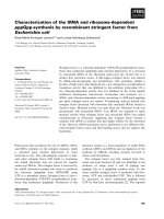
The actuarial estimates were plotted (Figure 3). The
relapse-free duration ranged from 2 to 357 months, with
a mean and median survival of 8 years and 4 months and
7 years and 3 months. At two, three, five and 10 years, the
probability of RFS was 89.5%, 87.4%, 86.2% and 85.0%,
respectively. The two-, three-, five-, and 10-year DRS rates
were 96.0%, 94.9%, 89.5% and 86.2%, respectively.
45 patients underwent external irradiation prior to brach-
ytherapy. The range of time interval between the first
external irradiation day and implant was 14–70 days
(median 34). 60Co external irradiation was used in 29
patients and 4 MV X-rays in 14. The total external radia-
tion dose ranged from 10–45 Gy (median 26), with the
daily fraction size varying from 2.0–2.5 Gy. Intra-oral
cone electron beam therapy (4 MeV) was administered in
2 patients; the total dose were 10 and 40 Gy, with the total
treatment time 5 and 46 days respectively. In these
patients, the median brachytherapy dose was 60 Gy. At
two and five years, the probability of RFS was 90.1% and
86.6% with brachytherapy only and 86.4% and 84.1%
with external irradiation prior to brachytherapy (p = 0.75)
(Figure 4).
76 patients (27%) relapsed or had an event that removed
them from the disease-free category. Those events were
distributed as follows; local failure only, 15 patients (5%);
regional failure only, 41 patients (15%); distant failure
only, 2 patients (1%); and local plus regional and/or dis-
tant failure, 40 patients (14%). The failure pattern was fur-
ther analyzed to assess specific 5-year cumulative survival
rates according to the sites of relapse as follows; regional,
63.7% and distant 97.6% (Figure 5).
At the last follow-up, 207 patients were still alive without
disease. 70 were dead, of which 29 died of their disease
(Table 2); of these 29, 15 died within 3 years of the diag-
nosis, 12 between 3 to 6 years of the diagnosis, and 2
between 6 to 8 years of the diagnosis. Three of these
patients received salvage brachytherapy with
198
Au seeds.
The dose of the
198
Au seeds was 61–73 Gy (median 66 Gy)
administered over seven days. Salvage external irradiation
was given with
60
Co in 4 cases. The total dose of external
irradiation by
60
Co was 12.5–50.2 Gy (median 20 Gy),
with the daily dose varying between 2.0–2.5 Gy. A 10 MeV
electron beam was prescribed in one case, with a total
dose 46 Gy and the dose per day of 2 Gy. Intra-oral cone
electron beam therapy was administered in 4 cases; the
total dose was 29–30 Gy (median 30 Gy) and the total
treatment time ranged from 8 to 15 days (mean 13). 26
patients underwent surgery only. No further aggressive
management was attempted in 10 patients. Twenty-one
patients were salvaged by surgery, 3 by brachytherapy
alone, 4 by intra-oral irradiation, and 1 by external irradi-
ation. None of the patients who received no further treat-
ment could be salvaged. The overall final primary control
rate was 95%. Table 3 shows the results of the univariate
analysis of RFS and DRS for 8 variables. There were no sig-
nificant differences in the sex distribution between those
showing RFS and DRS. There was no significant difference
in the age of the patients showing RFS or DRS. The growth
pattern was the superficial type in 81 patients (29%), the
exophytic type in 121 patients (44%), and the invasive
type in 75 patients (27%). Patients with the exophytic and
invasive type of tumor growth carried a 2.8- and 3.9 -fold
increased risk of local recurrence, and a 5.1- and 7.5- fold
increased risk of death as compared to those with the
superficial type of tumor growth. Leukoplakia around the
tumor was present in 67 patients (24%) and absent in 210
(76%). Its presence did not influence the RFS or DRS
rates. The tumor thickness was ≤5 mm in 106 (38%)
patients, 5–10 mm in 97 (35%), 10–15 mm in 40 (14%)
and > 15 mm in 34 (12%) patients. Those with a tumor
thickness measuring 10–15 mm had a 3-fold risk of local
recurrence as compared to those with a tumor thickness of
< 5 mm. Patients with a tumor thickness measuring 10–
15 mm or >10 mm had a 3.0- and 3.5-fold greater risk of
local recurrence as compared to those with a tumor thick-
ness of ≤5 mm. Patients with a tumor thickness measuring
Radiation Oncology 2006, 1:21 />Page 4 of 8
(page number not for citation purposes)
> 15 mm had a 3.9-fold greater risk of death as compared
to those with a tumor thickness of = 5 mm. The maximum
tumor diameter was ≤3 cm in 81 patients (29%) and > 3
cm in 196 patients (71%); this variable was not found to
affect the RFS or DRS rates. The difference of brachyther-
apy source,
192
Ir hairpin,
137
Cs needle or
226
Ra needle, was
not found to influence the risk of local recurrence or
death. The median brachytherapy dose was 70 Gy; the
dose was < 70 Gy in 216 patients (78%) and > 70 Gy in
61 patients (22%). No significant difference in the risk of
local recurrence or death was detected between these two
groups.
The multivariate analysis for local recurrence revealed the
invasive growth pattern of the tumor to be a significant
risk factor. For overall survival, old age and the invasive
growth pattern of the tumor were found to be the most
important prognostic factors. The invasive growth pattern
and tumor thickness were revealed to be significant factors
for cervical node metastasis (Table 4).
Grade 1–2 mucositis was seen in all the patient as early
complication and no patient showed toxicity greater than
Grade 3. Mucositis was ameliorated with median period
of 3 months. Prolonged complication more than 6
months was seen in 26 patients (9%). 6 of them had a
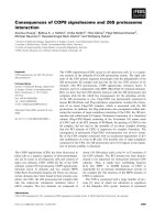
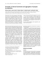
problem of compression by adjacent teeth. QOL of the
patients became remarkably better after the introduction
of a spacer between the tongue and the mandible (Fig 1
and 2). Formerly, the incidence of Grade 4 mandibular
Table 2: Patients' Current Status
Status Frequency (277 patients)
Alive, with no evidence of disease 207
Alive with clinical evidence of disease 0
Dead of disease 29
Dead of other causes 41
Table 1: Variables and Relative Classes for 277 patients treated with linear sources
Variables Classes No. %
Sex Male 169 61
Female 108 39
Age at diagnosis (yrs) ≤60 174 63
> 60 103 37
Growth pattern Superficial 81 29
Exophytic 121 44
Invasive 75 27
Site Side 254 92
Tip 1 0
Lower 20 7
Upper 2 1
Leukoplakia Absent 67 24
Present 210 76
Tumor thickness (mm) ≤510638
6–10 97 35
11–15 40 14
> 15 34 12
Maximum diameter (mm) ≤30 196 71
> 30 81 29
Brachytherapy source 192Ir hairpin 104 38
226Ra needle 162 58
137Cs needle 11 4
Brachytherapy dose (Gy) ≤70 216 78
> 70 61 22
Brachytherapy plane Single 242 87
Double 32 12
Volume 3 1
External irradiation + 45 16
-23284
Radiation Oncology 2006, 1:21 />Page 5 of 8
(page number not for citation purposes)
complication was high (22% = 23/105) in the cases with-
out local recurrence and 8 cases required operation. After
the use of spacers began, the incidence of mandibular
complication reduced (6% = 7/119), and no salvage oper-
ation was performed. In addition, salvage operation for
Grade 4 mucositis was necessitated in 3 cases of 103
patients treated without a spacer, while no operation was
required by patients treated with a spacer. The interval of
radiation mucositis after implantation was longer in the
non-spacer group (110 days) compared to the spacer
group (84 days), although it was not significant (p =
0.07). After the introduction of computer dosimetry, local
ulcers caused by overdose have decreased to zero.
Second tumors occurred in 73 patients and in 53 of them
primary tumors and regional metastases were clinically
controlled.
Some cases showed co-existing cancers including 20 oral
cancers, 15 esophageal cancers, 11 lung cancers, 8 oro-
hypopharyngeal cancers, 5 stomach cancers and another 8
tumors in the treated area with long latent periods over 8
years.
Discussion
The 5-year local control rate determined in the current
patient series was not significantly different from the fig-
ures published during the last 2 decades [6-11], although
it was slightly higher [12]. The current study, based on the
largest series so far, whose protocol included a relatively
long follow-up period and consisted of subjects that fell
under a unified treatment policy, was performed with the
aim of drawing conclusions on the natural history of the
disease and the post-treatment condition.
It was previously reported that the growth pattern of the
carcinoma did not have a statistically significant effect on
Table 4: Multivariate Analysis; significant factors
Relapse free
survival
Overall survival Cervical metastasis
free survival
Variables AHR P value AHR P value AHR P value
Growth pattern
Superficial vs. exophytic 2.17 0.84 4.93 0.06 0.70 0.15
Superficial vs. invasive 3.15 0.02 7.69 0.02 1.26 <0.001
Tumor thickness (mm)
≤5 vs. 6–10 0.82 0.62 1.02 0.98 2.85 <0.001
≤5 vs. 10–15 2.10 0.09 1.25 0.73 3.02 <0.001
≤5 vs. > 15 1.24 0.66 1.93 0.33 5.47 <0.001
AHR: adjusted hazard ratio
Table 3: Univariate Analysis; significant factors
Relapse free
survival
Overall survival Cervical
metastasis free
survival
Variables UHR P value UHR P value UHR P value
Growth pattern
Superficial vs. exophytic 2.84 <0.001 5.16 0.03 1.49 0.15
Superficial vs. invasive 3.92 <0.001 7.54 0.01 3.39 <0.001
Tumor thickness (mm)
≤5 vs. 6–10 1.76 0.19 2.08 0.15 2.87 <0.001
≤5 vs. 10–15 3.02 0.02 2.80 0.08 3.10 <0.001
≤5 vs. > 15 3.57 <0.001 3.93 0.01 5.02 <0.001
Brachytherapy source
192Ir vs. 137Cs 1.25 0.76 1.29 0.81 3.74 <0.001
192Ir vs. 226Ra 0.81 0.52 1.22 0.61 1.23 0.33
Brachytherapy dose (Gy)
≤70vs. > 70 1.24 0.54 1.24 0.61 1.03 0.91
UHR: unadjusted hazard ratio
Radiation Oncology 2006, 1:21 />Page 6 of 8
(page number not for citation purposes)
the disease-specific survival, local recurrence or overall
survival of the patients [13]. In an early report by Pater-
son, the tumor growth type was categorized into four
types; 1) papillary or fungating, 2) superficial, 3) ulcera-
tive and 4) infiltrating [14]. It was clinically difficult to
distinguish the ulcerative type from the infiltrating type by
appearance because they frequently grow into deep tissue
invasively. It has been suggested that the ulcerative or
infiltrating types of tumors are difficult to treat, because of
their tendency to invade deeply into adjacent tissue. In the
current study, the results appeared to support the impres-
sion that the invasive growth type affects the frequency of
local recurrence and survival unfavorably.
In contrast to the recent results reported by some groups,
the tumor thickness was not found to be a predictive fac-
tor for local recurrence or survival in the final model [[13]
and [15]]. The treatment policy of prescribing extra
Survival curves for 277 patients with T2 tongue carcinoma
treated with an
192
Ir,
137
Cs or
226
Ra needle; Disease- related
survival (); and relapse- free survival ()
Figure 3
Survival curves for 277 patients with T2 tongue carcinoma
treated with an
192
Ir,
137
Cs or
226
Ra needle; Disease- related
survival ( ); and relapse- free survival ( ).
5748+8#.
/106*5
A spacer attached to the lower teethFigure 2
A spacer attached to the lower teeth.
An acrylic resin spacerFigure 1
An acrylic resin spacer.
Relapse free survival for 277 patients treated with brachy-
therapy alone (); and with brachytherapy following external
irradiation ()
Figure 4
Relapse free survival for 277 patients treated with brachy-
therapy alone ( ); and with brachytherapy following
external irradiation ( ).
5748+8#.
/106*5
Radiation Oncology 2006, 1:21 />Page 7 of 8
(page number not for citation purposes)
implants for thick tumors may have influenced the result.
In addition, it is also possible that the tumor thickness did
not exert any significant influence in our series, because
exophytic tumors, with relatively better prognosis, and
invasive tumors, with a worse prognosis, were analyzed
together. However, further analysis was performed for
both the growth patterns, and the results showed no sig-
nificant influence of tumor thickness on the frequency of
local recurrence or survival in either group.
Age has been both claimed and denied as a predictor of
the prognosis [15-17]. In the current study, old patients
were found to be at the risk of dying prematurely,
although no significant influence of this parameter was
seen on the frequency of local recurrence.
Leukoplakia is known as a premalignant or potentially
malignant lesion of the oral mucosa [[2,9] and [18]].
Mucosal carcinomas associated with leukoplakia appear
to be only superficially invasive and carry a better progno-
sis than similar carcinomas not associated with leukopla-
kia. In the current study, leukoplakia at the periphery of
the tongue carcinoma was included in the target area of
treatment, as a not significant predictive factor.
The tumor size or the T factor has been evaluated in previ-
ous studies and been shown to be an important predictor
of local control [19,20]. In the current study, however,
consistent with some previous reports, it was not found to
affect survival or local control [15]. Since only patients
with T2 disease were included in the analysis, the tumor
diameter range was more limited than that in studies ana-
lyzing different T factors.
The higher death rate in cases with exophytic and invasive
tumors was assumed to be a reflection of the associated
increased rate of local recurrence. Nakagawa et al.
reported that the invasive growth pattern was related to
the risk of neck node metastasis and a higher death
rate[21].
Contribution of external irradiation prior to brachyther-
apy thought to be small and brachytherapy dose should
not be reduced.
In conclusion, the invasive growth pattern was found to
be a strong predictive factor of local recurrence. Most of
the other variables investigated in this study did not have
any prognostic implications. Attempts have also been
made to define predictive factors from the aspect of his-
topathology [[13,21] and [22]]. In addition, computer
dosimetry and spacer use are indispensable procedures.
However, QOL of oral cancer patients mainly depends on
mucous and mandibular status, proper measurement
using the EORTC QLQ-C30 questionnaire provides clear
evaluation from various aspects. [23] A recent study
revealed that high dose rate brachytherapy has advantage
to concentrate high dose to the target and reduce the risk
of normal tissue injury by optimization using tomo-
graphic anatomical information [[24] and [25]]. Modern
technique such as 3D optimised target oriented dose
application can also be applied to improve low dose rate
brachytherapy.
Competing interests
The author(s) declare that they have no competing inter-
ests.
References
1. WHO collaborating centre for oral precancerous lesions: Definition
of leukoplakia and related lesions: an aid to studies on oral
precancer. Oral Surg Oral Med Oral Pathol 1978, 46:518-539.
2. Waar van der I, Schepman KP, Meij van der EH, et al.: Oral leuko-
plakia: a clinicopathological review. Oral Oncology 1997,
33(5):291-307.
3. Shibuya H, Hoshina M, Takeda M, et al.: Brachytherapy for stage I
& II oral tongue cancer: an analysis of past cases focusing on
control and complications. Int J Radiat Oncol Biol Phys 1993,
26(1):51-58.
4. Miura M, Takeda M, Sasaki T, et al.: Factors affecting mandibular
complications in low dose rate brachytherapy for oral
tongue carcinoma with special reference to spacer. Int J Radiat
Oncol Biol Phys 1998, 41(4):763-770.
Survival curves for 277 patients with T2 tongue carcinoma
treated with an
192
Ir,
137
Cs or
226
Ra needle; Regional disease-
free survival (); and distant metastasis-free survival ()
Figure 5
Survival curves for 277 patients with T2 tongue carcinoma
treated with an
192
Ir,
137
Cs or
226
Ra needle; Regional disease-
free survival ( ); and distant metastasis-free survival
().
5748+8#.
/106*5
Publish with BioMed Central and every
scientist can read your work free of charge
"BioMed Central will be the most significant development for
disseminating the results of biomedical research in our lifetime."
Sir Paul Nurse, Cancer Research UK
Your research papers will be:
available free of charge to the entire biomedical community
peer reviewed and published immediately upon acceptance
cited in PubMed and archived on PubMed Central
yours — you keep the copyright
Submit your manuscript here:
/>BioMedcentral
Radiation Oncology 2006, 1:21 />Page 8 of 8
(page number not for citation purposes)
5. Delclos L, Lindberg RD, Fletcher GH: Squamous cell carcinoma
of the oral tongue and floor of mouth: Evaluation of intersti-
tial radium therapy. Am J Roentgenol 1976, 126(2):223-228.
6. Lefebvre JL, Coche-Dequeant B, Castelain B, et al.: Interstitial
brachytherapy and early tongue squamous cell carcinoma
management. Head Neck 1990, 12:232-236.
7. Leung TW, Wong VY, Kwan KH, et al.: High dose rate brachy-
therapy for early stage oral tongue cancer. Head Neck 2002,
24(3):274-81.
8. Mazeron JJ, Crook JM, Marinello G, et al.: Prognostic factors of
local outcome for T1, T2 carcinomas treated by iridium 192
implantation. Int J Radiat Oncol Biol Phys 1990, 19:281-285.
9. Shibuya H, Amagasa T, Seto K, et al.: Leukoplakia-associated mul-
tiple carcinomas in patients with tongue carcinoma. Cancer
1986, 57:843-846.
10. Bourgier C, Coche-Dequeant B, Fournier C, et al.: Exclusive low-
dose rate brachytherapy in 279 patients with T2N0 mobile
tongue carcinoma. Int J Radiat Oncol Biol Phys 2005, 63:434-40.
11. Pourel N, Peiffert D, Lartigau E, et al.: Quality of life in long-term
survivors of oropharynx carcinoma. Int J Radiat Oncol Biol Phys
2002, 54:742-51.
12. Wendt CD, Peters LJ, Delclos L, et al.: Primary radiotherapy in
the treatment of stage I and II oral tongue cancers: impor-
tance of the proportion of therapy delivered with interstitial
therapy. Int J Radiat Oncol Biol Phys 1990, 18(6):1287-92.
13. Yuen APW, Lam KY, Lam LK, et al.: Prognostic factors of clini-
cally stage I and II oral tongue carcinoma-a comparative
study of stage, thickness, shape, growth pattern, invasive
front malignancy grading, Martinez-Gimeno score, and path-
ologic features. Head Neck 2002, 24:513-520.
14. Paterson R: The treatment of malignant disease by radiother-
apy. Edited by: Paterson R, Stewart JG. London: Edward Arnold Pub-
lishers; 1963:204-234.
15. Al-Rajhi N, Khafaga Y, El-Husseiny J, et al.: Early stage carcinoma
of oral tongue: prognostic factors for local control and sur-
vival. Oral Oncology 2000, 36:508-514.
16. Siegelmann-Danieli N, Hanlon A, Ridge JA, et al.: Oral tongue can-
cer in patients less than 45 years old: institutional experience
and comparison with older patients. J Clin Oncol 1998,
16:745-753.
17. Yoshida K, Koizumi M, Inoue T, et al.: Radiotherapy of early
tongue cancer in patients less than 40 years old. Int J Radiat
Oncol Biol Phys 1999, 45(2);:367-371.
18. Sudbø J, Bankfalvi A, Bryne M, et al.: Prognostic value of graph
theory-based tissue architecture analysis in carcinomas of
the tongue. Lab Invest 2000, 80(12):1881-1889.
19. Hos¸ai AŞ, Ünal ÖF, Ayhan A: Possible prognostic value of his-
topathologic parameters in patients with carcinoma of the
oral tongue. Eur Arc Otorhinolaryngol 1998, 255:216-219.
20. Nyman J, Mercke C, Lindström J: Prognostic factors for local con-
trol and survival of cancer of the oral tongue A retrospective
analysis of 230 cases in Western Sweden. Acta oncologica 1993,
32(6):667-673.
21. Nakagawa T, Shibuya H, Yoshimura R, et al.: Neck node metastasis
after successful brachytherapy for early stage tongue carci-
noma. Radiother Oncol 2003, 68(2):129-35.
22. Silverman S, Gorsky M, Lozada F: Oral leukoplakia and malignant
transformation. Cancer 1984, 53:563-568.
23. Pernot M, Malissard L, Hoffstetter S, et al.: The study of tumoral,
radiobiological, and general health factors that influence
results and complications in a seriese of 448 oral tongue car-
cinomas treated exclusively by irradiation. Int J Radiat Oncol Biol
Phys 1994, 29:673-9.
24. Kolotas C, Baltas D, Zamboglou N, et al.: CT-Based interstitial
HDR brachytherapy. Strahlenther Onkol 1999, 175:419-27.
25. Umeda M, Komatsubara H, Nishimatsu N, et al.: High-dose rate
interstitial brachytherapy for stage I-II tongue cancer. Oral
Surg Oral Med Oral Pathol Oral Radiol Endod 2000, 90:667-70.