Báo cáo khoa học: " Radiation therapy planning with photons and protons for early and advanced breast cancer: an overview" ppt
Bạn đang xem bản rút gọn của tài liệu. Xem và tải ngay bản đầy đủ của tài liệu tại đây (431.4 KB, 11 trang )
Radiation Oncology
BioMed Central
Open Access
Review
Radiation therapy planning with photons and protons for early and
advanced breast cancer: an overview
Damien C Weber*1,2, Carmen Ares1, Antony J Lomax1 and John M Kurtz2
Address: 1Department of Radiation Medicine, Paul Scherrer Institute, Villigen-PSI, Switzerland and 2Department of Radiation Oncology, Geneva
University Hospital, Switzerland
Email: Damien C Weber* - ; Carmen Ares - ; Antony J Lomax - ;
John M Kurtz -
* Corresponding author
Published: 20 July 2006
Radiation Oncology 2006, 1:22
doi:10.1186/1748-717X-1-22
Received: 16 June 2006
Accepted: 20 July 2006
This article is available from: />© 2006 Weber et al; licensee BioMed Central Ltd.
This is an Open Access article distributed under the terms of the Creative Commons Attribution License ( />which permits unrestricted use, distribution, and reproduction in any medium, provided the original work is properly cited.
Abstract
Postoperative radiation therapy substantially decreases local relapse and moderately reduces
breast cancer mortality, but can be associated with increased late mortality due to cardiovascular
morbidity and secondary malignancies. Sophistication of breast irradiation techniques, including
conformal radiotherapy and intensity modulated radiation therapy, has been shown to markedly
reduce cardiac and lung irradiation. The delivery of more conformal treatment can also be achieved
with particle beam therapy using protons. Protons have superior dose distributional qualities
compared to photons, as dose deposition occurs in a modulated narrow zone, called the Bragg
peak. As a result, further dose optimization in breast cancer treatment can be reasonably expected
with protons. In this review, we outline the potential indications and benefits of breast cancer
radiotherapy with protons. Comparative planning studies and preliminary clinical data are detailed
and future developments are considered.
Background
Postoperative radiation therapy very substantially
improves local control in the treatment of both early and
locally-advanced breast cancer. Trial overviews indicate
that for every four local failures prevented, one fewer
death from breast cancer can be expected. However, this
long-term benefit can be mitigated somewhat by excess
mortality due to cardiovascular disease and secondary
malignancies [1]. Although local radiotherapy limited to
the breast or chest wall can usually be administered using
simple planning techniques with minimal late toxicity,
regional treatment including lymph nodal areas can
expose non-target organs to substantial radiation doses.
One of the principal goals of treatment planning is thus to
reduce any potential negative consequences of radiotherapy on long-term morbidity and mortality. This repre-
sents a particularly difficult challenge in the setting of
loco-regional radiotherapy.
In recent years, great advances have been made in the
planning and delivery of radiotherapy, as well as the
development of existing imaging modalities. Computerized planning systems in conjunction with modern imaging studies are routinely used in breast cancer treatments.
Three-dimensional conformal radiotherapy and, more
recently, intensity modulated radiation therapy (IMRT)
are being implemented increasingly in clinical use [2-6].
The delivery of optimal dose conformation can also be
achieved with protons. Proton beam therapy is characterized by remarkable depth-dose distributions that have a
low to median entrance dose, followed by a unified highdose region (Bragg peak region) in the tumor area, folPage 1 of 11
(page number not for citation purposes)
Radiation Oncology 2006, 1:22
lowed by a steep fall-off to zero-dose distal to the target.
As a result, physical dose distributions with protons are
both highly conformal and homogeneous. Several proton
facilities are currently operating worldwide and many
more are scheduled to open in coming years. Proton beam
therapy, however, is more costly than conventional treatment, and any potential benefits must be assessed in the
light of the associated costs to the health-care system.
Although comparative treatment-planning studies have
demonstrated the superior dose conformation achievable
with proton beams, it remains unclear whether protons
can achieve substantial clinical gains in cancer types other
than ocular melanoma or skull-base tumors. The industry-driven enthusiasm generated by proton dose distributions should not be allowed to outpace the clinical data
investigating efficacy and safety in specific tumor sites.
This review details the different proton beam delivery systems, with special emphasis upon the technical challenges
of producing and delivering proton treatment beams for
breast tumors. Dose-comparison studies of proton and
photon beam therapy for breast cancers are reviewed, preliminary clinical data are detailed and future development
considered.
Proton beam therapy: delivery systems and biologic effects
The beam delivery system is the technical component that
lies between the cyclotron and the patient. This system
monitors patient dose, generates the desired 3D dose distribution within the patient and may also provide
dynamic monitoring of its beam spreading and range control functions (see dynamic scanning technique). Two
beam line designs are commonly used for proton therapy
[7]. The scatter foil technique utilizes beam-flattening
devices, collimators, scatterers, and energy modulation
devices in the beam line to obtain a homogeneous dose in
the target and sharp lateral penumbra [8]. Additionally,
for each proton field, an individual aperture and compensator is manufactured and positioned in the proton beam
[9]. Compensators will conform the distal dose fall-off to
the target volume. In essence, it is a passive delivery system that relies on multiple coulombic scattering within
the scattering foil devices for lateral beam spreading. A
disadvantage of passive spreading is the interdependence
of beam range and field size [8]. As field size increases, the
scattering foil thickness must increase accordingly, resulting in loss of maximum treatment range. Most of the proton treatment facilities employ this simple and reliable
delivery system.
As opposed to photons, protons are charged particles and
can be easily deflected by the action of magnetic fields
under computer control [7]. This opens the possibility for
dynamic scanning, which can provide beam spread-out
modulation by magnetically scanning the protons, with
external apertures and compensators to conform the dose
/>
distribution. In dynamic scanning, no inherent interdependence of beam range and field size is observed. The
ultimate dynamic scanning system is voxel scanning ('spot'
or 'raster' scanning) [10-12], in which the beam is decomposed into multiple, three dimensionally distributed
Bragg peaks, which completely cover the target volume.
Each voxel is irradiated to the planned dose, and the beam
is switched off while moving to the adjacent voxel (spot
scanning) [13]. This system is currently used at the Paul
Scherrer Institut (PSI). Another active delivery system is
the raster scan system that is used for carbon-ion radiotherapy at the Gesellschaft für Schwerionenforschung mbH,
Darmstadt, Germany [14]. This active scanning system is
based on the continuous irradiation with a radiation pencil beam through the target volume. A Belgian manufacturer (Ion Beam Applications) is currently implementing
this delivery system for clinical use in the Boston proton
beam facility. At PSI 3D dose conformation is generated
without the need of external devices. Potential disadvantages include loss of precise tissue inhomogeneity compensation and potential increase in the lateral dose fall-off
for beams that are conformed without external apertures.
Furthermore, quality assurance is a more complex process
for dynamic systems. External apertures, compensators
and modulator wheels can be readily coded and identified
in passive systems, but higher technology is involved to
monitor beam spot motion and field uniformity. Noteworthy, the secondary neutron dose given to the patient
with this beam delivery method might be lower by a factor
of 10, when compared to the scatter foil technique. Various dose comparative studies have shown undisputedly
that protons, when compared to photons, administer a
lower integral dose to the patient [15,16]. This integral
dose may cause secondary cancers. The production of secondary neutrons by the proton beam could however
increase this integral dose and thus abrogate substantially
the advantage of proton beam therapy for breast cancer.
As such, the neutron dose has to be kept as minimal as
possible. With spot scanning, the neutron dose in the
Bragg Peak region can reach 1% of the treatment dose, but
in the non-target volume this dose is roughly 2 – 4 × 10⁻³
equivalent-dose (sievert) per Gy with the spot scanning
technique and can be considered negligible [17]. Secondary neutrons are produced as a result of patient and material located in the proton beam path interaction,
respectively. Hence, the production of these particles is
dependent on the design of the beam line. Improving it
(particularly the design and geometry of the Gantry's nozzle) might however decrease substantially the neutron
dose with the scatter foil technique (A Thornton, PTCOG
44, personal communication). The neutron issue has
been recently assessed in a review on IMRT and proton
beam therapy [18].
Page 2 of 11
(page number not for citation purposes)
Radiation Oncology 2006, 1:22
It must be emphasized that protons have biologic effects
in tissue similar to those of the megavoltage photons used
in conventional therapy. They are regarded as low linear
energy transfer particles, unlike other non-conventional
radiotherapy particles, such as neutrons or carbon ions.
The Relative Biological effectiveness of protons is defined
as the ratio of the dose of a reference beam (usually 60Co
or 6 MV) required to produce a specific effect in a biological system to the physical dose of proton radiation
required to produce the same effect [19]. Its value is not
fixed, but for 70 – 250 MeV protons range typically form
0.9 to 1.9, with an accepted 'generic' value of 1.1 in clinical proton therapy [20]. Consequently, the equivalent
60Co photon dose is the proton dose multiplied by 1.1.
This calculated dose is defined as the Cobalt Gray Equivalent (CGE) dose. On behalf of the International Commission on Radiation Units and Measurements and the
International Atomic Energy Agency, a committee will
submit a report on Prescribing, Recording and Reporting
Proton beam therapy in early spring 2006. It is proposed
that the unit of Gy-isoeffective will be designated Gy(I).
The full report will be published early 2007 (Dan Jones,
personal communication 2006).
Rationale for proton beams for breast cancer therapy
Photon whole breast irradiation (WBI) with two tangential fields sometimes administers substantial dose to the
lung and, for left-sided breast cancers, to portions of the
heart. When regional irradiation is indicated, the dose
administered to these and other organs-at-risk (OARs) can
be substantially increased. For this reason, a mixture of
photon and electron beams is often used to treat the internal mammary nodes. Because of the need to match the
electron and photon fields, this technique is characterized
by considerable target dose inhomogeneity. Moreover,
photon-beam irradiation of axillary lymph nodes also
produces substantial dose inhomogeneities regardless of
the technique used [21]. Newer radiotherapy techniques
have permitted dose delivery to be conformed more precisely to the target volume. Tangential IMRT improves the
dose homogeneity of WBI and reduces the dose to the
heart or lung [2,3]. Similarly, IMRT techniques can
improve homogeneity of dose delivery to the chest wall
and internal mammary nodes for post-mastectomy radiotherapy, albeit at a cost of an increased dose to portions of
the contra-lateral lung and breast [4]. Additionally, IMRT
may decrease the administered dose to the abdominal
organs when compared with conventional radiotherapy
using physical wedges [6]. Using automated beam orientation and modality selection (electrons vs. IMRT), modulated electron radiotherapy has also resulted in an
increased dose sparing to OARs with a somewhat less
homogeneous target-dose delivery when compared to
photon beams only [22]. Proton planning can also result
in unparalleled homogeneous dose distributions within
/>
complex target volumes, while simultaneously sparing
neighboring OARs. Comparative treatment planning
studies have shown consistently that proton beam therapy
can substantially decrease dose to OARs for various
tumors [23-29]. This radiation modality could thus be
delivered for the treatment of early or locally-advanced
breast cancers. This review discussed several potential
indications for the use of proton beams in breast cancer
therapy.
Methods
This review is based on Medline and PubMed literature
searches using the key words 'breast neoplasm', 'radiotherapy', 'proton beam therapy', and the authors' clinical
experience.
Whole breast and loco-regional irradiation with protons
Meta-analyses of available randomized data by the Early
Breast Cancer Trialists Collaborative Group have shown
that radiation therapy decreases local recurrence rates by
about 70% compared with surgery alone [1]. Absolute
reductions of around 5% in 15-year breast-cancer mortality have been demonstrated both for patients treated with
breast irradiation following conservation surgery and for
node-positive patients treated with loco-regional irradiation following mastectomy. Although irradiation limited
to the breast has not been shown to be associated with
excess intercurrent mortality, about 1% more deaths due
to causes other than breast cancer were observed among
patients having receiving loco-regional post-mastectomy
radiotherapy. This excess mortality was principally due to
cardiac and other vascular causes, and to a lesser extent to
secondary malignancies, particularly pulmonary [1]. An
increased incidence of contralateral breast cancers was
also observed in irradiated patients. Photon radiotherapy
has also been associated with a small but incremental
increase of long-term risk of contralateral breast cancer in
a large SEER series [30] and data stemmed from randomized trials (Early Breast Cancer Trialists' Collaborative
Group overview) [1]. Interestingly, the use of techniques
that minimize cardiac dose, such as the use of electron
beams to treat the mammary nodes and the chest wall,
have been specifically used in two more recent post-mastectomy trials [31,32]; these particular studies do not
show any deleterious effect of radiotherapy on cardiovascular mortality. These considerations demonstrate that
maximizing dose sparing to the heart, or other OARs, such
as the lung and contralateral breast, is of paramount
importance both in early and locally-advanced breast cancer.
In the irradiation of breast and regional lymph nodes, we
have previously shown that protons, when compared to
conventional or IMRT, deliver a highly homogeneous
treatment with a substantial decrease of the mean dose
Page 3 of 11
(page number not for citation purposes)
Radiation Oncology 2006, 1:22
delivered to the heart and contralateral lung alike [33]. In
the PSI study, a two-field spot-scanned proton (left and
anterior oblique fields), 9-fields (coplanar) IMRT (15 MV)
and conventional plans (wedged 6 MV opposed tangential fields with anterior field to treat the internal mammary nodes using 26 Gy with 6 MV photons and 24 Gy
with 12 MeV electrons) were computed and compared for
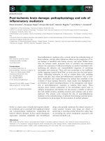
a breast cancer patient. Mean doses delivered to the ipsilateral lung and heart were lower with protons. Moreover,
the dose delivered to the contralateral breast was substantially reduced with protons, when compared to IMRT. For
this OAR, the average values of the mean and maximum
doses were 0.02 – 1.4 and 8.0 – 21.6 CGE-Gy for the proton and IMRT planning, respectively. This can be observed
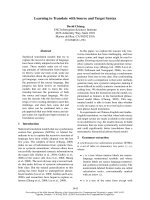
in the dose-volume histogram of the planned target volume (Fig. 1) and the OARs in the vicinity of the target volume (Fig. 2a, 2b). Likewise, Johansson et al. [34] reported
on 11 node positive left-sided breast cancer patients for
which one proton, one IMRT and two conventional plans
were computed, respectively, for each patient. Irradiation
techniques consisted on one single lateral oblique beam
(30°), 6-fields (coplanar) 6 MV photon beams and tangential beams, with or without electron fields, for the proton (passive delivery technique), IMRT and conventional
plans, respectively. The target volumes included the
remaining breast parenchyma, the internal mammary
nodes, and the supraclavicular-axillary lymph node
regions. The prescribed dose was 50 CGE-Gy. According
to a normal tissue complication probability (NTCP)
model, protons reduced the NTCP for heart by a factor of
4 and for the lung by a factor of >20, when compared to
the best photon plans. Although radiation pneumonitis
generally represents a relatively minor clinical problem,
CONVENTIONAL
IMRT - PLAN A
IMRT - PLAN B
PROTONS
Figure breast and proton intensity plans conventional
and the 1
(IMRT (Conventional), the (Protons)modulated
photon1–2) and theregional lymph nodes [33] treatment
Cumulative dose-volume histograms for thefor the breast
Cumulative dose-volume histograms for the conventional
photon (Conventional), the intensity modulated treatment
(IMRT 1–2) and the proton (Protons) plans for the breast
and the breast and regional lymph nodes [33].
/>
A
CONVENTIONAL
IMRT - PLAN A
IMRT - PLAN B
PROTONS
B
CONVENTIONAL
IMRT - PLAN A
IMRT - PLAN B
PROTONS
Figure 2a
(IMRT (Conventional), the intensity plans for the heart [33]
photon1–2) and theproton (Protons) modulated treatment
Cumulative dose-volume histograms for the conventional
Cumulative dose-volume histograms for the conventional
photon (Conventional), the intensity modulated treatment
(IMRT 1–2) and theproton (Protons) plans for the heart [33].
(B) Cumulative dose-volume histograms for the conventional photon (Conventional), the intensity modulated treatment (IMRT 1–2) and the proton (Protons) plans for the
ipsilateral lung [33].
potentially reducing the cardiac mortality from 6.7%,
with the tangential technique, to only 0.5% with protons
is likely to be clinically relevant, as a substantial number
of patients, even those with positive nodes, will remain
alive to be at risk for long-term morbidity [34]. Moreover,
modern systemic adjuvant treatments, such as anthracycline-based chemotherapy, with or without taxanes, or
trastuzumab [35], are associated with cardiotoxicity.
High-dose delivery to the heart may further increase this
risk in combination with these chemotherapy agents.
Maximum heart distance and mean lung dose has been
associated with cardiotoxicity in photon radiotherapy
series [36]. IMRT significantly reduces the mean dose of
the contralateral breast when compared to non-IMRT conventional tangential techniques [37], albeit at a cost of
increased normal tissue radiation exposure [18]. Proton
Page 4 of 11
(page number not for citation purposes)
Radiation Oncology 2006, 1:22
beam therapy further decreases the parasitic dose to the
contralateral breast and nullifies the integral dose delivered to the patient [33]. Consequently, the implementation of radiation techniques that lower the integral dose of
OARs in vicinity of the breast, such as protons, could be
recommended for certain clinical situation (e.g., node
positive left-sided tumors or inner tumor quadrant localization for young patients with large breasts).
Using biological parameters among other factors and a
simple spot-scanned proton beam therapy technique (single-field), Fogliata et al. have demonstrated that protons
reduce the lung equivalent uniform dose (EUD) significantly in both right- and left-sided tumors, when compared to other non-proton techniques (including IMRT)
for postoperative whole breast radiotherapy [38]. Unlike
the PSI [33] and Uppsala [34] study, the internal mammary chain, supraclavicular and axilla region was not part
of the treatment volume for this planning-comparison
exercise involving 5 patients with early breast cancer.
Interestingly, the mean heart dose for the subset of
patients with left-sided tumors was identical (mean, 2.6
CGE-Gy; range 2.2 CGE – 2.9 Gy). Maximum heart dose,
however, was reduced with protons: a 40% absolute dosedecrease in hot spots was calculated with a single 100 MeV
proton beam when compared to non-proton techniques.
This derives from the heavily weighted heart-dose constraints applied to the optimization process of the IMRT
planning with its consequential increased dose administered in the lung when compared to proton planning
(lung volume receiving 20 CGE-Gy: 6% vs . 20% for protons and IMRT, respectively).
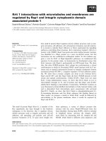
Table 1 details the planning target volume and doses
administered to OARs for 17 breast cancer patients
planned with protons and photons, with or without
IMRT. On the average, 97% of the PTV receives 95% of the
prescribed dose with protons compared to only 89% with
conventional photon techniques. With protons, the mean
dose to the heart is reduced by a factor of two to three
when compared to photon planning, with or without
IMRT. In these published studies proton plans have been
calculated using only one [34,38] or two [33] fields. Such
simple techniques could be easily used in a busy radiation
oncology department. In contrast, for IMRT plans, sophisticated techniques were required in order to meet the
planning goals and OAR's dose-constraints, resulting in
an increased number (mean, 5) of beams. Overall, comparative planning studies have shown consistently that
protons can reduce the administered dose to the heart,
lung and contralateral breast in the treatment of breast
with or without regional irradiation. It is possible that further proton dose optimization could be achieved by
added proton field directions, resulting in an additional
degree of dosimetric freedom.
/>
Partial breast irradiation with protons
Whole-breast irradiation with tangential photon beams is
considered standard treatment following breast-conserving surgery. However, the inconvenience associated with
conventional fractionation, and the substantial workload
that breast cancer represents in busy radiation oncology
departments, have led to increasing interest in other
options for these patients. This subject has been reviewed
elsewhere [39]. As most local failures after conservation
surgery occur in the vicinity of the primary tumor bed,
limiting the target volume to this area might achieve an
acceptable degree of local control for selected patients
whose tumors seem unlikely to be multifocal. The smaller
irradiated volume may also more readily allow radiotherapy to be markedly accelerated, or even to be applied in a
single fraction. This would substantially reduce the inconvenience associated with WBI, particularly for patients living far from treatment centers. Some of the acute and
chronic toxicity of WBI might also be avoided, thereby
improving patient satisfaction with treatment. Several retrospective accelerated partial breast irradiation (APBI)
series [40-43] have appeared in the literature, and prospective randomized trials comparing WBI vs. APBI are
ongoing (RTOG, GEC-ESTRO, Targit trial). APBI can be
delivered using several techniques, namely low- and highdose rate (HDR) brachytherapy using interstitial implantation [41,43-45] or a balloon catheter (MammoSite Radiation Therapy System; Cytyc Corp. Alpharetta, GA, USA)
[46], 3D external beam conformal radiation therapy [47]
or intraoperative radiotherapy (electrons or soft X-rays)
[48,49]. Biologic comparison of APBI protocols has been
recently reviewed [50]. Similarly with WBI, APBI could
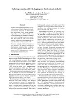
also be delivered using protons. Fig. 3 shows the dose distribution in an axial CT slice through the center of the
breast using spot-scanning proton beam technology and a
1 field (direct) beam arrangement. This proton therapy
planning was done on a patient treated at the Massachusetts General Hospital. The defined target volume consisted of the lumpectomy cavity plus a 20 mm margin.
Taghian et al. have published the dosimetric comparison
of APBI using protons with 3D conformal photon/electron based radiotherapy in 17 patients with early breast
cancer [51]. PTV coverage for both modalities was equivalent. The maximum and median dose delivered to the
heart, ipsilateral lung and non target breast tissue was
however significantly decreased with protons for all
patients. The Boston cohort has been recently updated
and the initial clinical experience of 25 patients treated
with APBI using proton beam therapy reported [52].
Using BID fractionation, 32 CGE was delivered to in 4
days, using 1 to 3 protons fields. To be enrolled in this
phase I/II clinical trial, breast cancer patients had to have
unifocal ≤2 cm tumors, negative margins (>2 mm) and
pathologically negative axillary lymph nodes. The median
volume of nontarget breast tissue receiving 50% of the
Page 5 of 11
(page number not for citation purposes)
Radiation Oncology 2006, 1:22
/>
Table 1: Overview of dose-volume histograms with proton, IMRT and photon conventional planning for the PTV and OARs in the
proton-photon planning comparison literature
PTV/OARs
Series (ref. no.)
PTV (breast only)
Lomax et al. [33]
Johansson et al.
[34]
Fogliata et al. [38]
V95% Protons
(mean)
V95% IMRT (mean)
V95% Photons
(mean)
97.1
94.0
92.2
85.9
86.6
88.8
99.8
95.5
92.2
Heart
Lomax et al. [33]
Johansson et al.
[34]
Fogliata et al. [38]
Lung (ipsilateral)
Lomax et al. [33]
Johansson et al.
[34]
Fogliata et al. [38]
Mean Dose (%)
Protons
Mean Dose (%)
IMRT
Mean Dose (%)
Photons
11.6
21.0*
24.0
41.0*
29.3
61.0*
4.4
5.6
5.0
25.0
1.0*
33.0
18.0*
33.3
29.0*
7.0
17.1
22.5
IMRT, intensity modulated radiotherapy; PTV, planning target volume; OAR, organ at risk; V95%, volume (in percentage) receiving 95% of the
prescribed dose.
*estimated % of the prescribed dose from the dose-volume histograms administered to the heart and lung
prescribed dose was 23% and the median dose received by
5% of the ipsilateral lung was only 1.3 CGE. The controlateral lung and heart received essentially no irradiation.
After observing acute moist desquamation at the treatment site in 3 patients treated with a single proton field,
Figure 3
treated with breast for an in an axial
center of thepartial breast irradiation cancer through
Dose distribution (protons)early breastCT slicepatient the
Dose distribution (protons) in an axial CT slice through the
center of the breast for an early breast cancer patient
treated with partial breast irradiation. The isodose contours
are represented by different colors (corresponding values
are displayed on the upper-right border of the figure).
the treatment technique was refined and skin sparing was
improved by the use of multiple (2–3) fields. These clinical data from Boston suggest that APBI using protons is
technically feasible and provide optimal OAR sparing.
Similarly, proton beam therapy could be delivered for
simultaneous integrated boost delivery (SIB) during WBI.
Notwithstanding the importance of the boost delivery on
local control [53,54], this additional radiation dose could
be delivered not sequentially but concomitantly to the
WBI. This would allow reduction of the overall treatment
time by 1.5 – 2 weeks by delivering the boost to the tumor
bed simultaneously with the whole breast schedule. Giving higher fractional boost doses (≈2.2 – 2.4 CGE-Gy/fraction) will administer higher biological equivalent dose
(BED) to the target volume. As the dose distributions
achieved with IMRT or protons are highly conformal,
OARs (heart, lung) that are not directly surrounding the
target regions will not receive a higher dose per fraction
and are therefore not at greater risk for late toxicity. Furthermore, this type of concomitant boost schedule is a
more efficient way of planning and radiation delivery as it
involves the use of the same plan for the entire course of
treatment. This SIB strategy is however a significant departure from conventional radiotherapy experience. Radiation therapy schedules are aimed at giving a high uniform
dose to the target volume for every fraction and then
reducing the volume to the boost portion. SIB has been
mostly studied for head and neck and prostate cancers
and occasionally for breast cancer in recent years [55]. A
Stanford study, however, has demonstrated that a SIB-
Page 6 of 11
(page number not for citation purposes)
Radiation Oncology 2006, 1:22
IMRT schedule for breast cancer increases the heart and
lung volumes receiving low-dose irradiation, indicating
that caution must be observed with regard to the OARs
when attempting to escalate the target dose [56]. Such an
increase in dose to the non-target breast tissue, heart and
lung would not be observed with protons. It can be
hypothesized that using a proton-SIB strategy, shorter biologically equivalent schedules could be calculated and
possibly implemented in clinical use. If a planning target
volume is defined by a 1-cm margin around the surgical
cavity, the radiobiological aspects of such a strategy will
be favorable, as only a limited volume of non-involved
breast tissue (within the planning target volume) will be
treated with a high fractional dose. Parenthetically,
administering a higher fractional boost dose with protons
can be achieved with or without intensity modulation.
Using the spot scanning technology, which dynamically
position Bragg peaks, differential weights could be individually defined within the target volume. This will allow
using these dose spots (i.e. Bragg peaks) to 'paint' the dose
as required with full flexibility. Theoretically, using intensity-modulated proton therapy (IMPT), with its ability to
deliver fields of arbitrary complex fluence profiles, will
probably result in more homogeneous dose deposition
when compared to non-IMPT plans. This derives from the
fact that the highly inhomogeneous individual IMPT
fields, which when combined produces a homogeneous
dose distribution, will compensate for the dose deposition of the other field's complex 3-D dose distributions in
the optimization process. In other words, the IMPT plans
will ultimately balance more evenly the high-dose regions
around the target volume than could the non-intensity
modulated protons. No proton-SIB data for breast cancer
have been yet published. A radiobiological and treatment
planning study for breast cancer is currently being carried
out at PSI, comparing conventional schedules with IMRTand proton-SIB treatments. These calculations could be
useful as means of designing fractionation strategies for
use in clinical protocols with SIB with or without protons.
Finally, protons could be used for sequential boost radiotherapy after WBI. Randomized trials have demonstrated
that local control can be significantly improved by addition of a localized tumor-bed boost delivered following
standard WBI [53,54]. In the large trial by the European
Organization for Research and Treatment of Cancer the
addition of a 16 Gy boost reduced the local failure rate by
a factor of almost 2, compared with 50 Gy WBI alone,
albeit at the cost of a greater number of fair-poor cosmetic
results [57]. Although most patients received electronbeam boosts, results seemed similar using brachytherapy
or external photon beams. It could be argued that the lateral dose fall-off may be an advantage with protons. As the
mass of protons is larger, when compared to electrons, the
angles of Coulomb interaction scattered particles are
/>
smaller. It could be counter-argued that a larger lateral
dose fall-off could be however beneficial if the target volume is ill-defined, which is usually the case for the clinical
planning of the boost. Additionally, the logistical problems associated with a proton-boost only delivery after
breast radiotherapy with photons would be surely prohibitive. Protons will surely play a minor role, if any at all, in
the development of sequential boost protocols.
All forms of partial breast treatment, namely, the APBI,
sequential boost and SIB, using protons is surely a very
effective means of limiting doses to normal structures, but
this modality has a number of potential shortcomings
that must be carefully considered. First, inter- and intrafraction tumor motion may abrogate any ballistic advantage of protons and mitigate any potential clinical benefit.
These motions during proton beam therapy can introduce
substantial unplanned heterogeneities in the dose distribution throughout the target volume [58]. Specific methods of breast-dose delivery, similar to those implemented
with photon radiotherapy [59], mitigating the effects of
organ motion, should thus be actively pursued, such as
breath hold and gating methods [60]. Second, the availability of proton beam therapy for this prevalent disease is
questionable. Photons and electrons are available worldwide and have been used in this setting for many years
unlike protons, which are restricted to a very few centers.
Third, the excellent cosmetic results achieved with modern photon therapy will not be improved with protons,
which do not deliver a lower skin dose when compared to
electrons. As mentioned earlier, the initial superficial dose
proximal to the target volume is generically 30 – 40% of
the maximum prescribed dose. More specifically, for a
superficial tumor, located 20 mm from the skin surface
and a 20 mm diameter (pT1c), the percentage of the total
dose delivered to this region would be 85 – 90%, using a
160 MeV direct proton beam. This compares identically
with the electron dose deposition, where 95% of the total
dose would be administered (applicator 10 × 10 cm) on
the skin using a 6 MeV energy electron beam. In the phase
I/II APBI clinical trial from Boston, the first 3 patients
treated with one proton field experienced acute moist
desquamation at the treatment site [52]. Subsequently, all
patients were treated with a 2 – 3 fields treatment technique. As such, sophistication of the radiation technique
using 1 proton field will not improve the cosmetic outcome unlike photon radiotherapy for which cosmesis was
indeed favorably influenced by improved technical factors in radiation delivery in a recent series [61]. Finally,
the production of secondary neutrons produced by
nuclear interactions in the material in the beam line is a
concern with proton beam therapy. The dose produced by
these uncharged particles depends on the materials –
geometry of the beam material delivery system and the
energy of the primary proton beam [62]. Estimating the
Page 7 of 11
(page number not for citation purposes)
Radiation Oncology 2006, 1:22
neutron dose by performing measurements and Monte
Carlo simulations, Schneider et al. have demonstrated
that the contribution to the integral dose from neutrons is
very low (in the order of 2 × 10⁻³ Sv per delivered Gy)
using the spot scanning technique [17]. This neutron-integral dose contribution, however, could be much higher
(by a factor of ten) using passive delivery systems, as a
result of the various scatterers, beam-flattening devices,
collimators and compensators that are hit by the primary
proton beam. Thus, the proton's scatterer foil technique
could substantially increase the high-LET neutron delivered integral dose, although this leakage neutron-radiation could be substantially decreased with improvement
in the nozzle design. Such a nozzle-design modification
has been undertaken at the Midwest Proton Therapy
Center (Bloomington, IN, USA), with measured neutron
doses substantially lower than those from other passive
scattering delivering systems (Allan Thornton, personal
communication, 2006). This additional dose with a large
biological factor could however consequentially translate
in an increase of radiation-induced cancers.
Cost and availability of proton beam therapy
In the United States, the costs of breast conservative treatment are significantly higher than those generated by
modified radical mastectomy, with or without breast
reconstruction [63]. The addition of radiation therapy
results in the higher costs of conservative surgery, representing roughly 70% of the total billing. Interestingly,
Palit et al. reported that the physician's fee for radiotherapy were significantly higher than the surgeon's and
amounted alone to roughly one-third of the total radiation therapy billing [63]. New technologies can contribute, at least theoretically, to reducing costs of breast cancer
radiotherapy; for example, multileaf collimation virtually
eliminates the need for beam blocking and reduces treatment time, and particle beam delivery systems reduce the
number of treatment portals required [64]. In the majority
of cases, however, emerging technologies will ultimately
translate into increased total billing as a result of increased
time dedicated to treatment planning and the obligate
acquisition of new planning and delivery equipment,
among other factors. In general, the additional cost factor
for proton therapy over that for intensity-modulated photons is now 2.4 – 3.0 [65]. For most of the treatment planning and treatment, the costs for protons and photons are
identical. The differential costs are accounted for by the
proton accelerator and the engineering staff required for
operating the facility. It is reasonable to assume that the
expense of proton therapy per patient will decrease, as
more facilities are built and greater numbers of patients
treated. A substantial number of proton beam facilities are
currently been planned and built worldwide [66]. In the
US, these proton beam therapy facilities involve major
cancer centers such as the M.D. Anderson Cancer Center,
/>
Houston TX, the Children's Hospital of Philadelphia,
Philadelphia PA and the University of Florida College of
Medicine, Gainesville FL, to name a few. Additionally,
accelerated proton beam therapy schedules (e.g. APBI,
SIB) may further decrease the treatment-related cost as
shown recently in a clinical trial [52]. Cost analysis of the
Boston cohort suggested that proton APBI was only modestly more expensive (25%) than traditional WBI with a
sequential boost. It must be stressed that these direct costs
do not account for other aspects of treatment, such as
patient's satisfaction or quality of care. Interestingly, a
cost-effectiveness analysis of proton radiation has been
published by the Karolinska Institute group [67]. This
group used a cohort-simulation mathematical model
comparing two hypothetical cohorts of women with
breast cancer receiving either proton beam therapy or conventional irradiation. The Markov-model simulated the
course of events in individual patients from diagnosis
until death or until age 100 years. Individuals were modelled in differential health states, each associated with a
certain cost and utility. In this study, proton beam therapy
provided an incremental benefit for an average breast cancer patient. The costs and quality adjusted-life years
gained was estimated to €67,000 for proton beam therapy. Base-case simulation suggested that a 2.4% and 13%
decrease of fatal cardiac disease and pneumonitis, respectively, should be observed with protons when compared
to conventional irradiation. These data suggests that proton beam therapy can be cost-effective and cost saving for
specific breast cancer indications, when compared to conventional radiotherapy. We now appear to be heading to
a watershed where an increased therapeutic index and
cost-effectiveness of protons come together. Although not
formally studied in a clinical setting, it is reasonable to
hypothesize that the use of proton beam therapy for highrisk breast cancer patients could translate into less late
radiation-induced toxicity, thus improving the overall
quality of care for these patients. Likewise, decreasing the
acute side effects of radiotherapy will promote the physical well-being and early return to occupational/social
activities after treatment. Similarly, the administration of
APBI or SIB with protons could potentially decrease the
overall-treatment time and thus improve the patient's burden associated with the long course of radiotherapy. The
perception that proton radiation therapy is less cost-effective than non-proton radiotherapy in specific clinical situations may be challenged by the potential for
improvements in clinical outcomes for advanced breast
cancer patients with extensive nodal involvement requiring regional radiotherapy or shortened adjuvant radiation
courses (e.g. APBI or SIB) for early breast cancers.
Conclusion
Based on the analysis presented in this paper, we believe
that proton irradiation may have some potential for
Page 8 of 11
(page number not for citation purposes)
Radiation Oncology 2006, 1:22
improving the outcome for patients with early and highrisk patients alike. However, the increased cost factor and
the questionable availability of protons for such a common disease could seriously hamper their routine use for
breast cancer. Substantial additional research will be
required before a role for proton therapy in this setting
can be established. Using the methodology of dose-comparison analysis, the impact of protons on dose deposition for certain clinical situations should be more
thoroughly assessed, and the functional effects of dose
sparing to OAR's should be formally investigated.
/>
8.
9.
10.
11.
12.
13.
Abbreviations
IMRT, intensity modulated radiotherapy; PSI, Paul Scherrer Institut; RBE; CGE, Cobalt Gray Equivalent; Gy(I), Gyisoeffective; WBI, whole breast irradiation; OAR, organ at
risk; NTCP, Normal Tissue Complication Probability;
EUD, equivalent uniform dose; APBI, accelerated partial
breast irradiation; BED, biologic equivalent dose; SIB,
simultaneous integrated boost; IMPT, intensity-modulated proton radiation therapy; Sv, Sievert.
14.
15.
16.
17.
Competing interests
The author(s) declare that they have no competing interests.
Authors' contributions
18.
19.
20.
DCW conceived and wrote the review, CA, AJL and JMK
reviewed the manuscript.
21.
Acknowledgements
Authors would like to thank Dr Hanne Kooy, Massachusetts General Hospital, Boston, for allowing use of the partial breast proton irradiation data.
22.
References
1.
2.
3.
4.
5.
6.
7.
Clarke M, Collins R, Darby S, Davies C, Elphinstone P, Evans E, Godwin J, Gray R, Hicks C, James S, MacKinnon E, McGale P, McHugh T,
Peto R, Taylor C, Wang Y: Effects of radiotherapy and of differences in the extent of surgery for early breast cancer on local
recurrence and 15-year survival: an overview of the randomised trials. Lancet 2005, 366:2087-106.
Li J, Freedman G, Price R, Wang L, Anderson P, Chen L, Xiong W,
Yang J, Pollack A, Ma C: Clinical implementation of intensitymodulated tangential beam irradiation for breast cancer.
Med Phys 2004, 31:1023-31.
Thilmann C, Sroka-Perez G, Krempien R, Hoess A, Wannenmacher
M, Debus J: Inversely planned intensity modulated radiotherapy of the breast including the internal mammary chain: a
plan comparison study. Technol Cancer Res Treat 2004, 3:69-75.
Krueger EA, Fraass BA, McShan DL, Marsh R, Pierce LJ: Potential
gains for irradiation of chest wall and regional nodes with
intensity modulated radiotherapy. Int J Radiat Oncol Biol Phys
2003, 56:1023-37.
Freedman GM, Anderson PR, Li J, Eisenberg DF, Hanlon AL, Wang L,
Nicolaou N: Intensity modulated radiation therapy (IMRT)
decreases acute skin toxicity for women receiving radiation
for breast cancer. Am J Clin Oncol 2006, 29:66-70.
Woo TC, Pignol JP, Rakovitch E, Vu T, Hicks D, O'Brien P, Pritchard
K: Body radiation exposure in breast cancer radiotherapy:
impact of breast IMRT and virtual wedge compensation
techniques. Int J Radiat Oncol Biol Phys 2006, 65:52-8.
Goitein M, Lomax A, Pedroni E: Treating Cancer with Protons.
Physics Today 2002, 55:45-50.
23.
24.
25.
26.
27.
28.
Koehler AM, Schneider RJ, Sisterson JM: Flattening of proton dose
distributions for large-field radiotherapy. Med Phys 1977,
4:297-301.
.
Kanai T, Kawachi K, Kumamoto Y, Ogawa H, Yamada T, Matsuzawa
H, Inada T: Spot scanning system for proton radiotherapy.
Med Phys 1980, 7:365-9.
Kramer M, Jakel O, Haberer T, Kraft G, Schardt D, Weber U: Treatment planning for heavy-ion radiotherapy: physical beam
model and dose optimization. Phys Med Biol 2000, 45:3299-317.
Pedroni E, Bacher R, Blattmann H, Bohringer T, Coray A, Lomax A,
Lin S, Munkel G, Scheib S, Schneider U, et al.: The 200-MeV proton
therapy project at the Paul Scherrer Institute: conceptual
design and practical realization. Med Phys 1995, 22:37-53.
Weber DC, Lomax AJ, Rutz HP, Stadelmann O, Egger E, Timmermann B, Pedroni ES, Verwey J, Miralbell R, Goitein G: Spot-scanning
proton radiation therapy for recurrent, residual or
untreated intracranial meningiomas. Radiother Oncol 2004,
71:251-8.
Haberer T, Becher W, Schardt D: Magnetic scanning systems for
heavy ion therapy. Nucl Instr Meth Phys Res 1993, 330:3299-3317.
Schneider U, Lomax A, Lombriser N: Comparative risk assessment of secondary cancer incidence after treatment of
Hodgkin's disease with photon and proton radiation. Radiat
Res 2000, 154:382-8.
Miralbell R, Lomax A, Cella L, Schneider U: Potential reduction of
the incidence of radiation-induced second cancers by using
proton beams in the treatment of pediatric tumors. International Journal of Radiation Oncology*Biology*Physics 2002, 54:824-829.
Schneider U, Agosteo S, Pedroni E, Besserer J: Secondary neutron
dose during proton therapy using spot scanning. Int J Radiat
Oncol Biol Phys 2002, 53:244-51.
Hall EJ: Intensity-modulated radiation therapy, protons, and
the risk of second cancers. Int J Radiat Oncol Biol Phys 2006, 65:1-7.
Raju MR: Proton radiobiology, radiosurgery and radiotherapy. Int J Radiat Biol 1995, 67:237-59.
Paganetti H, Niemierko A, Ancukiewicz M, Gerweck LE, Goitein M,
Loeffler JS, Suit HD: Relative biological effectiveness (RBE) values for proton beam therapy. Int J Radiat Oncol Biol Phys 2002,
53:407-21.
Jephcott C, Tyldesley S, Swift C: Regional radiotherapy toaxilla
and supraclavicular fossa for adjuvant breast treatment: a
comparison of four techniques. Int J Radiat Oncol Biol Phys 2004,
60:103-10.
Das SK, Bell M, Marks LB, Rosenman JG: A preliminary study of
the role of modulated electron beams in intensity modulated radiotherapy, using automated beam orientation and
modality selection. Int J Radiat Oncol Biol Phys 2004, 59:602-17.
Weber DC, Trofimov AV, Delaney TF, Bortfeld T: A treatment
planning comparison of intensity modulated photon and proton therapy for paraspinal sarcomas. Int J Radiat Oncol Biol Phys
2004, 58:1596-606.
Cozzi L, Fogliata A, Lomax A, Bolsi A: A treatment planning comparison of 3D conformal therapy, intensity modulated photon therapy and proton therapy for treatment of advanced
head and neck tumours. Radiotherapy and Oncology 2001,
61:287-297.
Miralbell R, Cella L, Weber DC, Lomax A: Optimizing radiotherapy of orbital and paraorbital tumors: intensity-modulated
X-ray beams vs. intensity-modulated proton beams. International Journal of Radiation Oncology*Biology*Physics 2000, 47:1111-1119.
Baumert BG, Lomax AJ, Miltchev V, Davis JB: A comparison of
dose distributions of proton and photon beams in stereotactic conformal radiotherapy of brain lesions. International Journal
of Radiation Oncology*Biology*Physics 2001, 49:1439-1449.
Lin R, Hug EB, Schaefer RA, Miller DW, Slater JM, Slater JD: Conformal proton radiation therapy of the posterior fossa: a study
comparing protons with three-dimensional planned photons
in limitingdose to auditory structures. International Journal of
Radiation Oncology*Biology*Physics 2000, 48:1219-1226.
Fuss M, Hug EB, Schaefer RA, Nevinny-Stickel M, Miller DW, Slater
JM, Slater JD: Proton radiation therapy (prt) for pediatric optic
pathway gliomas: comparison with 3d planned conventional
photons and a standard photon technique. International Journal
of Radiation Oncology*Biology*Physics 1999, 45:1117-1126.
Page 9 of 11
(page number not for citation purposes)
Radiation Oncology 2006, 1:22
29.
30.
31.
32.
33.
34.
35.
36.
37.
38.
39.
40.
41.
42.
43.
44.
45.
46.
Isacsson U, Hagberg H, Johansson K-A, Montelius A, Jung B, Glimelius
B: Potential advantages of protons over conventional radiation beams for paraspinal tumours. Radiotherapy and Oncology
1997, 45:63-70.
Gao X, Fisher SG, Emami B: Risk of second primary cancer in the
contralateral breast in women treated for early-stage breast
cancer: a population-based study. Int J Radiat Oncol Biol Phys
2003, 56:1038-45.
Overgaard M, Hansen P, Overgaard J, Rose C, Andersson M, Bach F,
Kjaer M, Gadeberg C, Mouridsen H, Jensen M, Zedeler K: Postoperative radiotherapy in high-risk premenopausal women with
breast cancer who receive adjuvant chemotherapy. Danish
Breast Cancer Cooperative Group 82b Trial. N Engl J Med
1997, 337:949-55.
Overgaard M, Jensen MB, Overgaard J, Hansen PS, Rose C, Andersson M, Kamby C, Kjaer M, Gadeberg CC, Rasmussen BB, BlichertToft M, Mouridsen HT: Postoperative radiotherapy in high-risk
postmenopausal breast-cancer patients given adjuvant
tamoxifen: Danish Breast Cancer Cooperative Group DBCG
82c randomised trial. Lancet 1999, 353:1641-8.
Lomax AJ, Cella L, Weber D, Kurtz JM, Miralbell R: Potential role
of intensity-modulated photons and protons in the treatment of the breast and regional nodes. Int J Radiat Oncol Biol Phys
2003, 55:785-92.
Johansson J, Isacsson U, Lindman H, Montelius A, Glimelius B: Nodepositive left-sided breast cancer patients after breast-conserving surgery: potential outcomes of radiotherapy modalities and techniques. Radiother Oncol 2002, 65:89-98.
Seidman A, Hudis C, Pierri MK, Shak S, Paton V, Ashby M, Murphy M,
Stewart SJ, Keefe D: Cardiac dysfunction in the trastuzumab
clinical trials experience. J Clin Oncol 2002, 20:1215-21.
Mantini G, Smaniotto D, Balducci M, Dinapoli N, Campitelli M, Corvari B, Simili A, Ciarniello V: Radiation-induced cardiovascular
disease: impact of dose and volume. Rays 2005, 30:157-68.
Bhatnagar AK, Brandner E, Sonnik D, Wu A, Kalnicki S, Deutsch M,
Heron DE: Intensity-modulated radiation therapy (IMRT)
reducesthe dose to the contralateral breast when compared
to conventional tangential fields for primary breast irradiation: initial report. Cancer J 2004, 10:381-5.
Fogliata A, Bolsi A, Cozzi L: Critical appraisal of treatment techniques based on conventional photon beams, intensity modulated photon beams and proton beams for therapy of intact
breast. Radiother Oncol 2002, 62:137-45.
Sarin R: Partial-breast treatment for early breast cancer:
emergence of a new paradigm. Nat Clin Pract Oncol 2005, 2:40-7.
Benitez PR, Chen PY, Vicini FA, Wallace M, Kestin L, Edmundson G,
Gustafson G, Martinez A: Partial breast irradiation in breast
conserving therapy by way of intersitial brachytherapy. Am J
Surg 2004, 188:355-64.
King TA, Bolton JS, Kuske RR, Fuhrman GM, Scroggins TG, Jiang XZ:
Long-term results of wide-field brachytherapy as the sole
method of radiation therapy after segmental mastectomy
for T(is,1,2) breast cancer. Am J Surg 2000, 180:299-304.
Vicini FA, Baglan KL, Kestin LL, Mitchell C, Chen PY, Frazier RC,
Edmundson G, Goldstein NS, Benitez P, Huang RR, Martinez A:
Accelerated treatment of breast cancer. J Clin Oncol 2001,
19:1993-2001.
Perera F, Engel J, Holliday R, Scott L, Girotti M, Girvan D, Chisela F,
Venkatesan V: Local resection and brachytherapy confined to
the lumpectomy site for early breast cancer: a pilot study. J
Surg Oncol 1997, 65:263-7.
Krishnan L, Jewell WR, Tawfik OW, Krishnan EC: Breast conservation therapy with tumor bed irradiation alone in a selected
group of patients with stage I breast cancer. Breast J 2001,
7:91-6.
Polgar C, Sulyok Z, Fodor J, Orosz Z, Major T, Takacsi-Nagy Z, Mangel LC, Somogyi A, Kasler M, Nemeth G: Sole brachytherapy of
the tumor bed after conservative surgery for T1 breast cancer: five-year results of a phase I-II study and initial findings
of a randomized phase III trial. J Surg Oncol 2002, 80:121-8.
Keisch M, Vicini F, Kuske RR, Hebert M, White J, Quiet C, Arthur D,
Scroggins T, Streeter O: Initial clinical experience with the
MammoSite breast brachytherapy applicator in women with
early-stage breast cancer treated with breast-conserving
therapy. Int J Radiat Oncol Biol Phys 2003, 55:289-93.
/>
47.
48.
49.
50.
51.
52.
53.
54.
55.
56.
57.
58.
59.
60.
61.
62.
63.
64.
Vicini FA, Remouchamps V, Wallace M, Sharpe M, Fayad J, Tyburski L,
Letts N, Kestin L, Edmundson G, Pettinga J, Goldstein NS, Wong J:
Ongoing clinical experience utilizing 3D conformal external
beam radiotherapy to deliver partial-breast irradiation in
patients with early-stage breast cancer treated with breastconserving therapy. Int J Radiat Oncol Biol Phys 2003, 57:1247-53.
Vaidya JS, Hall-Craggs M, Baum M, Tobias JS, Falzon M, D'Souza DP,
Morgan S: Percutaneous minimally invasive stereotactic primary radiotherapy for breast cancer. Lancet Oncol 2002,
3:252-3.
Veronesi U, Orecchia R, Luini A, Gatti G, Intra M, Zurrida S, Ivaldi G,
Tosi G, Ciocca M, Tosoni A, De Lucia F: A preliminary report of
intraoperative radiotherapy (IORT) in limited-stage breast
cancers that are conservatively treated. Eur J Cancer 2001,
37:2178-83.
Rosenstein BS, Lymberis SC, Formenti SC: Biologic comparison of
partial breast irradiation protocols. Int J Radiat OncolBiol Phys
2004, 60:1393-404.
Taghian A, Kozak KR, Adams J, Doppke K, Nyamwanda J, Crowley E,
Smith B, Gadd M, Habin K, Katz A, Powell S, Lu H: Accelerated
Partial Breast Irradiation (APBI) Using Protons for Patients
with Early-Stage Breast Cancer: A Comparison with 3D
Conformal Photon/Electron Based Treatment. Int J Radiat Biol
2005, 63:S8. (Abstract 15)
Taghian AG, Kozak KR, Katz A, Adams J, Lu HM, Powell SN, Delaney
TF: Accelerated partial breast irradiation using proton
beams: Initial dosimetric experience. Int J Radiat Oncol Biol Phys
2006 in press.
Bartelink H, Horiot JC, Poortmans P, Struikmans H, Van den Bogaert
W, Barillot I, Fourquet A, Borger J, Jager J, Hoogenraad W, Collette
L, Pierart M: Recurrence rates after treatment of breast cancer with standard radiotherapy with or without additional
radiation. N Engl J Med 2001, 345:1378-87.
Romestaing P, Lehingue Y, Carrie C, Coquard R, Montbarbon X,
Ardiet JM, Mamelle N, Gerard JP: Role of a 10-Gy boost in the
conservative treatment of early breast cancer: results of a
randomized clinical trial in Lyon, France. J Clin Oncol 1997,
15:963-8.
Guerrero M, Li XA, Earl MA, Sarfaraz M, Kiggundu E: Simultaneous
integrated boost for breast cancer using IMRT: a radiobiological and treatment planning study. Int J Radiat Oncol Biol Phys
2004, 59:1513-22.
Smitt M, Li S, Shostak C, Chang W, Boyer A: Breast-conserving
radiation therapy: potential of inverse planning with intensitymodulation. Radiology 1997, 203:871-876.
Poortmans P, Bartelink H, Horiot JC, Struikmans H, Van den Bogaert
W, Fourquet A, Jager J, Hoogenraad W, Rodrigus P, Warlam-Rodenhuis C, Collette L, Pierart M: The influence of the boost technique on local control in breast conserving treatment in the
EORTC 'boost versus no boost' randomised trial. Radiother
Oncol 2004, 72:25-33.
Lambert J, Suchowerska N, McKenzie DR, Jackson M: Intrafractional motion during proton beam scanning. Phys Med Biol
2005, 50:4853-62.
Korreman SS, Pedersen AN, Nottrup TJ, Specht L, Nystrom H:
Breathing adapted radiotherapy for breast cancer: comparison of free breathing gating with the breath-hold technique.
Radiother Oncol 2005, 76:311-8.
Inada T, Tsuji H, Hayakawa Y, Maruhashi A, Tsujii H: [Proton irradiation synchronized with respiratory cycle]. Nippon Igaku
Hoshasen Gakkai Zasshi 1992, 52:1161-7.
Palazzi M, Tomatis S, Valli MC, Guzzetti R, Tonoli S, Bertoni F, Magrini
SM, Meregalli S, Asnaghi D, Arienti V, Pradella R, Cafaro I: Impact of
Radiotherapy Technique on the Outcome of Early Breast
Cancer Treated With Conservative Surgery: A Multicenter
Observational Study on 1,176 Patients. Int J Radiat Oncol Biol
Phys 2006.
Agosteo S, Birattari C, Caravaggio M, Silari M, Tosi G: Secondary
neutron and photon dose in proton therapy. Radiother Oncol
1998, 48:293-305.
Palit TK, Miltenburg DM, Brunicardi FC: Cost analysis of breast
conservation surgery compared with modified radical mastectomy with and without reconstruction. Am J Surg 2000,
179:441-5.
Castro JR, Petti PL, Daftari IK, Collier JM, Renner T, Ludewigt B, Chu
W, Pitluck S, Fleming T, Alonso J, et al.: Clinical gain from
Page 10 of 11
(page number not for citation purposes)
Radiation Oncology 2006, 1:22
65.
66.
67.
/>
improved beam delivery systems. Radiat Environ Biophys 1992,
31:233-40.
Goitein M, Jermann M: The relative costs of proton and X-ray
radiation therapy. Clin Oncol (R Coll Radiol) 2003, 15:S37-50.
[ />Lundkvist J, Ekman M, Ericsson SR, Isacsson U, Jonsson B, Glimelius B:
Economic evaluation of proton radiation therapy in the
treatment of breast cancer. Radiother Oncol 2005, 75:179-85.
Publish with Bio Med Central and every
scientist can read your work free of charge
"BioMed Central will be the most significant development for
disseminating the results of biomedical researc h in our lifetime."
Sir Paul Nurse, Cancer Research UK
Your research papers will be:
available free of charge to the entire biomedical community
peer reviewed and published immediately upon acceptance
cited in PubMed and archived on PubMed Central
yours — you keep the copyright
BioMedcentral
Submit your manuscript here:
/>
Page 11 of 11
(page number not for citation purposes)