Báo cáo khoa học: " Absence of toxicity with hypofractionated 3-dimensional radiation therapy for inoperable, early stage non-small cell lung cancer" pot
Bạn đang xem bản rút gọn của tài liệu. Xem và tải ngay bản đầy đủ của tài liệu tại đây (369.59 KB, 6 trang )
BioMed Central
Page 1 of 6
(page number not for citation purposes)
Radiation Oncology
Open Access
Research
Absence of toxicity with hypofractionated 3-dimensional radiation
therapy for inoperable, early stage non-small cell lung cancer
Sergio L Faria*
1
, Luis Souhami
1
, Lorraine Portelance
1
, Marie Duclos
1
,
Te Vuong
1
, David Small
2
and Carolyn R Freeman
1
Address:
1
Department of Radiation Oncology, McGill University Health Centre, Montreal, Canada and
2
Pulmonary Division of the Jewish General
Hospital, Montreal, Canada
Email: Sergio L Faria* - ; Luis Souhami - ;
Lorraine Portelance - ; Marie Duclos - ; Te Vuong - ;
David Small - ; Carolyn R Freeman -
* Corresponding author
Abstract
Purpose: Hypofractionated radiotherapy may overcome repopulation in rapidly proliferating
tumors such as lung cancer. It is more convenient for the patients and reduces health care costs.
This study reports our results on patients with medically inoperable, early stage, non-small cell lung
cancer (NSCLC) treated with hypofractionation.
Materials and methods: Stage T1-2N0 NSCLC patients were treated with hypofractionation
alone, 52.5 Gy/15 fractions, in 3 weeks, with 3-dimensional conformal planning. T1-2N1 patients
with the hilar lymphnode close to the primary tumor were also eligible for this treatment. We did
not use any approach to reduce respiratory motion, but it was monitored in all patients. Elective
nodal radiotherapy was not performed. Routine follow up included assessment for acute and late
toxicity and radiological tumor response. Median follow up time was 29 months for the surviving
patients.
Results: Thirty-two patients with a median age of 76 years, T1 = 15 and T2 = 17, were treated.
Median planning target volume (PTV) volume was 150cc and median V16 of both lungs was 13%.
The most important finding of this study is that toxicity was minimal. Two patients had grade ≤ 2
acute pneumonitis and 3 had mild (grade 1) acute esophagitis. There was no late toxicity. Actuarial
1 and 2-year overall survival rates are 78% and 56%, cancer specific survival rates (CSS) are 90%
and 74%, and local relapse free survival rates are 93% and 76% respectively.
Conclusion: 3-D planning, involved field hypofractionation at a dose of 52.5 Gy in 15 daily
fractions is safe, well tolerated and easy radiation treatment for medically inoperable lung cancer
patients. It shortens by half the traditional treatment. Results compare favorably with previously
published studies. Further studies are needed to compare similar technique with other treatments
such as surgery and stereotactic radiotherapy.
Published: 01 November 2006
Radiation Oncology 2006, 1:42 doi:10.1186/1748-717X-1-42
Received: 14 August 2006
Accepted: 01 November 2006
This article is available from: />© 2006 Faria et al; licensee BioMed Central Ltd.
This is an Open Access article distributed under the terms of the Creative Commons Attribution License ( />),
which permits unrestricted use, distribution, and reproduction in any medium, provided the original work is properly cited.
Radiation Oncology 2006, 1:42 />Page 2 of 6
(page number not for citation purposes)
Background
The use of accelerated hypofractionated radiation therapy
is an attractive option for patients with early stage non-
small cell lung cancer (NSCLC) who are not surgical can-
didates. Although primarily appealing for patients with
tumors that are rapidly proliferating such as NSCLC
because of a possible biologic advantage [1-4], hypofrac-
tionation is also of interest because it requires half of the
number of hospital visits, making it very convenient for
sick/elderly patients, reduces health care costs and frees up
resources for other patients
The optimal accelerated regimen of conformal 3-dimen-
sional hypofractionated radiation therapy (3DHRT) for
this group of patients is yet to be defined. Hypofractiona-
tion may result in an increase of normal tissue effects and
a careful evaluation of acute and late toxicity is essential.
The current challenge is to find the best balance between
an optimal tumoricidal dose and an acceptable toxicity
rate.
Since 2002 we have treated patients with inoperable early
stage NSCLC with 3DHRT. This study reports the results of
this experience.
Materials and methods
All stage T1-2, N0 lung cancer patients, medically unfit for
surgery, received this 3DHRT. T1-2, N1 patients were also
allowed to receive the hypofractionated regimen if the
hilar region was in close proximity to the primary tumor.
There was no exclusion based on age, size of the tumor or
respiratory function. They were staged with chest X-ray
and CT-scan of the chest including the upper part of the
abdomen. Eight patients only had also PET/CT scans pre-
treatment. CT-scan of brain, abdominal imaging and
bone scans were ordered only for patients with a suspi-
cious sign or symptom of distant metastases.
They all received 3DHRT alone to a total dose of 52.5 Gy
given in 3 weeks, in 15 daily fractions of 3.5 Gy prescribed
at the isocenter, using 18 MV photons, without any device
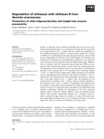
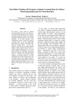
to reduce respiratory motion. Breathing motion was mon-
itored in all patients by fluoroscopy (Figure 1) and/or by
using multiple electronic cine-portals during treatment.
Dose was prescribed to the isocenter without lung correc-
tion for inhomogeneity (it is the routine practice in our
Department). Gross tumor volume (GTV) encompassed
only the radiologically visible tumor as seen by the chest
CT with the lung window. Planning target volume (PTV)
was GTV plus a 10–15 mm margin in all directions. Elec-
Example of a lung tumor in the right lower lobe easily seen by fluoroscopy with normal breathing during expiration (A) and inspiration (B)Figure 1
Example of a lung tumor in the right lower lobe easily seen by fluoroscopy with normal breathing during expiration (A) and
inspiration (B). The radiation field, in spite of the motion, encompasses appropriately the tumor. The maximum displacement is
only 4 mm.
A = expiration B = inspiration
Radiation Oncology 2006, 1:42 />Page 3 of 6
(page number not for citation purposes)
tive nodal radiotherapy was not performed. The treatment
planning ensured that the esophagus, heart and spinal
cord received the minimum possible dose, but always less
than 50% of the total prescribed tumor dose. Acute toxic-
ity was prospectively assessed for lung, esophagus, and
skin using the RTOG acute radiation morbidity scoring
criteria every week during treatment. Late lung toxicity
was evaluated with a modified scoring system considering
only the lung symptoms, as summarized in Table 1. Radi-
ographic abnormalities alone in asymptomatic patients
were not considered to represent late lung toxicity. Late
toxicity for other organs was evaluated using the Com-
mon Toxicity Criteria (CTC) version 2.
Follow-up was done every 3–4 months with chest X-rays
performed routinely at all visits. CT scans of the chest were
performed in patients with abnormal x-ray findings,
symptoms, or suspicion of disease progression. Patients
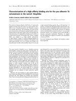
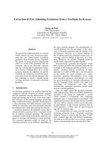
Example of typical 3-dimensional planning with 2 fields avoiding the esophagus, heart and spinal cordFigure 2
Example of typical 3-dimensional planning with 2 fields avoiding the esophagus, heart and spinal cord.
Table 1: Symptoms only scoring used for late lung toxicity (modified from the Common Toxicity Criteria (CTC) version 2 – Lung)
012345
No increase in lung
symptoms
Increase in lung
symptoms due to RT
but not requiring
steroids
Same but steroids are
required
Oxygen is needed Assisted ventilation is
required
Death related to
radiation
Radiation Oncology 2006, 1:42 />Page 4 of 6
(page number not for citation purposes)
were considered as dying from NSCLC if they had evi-
dence of active cancer, either locally or distantly, before
death.
First recurrence was classified as 1) local if it was at the site
of the primary tumor, 2) regional if it was inside the chest
but not local, or 3) distant when it occurred in any other
site outside the chest. The date of first recurrence was that
of the confirmation of recurrence (usually by imaging).
Time zero was the date of the first radiotherapy day. Sur-
vival curves were constructed using the Kaplan-Meier
method. This retrospective study was performed accord-
ing to the guidelines of the McGill University Health Cen-
tre Ethical Committee.
Results
Between October 2002 and June 2004, 32 patients
entered in the 3DHRT program. None received chemo-
therapy. There were 23 (72%) males and 9 (28%) females.
The median age at diagnosis was 76 years (range 56–90).
Median FEV1 = 1.08 (range: 0.51–2.80). Fifteen cases
(47%) were T1 and 17 (53%) were T2 because had more
than 3 cm in greatest dimension. Twenty patients had
confirmed histology of NSCLC. For the remaining 12
patients, the presumptive diagnosis of NSCLC was made
by a multidisciplinary team based on the medical history
and imaging findings. In these 12 cases biopsy was either
inconclusive or was not tried because the procedure was
felt to carry a high risk of complication in these patients.
The majority of patients (94%) were N0 by CT scan. Only
two patients (6%) were considered to have N1 disease
close to the primary lung tumor. In 19 cases (59%) the
tumor was in the right lung. In 22 cases (69%) the tumor
was located in the upper lobes.
Radiotherapy was delivered mostly with either two fields
(either opposed or wedge pair) (Figure 2) in 17 cases
(53%) or 3 fields in 13 (41%) cases. Although less confor-
mal, we preferred using 2 or 3 fields to avoid irradiating
more normal lung and to facilitate monitoring the respi-
ratory motion.
The median area of the fields was 72 cm
2
(typically 8 cm
× 9 cm) and the median PTV volume was 150 cm
3
(range:
28–1110). The median V16 value for both lungs (PTV/
GTV was not excluded from the V16 determination) was
13% (range: 3 – 29).
No patient was lost to follow up. As May 2006, the
median follow-up time was of 29 months for the surviv-
ing patients and 21 months (range: 3 – 41 months) for the
whole group. Eighteen patients had died, but only 10 due
to lung cancer progression. Sixteen patients developed
some form of recurrent disease. The first site of failure was
as follows: 5 local only, 2 local + regional, 5 regional only,
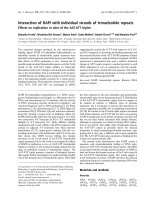
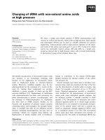
1 regional + distant and 3 distant only. Actuarial 1 and 2-
year overall survival rates are 78% and 56%, cancer spe-
cific survival rates (CSS) are 90% and 74% (Figure 3), and
local relapse free survival rates are 93% and 76% respec-
tively. Median overall survival and cancer specific survival
are 29 and 35 months respectively.
Toxicity was mostly non-existent. One patient had grade 1
and another grade 2 acute pneumonitis and 3 patients
had very mild (grade 1) acute esophagitis. There was no
acute skin toxicity. Concerning late toxicity results were
even better. No late toxicity has been observed in the
esophagus, skin, subcutaneous tissue and even lungs
(remembering that radiographic abnormalities alone
were not considered as late lung toxicity).
Discussion
According to the model suggested by Abratt and Hunter
[5], assuming an effective doubling time of 3 days and
that repopulation in NSCLC commences in 21 days, 52.5
Gy in 15 daily fractions of 3.5 Gy given in 3 weeks would
be biologically equivalent to 78 Gy in 39 daily fractions of
2 Gy, given over 8 weeks. Table 2 compares some hypof-
ractionated regimens according to that model.
The contribution of the present study is mostly related to
toxicity, which has always been a concern with any hypof-
ractionated radiation treatment. In our present series
acute toxicity was prospectively evaluated and found to be
minimal with the dose of 52.5 Gy delivered in 3 weeks. To
date there has been no late toxicity, showing that this reg-
imen is a very safe radiation treatment. The fact that the
Overall (OS) (lower) and Cancer Specific (CSS) (upper) actu-arial survival curvesFigure 3
Overall (OS) (lower) and Cancer Specific (CSS) (upper) actu-
arial survival curves.
Survival of Hypofractionation-Lung
0 10 20 30 40 50
0.0
0.1
0.2
0.3
0.4
0.5
0.6
0.7
0.8
0.9
1.0
1.1
Time
Fraction survival
Radiation Oncology 2006, 1:42 />Page 5 of 6
(page number not for citation purposes)
patients did not have elective mediastinal nodal irradia-
tion [6,7] and received essentially no irradiation to the
esophagus, heart, and spinal cord, likely explains their
very good tolerance to the treatment in spite of being
patients with co-morbid diseases.
Co-morbid conditions have been shown to affect progno-
sis in early stage NSCLC [8]. Unfortunately, the typical
early stage NSCLC patients referred for curative radiation
therapy have grave impairment of pulmonary function,
serious cardiovascular disease [9], are often imprecisely
staged [10], and up to 43% of the patients die from inter-
current diseases [11]. In our present cohort of patients the
situation is not different. Some of our patients were so
frail that procedures like a needle biopsy could not be per-
formed.
In general, patients treated with radiation alone are older
when compared to patients treated by surgery: the median
age in our present group is 76 years compared to 65 years
in surgical series [12]. This poor general physical condi-
tion explains the fact that 7 of our patients died within
only 12 months after the radiation therapy (four of them
with no evidence of tumor progression) making it difficult
to evaluate long-term outcome in this group of patients.
Local control is the main purpose of localized radiation
therapy, particularly for early stage NSCLC patients where
local relapse is considered to be the most common failure
pattern after radiation therapy alone [9,11]. We need to
have in mind that lung is very sensitive to radiation and
some times it is difficult to differentiate fibrosis from local
relapse. This difficulty likely explains the wide range of
local failure, reported from 6.4% to 70% [9]. In our
present series 7/32 patients (22%) were considered to
have local relapse (alone or with other metastases), sug-
gesting that this regimen seems to give local control at
least similar to what has been reported with standard frac-
tionation [9-11].
Cheung et al [13] reported 1 and 2-year CSS of 89.8% and
54.1% respectively, with the use of hypofractionated radi-
ation therapy alone in early stage NSCLC, dose of 48 Gy
in 12 fractions, using 2D and 3DHRT in 33 patients.
The largest series of early stage NSCLC patients treated
with accelerated hypofractionated radical radiotherapy
using 3D planning comes from Wales [14]. Using 2-D and
3-D planning, 112 patients clinically staged as I/II, most
of them receiving the dose of 50 Gy in 20 daily fractions,
had an overall median survival of 23.5 months. The
authors do not mention the rate of local control for this
early stage group of patients; toxicity was evaluated retro-
spectively and no severe grade 3/4 toxicity was recorded.
We did not use any special device to either decrease inspir-
atory motion or to deliver radiation in fixed phases of the
breathing cycle. However, we carefully monitored the
tumor motion of our patients with fluoroscopy or elec-
tronic portal images to confirm that the GTV was inside
the treatment fields (Figure 1). We cannot compare this
relatively simple 3-D planning technique with stereotactic
body radiation therapy. The latter is a promising alterna-
tive, but it requires significant technical advances to min-
imize organ motion due to respiration and in the use of
tumor imaging to guide the administration of the radia-
tion treatment [15-17]. It is still not available in most
institutions and for this reason we believe there is plenty
of space for 3DHRT.
Conclusion
Three-dimensional involved field hypofrationated radio-
therapy, at a dose of 52.5 Gy in 15 fractions of 3.5 Gy,
given in 3 weeks, shortens by half the traditional treat-
ment duration, is safe and very well tolerated by patients
with medically inoperable early NSCLC. The technique
does not require any special device to deliver the radiation
treatment allowing any service of radiation oncology with
3-D planning to do it. Results of 2-year local relapse free
survival of 76% and CSS of 74% respectively, compare
favorably with other published results. Based on this
present and on other Canadian experiences [13] the
National Cancer Institute of Canada (NCIC) recently
started a phase II trial (BR-25) using similar hypofraction-
Table 2: Comparison of different radiation therapy fractionations according to Abratt model.
Dose per
fraction
No of
fractions
Time (days) Total Dose
(Gy)
ID2 acute
reaction
TED ID2 late
reaction
McGill 3.5 Gy 15 19 52.5 59 59 68
Standard 2 Gy 2 Gy 39 53 78 78 59 78
Sunnybrook
13
4 Gy 12 16 48 56 56 67
Wales
14
2.75 20 26 55 58 55 60
ID2 = isoeffective dose at 2 Gy per fraction; acute reaction α/β = 10; late reaction α/β = 3. TED = tumor effect dose (used to indicate the ID2
modified for overall treatment time to account for repopulation) (5).
Publish with BioMed Central and every
scientist can read your work free of charge
"BioMed Central will be the most significant development for
disseminating the results of biomedical research in our lifetime."
Sir Paul Nurse, Cancer Research UK
Your research papers will be:
available free of charge to the entire biomedical community
peer reviewed and published immediately upon acceptance
cited in PubMed and archived on PubMed Central
yours — you keep the copyright
Submit your manuscript here:
/>BioMedcentral
Radiation Oncology 2006, 1:42 />Page 6 of 6
(page number not for citation purposes)
ated technique giving the dose of 60 Gy in 15 fractions for
this group of NSCLC patients.
Competing interests
The author(s) declare that they have no competing inter-
ests.
References
1. Fowler JF, Chappell R: Non-small cell lung tumors repopulate
rapidly during radiation therapy. Int J Radiat Oncol Biol Phys 2000,
46:516-7.
2. Mehta M, Scrimger R, Mackie R, Paliwal B, Chappell R, Fowler J: A
new approach to dose escalation in non-small-cell lung can-
cer. Int J Radiat Oncol Biol Phys 2001, 49:23-33.
3. Abratt RP, Bogart JA, Hunter A: Hypofractionated radiation for
non-small cell lung cancer. Lung Cancer 2002, 36:225-33.
4. Mehta M, Fowler J: Accelerated hypofractionation. Int J Radiat
Oncol Biol Phys 2005, 61:299-300.
5. Abratt RP, Hunter A: Accelerated hypofractionation. Int J Radiat
Oncol Biol Phys 2003, 56:1206-1207.
6. Hayman JA, Martel MK, Ten Haken RK, Ten Haken RK, Normolle DP,
Todd RF 3rd, Littles JF, Sullivan MA, Possert PW, Turrisi AT, Lichter
AS: Dose-escalation in non-small cell lung cancer using three-
dimensional conformal radiation therapy: update of a phase
I trial. J Clin Oncol 2001, 19:127-36.
7. Rosenzweig KE, Sim SE, Mychalczak B, Braban LE, Schindelheim R,
Leibel SA: Elective nodal irradiation in the treatment of non-
small-cell lung cancer with three-dimensional conformal
radiation therapy. Int J Radiat Oncol Biol Phys 2001, 50(3):681-5.
8. Firat S, Bousamra M, Gore E, Byhart R: Comorbidity and KPS are
independent prognostic factors in stage I non-small cell lung
cancer. Int J Radiat Oncon Biol Phys 2002, 52:1047-57.
9. Qiao X, Tullgren O, Lax I, Sirzen F, Lewensohn R: The role of radi-
ation therapy in treatment of stage I non-small cell lung can-
cer. Lung Cancer 2003, 41:1-11.
10. Zimmermann FB, Bamberg M, Molls M, Jeremic B: Radiation ther-
apy alone in early stage non-small cell lung cancer. Semin Surg
Oncol 2003, 21:91-97.
11. Sibley GS: Radiation therapy for patients with medically inop-
erable Stage I non small cell lung carcinoma: smaller vol-
umes and higher doses-a review. Cancer 1998, 82:433-8.
12. Gajra A, Newman N, Gamble GP, Abraham NZ, Kohman LJ, Graziano
SL: Impact of tumor size on survival in stage IA non-small cell
lung cancer: a case for subdividing stage IA disease. Lung Can-
cer 2003, 42:51-7.
13. Cheung PC, Yeung LT, Basrur V, Ung YC, Balogh J, Danjoux CE:
Accelerated hypofractionation for early-stage non-small-cell
lung cancer. Int J Radiat Oncol Biol Phys 2002, 54:1014-23.
14. Lester JF, Macbeth FR, Brewster AE, Court JB, Iqbal N: CT planned
accelerated hypofractionated radiotherapy in the radical
treatment of non-small cell lung cancer. Lung Cancer 2004,
45:237-42.
15. Hara R, Itami J, Kondo T, Aruga T, Abe Y, Ito M, Fuse M, Shinohara
D, Nagaoka T, Kobiki T: Stereotactic single high dose irradia-
tion of lung tumors under respiratory gating. Radiother Oncol
2002, 63:159-63.
16. Whyte RI, Crownover R, Murphy MJ, Martin DP, Rice TW, DeCamp
MM, Rodebaugh Rm Le QT: Stereotactic radiosurgery for lung
tumors:preliminary report of a phase I trial. Ann Thorac Surg
2003, 75:1097-101.
17. McGarry RC, Papiez L, Williams M, Whitford T, Timmerman RD:
Stereotactic body radiation therapy of early-stage non-
small-cell lung carcinoma: Phase I study. Int J Radiat Oncol Biol
Phys 2005, 63:1010-5.