Báo cáo khoa học: " Intensity Modulated Radiotherapy (IMRT) in the postoperative treatment of an adenocarcinoma of the endometrium complicated by a pelvic kidney" pot
Bạn đang xem bản rút gọn của tài liệu. Xem và tải ngay bản đầy đủ của tài liệu tại đây (981.8 KB, 5 trang )
BioMed Central
Page 1 of 5
(page number not for citation purposes)
Radiation Oncology
Open Access
Methodology
Intensity Modulated Radiotherapy (IMRT) in the postoperative
treatment of an adenocarcinoma of the endometrium complicated
by a pelvic kidney
Marcus S Castilho*, Alexandre A Jacinto, Gustavo A Viani, Andre Campana,
Juliana Carvalho, Robson Ferrigno, Paulo ERS Novaes, Ricardo C Fogaroli
and Joao V Salvajoli
Address: Department of Radiation Oncology, Hospital do Câncer A C Camargo, São Paulo, Brazil
Email: Marcus S Castilho* - ; Alexandre A Jacinto - ; Gustavo A Viani - ;
Andre Campana - ; Juliana Carvalho - ; Robson Ferrigno - ;
Paulo ERS Novaes - ; Ricardo C Fogaroli - ; Joao V Salvajoli -
* Corresponding author
Abstract
Background: Pelvic Radiotherapy (RT) as a postoperative treatment for endometrial cancer
improves local regional control. Brachytherapy also improves vaginal control. Both treatments
imply significant side effects that a fine RT technique can help avoiding. Intensity Modulated RT
(IMRT) enables the treatment of the target volume while protecting normal tissue. It therefore
reduces the incidence and severity of side effects.
Case: We report on a 50 year-old patient with a serous-papiliferous adenocarcinoma of the uterus
who was submitted to surgical treatment without lymph node sampling followed by Brachytherapy,
and Chemotherapy. The patient had a pelvic kidney, and was therefore treated with IMRT.
So far, the patient has been free from relapse and with normal kidney function.
Conclusion: IMRT is a valid technique to prevent the kidney from radiation damage.
Background
Randomized trials have shown that Pelvic Radiotherapy
(RT) as a postoperative treatment for intermediate and
high risk endometrial cancer improves local regional con-
trol. Its impact on overall survival is still unknown. Intra-
cavitary Brachytherapy also improves vaginal control.
Both treatments, however, imply significant side effects
that a fine technique can help avoiding. Intensity Modu-
lated RT (IMRT) is the most efficient external beam RT
delivery technique nowadays. Using a high gradient of
radiation dose enables the treatment of the target volume
while protecting normal tissues in an attempt to reduce
the incidence and severity of side effects.
Patient history
A 50-year old Caucasian woman was referred to the Radi-
ation Oncology Department of Hospital do Cancer A C
Camargo, São Paulo, Brazil, with Endometrial Cancer.
Due to bilateral ovary mass she was submitted to explora-
tory laparotomy. During the surgical procedure, Total
Abdominal Hysterectomy and Bilateral Salpingectomy
and Oophorectomy (TAH/BSO) were performed. The
Published: 20 November 2006
Radiation Oncology 2006, 1:44 doi:10.1186/1748-717X-1-44
Received: 30 August 2006
Accepted: 20 November 2006
This article is available from: />© 2006 Castilho et al; licensee BioMed Central Ltd.
This is an Open Access article distributed under the terms of the Creative Commons Attribution License ( />),
which permits unrestricted use, distribution, and reproduction in any medium, provided the original work is properly cited.
Radiation Oncology 2006, 1:44 />Page 2 of 5
(page number not for citation purposes)
pathological analysis revealed a mucinous cystic adenoma
in her left ovary and an endometrioid cyst in her right
ovary (no evidence of malignancy). The endometrium
presented a solid, Serous Papiliferous Adenocarcinoma,
poorly differentiated, compromising the inner half of the
myometrium with extension to the upper endocervix.
There was no lymph vascular space invasion and the mar-
gins were not compromised.
She was classified as IIA by FIGO criteria [1] and received
6 cycles of Carboplatin and Paclitaxel, followed by 29 Gy
of High Dose Rate Brachytherapy (HDR BT) prescribed on
the vaginal surface, divided in 4 fractions, with median
dose to the rectum and bladder reference points of respec-
tively 48 and 58%.
She was referred to our Institution because she had a Con-
genital Pelvic Kidney.
Static and dynamic Scintigrafic renal function studies were
performed. They showed that the pelvic kidney was func-
tioning perfectly – it absorbed 45% of the injected radio-
active isotope.
A study plan for IMRT was led. It showed the dose to nor-
mal tissue and kidney was kept under tolerable limits. The
patient was informed of the risks and benefits of proceed-
ing with the treatment. The prescribed dose to cover 95%
of the target volume (whole pelvic drainage and vaginal
vault) was 45 Gy at 1.8 Gy per fraction.
Seven co-planar fields were chosen at an interval rotation
of 50 degrees. Dynamic Multileaf Collimation was used.
The target volume excluded the entire pelvic kidney and
covered pelvic lymphatics from L5 down.
RT field fluency is presented in figure 1, and Dose Distri-
bution is presented in figure 2. The dose volume analysis
(DVH) is presented in figure 3.
Planned dose distribution was verified dosimetrically and
matched the software's calculation. The qualitative analy-
sis of isodose curves was satisfactory too.
During treatment, the patient presented peri-anal radio-
dermitis (RTOG grade 1), increased bowel movements
(up to 3 times/day), and a lowering in platelet count lev-
els (75,000/mm3) which led to a 7 day treatment inter-
ruption at 37.8 Gy. She subsequently recovered with a
platelet rise to 90,000/mm3 and the treatment was
resumed. The renal function panel was unaltered during
the whole RT course.
When last seen – 18 months after the end of RT – the
patient was free from disease. She had normal kidney
function, both by serum panel and isotopic nephrogram
evaluation. The nephrogram did not show any changes
compared to the initial exam.
Discussion
Patients with a pelvic kidney should not receive RT unless
it is a mainstream in the treatment of that type of tumor.
There are very few reports on treating pelvic kidneys
patients with EBRT [2-8].
It is important to establish the need, and the benefits of RT
to any patient with in such a condition.
Pelvic kidney function
We used renal blood tests and isotopic nephrogram to
access the patient's renal function. The scintigrafic study
used static and dynamic assessment of glomerular and
tubular function. Her right pelvic kidney took 45% of the
radio labeled marker (DMSA/DTPA), and had normal
excretion of it. Eighteen months after the treatment the
kidney' uptake was unchanged.
Scintigraphic renograms have correlated with biochemical
and clearance end points [9], and are adequate for this sit-
uation, as the other kidney is functioning well, and any
effect on the pelvic kidney would be better seen with func-
tional images rather than with functional biochemical
exams.
Benefit of adjuvant radiation and chemotherapy
The standard surgical treatment for uterine neoplasia con-
sists of Radical Hysterectomy, bilateral salpingo
oophorectomy, and lymphadenectomy or lymph node
sampling.
In this case, the surgical approach was not radical in intent
because the uterine neoplasia was an incidental finding.
Therefore, the lymph node status was not known. In this
setting, the benefit of re-operation is unclear and not evi-
dence-based. The prospective PORTEC trial [10] has
directly tested the benefit of RT for patients without
lymph node information. Patients with endometrial ade-
nocarcinoma were randomized to receive postoperative
pelvic EBRT, or no adjuvant therapy. They noticed a signif-
icant advantage in pelvic control for the adjuvant treat-
ment arm with risk features (deep myometrial invasion,
cervical canal extension, high grade histology, or lymph
vascular space invasion), though not translated into sur-
vival benefit. The majority of failures occurred at the vag-
inal vault. This study did not evaluate specifically serous
papiliferous tumors, but this subset of tumors is known to
have a worse prognosis. This patient is classified as having
a high risk tumor. It is considered a non- endometrioid
tumor, not responsive to estrogenic castration. Metha and
Radiation Oncology 2006, 1:44 />Page 3 of 5
(page number not for citation purposes)
cols [11] have studied a group of women with stage I-II
serous papiliferous tumors treated with surgery followed
or not by adjuvant therapy. Though no variables were sta-
tistically correlated to prognosis, out of 13 women who
did not receive RT/BT, 5 recurred in the pelvis (4 in the
vagina, 1 in the lateral pelvis). In contrast, none of the
patients who received RT/BT (total of 10) recurred in the
pelvis. The 5-year pelvic recurrence free survival was 100
vs. 57%, with a p = 0.06.
This information and other published results suggesting a
benefit of carboplatin/paclitaxel based chemotherapy for
this histological type and the fact that this histological
type of tumor carries a high risk of recurrence makes us
believe that our patient did benefit from the adjuvant
chemo-radiotherapy, including vaginal vault BT.
Expected risks, side effects, and tolerance
Kidney tolerance to radiation dose highly depends on the
irradiated volume.
Tolerance dose for a 5% chance of late adverse effect at 5
years is estimated to be 50 Gy for one third of the kidney,
30 Gy for two thirds, and 23 Gy for the whole kidney [12].
It increases to 50% late toxicity if two thirds are irradiated
to a dose of 40 Gy or one third to a dose of 28 Gy.
As noted on the DVH (figure 3) these parameters have
been respected in the present case.
The literature does not define the optimal treatment for
patients with pelvic kidneys who need to undergo pelvic
RT. We could find 7 case reports concerning this subject
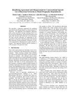
Radiation fluenceFigure 1
Radiation fluence. shows radiation fields, their fluence maps, and the resulting dose distribution on a section plane that
includes the pelvic kidney.
Radiation Oncology 2006, 1:44 />Page 4 of 5
(page number not for citation purposes)
[2,4-8]. In 5 cases the primary tumor being treated was a
uterine cervix carcinoma [2,6-8]. In 3 of them, the kidney
was transplanted outside the pelvis, away from the RT tar-
get volume [2,7,8]. However, there was significant mor-
bidity related to the procedure, especially regarding the
graft vasculature, and the urinary tract. In one case an ade-
nocarcinoma of the uterine cervix in a transplanted
patient was treated initially with Intracavitary BT (low
dose rate) followed by a modified field pelvic RT protect-
ing the kidney, but partially compromising the RT target
volume [6]. This patient relapsed on the border of the RT
field.
Other reports of auto-transplantation followed by RT for
inguinal-pelvic irradiation in a vulvar cancer patient, and
for adjuvant treatment of a stage III operated rectal aden-
ocarcinoma exists [4,5].
Although the preferred approach has not been estab-
lished, no report exists on the use of high technology RT
in an attempt to accomplish an adequate plan without
moving the kidney out of the RT field. Conformal 3D RT
has been developed to precisely study the combination of
RT fields, and properly match the dose distribution to the
CT visible tumor, while evaluating dose received by nor-
mal tissue, therefore predicting treatment tolerance. It is
however limited in achieving these goals when the tumor
is surrounded by normal tissues with low radiation resist-
ance, or when the normal organ is in the middle of the RT
port. In this setting IMRT has been shown effective, and its
use for head and neck, thoracic, and abdominal treat-
ments have been increasing.
We showed that IMRT is also a good alternative in such a
complex situation. It has prevented the patient from
undergoing an auto-transplantation procedure.
During treatment, this patient presented mild (common
toxicity criteria grade 1) platelet complication. Lately
there has been an increase in the use of IMRT to spare the
blood marrow, providing that, in case of a relapse and
need for new chemotherapy regimens, maintaining as
much functioning marrow as possible presents another
advantage of using IMRT. Roeske and cols have shown the
main location of blood elements production in the pelvis
Dose distributionFigure 2
Dose distribution. shows the dose distribution for the 45 Gy prescribed dose.
Radiation Oncology 2006, 1:44 />Page 5 of 5
(page number not for citation purposes)
[13], and it is possible to define these points as dose
restriction points for the IMRT planning.
To our knowledge, this is the first report on the use of
IMRT to spare a pelvic kidney without compromising a
pelvic RT plan.
IMRT was a valid radiation technique to keep the pelvic
kidney dose under acceptable dose volume constraints
without compromising the target volume.
IMRT should be considered an option for treating pelvic
fields in patients who present a pelvic kidney.
References
1. Benedet JL, Bender H, Jones H III, Ngan HY, Pecorelli S: FIGO stag-
ing classifications and clinical practice guidelines in the man-
agement of gynecologic cancers. FIGO Committee on
Gynecologic Oncology. Int J Gynaecol Obstet 2000, 70:209-262.
2. Abouna GM, Micaily B, Lee DJ, Kumar MS, Jahshan AE, Lyons P: Sal-
vage of a kidney graft in a patient with advanced carcinoma
of the cervix by reimplantation of the graft from the pelvis
to the upper abdomen in preparation for radiation therapy.
Transplantation 1994, 58:520-522.
3. Bakri YN, Mansi M, Sundin T: Stage IIB carcinoma of the cervix
complicated by an ectopic pelvic kidney. Int J Gynaecol Obstet
1993, 42:174-176.
4. Bokhari MB, Hostetter RB, Auber ML, Ulewicz DE: Locally
advanced rectal cancer with a pelvic kidney complicating
adjuvant radiation therapy. J Surg Oncol 1996, 63:57-60.
5. DeRoover A, Verni MP, Taylor RJ: Renal allograft autotransplan-
tation before pelvic irradiation. Transplantation 2000,
70:844-846.
6. Ripley D, Levenback C, Eifel P, Lewis RM: Adenocarcinoma of the
cervix in a renal transplant patient. Gynecol Oncol 1995,
59:151-155.
7. Rosenshein NB, Lichter AS, Walsh PC: Cervical cancer compli-
cated by a pelvic kidney. J Urol 1980, 123:766-767.
8. Roth TM, Woodring CT, McGehee RP: Stage II-B carcinoma of
the cervix complicated by bilateral pelvic kidneys. Gynecol
Oncol 2004, 92:376-379.
9. Dewit L, Anninga JK, Hoefnagel CA, Nooijen WJ: Radiation injury
in the human kidney: a prospective analysis using specific
scintigraphic and biochemical endpoints. Int J Radiat Oncol Biol
Phys 1990, 19:977-983.
10. Scholten AN, van Putten WL, Beerman H, Smit VT, Koper PC,
Lybeert ML, Jobsen JJ, Warlam-Rodenhuis CC, De Winter KA, Lut-
gens LC, van Lent M, Creutzberg CL: Postoperative radiotherapy
for Stage 1 endometrial carcinoma: long-term outcome of
the randomized PORTEC trial with central pathology
review. Int J Radiat Oncol Biol Phys 2005,
63:834-838.
11. Mehta N, Yamada SD, Rotmensch J, Mundt AJ: Outcome and pat-
tern of failure in pathologic stage I-II papillary serous carci-
noma of the endometrium: implications for adjuvant
radiation therapy. Int J Radiat Oncol Biol Phys 2003, 57:1004-1009.
12. Emami B, Lyman J, Brown A, Coia L, Goitein M, Munzenrider JE, Shank
B, Solin LJ, Wesson M: Tolerance of normal tissue to therapeu-
tic irradiation. Int J Radiat Oncol Biol Phys 1991, 21:109-122.
13. Roeske JC, Lujan A, Reba RC, Penney BC, Diane YS, Mundt AJ: Incor-
poration of SPECT bone marrow imaging into intensity
modulated whole-pelvic radiation therapy treatment plan-
ning for gynecologic malignancies. Radiother Oncol 2005,
77:11-17.
Dose Volume HistogramFigure 3
Dose Volume Histogram. shows the graphic of Dose Volume Histogram. The curves show the distribution for the PTV,
rectum, bladder, intestines, pelvic kidney and left topic kidney.