Báo cáo khoa học: " On the visualization of universal degeneracy in the IMRT problem" ppt
Bạn đang xem bản rút gọn của tài liệu. Xem và tải ngay bản đầy đủ của tài liệu tại đây (1.59 MB, 7 trang )
BioMed Central
Page 1 of 7
(page number not for citation purposes)
Radiation Oncology
Open Access
Methodology
On the visualization of universal degeneracy in the IMRT problem
Markus L Alber*
1
and Gustav Meedt*
2
Address:
1
Section for Biomedical Physics, Clinic for Radiooncology, University of Tübingen, Germany and
2
Computerized Medical Systems, St.
Louis, USA
Email: Markus L Alber* - ; Gustav Meedt* -
* Corresponding authors
Abstract
Background: In general, the IMRT optimisation problem possesses many equivalent solutions.
This makes it difficult to decide whether a result produced by an IMRT planning algorithm can be
further improved, e.g. by adding more beams, or whether it is close to the globally best solution.
Results: It is conjectured that the curvature properties of the objective function around any
globally optimum dose distribution are universal. This allows an assessment of optimality of dose
distributions that are generated by different beam arrangements in a complementary manner to the
objective function value alone. A tool to visualize the curvature structure of the objective function
is devised.
Conclusion: In an example case, it is demonstrated how the assessment of the curvature space
can indicate the equivalence of rival beam configurations and their proximity to the global optimum.
Background
Because of the dependence on optimisation algorithms,
treatment planning for intensity modulated photon radi-
otherapy (IMRT) frequently evades intuition. This is par-
ticularly true for the issue of beam placement in complex
cases that may even require non-coplanar beam direc-
tions, where it is hard to decide whether both the number
and the directions of beams are optimum in the sense that
the resulting dose distribution cannot be improved fur-
ther with reasonable effort. The purpose of this note is to
give an intuitive picture and a tool for visualization of a
central property of the IMRT optimisation problem,
which is the degeneracy of the solution space.
It is a practical wisdom of the field, that there exists a very
large number of virtually equivalent solutions for an
IMRT optimisation problem. Due to this degeneracy of
the solution space, it is not necessary to find one solitary
best set of beam directions and fluence weight profiles,
but merely a set that is equivalent (i.e. in practice: close
enough) to the global optimum. It is fair to say that the
true optimum of an IMRT problem is virtually never
sought: usually, a few dozen iterations of a gradient algo-
rithm are deemed sufficient, while a non-linear optimisa-
tion problem of a few thousand parameters would
typically require thousands of iterations. Eventually, it is
degeneracy that makes beamlet-based IMRT optimisation
tractable.
The degeneracy of the objective function is inherent to the
problem and does not arise spuriously from any particular
mathematical formulation. In a previous paper [1], a con-
jecture was made about the universality of the curvature
properties in optimisation problems that differed in the
number and arrangement of beams. It is helpful to imag-
ine the situation in 2D as a long, narrow valley that is
Published: 18 December 2006
Radiation Oncology 2006, 1:47 doi:10.1186/1748-717X-1-47
Received: 01 June 2006
Accepted: 18 December 2006
This article is available from: />© 2006 Alber and Meedt; licensee BioMed Central Ltd.
This is an Open Access article distributed under the terms of the Creative Commons Attribution License ( />),
which permits unrestricted use, distribution, and reproduction in any medium, provided the original work is properly cited.
Radiation Oncology 2006, 1:47 />Page 2 of 7
(page number not for citation purposes)
absolutely flat along its floor, but rises sharply perpendic-
ular to it. The steep slopes on either side correspond to the
high costs for increasing the dose, imposed by some nor-
mal tissue objective, and for decreasing dose, imposed by
some target objective. The minimum in this direction is a
finely tuned balance between these objectives. Since these
objectives oppose each other for all reasonable beam
arrangements, it can be assumed that the balance is
sought in a very similar way for all of them. This means,
that the solution space for all beam arrangements should
show the same pattern of directions of great curvature,
which is characteristic for the given case. It would be
intriguing if this universal curvature pattern could be vis-
ualized.
In order to compare the curvature properties of the opti-
mum dose distributions of different beam arrangements,
the matrix of second derivatives for all beamlets of the
union of the two beam arrangements would have to be
computed. This can quickly become an onerous task. In
this manuscript, we try to overcome this obstacle by intro-
ducing an independent set of arbitrary, but well chosen
probing fluence elements which can be shared between dif-
ferent beam arrangements and are solely used for visuali-
zation. These probing fluence elements define a curvature
map. In case the objective function of the optimisation
problem is convex, it can be shown that all globally opti-
mum solutions share the same subspace of zero curvature.
Further, it is conjectured that the subspace of non-vanish-
ing curvature is also an invariant. An example of this cur-
vature map and its application in the context of manual
and computerized beam direction optimisation is given
for a head and neck tumour case.
Methods
The notation of this development is introduced as fol-
lows. Let F(D) be a twice continuously differentiable
objective function of the dose distribution D = (D
j
)
j = 1 m
in the patient volume V consisting of m voxels, whose
minimum defines the desired solution. Further, let
for i ≠ j. All the treatment goals with respect
to target coverage, target dose homogeneity and normal
tissue sparing are assumed to be incorporated in this
objective function. For simplicity, let F be a sum of local
objective function densities f(D
j
), so that
. This does not restrict generality unduly,
since most objective functions proposed for IMRT can be
written in this form and fulfill the condition on the sec-
ond derivative. For a proof see Romeijn et al. [2]. For
example, the classical one- and two-sided quadratic pen-
alty functions f(D) = (D - D
0
)
2
Θ (D, D
0
), where Θ (D, D
0
)
determines whether the penalty applies to doses greater
and/or smaller than a threshold D
0
, are of this shape
(Here, it is convenient to postulate that Θ is not a step
function, but a steep yet differentiable sigmoid so as to
ensure the existence of a second derivative). Also, biolog-
ical indices based on sums of local dose-response func-
tions like EUD (f = , k > 0), TCP (f = exp(-
α
D
j
)) and
NTCP (e.g. f = , k ≥ 1), as well as DVH penalties (e.g. f
= 1/(1 + (D
0
/D)
k
), k > 1) can be transformed into this
'standard form' without loss of generality [3]. The dose
distribution is generated by a fluence distribution, which
is by definition composed of a weighted sum of elemen-
tary fluence distributions. Let be a basis set of elemen-
tary fluence distributions (fluence elements) (
η
i
)
i = 1 n
,
and (
φ
i
)
i = 1 n
≤ 0 the associated weights (A fluence element
can, but need not be a beamlet in the common under-
standing as a constituent of an intensity modulated field.
A beamlet is an elementary fluence distribution with the
property that a finite superposition yields a deliverable
intensity modulated field). For example, can be the set
of all beamlets of an arrangement of intensity modulated
beams, or the set of all arclets of radiosurgery arcs. The
basis set defines the parameter space of the optimisation
as the space of the weights that are associated with the flu-
ence elements. By means of the basis , a subset of the
abstract space of all physically feasible fluence distribu-
tions is mapped onto . This is in analogy to geometry,
where e.g. a point in space can be identified by its coordi-
nates relative to a set of basis vectors.
Let (T
j
)
j = 1 m
be the (linear) dose deposition operator that
associates the fluence element
η
i
with its dose distribution
(T
j
)
j = 1 m
η
i
= (T
ji
)
j = 1 m
. Hence,
The solution of the IMRT problem is a vector of fluence
weights ( )
i = 1 n
which obtains from the minimization
of F under the constraints
φ
i
≥ 0. Given that F is assumed
to be twice continuously differentiable, the necessary opti-
mality conditions
∂
∂∂
=
2
0
FD
DD
ij
()
FfD
j
j
m
=
=
∑
()
1
D
j
k
D
j
k
0
||
DT
jji
i
n
i
=
()
=
∑
1
1
φ
.
φ
i
∗
∂
∂
==∀∈
()
∗
∗
FD
i
ii
()
φ
φη
00 2or
Radiation Oncology 2006, 1:47 />Page 3 of 7
(page number not for citation purposes)
hold. If all f are convex, F is also convex, and by virtue of
the convexity of the feasible set = {
φ
i
∈ :
φ
i
≥ 0},
only a single global minimum exists (generally, the dose
limiting objectives ensure that F is bounded from below
and F → ∞ for all
φ
i
→ ∞). However, this does not mean
that the minimum is attained in a single point. In the case
of degeneracy, as commonly present in the IMRT setting,
the set of minimizers is a sizeable, high dimensional, closed
and connected subset of the parameter space. Any such
point in this solution space is indistinguishable from any
other with respect to its objective function value. The
uniqueness of the minimizer cannot be guaranteed even if
the objective densities f are strictly convex, because F(
φ
) is
a composition of monotonously increasing and decreas-
ing convex functions. In practice, this causes IMRT optimi-
sation algorithms to terminate at different solutions if
started from slightly different points, which may be mis-
taken for local, isolated minima. The situation is different
in case either f or are non-convex, which can occur
when dose-volume objectives are employed or MLC con-
straints need to be considered. In this case the multiple
global minimizers can lie in disconnected and non-con-
vex sets, or local minima may exist. Notice that these are
possibilities, not necessities.
If two different dose distributions, possibly generated by
different beam arrangements, have the same objective
function value, they may be degenerate to the global solu-
tion, or merely be local solutions that share the same
objective function value by coincidence. By virtue of the
linearity of the dose operator T, there exists an easy test to
investigate this situation. Let
1
,
2
be two fluence basis
sets, for example the sets of all beamlets of two arrange-
ments of beam directions, and , be the associated
optimum dose distributions. Further, let F() = F().
For 0 ≤
λ
≤ 1, we define
F(
λ
) = F(
λ
+ (1 -
λ
)) (3)
and compute F(
λ
) for a small number of
λ
∈ [0, 1]. This
test does not entail more than the weighted addition of
two dose distributions and repeated evaluations of the
objective function, but no operations in fluence weight
space.
If there exists one
λ
' with F(
λ
') >F( ), then obviously
both dose distributions do not belong to the same mini-
mum. This situation can only occur if F is non-convex. If
F is convex, and , are degenerate to the global min-
imum, then
F() = F(D*) = F(
λ
+ (1 -
λ
)D*). (4)
By definition, F is always locally convex in a certain envi-
ronment of the global minimum (otherwise it would not
be a minimum), which makes the following argumenta-
tion applicable to some extent also to the globally non-
convex case. In the following, we use eq. 4 to motivate a
conjecture about universal curvature properties of all solu-
tions that are degenerate to the global optimum. In all but
the most contrived cases, the comprehensive basis * of
all globally optimum dose distributions, i.e. the union of
all basis sets that generate a global optimum, is very large.
Simultaneously, a very large number of degenerate and
near-degenerate solutions exist. Assume that some dose
distribution is degenerate to the global optimum D*,
i.e. F() = F(D*). By the above definition,
1
⊂ *.
The optimality conditions have to be satisfied for all ele-
ments of *. We apply the optimality conditions eq. 2 to
the right hand side of eq. 4
We expand the left hand side to first order in
λ
, using that
Since is degenerate to D*, the left hand side vanishes,
i.e.
In order to interprete this condition, it is helpful to
observe the Hessian matrix of second derivatives with
respect to the fluence weights
||
D
1
∗
D
2
∗
D
1
∗
D
2
∗
D
1
∗
D
2
∗
D
1
∗
D
1
∗
D
2
∗
D
1
∗
D
1
∗
D
1
∗
D
1
∗
∂+−
∂
=
∂+−
∂
⋅=∀ ∈
∗∗
∗∗
=
∗
∑
fD D
fD D
D
T
i
j
j
m
ji i
(())
(())
.
λλ
φ
λλ
η
1
1
1
1
1
0
5
(()
∂
∂∂
=
2
0
fD
DD
ij
()
lhs =+ −
∂
∂
+∀∈
()
∗∗
=
∗
∗
∑
06
1
1
2
2
2
λλη
()
()
() .
,
DD
fD
D
T
jj
j
m
j
ji i
D
1
∗
()
()
.
,
DD
fD
D
T
jj
j
m
j
ji i1
1
2
2
07
∗∗
=
∗
∗
−
∂
∂
=∀ ∈
()
∑
η
Radiation Oncology 2006, 1:47 />Page 4 of 7
(page number not for citation purposes)
Notice that H
ij
is a symmetric, but not diagonal matrix,
while the inner second derivative of the objective function
with respect to the local dose is diagonal by definition.
Commonly, the vast majority of eigenvalues of this matrix
is zero, owing to the high degeneracy of the IMRT prob-
lem, while the solution is characterized by the (smaller)
subspace of non-vanishing curvature [1]. By comparison
with eq. 7, it becomes apparent that this criterion evalu-
ates whether the difference between two degenerate glo-
bal solutions ( - D*)
j
= Σ
i
( -
φ
*)
i
T
ji
, lies entirely in
the subspace of zero curvature of the global optimum.
Since this has to hold for any two globally optimum dose
distributions, their curvature matrices H
ij
(D*) share the
same subspace of zero curvature (To see this, assume there
exists a direction
η
Δ = - for which the curvature
around is zero, but around is greater than zero. In
a convex setting, it is possible to go from to along
a straight line without leaving the solution space, but the
gradient of F in the direction of
η
Δ has to increase when
going from to . This is in contradiction with the
assumption that both dose distributions are globally opti-
mum).
In [1], it was conjectured that because all globally degen-
erate optima share the same modes of solving the funda-
mental conflicts of the problem between target and
normal tissue objectives, the structure of the subspace of
high curvature around them is universal. Together with
the universality of the subspace of zero curvature, this ear-
lier conjecture would be a consequence of the following,
more general formulation: Let * be the set of all glo-
bally optimum dose distributions and let Φ* be the asso-
ciated set of all fluence weight distributions with respect
to the basis *. Let the set Φ
0
span the subspace of zero
curvature of F(D*), D* ∈ *, which is assumed to be
locally convex around *. It is conjectured that all third
and higher order derivatives of F with respect to any fluence
vector in Φ
0
vanish for all dose distributions in *. The first
and second order derivatives vanish by virtue of the opti-
mality conditions and the above considerations following
eq. 7.
A direct consequence of this conjecture is, that the sub-
space of non-vanishing curvature of H
ij
(D*) is also an
invariant and the matrix H
ij
(D*) is constant for all dose
distributions in *. The parameter space becomes a
direct product of the space of vanishing curvature
0
and
the space of positive curvature
+
. In practice, three
obstacles to validating this conjecture for a given example
exist. Firstly, the basis set * can only be approximated
by a finite basis set. Secondly, the computation of the Hes-
sian matrix is extremely time consuming for large basis
sets, since it grows quadratically with the number of basis
fluence elements. Thirdly, the conjecture cannot be
proven numerically because the size of the derivative ten-
sors grows exponentially with the order of the derivative.
In order to visualize the putatively universal curvature
properties with a minimum of computational effort, the
concept of the curvature map is introduced, which avoids
these problems. Let D* be a globally optimum dose distri-
bution and let
p
be any set of arbitrarily chosen probing
fluence elements, then the curvature map (CM) is defined as
The finite set of probing fluence elements is assumed to be
a subset of the fictitious basis set * and is prompted by
practical reasons only. This set can be chosen freely.
Notice that due to the (local) convexity of F, the CM can-
not be negative. If the conjecture holds, the curvature
maps of all dose distributions that are degenerate to the
global optimum are equivalent.
The globally optimum solution is essentially determined
by those volumes in the patient where it is hard to meet
target volume or normal tissue objectives. If a probing flu-
ence element
η
passes through these volumes where the
dose distribution is a forced compromise, its correspond-
ing CM-value M(
η
) will be high. In contrast, if a probing
fluence element passes only through volumes where the
target and normal tissue objectives can be met, the CM-
value will be close to zero.
One advantageous choice of probing fluence elements for
beam direction optimisation could be the 'generalized
Gamma knife basis set', a set of conical beams of a certain
diameter of 15–20 mm say, which impinge on the isocen-
tre from about 5000 directions distributed uniformly
across the unit sphere. The curvature map would then
show beam directions which pass through or avoid areas
HD T
fD
D
T
ij ki
T
k
m
k
kj
()
()
.=
∂
∂
()
=
∑
1
2
2
8
D
1
∗
φ
i
∗
D
1
∗
D
2
∗
D
1
∗
D
2
∗
D
1
∗
D
2
∗
D
2
∗
D
1
∗
MD
fD
D
T
ip j
j
m
j
ji
():
()
.
ηη
∈=
∂
∂
()
∗
=
∗
∑
1
2
2
9
Radiation Oncology 2006, 1:47 />Page 5 of 7
(page number not for citation purposes)
of conflicts between dose prescriptions. There are no lim-
its to the choice of * other than practicality and visual-
ization.
Results and Discussion
The example case presented here is a paranasal sinus
tumour, see figure 1. The obvious obstacle for achieving
the target dose is the proximity of the optical pathway and
to a much smaller degree the brain. The setup of the opti-
misation problem included hard constraints on the maxi-
mum equivalent uniform dose (EUD) [3,4] of the organs
at risk (optical pathways, brain, brainstem, unspecified
tissue), and a hard constraint for the maximum dose to
the target. For the target, the objective was to maximize
EUD under the given constraints. The sum of these con-
stituent objective functions, together with their appropri-
ate Lagrange multipliers, yields the objective function F.
With this combination of physical and EUD constraints,
the objective function is convex. The Lagrange multipliers
are fixed such that the constraints are obeyed for the solu-
tion D*, with the consequence that the sole floating figure
of merit is the EUD of the target volume. In figure 2, cur-
vature maps for several beam configurations are shown.
The CM value is colour coded, starting from zero in blue
to the common maximum in red, i.e. all CMs cover the
same range of curvatures. Beam directions are indicated as
dots on the sphere.
Several observations can be made.
• The rows two through four show CMs of a five, six and
seven IMRT beam configuration created by a beam direc-
tion optimisation (BDO) algorithm [5]. The first row
shows a beam configuration which was chosen manually
(6 MAN). The difference between 5 BDO and 6 BDO is
somewhat greater than between 6 BDO and 7 BDO, but
the general distribution of curvatures is rather constant,
with a noticable shrinkage of the total area covered in red
and yellow. This impression of increasing proximity to the
global optimum is also supported by their final objective
function values, which were 97 per cent and 99 per cent
respectively of the EUD of the 7 BDO configuration.
Notice, that each configuration has no more than one
beam in common. The difference between the BDO plans
would be greater if they were scaled to their common
maximum, rather than the maximum of 6 MAN.
• The first row shows the CM of a manually selected 6 field
configuration. The CM differs noticeably from the 7 BDO
CM. At the same time, only 94 per cent of the EUD of 7
BDO could be reached. The frontal red region is more
extensive in the 6 MAN configuration, which shows that a
larger sector of the unit sphere is affected by conflict vol-
umes in the patient. There is one beam direction which
originates from a blue region. Blue sectors indicate that
directions lie in the subspace of vanishing curvature and
thus can be easily substituted. If possible, the BDO algo-
rithm eliminates these directions. In contrast, the human
expert picked this direction on the basis of intuition,
which was misleading in this case.
• The fifth row shows the CM for the combined 5 BDO
and 6 BDO configuration (i.e. an optimized dose distribu-
tion of 11 beams). Notice that there is virtually no differ-
ence to the 7 BDO configuration. The combined
configuration yields the same EUD in the target as 7 BDO.
This indicates, that about 7 beams suffice to generate a
globally optimum dose distribution for this case, and that
a 11 beam arrangement can contain several superfluous
beams.
• Notice that in any case, since the objective function in
this example is convex, at least the blue areas of the CM of
two globally optimum dose distributions have to agree.
Naturally, the basis of probing fluence elements is never
complete. In this example, it was chosen such that the full
extent of the patient is covered. For more extensive target
volumes, different choices may be more appropriate.
Notice, that according to the above conjecture, the CM of
globally optimum dose distributions should be equiva-
lent for any choice of basis.
If the CM is applied in beam direction optimisation, it
provides complementary information to the objective
function value. Iterative beam selection has to continue as
long as the CM changes between successive configura-
tions. If two rival configurations differ in their CMs, nei-
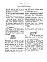
Geometry of the example caseFigure 1
Geometry of the example case. Transversal and coronal
sections of the example case, a paranasal sinus tumour. Tar-
get volume outlined in black/dark grey, optical pathway light
grey.
Radiation Oncology 2006, 1:47 />Page 6 of 7
(page number not for citation purposes)
Curvature Maps for the example caseFigure 2
Curvature Maps for the example case. Curvature maps of a manually optimised 6 beam configuration, a 5, 6, and 7 beam
direction optimised configuration, and a 11 field configuration consisting of the union of the 5 BDO and 6 BDO configurations.
The CM values were mapped onto a sphere for 5000 rays which sample all feasible directions of incidence. Left column: cranial
view, second column: caudal view, third column: frontal view. The black excision corresponds to beam angles that were
excluded due to couch/gantry collisions. Blue sectors correspond to a CM-value of zero, red and yellow regions display zones
of conflicts between target objectives and normal tissue constraints. The colour scale is equivalent for all rows. Beam direc-
tions are given as red dots.
Publish with BioMed Central and every
scientist can read your work free of charge
"BioMed Central will be the most significant development for
disseminating the results of biomedical research in our lifetime."
Sir Paul Nurse, Cancer Research UK
Your research papers will be:
available free of charge to the entire biomedical community
peer reviewed and published immediately upon acceptance
cited in PubMed and archived on PubMed Central
yours — you keep the copyright
Submit your manuscript here:
/>BioMedcentral
Radiation Oncology 2006, 1:47 />Page 7 of 7
(page number not for citation purposes)
ther of them is truly equivalent to the global optimum. By
virtue of the above conjecture, the CM contributes supple-
mental information in case the beam selection process
gets stuck with two rival beam arrangements which pro-
duce the same objective function value, yet could be
improved further. Once a final configuration was found,
beams originating from blue zones are most likely redun-
dant and can be removed from the configuration. Notice,
that the CM cannot be used to guide the search for addi-
tional beam directions at intermediate stages: since it is
subject to significant change while the beam configura-
tion evolves, the indication of redundant beams is not sta-
ble. The first derivative with respect to the weight of a
probing fluence element may offer more information for
this purpose.
Conclusion
It is conjectured for the IMRT optimisation problem, that
in the proximity of the global optimum, the second and
all higher order derivatives vanish in a subspace of sizea-
ble dimension. As a consequence, the curvature of the
objective function for any globally optimum dose distri-
bution is equivalent. In order to verify this property, the
tool of a curvature map was introduced which relies on a
freely choosable set of probing fluence elements. This
allows to compare the curvature properties of dose distri-
bution which have no beams in common. These consider-
ations about curvature are intended to highlight the
special properties of the IMRT optimisation problem,
which may further its understanding or be used to advan-
tage in algorithm design.
Authors' contributions
MA and GM developed the maths and prepared the man-
uscript. GM coded the software tools and prepared the
example.
Acknowledgements
This research was supported by Computerized Medical Systems, St. Louis,
USA.
References
1. Alber M, Meedt G, Reemtsen R, Nüsslin F: On the degeneracy of
the IMRT optimisation problem. Med Phys 2002,
29(11):2584-2589.
2. Romeijn HE, Dempsey JF, Li JG: A unifying framework for multi-
criteria fluence map optimization models. Phys Med Biol 2004,
49(10):1991-2013.
3. Alber M, Nüsslin F: An objective function for radiation treat-
ment optimization based on local biological measures. Phys
Med Biol 1999, 44(2):479-493.
4. Niemierko A: Reporting, and analyzing dose distributions: A
concept of equivalent uniform dose. Med Phys 1997,
24:103-110.
5. Meedt G, Alber M, Nüsslin F: Non-coplanar beam direction opti-
mization for intensity modulated radiotherapy. Phys Med Biol
2003, 48(18):2999-3019.