Báo cáo khoa học: " The role of PDGF in radiation oncology" ppsx
Bạn đang xem bản rút gọn của tài liệu. Xem và tải ngay bản đầy đủ của tài liệu tại đây (347.87 KB, 9 trang )
BioMed Central
Page 1 of 9
(page number not for citation purposes)
Radiation Oncology
Open Access
Review
The role of PDGF in radiation oncology
Minglun Li*, Verena Jendrossek and Claus Belka
Address: Department of Radiation Oncology, University Hospital Tuebingen, Germany
Email: Minglun Li* - ; Verena Jendrossek - ;
Claus Belka -
* Corresponding author
Abstract
Platelet-derived growth factor (PDGF) was originally identified as a constituent of blood serum and
subsequently purified from human platelets. PDGF ligand is a dimeric molecule consisting of two
disulfide-bonded chains from A-, B-, C- and D-polypeptide chains, which combine to homo- and
heterodimers. The PDGF isoforms exert their cellular effects by binding to and activating two
structurally related protein tyrosine kinase receptors. PDGF is a potent mitogen and
chemoattractant for mesenchymal cells and also a chemoattractant for neutrophils and monocytes.
In radiation oncology, PDGF are important for several pathologic processes, including oncogenesis,
angiogenesis and fibrogenesis. Autocrine activation of PDGF was observed and interpreted as an
important mechanism involved in brain and other tumors. PDGF has been shown to be fundamental
for the stability of normal blood vessel formation, and may be essential for the angiogenesis in
tumor tissue. PDGF also plays an important role in the proliferative disease, such as atherosclerosis
and radiation-induced fibrosis, regarding its proliferative stimulation of fibroblast cells. Moreover,
PDGF was also shown to stimulate production of extracellular matrix proteins, which are mainly
responsible for the irreversibility of these diseases. This review introduces the structural and
functional properties of PDGF and PDGF receptors and discusses the role and mechanism of PDGF
signaling in normal and tumor tissues under different conditions in radiation oncology.
Background
PDGF was originally identified as a constituent of whole
blood serum that was absent in cell-free plasma [1,2] and
subsequently purified from human platelets [3,4].
Although the α-granules of platelets are a major storage
site for PDGF, recent studies have shown that PDGF can
be synthesized by a number of different cell types such as
macrophages, epithelial and endothelial cells [5-8]. Stud-
ies have shown that PDGF has important physiologic
functions in organ development [9,10]. PDGF has also
been implicated in a wide variety of pathological proc-
esses, including fibrosis, atherosclerosis, glomerulone-
phritis and aggressive fibromatosis [11-15]. Moreover,
aberrant production of PDGF and autocrine stimulation
may be an important mechanism in the neoplastic con-
version of PDGF receptor-positive cells [16-18]. Here, we
point out the most important features of PDGF and PDGF
receptors concerning their roles in radiation oncology.
PDGF structure and signaling
PDGF is a disulfide-linked dimer of two related polypep-
tide chains, designated A, B, C and D, which are assem-
bled as heterodimers (PDGF-AB) or homodimers (PDGF-
AA, PDGF-BB, PDGF-CC and PDGF-DD) [19-21]. PDGF
exerts its biological activity by binding to structurally sim-
ilar PDGF receptors (PDGFR-α and -β). The PDGFR-α
Published: 11 January 2007
Radiation Oncology 2007, 2:5 doi:10.1186/1748-717X-2-5
Received: 15 November 2006
Accepted: 11 January 2007
This article is available from: />© 2007 Li et al; licensee BioMed Central Ltd.
This is an Open Access article distributed under the terms of the Creative Commons Attribution License ( />),
which permits unrestricted use, distribution, and reproduction in any medium, provided the original work is properly cited.
Radiation Oncology 2007, 2:5 />Page 2 of 9
(page number not for citation purposes)
binds to A-, B- and C- chains with high affinity, whereas
PDGFR-β only binds the B- and D- chains [22-25]. Differ-
ent from PDGF-A and -B, PDGF-C and PDGF-D require
proteolytic activation before binding to and activation of
PDGFR [19,20]. PDGF ligand dimer induces dimerization
of both receptors and subsequently autophosphorylation
of the PDGF receptor tyrosine kinase (RTK). Activated RTK
phosphorylates numerous signaling molecules that initi-
ate intracellular signaling cascades (Reviewed in Ref.
[31]).
The best characterized mechanisms by which PDGF
down-streaming signaling mediates cellular responses
involve the activation of the ras/MAPK pathway, which
can functionally increase cellular proliferation, migration
and differentiation [26], and the PI3K/Akt pathway,
which promotes cell survival [27]. Both pathways are of
crucial importance for tumor resistance to radiotherapy
and chemotherapy. Furthermore, platelet-derived growth
factor (PDGF) exerts its potent mitogen and chemotactic
effects in a variety of mesenchymal cells such as fibrob-
lasts, vascular smooth muscle cells, glomerular mesangial
cells, and brain glial cells [14,28-30] making PDGF a
potential key molecule for tissue rebuilding in response to
physiological and non-physiological conditions.
PDGF in oncology
Many investigators have shown that autocrine activation
of PDGF was interpreted to be an important pathogenetic
mechanism involved in different brain tumors [16-18].
In gliomas, analysis of PDGF/PDGFR expression sug-
gested the presence of autocrine and paracrine loops of
PDGF in glioma activating PDGFR-α in glioma cells,
while PDGFR-α expression was higher in malign gliomas
than in benign gliosis [17].
Moreover, the recently identified new PDGF isoforms,
PDGF-C and -D are also detectable in glioblastoma cell
lines and primary human tumor tissues [31].
On the other hand, treatment with a PDGFR antagonist
interrupted autocrine growth stimulation and thus inhib-
ited survival and mitogenesis in glioblastoma cells and
prevented glioma formation in a mouse xenograft model
[31,32].
In the case of meningioma, Adam and his colleagues pro-
vided evidence that cytokines secreted by meningioma
cells profoundly stimulated growth of meningioma and
neuroblastoma cells in vitro, while this growth stimula-
tion was completely abolished by a neutralizing antibody
against PDGF [16]. Todo et al showed DNA synthesis in
tumor cells could be inhibited through an antagonist of
PDGF in three of seven meningiomas cell lines [32].
Similarly, autocrine loops involving PDGF-A or -B and
their respective receptors was also observed in many
malignant and low-grade astrocytomas, while the activa-
tion of PDGF autocrine loops was suggested to be an early
event in the pathogenesis of malignant astrocytomas [33].
Aggressive fibromatosis also referred to as desmoid tumor
develops from muscle connective tissue, fasciae and
aponeuroses. The neoplasm is composed of fibrocyte-like
cells, and characterized by local infiltrative growth and
high risk of recurrence (~70%) after surgical treatment
[34]. Depending on the location and extent of the tumor,
radiotherapy is indicated for patients with unresectable
tumors or those with positive resection margins. Overex-
pression of PDGF were observed in desmoid tumors,
while inhibition of PDGF signaling by imatinib induced
overall 1 year tumor control rate of 36.8% in a phase II
clinical study [15]. Thus, inhibition of PDGF may be an
attractive therapy option, alone or combined with surgery
or/and radiotherapy in refractory cases.
Another example for an important role of PDGF in onco-
genesis is the so-called gastrointestinal stromal tumors
(GISTs). Many GISTs have gain-of-function mutations of
c-kit receptor tyrosine kinase (KIT) gene. Approximately
35% of GISTs lacking KIT mutations have intragenic acti-
vation mutations in PDGFR-α [35].
However, the alternative defects lead to similar alterations
of the downstream signaling cascades and cytogenetic
changes. Therefore the defects (gain-of-function through
mutated-KIT or mutated-PDGFR-α) appear to be alterna-
tive and mutually exclusive [35].
Likewise, overexpression of PDGF and c-kit was also
observed in Leydig tumors. Treatment with imatinib
almost completely inhibited Leydig tumor growth in an
allograft mouse model by inhibition of PDGF and c-kit
signaling with no drug-resistance development during
imatinib treatment [36].
The clinical success of imatinib/gleevec, a triple tyrosine
kinase inhibitor of c-kit, PDGF and c-Abl signaling, in
chronic myeloid leukemia [37] and gastrointestinal stro-
mal tumors [38] has accelerated the development of
molecular targeted cancer therapy. It is highly likely that
many more antitumoral substances of this class will be
developed and discovered in the near future.
PDGF and angiogenesis
In addition to its direct tumor growth promoting effect,
the importance of PDGF in tumor propagation relates to
the inherent angiogenic activity [39]. In this regard, PDGF
has been shown to be essential for the stability of normal
blood vessel formation by recruiting pericytes and
Radiation Oncology 2007, 2:5 />Page 3 of 9
(page number not for citation purposes)
smooth muscle cells [40]. PDGF-B expression by endothe-
lial cells recruits pericytes through a short-range paracrine
mode [41]. Pericytes expressing PDGFRs migrate along
steep gradient of PDGF-B in the peri-endothelial compart-
ment to endothelial cells and thus initiate intimate asso-
ciation with the abluminal surface of the endothelial cells
[41]. Pericyte-deficiency promotes a range of microvascu-
lar changes, such as endothelial hyperplasia, vessel dila-
tion, leakage and rupture, leading to capillary
microaneurysms, and lethal microhemorrhage [40].
Despite structural and functional abnormalities in the
microvasculature, mice embryos deficient of up to 90%
pericytes are compatible with embryonic and postnatal
survival, while loss of more than 95% of the pericytes is
lethal [40,42]. This suggests that a rather low threshold
density of pericytes is required for basal microvascular
function.
Angiogenesis is an important event in tumor growth, since
tumors located more than 100–200 μm distant from a
blood vessel need neovascular formation to ensure a suf-
ficient supply of nutrients and oxygen [43]. Tumor cells in
hypoxia secrete cytokines, including VEGF, PDGF, basic
fibroblast growth factor (bFGF), insulin growth factor
(IGF), to stimulate neovascular formation [43].
However, neovasculature in tumors differs strikingly from
normal physiologic vessels. The badly coordinated growth
leads to vessel malformation including vessel dilation,
tortuosity, leakage, rupture and formation of microaneu-
rysms [40]. Interestingly, these hallmarks of microvascu-
lar malformation in tumors were found to be identical
with the alterations found in pericyte-deficient mice
(PDGF-B -/- or PDGFR-β -/-), pointing to a pericyte-defi-
ciency in the disordered neovascular formation in tumors
[41].
Since small numbers of pericytes in tumor vessels may be
critical for vessel integrity and function [40], targeting per-
icytes in tumors may be an attractive and efficacious way
for anti-angiogenic therapy.
Recent data from experiments in vivo imply that targeting
pericytes actually provides additional benefits [44]. Tradi-
tionally, endothelial cells as a host component in the
tumors with normal genome are suggested to be the pri-
mary target for anti-angiogenic therapies [45]. Inhibiting
VEGF in endothelial cells reduced endothelial cell sur-
vival, proliferation, tube formation and invasion in vitro
[45]. However, Erber and his colleagues demonstrated
that endothelial cells were resistant to the inhibitory effect
of SU5416 by blocking VEGFR in vivo through pericyte
mediated escape strategies via the Ang-1/Tie2 pathway
[46]. Combined inhibition of VEGF and PDGF signaling
enforces tumor vessel regression by direct anti-angiogenic
effect to endothelial cells and pericytes and by inhibiting
pericyte mediated endothelial cell survival mechanisms
[46].
This view is also supported by other studies showing that
tumor vessels lacking pericytes are more dependent on
VEGF for their survival than are vessels invested by peri-
cytes [44]. In fact, sorafenib and sunitinib/SU11248 act as
anti-angiogenic agents by inhibiting VEGFR-2/-3, PDGFR-
β, Flt-3, and c-KIT. Both drugs exert clear clinical effects in
patients with renal cell carcinoma which are most likely
mediated via anti-angiogenic effects [47,48]. The thera-
peutic efficacy to other tumors is currently under investi-
gation [48].
In conclusion, PDGF has at least two distinct functions in
pro-angiogenic signaling. On the one hand PDGF
increases survival and proliferation of endothelial cells
and on the other hand, PDGF regulates vessel growth via
pericyte recruitment and association to newly formed ves-
sels.
PDGF inhibition in combination with
radiotherapy
Ionising radiation causes miscellaneous effects to the
tumor mass. It exerts a direct antitumoral effect on tumor
cells, for example through DNA double-strand-break lead-
ing to failure of DNA transcription and duplication and
eventual death of tumor cells [49]. However, radiation
induced damage of endothelial cells plays a major role in
tissue damage and antitumoral efficacy [45]. In this
regard, within hours after ionising radiation, lesions with
structural changes could be observed in endothelial cells
by using electron microscopy [50]. Thus, ionising radia-
tion can be also considered as a potent anti-angiogenic
agent [45].
On the other hand, it was shown that tumor cells are able
to produce pro-angiogenic cytokines including VEGF,
PDGF and FGF in response to ionising radiation. These
pro-angiogenic cytokines could protect endothelial cells
and vessels from radiation-induced damage and conse-
quently ensure supply of oxygen and nutrients for tumor
cells [9,11,18]. The secretion of PDGF could also be stim-
ulated in irradiated stromal cells, such as endothelial and
fibroblast cells [51]. Elevated expression of these growth
factors correlates with higher vessel density and negative
clinically prognosis in various tumors [52]. Usually, such
tumors possess a relative resistance to radiation therapy
[53].
Inhibition of pro-angiogenic signaling by tyrosine kinase
inhibitors can therefore augment the radiation induced
damage to endothelial cells and abolishes the tumor cells
mediated protection. Moreover, these inhibitors can pre-
Radiation Oncology 2007, 2:5 />Page 4 of 9
(page number not for citation purposes)
vent the re-growth of endothelial cells and neovascular
formation.
Consequently, anti-angiogenic substances targeting VEGF
and PDGF may increase anti-angiogenic activity of ionis-
ing radiation and possess a potent antitumoral synergy
with radiation.
Glioma is a good example for demonstration of the dual
role of PDGF signaling in the oncogenesis and angiogen-
esis in tumor mass.
Using in situ hybridization and immunohistochemistry
techniques, Hermanson et al demonstrated the presence
of autocrine and paracrine loops in gliomas, activating the
PDGFR-α in glioma cells. The activation of PDGFR-β in
endothelial cells was also observed in the tumor mass,
pointing to the dual role of PDGF signaling in oncogene-
sis and angiogenesis in glioma tumors [17].
On the one hand, treatment with imatinib/gleevec dis-
rupted an autocrine PDGF/PDGFR loop by specifically
inhibiting phosphorylation of PDGFR and thus exerted a
synergistic antitumoral effect with ionising radiation as
radiosensitizer [54]. And on the other hand, targeting
PDGF signaling inhibits the hypoxia-induced angiogen-
esis and strengthens the anti-angiogenic effect of radiation
[46].
PDGF in radiotherapy-induced fibrogenesis
The development of acute inflammation and chronic
fibrosis is a frequent side effect of ionising radiation and
thus a dose-limiting factor for treatment efficacy [55].
In the case of lung tumors, the dose limitation imposed by
normal tissue tolerance presently precludes successful
radiotherapeutic treatment in many patients [56]. Pulmo-
nary fibrosis is a progressive condition, characterized by
mesenchymal cell proliferation, the subsequent deposi-
tion of extracellular matrix proteins and extensive remod-
eling of the pulmonary parenchyma [57]. In both human
and animal model systems, acute pneumonitis and late
fibrosis are directly dependent upon total irradiation
dose, fraction size, and lung volume irradiated [58-60].
New precise radiotherapy techniques can spare more nor-
mal tissue around tumor volume and thus reduce the
intensity of side effects. However a recent study has shown
that 14.6 % patients with lung cancer still developed inter-
mediate grade radiogenic pneumonitis after primary radi-
otherapy with dose escalation using 3D conformal
techniques and 13.8 % patients developed fibrosis [61].
The treatment of fibrosis remains still elusive, since the
exact mediators and mechanisms involved in fibrogenesis
are not completely understood [57]. The traditional inter-
pretation of radiation-induced fibrosis as a consequence
of acute inflammation has been questioned in recent
years, because clinical measures of inflammation do not
correlate well with fibrotic progression and because anti-
inflammatory drugs do not significantly affect clinical
outcome [56,62,63]. New evidence suggests that immedi-
ate intercellular communications through regulation of
cytokines happens within hours to days after irradiation
[64].
A number of investigations provided clear evidence for
increased expression of various cytokines including
PDGF, transforming growth factor-β, tumor necrosis fac-
tor-α and interleukin-1 in response to ionising radiation
[22,65-67]. In this regard, some pro-inflammatory
cytokines seem to be important for the acute impairment
in the pneumonitis phase, for example TNF-α and CD95-
ligand [66,68], whereas others are involved in the regula-
tion of the fibrotic response. For the development of
fibrosis, transforming growth factor-β is till now a widely
accepted key player [69].
Moreover, recent evidence supports an important role of
PDGF for the development of lung fibrosis in response to
ionising radiation. Firstly, PDGF and PDGFR are
expressed at low levels in normal adults, while elevated
levels are detected in lungs of patients with radiation-
induced pulmonary fibrosis [70]. Augmented expression
of PDGF is further observed in asbestos-, bleomycin- and
idiopathic pulmonary fibrosis [71-73]. Increased expres-
sion of PDGF in rat lungs by adenoviral delivery or lung-
specific over-expression in mice is associated with pro-
nounced lung fibrosis [74,75]. Moreover, inhibiting the
PDGF pathway with neutralising antibodies to PDGF or
administration of soluble extracellular region of PDGFR-
β could attenuate fibrotic development [76,77].
Recently it has been shown that three distinct receptor
tyrosine kinase inhibitors (RTKI), overlapping in inhibi-
tion of PDGF signaling, attenuated radiation-induced pul-
monary fibrogenesis in vivo [78]. The radiation-induced
overexpression of PDGF led to phosphorylation and acti-
vation of PDGFR in lungs of irradiated mice, while the
phosphorylation of PDGFR was strongly inhibited in both
irradiated groups treated with RTKIs. Accordingly, the
treatment with RTKIs attenuated the development of pul-
monary fibrosis in excellent correlation with clinical, his-
tological, and computed tomography results, although
the acute inflammatory response induced by radiation
injury was not completely abrogated. Moreover, all three
tyrosine kinase inhibitors reduced lung fibrosis after radi-
ation injury and prolonged animal survival. Thus, there is
hard evidence to support the important role of the PDGF/
PDGFR system for mesenchymal cells in proliferative dis-
eases.
Radiation Oncology 2007, 2:5 />Page 5 of 9
(page number not for citation purposes)
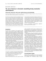
Schematic presentation of radiation induced fibrogenesis in lungsFigure 1
Schematic presentation of radiation induced fibrogenesis in lungs. Illustration of a microenvironment of gas-blood exchange
unit in lungs in the physiologic conditions (upper part) and radiation induced activation of PDGF pathways in the fibrogenesis in
lungs (lower part).
<
<<
<
<
<<
<
<
<<
<
Proliferation
Differentiation
Differentiation
Excessive Deposition of
ECM und irreversible
fibrotic lesion
<
<<
<
<
<<
<
<
<<
<
Ionizing
radiation
<
<<
<
<
<<
<
PDGF ligands
Macrophage
Lung epithelial cells
Endothelial cells
Other cytokines, e.g. TGF, TNF, IL
<
<<
<
PDGF receptor
Other leucocyte, e.g. Monocyte, neutrophil
Fibroblast
<
<<
<
Migration
<
<<
<
<
<<
<
Normal collagen
deposition
Physiological conditions
Radiation Oncology 2007, 2:5 />Page 6 of 9
(page number not for citation purposes)
Since fibroblasts are the putative effector cells, recruitment
and stimulation of fibroblasts should be the most impor-
tant event during development of fibrosis. In this regard,
PDGF may exert profibrotic effect through its mitogenic
and chemotactic stimulation to mesenchymal cells, such
as fibroblasts, myofibroblasts and smooth muscle cells
[79]. Moreover, PDGF was also shown to stimulate pro-
duction of extracellular matrix proteins, such as collagen,
hyaluronic acid, fibronectin and proteoglycan [80-83],
which are mainly responsible for the irreversibility of
fibrotic lesion.
The radiation-induced secretion of PDGF has been
assumed to derive solely from leucocytes. However, radi-
ation of stromal cells, such as fibroblasts and endothelial
cells, induced paracrine PDGF in co-culture systems
which substantially stimulated the proliferation of non-
irradiated fibroblasts [51].
In accordance with these results, endothelial cells were
reported as potential sources of PDGF after radiation in
vitro [84]. Moreover, the expression of c-sis mRNA in epi-
thelial cells was also observed in certain pulmonary
fibrotic diseases [85].
Other experiments demonstrated that anti-inflammatory
treatment with dexamethasone did not decrease the level
of PDGF-BB or the mitogenic activity of bronchial alveo-
lar lavage fluid for fibroblasts in the chronic lung disease
of prematurity [86]. Savikko and his colleagues also
showed that limiting the extent of inflammation by
cyclosporin A treatment did not inhibit the expression of
PDGF ligands and receptors [87]. Thus, stromal cells, such
as endothelial, fibroblasts cells, should be at least partially
responsible for the release of cytokines, including PDGF.
A schematic diagram depicts the suggested role of radia-
tion induced PDGF signaling in fibrogenesis (Fig. 1).
Conclusion and outlook
PDGF signaling plays an important role in radiation
oncology with respect to its oncogenic, angiogenic and
profibrotic effects. The rational of targeting PDGF signal-
ing in radiation oncology can arise in three ways: 1) the
direct antitumoral potential, 2) the anti-angiogenic
impact, and 3) the antifibrotic activity which protects nor-
mal tissue from the side effects of ionising radiation.
Suppression of PDGF is discussed as one potential mech-
anism of action of some novel antifibrotic drugs undergo-
ing clinical trials [88,89]. It has been suggested that
pirfenidone and interferon gamma, both ameliorate lung
fibrosis by downregulation of PDGF expression [72,90].
However, since diverse signaling pathways activated by
growth factor receptors induce broadly overlapping,
rather than independent sets of signaling, it's unlikely to
completely inhibit a biologic process by blocking a single
cytokine/growth factor. Thus multi-targeted agents may
be more effective in the oncological therapy.
At the same time, a special attention should be paid to the
side effects of this new class of molecular targeted agents,
since clinical experience is still sparse, especially in com-
bination with radiotherapy and chemotherapy.
Competing interests
The author(s) declare that they have no competing inter-
ests.
Authors' contributions
ML drafted the manuscript. CB and VJ critiqued the man-
uscript. All authors read and approved the final manu-
script.
References
1. Ross R, Glomset J, Kariya B, Harker L: A platelet-dependent
serum factor that stimulates the proliferation of arterial
smooth muscle cells in vitro. Proc Natl Acad Sci U S A 1974,
71:1207-1210.
2. Westermark B, Wasteson A: A platelet factor stimulating
human normal glial cells. Exp Cell Res 1976, 98:170-174.
3. Heldin CH, Westermark B, Wasteson A: Platelet-derived growth
factor: purification and partial characterization. Proc Natl Acad
Sci U S A 1979, 76:3722-3726.
4. Antoniades HN, Scher CD, Stiles CD: Purification of human
platelet-derived growth factor. Proc Natl Acad Sci U S A 1979,
76:1809-1813.
5. Demayo F, Minoo P, Plopper CG, Schuger L, Shannon J, Torday JS:
Mesenchymal-epithelial interactions in lung development
and repair: are modeling and remodeling the same process?
Am J Physiol Lung Cell Mol Physiol 2002, 283:L510-L517.
6. Zhang S, Smartt H, Holgate ST, Roche WR: Growth factors
secreted by bronchial epithelial cells control myofibroblast
proliferation: an in vitro co-culture model of airway remod-
eling in asthma. Lab Invest 1999, 79:395-405.
7. Mondy JS, Lindner V, Miyashiro JK, Berk BC, Dean RH, Geary RL:
Platelet-derived growth factor ligand and receptor expres-
sion in response to altered blood flow in vivo. Circ Res 1997,
81:320-327.
8. Lindroos PM, Coin PG, Badgett A, Morgan DL, Bonner JC: Alveolar
macrophages stimulated with titanium dioxide, chrysotile
asbestos, and residual oil fly ash upregulate the PDGF recep-
tor-alpha on lung fibroblasts through an IL-1beta-dependent
mechanism. Am J Respir Cell Mol Biol 1997, 16:283-292.
9. Ponten A, Li X, Thoren P, Aase K, Sjoblom T, Ostman A, Eriksson U:
Transgenic overexpression of platelet-derived growth fac-
tor-C in the mouse heart induces cardiac fibrosis, hypertro-
phy, and dilated cardiomyopathy. Am J Pathol 2003,
163:673-682.
10. Pinzani M, Milani S, Herbst H, DeFranco R, Grappone C, Gentilini A,
Caligiuri A, Pellegrini G, Ngo DV, Romanelli RG, Gentilini P: Expres-
sion of platelet-derived growth factor and its receptors in
normal human liver and during active hepatic fibrogenesis.
Am J Pathol 1996, 148:785-800.
11. Iida H, Seifert R, Alpers CE, Gronwald RG, Phillips PE, Pritzl P, Gor-
don K, Gown AM, Ross R, Bowen-Pope DF, .: Platelet-derived
growth factor (PDGF) and PDGF receptor are induced in
mesangial proliferative nephritis in the rat. Proc Natl Acad Sci
U S A 1991, 88:6560-6564.
12. Wilcox JN, Smith KM, Williams LT, Schwartz SM, Gordon D: Plate-
let-derived growth factor mRNA detection in human
Radiation Oncology 2007, 2:5 />Page 7 of 9
(page number not for citation purposes)
atherosclerotic plaques by in situ hybridization. J Clin Invest
1988, 82:1134-1143.
13. Rice AB, Moomaw CR, Morgan DL, Bonner JC: Specific inhibitors
of platelet-derived growth factor or epidermal growth factor
receptor tyrosine kinase reduce pulmonary fibrosis in rats.
Am J Pathol 1999, 155:213-221.
14. Yagi M, Kato S, Kobayashi Y, Kobayashi N, Iinuma N, Nakamura K,
Kubo K, Ohyama SI, Murooka H, Shimizu T, Nishitoba T, Osawa T,
Nagano N: Beneficial effects of a novel inhibitor of platelet-
derived growth factor receptor autophosphorylation in the
rat with mesangial proliferative glomerulonephritis. Gen
Pharmacol 1998, 31:765-773.
15. Heinrich MC, McArthur GA, Demetri GD, Joensuu H, Bono P, Her-
rmann R, Hirte H, Cresta S, Koslin DB, Corless CL, Dirnhofer S, van
Oosterom AT, Nikolova Z, Dimitrijevic S, Fletcher JA: Clinical and
molecular studies of the effect of imatinib on advanced
aggressive fibromatosis (desmoid tumor). J Clin Oncol 2006,
24:1195-1203.
16. Adams EF, Todo T, Schrell UM, Thierauf P, White MC, Fahlbusch R:
Autocrine control of human meningioma proliferation:
secretion of platelet-derived growth-factor-like molecules.
Int J Cancer 1991, 49:398-402.
17. Hermanson M, Funa K, Hartman M, Claesson-Welsh L, Heldin CH,
Westermark B, Nister M: Platelet-derived growth factor and its
receptors in human glioma tissue: expression of messenger
RNA and protein suggests the presence of autocrine and
paracrine loops. Cancer Res 1992, 52:3213-3219.
18. Nister M, Libermann TA, Betsholtz C, Pettersson M, Claesson-Welsh
L, Heldin CH, Schlessinger J, Westermark B: Expression of mes-
senger RNAs for platelet-derived growth factor and trans-
forming growth factor-alpha and their receptors in human
malignant glioma cell lines. Cancer Res 1988, 48:3910-3918.
19. Li X, Ponten A, Aase K, Karlsson L, Abramsson A, Uutela M, Back-
strom G, Hellstrom M, Bostrom H, Li H, Soriano P, Betsholtz C, Hel-
din CH, Alitalo K, Ostman A, Eriksson U: PDGF-C is a new
protease-activated ligand for the PDGF alpha-receptor. Nat
Cell Biol 2000, 2:302-309.
20. Bergsten E, Uutela M, Li X, Pietras K, Ostman A, Heldin CH, Alitalo
K, Eriksson U: PDGF-D is a specific, protease-activated ligand
for the PDGF beta-receptor. Nat Cell Biol 2001, 3:512-516.
21. Zhuo Y, Zhang J, Laboy M, Lasky JA:
Modulation of PDGF-C and
PDGF-D expression during bleomycin-induced lung fibrosis.
Am J Physiol Lung Cell Mol Physiol 2004, 286:L182-L188.
22. Heldin CH: Simultaneous induction of stimulatory and inhibi-
tory signals by PDGF. FEBS Lett 1997, 410:17-21.
23. Hammacher A, Mellstrom K, Heldin CH, Westermark B: Isoform-
specific induction of actin reorganization by platelet-derived
growth factor suggests that the functionally active receptor
is a dimer. EMBO J 1989, 8:2489-2495.
24. Kanakaraj P, Raj S, Khan SA, Bishayee S: Ligand-induced interac-
tion between alpha- and beta-type platelet-derived growth
factor (PDGF) receptors: role of receptor heterodimers in
kinase activation. Biochemistry 1991, 30:1761-1767.
25. Seifert RA, Hart CE, Phillips PE, Forstrom JW, Ross R, Murray MJ,
Bowen-Pope DF: Two different subunits associate to create
isoform-specific platelet-derived growth factor receptors. J
Biol Chem 1989, 264:8771-8778.
26. Schlessinger J: How receptor tyrosine kinases activate Ras.
Trends Biochem Sci 1993, 18:273-275.
27. Franke TF, Yang SI, Chan TO, Datta K, Kazlauskas A, Morrison DK,
Kaplan DR, Tsichlis PN: The protein kinase encoded by the Akt
proto-oncogene is a target of the PDGF-activated phosphati-
dylinositol 3-kinase. Cell 1995, 81:727-736.
28. Powell DW, Mifflin RC, Valentich JD, Crowe SE, Saada JI, West AB:
Myofibroblasts. I. Paracrine cells important in health and dis-
ease. Am J Physiol 1999, 277:C1-C9.
29. Siegbahn A, Hammacher A, Westermark B, Heldin CH: Differential
effects of the various isoforms of platelet-derived growth fac-
tor on chemotaxis of fibroblasts, monocytes, and granulo-
cytes. J Clin Invest 1990, 85:916-920.
30. Shih AH, Holland EC: Platelet-derived growth factor (PDGF)
and glial tumorigenesis. Cancer Lett 2006, 232:139-147.
31. Lokker NA, Sullivan CM, Hollenbach SJ, Israel MA, Giese NA: Plate-
let-derived growth factor (PDGF) autocrine signaling regu-
lates survival and mitogenic pathways in glioblastoma cells:
evidence that the novel PDGF-C and PDGF-D ligands may
play a role in the development of brain tumors. Cancer Res
2002, 62:3729-3735.
32. Todo T, Adams EF, Fahlbusch R: Inhibitory effect of trapidil on
human meningioma cell proliferation via interruption of
autocrine growth stimulation. J Neurosurg 1993, 78:463-469.
33. Guha A, Dashner K, Black PM, Wagner JA, Stiles CD: Expression of
PDGF and PDGF receptors in human astrocytoma opera-
tion specimens supports the existence of an autocrine loop.
Int J Cancer 1995, 60:168-173.
34. Abdelkader M, Riad M, Williams A: Aggressive fibromatosis of
the head and neck (desmoid tumours). J Laryngol Otol 2001,
115:772-776.
35. Heinrich MC, Corless CL, Duensing A, McGreevey L, Chen CJ, Joseph
N, Singer S, Griffith DJ, Haley A, Town A, Demetri GD, Fletcher CD,
Fletcher JA: PDGFRA activating mutations in gastrointestinal
stromal tumors. Science 2003, 299:708-710.
36. Basciani S, Brama M, Mariani S, De Luca G, Arizzi M, Vesci L, Pisano
C, Dolci S, Spera G, Gnessi L: Imatinib mesylate inhibits Leydig
cell tumor growth: evidence for in vitro and in vivo activity.
Cancer Res 2005, 65:1897-1903.
37. Kantarjian HM, Cortes JE, O'Brien S, Luthra R, Giles F, Verstovsek S,
Faderl S, Thomas D, Garcia-Manero G, Rios MB, Shan J, Jones D, Tal-
paz M: Long-term survival benefit and improved complete
cytogenetic and molecular response rates with imatinib
mesylate in Philadelphia chromosome-positive chronic-
phase chronic myeloid leukemia after failure of interferon-
alpha. Blood 2004, 104:1979-1988.
38. Melichar B, Voboril Z, Nozicka J, Ryska A, Urminska H, Vanecek T,
Michal M: Pathological complete response in advanced gas-
trointestinal stromal tumor after imatinib therapy. Intern
Med 2005, 44:1163-1168.
39. Risau W, Drexler H, Mironov V, Smits A, Siegbahn A, Funa K, Heldin
CH: Platelet-derived growth factor is angiogenic in vivo.
Growth Factors 1992, 7:
261-266.
40. Lindahl P, Johansson BR, Leveen P, Betsholtz C: Pericyte loss and
microaneurysm formation in PDGF-B-deficient mice. Science
1997, 277:242-245.
41. Abramsson A, Lindblom P, Betsholtz C: Endothelial and nonen-
dothelial sources of PDGF-B regulate pericyte recruitment
and influence vascular pattern formation in tumors. J Clin
Invest 2003, 112:1142-1151.
42. Enge M, Bjarnegard M, Gerhardt H, Gustafsson E, Kalen M, Asker N,
Hammes HP, Shani M, Fassler R, Betsholtz C: Endothelium-specific
platelet-derived growth factor-B ablation mimics diabetic
retinopathy. EMBO J 2002, 21:4307-4316.
43. Carmeliet P, Jain RK: Angiogenesis in cancer and other dis-
eases. Nature 2000, 407:249-257.
44. Bergers G, Song S, Meyer-Morse N, Bergsland E, Hanahan D: Bene-
fits of targeting both pericytes and endothelial cells in the
tumor vasculature with kinase inhibitors. J Clin Invest 2003,
111:1287-1295.
45. Abdollahi A, Lipson KE, Han X, Krempien R, Trinh T, Weber KJ, Hah-
nfeldt P, Hlatky L, Debus J, Howlett AR, Huber PE: SU5416 and
SU6668 attenuate the angiogenic effects of radiation-
induced tumor cell growth factor production and amplify the
direct anti-endothelial action of radiation in vitro. Cancer Res
2003, 63:3755-3763.
46. Erber R, Thurnher A, Katsen AD, Groth G, Kerger H, Hammes HP,
Menger MD, Ullrich A, Vajkoczy P: Combined inhibition of VEGF
and PDGF signaling enforces tumor vessel regression by
interfering with pericyte-mediated endothelial cell survival
mechanisms. FASEB J 2004, 18:338-340.
47. Motzer RJ, Rini BI, Bukowski RM, Curti BD, George DJ, Hudes GR,
Redman BG, Margolin KA, Merchan JR, Wilding G, Ginsberg MS, Bacik
J, Kim ST, Baum CM, Michaelson MD: Sunitinib in patients with
metastatic renal cell carcinoma. JAMA 2006, 295:2516-2524.
48. Strumberg D: Preclinical and clinical development of the oral
multikinase inhibitor sorafenib in cancer treatment. Drugs
Today (Barc ) 2005, 41:773-784.
49. Budach W, Taghian A, Freeman J, Gioioso D, Suit HD: Impact of
stromal sensitivity on radiation response of tumors.
J Natl
Cancer Inst 1993, 85:988-993.
50. Guerry-Force ML, Perkett EA, Brigham KL, Meyrick B: Early struc-
tural changes in sheep lung following thoracic irradiation.
Radiat Res 1988, 114:138-153.
Radiation Oncology 2007, 2:5 />Page 8 of 9
(page number not for citation purposes)
51. Li M, Ping G, Plathow C, Trinh T, Lipson KE, Hauser K, Krempien R,
Debus J, Abdollahi A, Huber PE: Small molecule receptor tyro-
sine kinase inhibitor of platelet-derived growth factor signal-
ing (SU9518) modifies radiation response in fibroblasts and
endothelial cells. BMC Cancer 2006, 6:79.
52. Kerbel R, Folkman J: Clinical translation of angiogenesis inhibi-
tors. Nat Rev Cancer 2002, 2:727-739.
53. Geng L, Donnelly E, McMahon G, Lin PC, Sierra-Rivera E, Oshinka H,
Hallahan DE: Inhibition of vascular endothelial growth factor
receptor signaling leads to reversal of tumor resistance to
radiotherapy. Cancer Res 2001, 61:2413-2419.
54. Holdhoff M, Kreuzer KA, Appelt C, Scholz R, Na IK, Hildebrandt B,
Riess H, Jordan A, Schmidt CA, Van Etten RA, Dorken B, le Coutre
P: Imatinib mesylate radiosensitizes human glioblastoma
cells through inhibition of platelet-derived growth factor
receptor. Blood Cells Mol Dis 2005, 34:181-185.
55. Plathow C, Li M, Gong P, Zieher H, Kiessling F, Peschke P, Kauczor
HU, Abdollahi A, Huber PE: Computed tomography monitoring
of radiation-induced lung fibrosis in mice. Invest Radiol 2004,
39:600-609.
56. Abratt RP, Morgan GW, Silvestri G, Willcox P: Pulmonary compli-
cations of radiation therapy. Clin Chest Med 2004, 25:167-177.
57. Trott KR, Herrmann T, Kasper M: Target cells in radiation pneu-
mopathy. International Journal of Radiation Oncology*Biology*Physics
2004, 58:463-469.
58. Chen ES, Greenlee BM, Wills-Karp M, Moller DR: Attenuation of
lung inflammation and fibrosis in interferon-gamma-defi-
cient mice after intratracheal bleomycin. Am J Respir Cell Mol
Biol 2001, 24:545-555.
59. Sunyach MP, Falchero L, Pommier P, Perol M, Arpin D, Vincent M,
Boutry D, Rebatu P, Ginestet C, Martel-Lafay I, Perol D, Carrie C:
Prospective evaluation of early lung toxicity following three-
dimensional conformal radiation therapy in non-small-cell
lung cancer: preliminary results. Int J Radiat Oncol Biol Phys 2000,
48:459-463.
60. Rosenzweig KE, Mychalczak B, Fuks Z, Hanley J, Burman C, Ling CC,
Armstrong J, Ginsberg R, Kris MG, Raben A, Leibel S: Final report
of the 70.2-Gy and 75.6-Gy dose levels of a phase I dose esca-
lation study using three-dimensional conformal radiother-
apy in the treatment of inoperable non-small cell lung
cancer. Cancer J 2000, 6:82-87.
61. Kong FM, Hayman JA, Griffith KA, Kalemkerian GP, Arenberg D,
Lyons S, Turrisi A, Lichter A, Fraass B, Eisbruch A, Lawrence TS, Ten
Haken RK: Final toxicity results of a radiation-dose escalation
study in patients with non-small-cell lung cancer (NSCLC):
Predictors for radiation pneumonitis and fibrosis. Int J Radiat
Oncol Biol Phys 2006, 65:1075-1086.
62. McBride WH: Cytokine cascades in late normal tissue radia-
tion responses. Int J Radiat Oncol Biol Phys 1995, 33:233-234.
63. Kamp DW: Idiopathic pulmonary fibrosis: the inflammation
hypothesis revisited. Chest 2003, 124:1187-1190.
64. Rubin P, Johnston CJ, Williams JP, McDonald S, Finkelstein JN: A per-
petual cascade of cytokines postirradiation leads to pulmo-
nary fibrosis. Int J Radiat Oncol Biol Phys 1995, 33:99-109.
65. Broekelmann TJ, Limper AH, Colby TV, McDonald JA: Transform-
ing growth factor beta 1 is present at sites of extracellular
matrix gene expression in human pulmonary fibrosis. Proc
Natl Acad Sci U S A 1991, 88:6642-6646.
66. Johnston CJ, Piedboeuf B, Rubin P, Williams JP, Baggs R, Finkelstein
JN: Early and persistent alterations in the expression of inter-
leukin-1 alpha, interleukin-1 beta and tumor necrosis factor
alpha mRNA levels in fibrosis-resistant and sensitive mice
after thoracic irradiation. Radiat Res 1996, 145:762-767.
67. Lindroos PM, Coin PG, Osornio-Vargas AR, Bonner JC: Interleukin
1 beta (IL-1 beta) and the IL-1 beta-alpha 2-macroglobulin
complex upregulate the platelet-derived growth factor
alpha-receptor on rat pulmonary fibroblasts. Am J Respir Cell
Mol Biol 1995, 13:455-465.
68. Heinzelmann F, Jendrossek V, Lauber K, Nowak K, Eldh T, Boras R,
Handrick R, Henkel M, Martin C, Uhlig S, Kohler D, Eltzschig HK,
Wehrmann M, Budach W, Belka C: Irradiation-induced pneumo-
nitis mediated by the CD95/CD95-ligand system. J Natl Cancer
Inst 2006, 98:
1248-1251.
69. Hill RP, Rodemann HP, Hendry JH, Roberts SA, Anscher MS: Nor-
mal tissue radiobiology: from the laboratory to the clinic. Int
J Radiat Oncol Biol Phys 2001, 49:353-365.
70. Tada H, Ogushi F, Tani K, Nishioka Y, Miyata J, Sato K, Asano T, Sone
S: Increased Binding and Chemotactic Capacities of PDGF-
BB on Fibroblasts in Radiation Pneumonitis. Radiation Research
2003, 159:805-811.
71. Bonner JC, Goodell AL, Coin PG, Brody AR: Chrysotile asbestos
upregulates gene expression and production of alpha-recep-
tors for platelet-derived growth factor (PDGF-AA) on rat
lung fibroblasts. J Clin Invest 1993, 92:425-430.
72. Gurujeyalakshmi G, Hollinger MA, Giri SN: Inhibitory effect of
interferon gamma, interleukin-1, interleukin-6 and platelet-
derived growth factor-A mRNA expression in bleomycin-
mouse model of lung fibrosis. Res Commun Pharmacol Toxicol
1996:1-15.
73. Liu JY, Morris GF, Lei WH, Hart CE, Lasky JA, Brody AR: Rapid acti-
vation of PDGF-A and -B expression at sites of lung injury in
asbestos-exposed rats. Am J Respir Cell Mol Biol 1997, 17:129-140.
74. Hoyle GW, Li J, Finkelstein JB, Eisenberg T, Liu JY, Lasky JA, Athas G,
Morris GF, Brody AR: Emphysematous lesions, inflammation,
and fibrosis in the lungs of transgenic mice overexpressing
platelet-derived growth factor. Am J Pathol 1999,
154:1763-1775.
75. Yoshida M, Sakuma J, Hayashi S, Abe K, Saito I, Harada S, Sakatani M,
Yamamoto S, Matsumoto N, Kaneda Y, .: A histologically distinc-
tive interstitial pneumonia induced by overexpression of the
interleukin 6, transforming growth factor beta 1, or platelet-
derived growth factor B gene. Proc Natl Acad Sci U S A 1995,
92:9570-9574.
76. Duan DS, Pazin MJ, Fretto LJ, Williams LT: A functional soluble
extracellular region of the platelet-derived growth factor
(PDGF) beta-receptor antagonizes PDGF-stimulated
responses. J Biol Chem 1991, 266:413-418.
77. Ferns GA, Raines EW, Sprugel KH, Motani AS, Reidy MA, Ross R:
Inhibition of neointimal smooth muscle accumulation after
angioplasty by an antibody to PDGF. Science 1991,
253:
1129-1132.
78. Abdollahi A, Li M, Ping G, Plathow C, Domhan S, Kiessling F, Lee LB,
McMahon G, Grone HJ, Lipson KE, Huber PE: Inhibition of plate-
let-derived growth factor signaling attenuates pulmonary
fibrosis. J Exp Med 2005, 201:925-935.
79. Heldin CH, Westermark B: Mechanism of action and in vivo role
of platelet-derived growth factor. Physiol Rev 1999,
79:1283-1316.
80. Blatti SP, Foster DN, Ranganathan G, Moses HL, Getz MJ: Induction
of fibronectin gene transcription and mRNA is a primary
response to growth-factor stimulation of AKR-2B cells. Proc
Natl Acad Sci U S A 1988, 85:1119-1123.
81. Heldin P, Laurent TC, Heldin CH: Effect of growth factors on
hyaluronan synthesis in cultured human fibroblasts. Biochem
J 1989, 258:919-922.
82. Canalis E: Effect of platelet-derived growth factor on DNA
and protein synthesis in cultured rat calvaria. Metabolism 1981,
30:970-975.
83. Schonherr E, Jarvelainen HT, Sandell LJ, Wight TN: Effects of plate-
let-derived growth factor and transforming growth factor-
beta 1 on the synthesis of a large versican-like chondroitin
sulfate proteoglycan by arterial smooth muscle cells. J Biol
Chem 1991, 266:17640-17647.
84. Zerwes HG, Risau W: Polarized secretion of a platelet-derived
growth factor-like chemotactic factor by endothelial cells in
vitro. J Cell Biol 1987, 105:2037-2041.
85. Antoniades HN, Bravo MA, Avila RE, Galanopoulos T, Neville-Golden
J, Maxwell M, Selman M: Platelet-derived growth factor in idio-
pathic pulmonary fibrosis. J Clin Invest 1990, 86:1055-1064.
86. Dik WA, Versnel MA, Naber BA, Janssen DJ, van Kaam AH, Zimmer-
mann LJ: Dexamethasone treatment does not inhibit fibro-
proliferation in chronic lung disease of prematurity. Eur Respir
J 2003, 21:842-847.
87. Savikko J, Taskinen E, Von Willebrand E: Chronic allograft neph-
ropathy is prevented by inhibition of platelet-derived growth
factor receptor: tyrosine kinase inhibitors as a potential
therapy. Transplantation 2003, 75:1147-1153.
88. Nicod LP: Pirfenidone in idiopathic pulmonary fibrosis. Lancet
1999, 354:268-269.
89. Ziesche R, Hofbauer E, Wittmann K, Petkov V, Block LH: A prelim-
inary study of long-term treatment with interferon gamma-
Publish with BioMed Central and every
scientist can read your work free of charge
"BioMed Central will be the most significant development for
disseminating the results of biomedical research in our lifetime."
Sir Paul Nurse, Cancer Research UK
Your research papers will be:
available free of charge to the entire biomedical community
peer reviewed and published immediately upon acceptance
cited in PubMed and archived on PubMed Central
yours — you keep the copyright
Submit your manuscript here:
/>BioMedcentral
Radiation Oncology 2007, 2:5 />Page 9 of 9
(page number not for citation purposes)
1b and low-dose prednisolone in patients with idiopathic pul-
monary fibrosis. N Engl J Med 1999, 341:1264-1269.
90. Gurujeyalakshmi G, Hollinger MA, Giri SN: Pirfenidone inhibits
PDGF isoforms in bleomycin hamster model of lung fibrosis
at the translational level. Am J Physiol 1999, 276:L311-L318.