Báo cáo y học: "Common interleukin-6 promoter variants associate with the more severe forms of distal interphalangeal osteoarthritis" pot
Bạn đang xem bản rút gọn của tài liệu. Xem và tải ngay bản đầy đủ của tài liệu tại đây (244.09 KB, 9 trang )
Open Access
Available online />Page 1 of 9
(page number not for citation purposes)
Vol 10 No 1
Research article
Common interleukin-6 promoter variants associate with the more
severe forms of distal interphalangeal osteoarthritis
Olli-Pekka Kämäräinen
1
, Svetlana Solovieva
2
, Tapio Vehmas
2
, Katariina Luoma
3
, Hilkka Riihimäki
2
,
Leena Ala-Kokko
1,4
, Minna Männikkö
1
and Päivi Leino-Arjas
2
1
Collagen Research Unit, Biocenter and Department of Medical Biochemistry and Molecular Biology, University of Oulu, 90220 Oulu, Finland
2
Centre of Expertise for Health and Work Ability, Finnish Institute of Occupational Health, 00250 Helsinki, Finland
3
Department of Radiology, Helsinki University Central Hospital, 00290 Helsinki, Finland
4
Connective Tissue Gene Tests, Allentown, PA 18103, USA
Corresponding author: Minna Männikkö,
Received: 29 Aug 2007 Revisions requested: 12 Oct 2007 Revisions received: 20 Dec 2007 Accepted: 8 Feb 2008 Published: 8 Feb 2008
Arthritis Research & Therapy 2008, 10:R21 (doi:10.1186/ar2374)
This article is online at: />© 2008 Kämäräinen et al.; licensee BioMed Central Ltd.
This is an open access article distributed under the terms of the Creative Commons Attribution License ( />),
which permits unrestricted use, distribution, and reproduction in any medium, provided the original work is properly cited.
Abstract
Introduction The objective of this study was to investigate the
relationship of the IL-6 promoter variants G-597A, G-572C and
G-174C (rs1800797, rs1800796 and rs1800795,
respectively), which have been shown to affect both the
transcription and secretion of IL-6, to symptomatic distal
interphalangeal (DIP) osteoarthritis (OA).
Methods A total of 535 women aged 45 to 63 years were
included. Radiographs of both hands were taken and each DIP
joint was evaluated (grade 0 to 4) for the presence of OA.
Information on symptoms (pain, tenderness) in each joint was
collected by using a self-administered questionnaire.
Symptomatic DIP OA was defined by the presence of both
radiographic findings of grade 2 or more and symptoms in at
least two DIP joints, and symmetrical DIP OA by the presence
of radiographic findings of grade 2 or more in at least one
symmetrical pair of DIP joints. Common polymorphic loci in the
IL-6 gene were amplified and the promoter haplotypes were
reconstructed from genotype data with the PHASE program.
Logistic regression analysis was used to examine the
association between the IL-6 genotypes/diplotypes and the DIP
OA outcome.
Results The G alleles of two promoter single nucleotide
polymorphisms (SNPs) G-597A and G-174C were more
common among the subjects with symptomatic DIP OA than
among those with no disease (P = 0.020 and 0.024, corrected
for multiple testing). In addition, the carriage of at least one G
allele in these positions increased the risk of disease (P = 0.006
and P = 0.008, respectively). Carrying a haplotype with the G
allele in all three promoter SNPs increased the risk of
symptomatic DIP OA more than fourfold (odds ratio (OR) 4.45,
P = 0.001). Carriage of the G-G diplotype indicated an
increased risk of both symmetrical DIP OA (OR 1.52, 95%
confidence interval 1.01 to 2.28) and symptomatic DIP OA (OR
3.67, 95% confidence interval 1.50 to 9.00).
Conclusion The present study showed that the presence of G
alleles at common IL-6 polymorphic promoter loci was
associated with the more severe DIP OA outcomes, symmetrical
and symptomatic.
Introduction
Osteoarthritis (OA) is a degenerative disorder of the synovial
joints causing pain and premature wear and loss of articular
cartilage. It is the most common form of arthritic disease, with
a strong genetic component [1-3]. The genetic etiology of this
complex disease is not well known, although genome-wide
linkage analyses and individual gene studies have recently
uncovered several genomic areas containing OA-associated
variants [4]. The joints of the hand are most commonly affected
by OA [5], and hand OA is highly prevalent particularly among
middle-aged women, often being polyarticular [6]. Further-
more, it has been demonstrated recently that the genetic
determinants of OA are sex-related and that a joint-specific
approach to the genetics of this condition may be more
rewarding than a global approach [7,8]. OA of the distal inter-
phalangeal (DIP) joints is a homogeneous form of OA [9,10]
that has yielded positive results in genome scans, which has
BMI = body mass index; CI = confidence interval; DIP = distal interphalangeal; IL-6 = interleukin-6; LD = linkage disequilibrium; OA = osteoarthritis;
OR = odds ratio; SNP = single nucleotide polymorphism.
Arthritis Research & Therapy Vol 10 No 1 Kämäräinen et al.
Page 2 of 9
(page number not for citation purposes)
previously been unsuccessful in studies that have treated
hand OA as a single entity [8,11].
IL-6 is believed to be one of the major factors in joint destruc-
tion, being a pleiotropic pro-inflammatory cytokine that is mark-
edly upregulated at times of tissue inflammation. A significant
increase in the level of IL-6 mRNA has been detected in OA-
affected cartilage, and the IL-6 levels in the serum and synovial
fluid have been reported to be elevated among OA patients
[12]. In addition, human recombinant IL-6 has been shown to
enhance human recombinant IL-1β-induced proteoglycan
degradation and to inhibit chondrocyte proliferation [13].
Known variations within the IL-6 gene have been repeatedly
screened in various association studies. According to the
reports, a common guanine/cytosine polymorphism at position
-174 in the promoter region of the IL-6 gene seems to have a
role in a variety of diseases and conditions [14]. This variation
regulates the transcription of the IL-6 gene and is associated
with plasma levels of IL-6 [15]. The activity of the promoter is
also affected by the nearby polymorphic sites at -597 and -
572, which seem to control the influence of the polymorphism
at position -174 [16].
Previous studies have suggested that the allelic variations and
common haplotypes of the IL-6 gene are related to cartilage-
degrading conditions [17,18]. Functional experiments to study
polymorphic effects on the synthesis of IL-6 have shown that
its transcription and synthesis are affected by the allelic varia-
tions within the gene, although the mechanism and level of the
contribution to the actual cartilage-degrading process is still
under debate. Another point of view is that IL-6 molecules
seem to contribute to the development of the pain sensation
and that this effect can be modified by genomic variations,
especially in the promoter area of the gene [19]. IL-6 is mark-
edly upregulated in various pathologic situations generally
associated with pain and hyperalgesia [20], and its administra-
tion on the skin provokes pain, which increases if it is injected
into the cerebrospinal fluid [21].
This study was undertaken to define how strongly the common
genetic variations within the IL-6 gene contribute to the differ-
ent forms of DIP OA. We report here that the presence of G
alleles at common IL-6 polymorphic promoter loci was associ-
ated with the more severe forms of DIP OA, symptomatic and
symmetrical, in our sample.
Materials and methods
Subjects
Potential subjects representing two occupational groups with
a similar socio-economic status but completely different hand
loads, namely dentistry and teaching, were identified through
the Finnish Dental Association and the Finnish Teachers'
Union. Four hundred and thirty-six women aged 45 to 63 years
were randomly selected from each occupational group, using
the place of residence as an inclusion criterion (Helsinki or its
neighboring cities) for participation in a study concerning
work-related factors and individual susceptibility in the etiology
of hand osteoarthritis. Of those who received the question-
naires, 295 dentists (67.7%) and 248 teachers (56.9%) par-
ticipated in a clinical examination. This participation was
voluntary, and signed informed consent was obtained from all
subjects. The study was approved by the local ethics commit-
tee for research in occupational health and safety.
Clinical and radiological assessments
Radiographs of both hands were taken for all the participants,
and each DIP joint was evaluated and analyzed by an experi-
enced radiologist who was blinded to all the data regarding
the subjects. The presence of hand OA was defined by using
a modified Kellgren and Lawrence system [22] based on the
following criteria: grade 0 = no OA (normal finding); grade 1 =
suspected OA; grade 2 = mild OA; grade 3 = moderate OA;
grade 4 = severe OA. Reference images, as described else-
where [6], were used. A second reading was performed inde-
pendently by the original radiologist and another radiologist for
46 randomly selected subjects. The intra-observer and inter-
observer agreements [23] indicated good reliability for the
readings and the grading of OA (from 0.73 to 0.88 and from
0.67 to 0.85, respectively) [6].
If the subject had at least two DIP joints with radiographic OA
of grade 2 to 4, she was classified as having finger DIP OA.
Symmetrical DIP OA was defined as a subcategory of DIP OA:
OA in at least one symmetrical pair of the DIP joints (if one DIP
joint of the hand is affected, the same joint of the opposite
hand is also affected).
Information on symptoms (pain, tenderness) in each joint stud-
ied was collected by means of the self-administered question-
naire, with the prompt: 'Please point out on the picture below
in which finger joint you have felt pain or tenderness during the
past 30 days.' The subjects were also asked to mark the inten-
sity of the pain: 0 = no pain, 1 = mild pain, 2 = moderate pain,
3 = severe pain. If the subject had both radiographic findings
(grade ≥ 2) and symptoms (grade ≥ 1) in at least two corre-
sponding DIP joints, she was classified as having symptomatic
DIP OA.
Data regarding individual characteristics were collected by
means of a self-administered questionnaire, which included
items on anthropometric measures and smoking history.
Weight was measured without shoes to an accuracy of 0.1 kg.
Body mass index (BMI) (weight (kg) divided by height squared
(m
2
)) was calculated on the basis of the subject's self-reported
height and the weight as measured at the clinical examination.
BMI was divided into tertiles for logistic regression analysis
(low, less than 22.5 kg/m
2
; medium, 22.5 to 25.5 kg/m
2
; high,
more than 25.5 kg/m
2
). In terms of their smoking history, the
subjects were classified into those who had never smoked and
Available online />Page 3 of 9
(page number not for citation purposes)
those who had smoked at some time (current or previous
smokers).
Genotyping of the IL-6 genomic variants
Three sites with a single nucleotide polymorphism (SNP) at
the promoter positions -597, -572 and -174 (G-597A, G-
572C and G-174C) were investigated. The corresponding
SNP reference numbers are rs1800797, rs1800796 and
rs1800795. Genomic DNA was prepared from EDTA-anti-
coagulated peripheral blood and used as a template for PCR.
The DNA was amplified in a total reaction volume of 23 μl.
Genomic DNA (20 ng) was used with 0.25 μM forward and
reverse primers, 1.5 μM MgCl
2
, 0.2 mM dNTPs and 1 U of
Ampli Tag Gold polymerase (Applied Biosystems, Foster City,
CA, USA). Polymorphic loci were amplified by using previously
described primer sequences [17]. PCR cycling conditions
were as follows: initial denaturation for 10 minutes at 95°C, 35
cycles at 95°C for 30 s, 60 to 68°C for 30 s and 72°C for 30
s, and a final extension at 72°C for 10 minutes. All the PCR
products were sequenced with an ABI PRISM™ 3100
sequencer and BigDye Terminator sequencing kit (Applied
Biosystems) to obtain adequate definition of the genotype for
all subjects with respect to the different polymorphic loci. The
genotyping and recording of the results took place in a double-
blinded manner without any personal information on the sub-
jects, for example age or occupation.
For technical reasons, eight blood samples were not geno-
typed. Of the 535 samples analyzed, we failed to obtain a gen-
otype at promoter position -174 for two subjects and at
positions -597 and -572 for three subjects.
Statistical methods
Student's t test or the χ
2
test was used to compare individual
characteristics between subjects with and without sympto-
matic DIP OA. Potential deviation from Hardy–Weinberg equi-
librium was tested with the χ
2
test. Fisher's exact probability
test or the χ
2
test was used to compare allele and genotype
frequencies between individuals with and without sympto-
matic DIP OA. The allelic association of each locus was first
investigated separately, and corrected P values (P
corr
) were
calculated by multiplying P by the number of alleles compared.
Because the loci are in close proximity to each other, haplo-
type analysis was performed to investigate whether underlying
linkage disequilibrium (LD) contributed to the non-independ-
ence of these associations. The degree of pairwise LD was
calculated for each pair of SNPs with the Haploview software
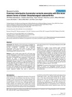
[24]. An LD plot for the SNPs studied here is presented in Fig-
ure 1 and the LD measures in Table 1. The promoter haplo-
types were reconstructed statistically from the population
genotype data by using the PHASE program with the Markov
chain method for haplotype assignments [25]. A set of logistic
regression analyses was performed to examine the associa-
tion between the IL-6 genotypes and symptomatic DIP OA.
Crude and adjusted odds ratios (ORs) and their 95% confi-
dence intervals (CIs) were calculated with the SPSS statistical
package (Statistical Package for the Social Sciences, version
14.0; SPSS Inc., Chicago, IL, USA). The ORs were adjusted
for age (in years), occupation (dentists versus teachers), BMI
(tertiles) and smoking history (never versus some time) as
potential confounders. Because the haplotype and genotype
analyses that followed the initial allelic associations were not
entirely independent tests, the P values were not corrected for
multiple testing.
Results
Clinical findings
Of the 535 successfully genotyped participants, 48 (9%)
were diagnosed as having symptomatic OA in at least two DIP
joints. The basic characteristics of the subjects by OA status
are presented in Table 2.
Genetic findings
All the genotype frequencies analyzed were in Hardy–Wein-
berg equilibrium. No statistically significant differences in the
frequencies of the genotypes or carriage rates of the IL-6 pol-
ymorphisms were observed between the two occupational
groups. Because the two occupational groups proved to be
homogeneous with regard to the polymorphisms of interest,
Figure 1
Haploview linkage disequilibrium plot of the IL-6 promoter single nucleotide polymorphisms rs1800797, rs1800796 and rs1800795Haploview linkage disequilibrium plot of the IL-6 promoter single
nucleotide polymorphisms rs1800797, rs1800796 and rs1800795.
Arthritis Research & Therapy Vol 10 No 1 Kämäräinen et al.
Page 4 of 9
(page number not for citation purposes)
they were pooled and the results presented here apply to the
whole series, except that the statistical calculations have been
adjusted for occupation.
No association was observed between DIP OA (radiographic
OA) and IL-6 promoter polymorphisms, but there was a statis-
tically significant association between the G alleles at pro-
moter positions -174 and -597 and symptomatic DIP OA
(Table 3), the G allele being seen more frequently among the
subjects with symptomatic DIP OA (P = 0.010, P
corr
= 0.020
and P = 0.012, P
corr
= 0.024, respectively). This difference
was also evident when comparing the carriers of the G allele
at polymorphic locations G-174C and G-597A, in that 67.6%
(n = 328) of the subjects without symptomatic DIP OA had at
least one G (-174) allele in comparison with 87.5% (n = 42)
of the subjects with the disease (P = 0.004), the correspond-
ing figures for the G (-597) allele being 68.2% (n = 331) and
87.2% (n = 41), respectively (P = 0.007). There were no dif-
ferences between the groups in allele frequencies or carriage
rates at the promoter location -572. At G-174C the combined
GG and GC genotypes increased the risk of the disease in
comparison with the CC genotype (P = 0.008), and similar
results were obtained for the G-597A polymorphism (P =
0.006). The genotypes at the G-572C polymorphic location
had no effect on the risk of symptomatic OA.
The three IL-6 promoter polymorphisms revealed a total of five
haplotypes. The most common was C-G-A (0.53), followed by
G-G-G (0.40), whereas the others occurred at a combined fre-
quency of only 0.07 (data not shown). No statistically signifi-
cant associations of a haplotype containing the G allele at
each locus with DIP OA and symmetrical DIP OA were
Table 1
Linkage disequilibrium measures between the studied IL-6 promoter single nucleotide polymorphisms
Measure rs1800797 and rs1800796 rs1800797 and rs1800795 rs1800796 and rs1800795
D' 1.0 (0.78–1.0) 0.968 (0.94–0.99) 1.0 (0.78–1.0)
LOD 7.53 184.12 7.79
r
2
0.045 0.885 0.048
Haplotypes AG 54.0% AC 53.3% GC 55.5%
GC 3.7% GC 2.2% CG 3.7%
GG 42.3% GG 43.8% GG 40.8%
AG 0.7%
D' – statistical measure of linkage disequilibrium (D' = 1 is known as complete linkage). The numbers in parentheses indicate 95% confidence
intervals for D'. The lod score (LOD) serves as a test of the null hypothesis of free recombination versus the alternative hypothesis of linkage. LOD
>3 is traditionally regarded as evidence for linkage.
r2 is a correlation coefficient between pairs of loci.
Table 2
Characteristics of the material
Characteristic Total Symptomatic DIP OA P
a
No Yes
Number (percentage) 535 (100) 487 (91.0) 48 (9.0)
Occupation 0.068
Dentists, n (percentage) 290 (54.2) 270 (55.4) 20 (41.7)
Teachers, n (percentage) 245 (45.8) 217 (44.6) 28 (58.3)
Age, years (mean ± SD) 53.9 ± 5.3 53.6 ± 5.2 57.7 ± 4.2 0.0001
BMI (mean ± SD) 24.5 ± 3.6 24.4 ± 3.6 25.0 ± 3.3 0.24
Smoking history 0.12
Never, n (percentage) 396 (74.0) 356 (73.1) 40 (83.3)
Some time, n (percentage) 139 (26.0) 131 (26.9) 8 (16.7)
BMI, body mass index; DIP OA, distal interphalangeal osteoarthritis. Symptomatic DIP OA was defined by the presence of both radiographic
findings of grade 2 or more and symptoms in at least two DIP joints.
a
P value for the comparison between DIP OA status groups (Student's t-test or chi-square test).
Available online />Page 5 of 9
(page number not for citation purposes)
observed (OR 1.24, 95% CI 0.85 to 1.83, and OR 1.42, 95%
CI 0.96 to 2.10; Table 4), whereas the haplotype G-G-G was
overrepresented in the women with symptomatic DIP OA in
comparison with those without the disease (0.51 versus 0.39,
P = 0.023). Analysis of the G-G-G haplotype pairs (diplo-
types) showed that the G-G-G/other diplotype was overrepre-
sented among the women with symptomatic DIP OA (70.8%
versus 45.8%, P = 0.001; Table 4). The risk of symptomatic
DIP OA was increased in the carriers of the G-G-G haplotype
(OR 4.45, 95% CI 1.82 to 10.88).
In addition, when the diplotypes for the -174 and -597 loci
were analyzed together, the findings indicated that carriage of
the G-G diplotype increased the risk of both symmetrical DIP
OA (OR 1.52, 95% CI 1.01 to 2.28) and symptomatic DIP OA
(OR 3.67, 95% CI 1.50 to 9.00; Table 5), although only the
statistically significant association between the G-G diplotype
and symptomatic DIP OA remained after correcting for multi-
ple testing.
Discussion
The present study showed an association between certain
promoter genotypes of IL-6 and the more severe outcomes of
DIP OA, namely symmetrical and symptomatic DIP OA. IL-6 is
one of the most important mediators of inflammatory reactions
in humans. At least 50 SNPs and five common haplotypes
have been identified so far in the IL-6 gene, and the genetic
variations, especially within the non-coding promoter
sequence, have been shown to have a powerful influence on
the expression of the gene [26-28]. Pain and inflammation
symptoms are known to be related to IL-6, and it was recently
reported by Oen and coworkers that the promoter genotype -
174GG has a positive correlation with pain in juvenile rheuma-
toid arthritis [19].
It has been reported previously that IL-6 production ex vivo is
greater in individuals who are homozygous for the haplotype
containing G at -597 and -174 [29]. This is interesting in the
light of the fact that both of these G alleles substantially
increased the risk of symptomatic OA in our material. Individu-
ally, the G allele of the G-174C variation has repeatedly been
Table 3
Frequency of the IL-6 (G-174C, G-572C, G-597A) genotypes, by DIP OA status
Genotype Symptomatic DIP OA P
c
Condition absent (n = 485) Condition present (n = 47–48)
n Percentage n Percentage
IL-6(G-174C)
a
0.016
GG 93 19.2 13 27.1
GC 235 48.5 29 60.4
CC 157 32.4 6 12.5
G allele carriage 328 67.6 42 87.5 0.004
G allele frequency 421 43.4 55 57.3 0.010
d
IL-6(G-572C)
b
0.362
GG 451 93.0 42 89.4
GC 34 7.0 5 10.6
C allele carriage 34 7.0 5 10.6 0.362
C allele frequency 34 3.5 5 5.3 0.382
IL-6(G-597A)
a
0.023
GG 103 21.2 14 29.8
GA 226 47.0 27 57.4
AA 154 31.8 6 12.8
G allele carriage 331 68.2 41 87.2 0.007
G allele frequency 432 44.5 55 58.5 0.012
e
DIP OA, distal interphalangeal osteoarthritis. Symptomatic DIP OA was defined by the presence of both radiographic findings of grade 2 or more
and symptoms in at least two DIP joints.
a
Genotypes were available for 533 subjects;
b
Genotypes were available for 532 subjects.
c
P value is given for the comparison between DIP OA
status groups (Chi-square test or Fisher's exact probability test);
d
P
corr
= 0.02, odds ratio 1.75 (95% confidence interval 1.15 to 2.67);
e
P
corr
=
0.024, odds ratio 1.76 (95% confidence interval 1.15 to 2.69).
Arthritis Research & Therapy Vol 10 No 1 Kämäräinen et al.
Page 6 of 9
(page number not for citation purposes)
shown to associate with increased expression and plasma
levels of IL-6 [17,30], and the same polymorphism has recently
been directly linked to hip OA, because the CC genotype was
significantly higher in the control population [18]. The three
promoter variations G-597A, G-572C and G-174C have been
shown to influence IL-6 transcription through a complex inter-
action determined by the haplotype, and the G alleles at these
loci have been found to associate with increased transcription
of IL-6 [16]. Our results strongly support this finding, because
the G-G-G haplotype was clearly overrepresented among
those with a symptomatic disease in our sample.
OA can be defined by symptoms or pathology (radiographic
features). Although osteoarthritis is regarded as a likely origin
of joint pain [31], the association between radiographic evi-
dence of OA and symptoms in the general population seems
to be rather poor, as many persons with radiographic OA do
not have any symptoms, and vice versa [32]. The American
College of Rheumatology criteria for the classification of OA
identify cases of persistent pain (most days for at least 1
month). The proportion of radiographic OA that is sympto-
matic has been estimated to be between 20% and 40%, and
most persons with radiographic OA do not have persistent
symptoms [32]. Although the American College of Rheumatol-
ogy criteria are the most frequently used definition of
symptomatic hand OA for clinical studies, their limitations for
epidemiological studies have been recognized [32]. The avail-
able evidence suggests that radiography is better than clinical
examination for defining hand OA in epidemiological studies,
and that it is possible to identify persons with clinically signifi-
cant OA by combining a radiographic criterion with self-
reported symptoms [32]. It has recently been shown in this
same population that the severity of finger joint pain is clearly
dependent on the severity of radiographic OA [33].
Table 4
Frequency of the IL-6 G-G-G diplotypes, by the DIP OA status (n = 533 to 535)
Condition Diplotypes Condition absent Condition present Odds ratio (95% confidence interval)
n Percentage n Percentage Crude Adjusted
DIP OA
Total n 309 224
other/other 117 37.9 74 33.0 1.00 1.00
G-G-G/other 148 47.9 107 47.8 1.14 (0.78–1.68) 1.17 (0.77–1.76)
G-G-G/G-G-G 44 14.2 43 19.2 1.54 (0.93–2.58) 1.48 (0.86–2.55)
G-G-G carriage 192 62.1 150 67.0 1.23 (0.86–1.77) 1.24 (0.85–1.83)
G-G-G frequency 236 38.2 193 43.1 1.22 (0.96–1.57) 1.20 (0.92–1.56)
Symmetrical DIP OA
Total n 329 205
other/other 127 38.6 64 31.2 1.00 1.00
G-G-G/other 155 47.1 101 49.3 1.29 (0.87–1.91) 1.35 (0.89–2.04)
G-G-G/G-G-G 47 14.3 40 19.5 1.69 (1.01–2.83) 1.65 (0.95–2.84)
G-G-G carriage 202 61.4 141 68.8 1.38 (0.96–2.00) 1.42 (0.96–2.10)
G-G-G frequency 229 35.9 181 42.1 1.30 (1.01–1.67)
a
1.29 (0.99–1.68)
Symptomatic DIP OA
Total n 487 48
other/other 185 38.0 6 35.7 1.00 1.00
G-G-G/other 223 45.8 34 70.8 4.70 (1.93–11.44)
a
5.03 (2.02–12.51)
a
G-G-G/G-G-G 79 16.2 8 16.7 3.12 (1.05–9.29) 3.01 (0.98–9.22)
G-G-G carriage 302 62.0 42 87.5 4.29 (1.79–10.28)
a
4.45 (1.82–10.88)
a
G-G-G frequency 381 39.1 50 51.0 1.83 (1.19–2.81)
a
1.62 (1.07–2.45)
a
Distal interphalangeal osteoarthritis (DIP OA) was defined by the presence of radiographic findings of grade 2 or more in at least two DIP joints.
Symmetrical DIP OA was defined by the presence of radiographic findings of grade 2 or more in at least one symmetrical pair of the DIP joints.
Symptomatic DIP OA was defined by the presence of both radiographic findings of grade 2 or more and symptoms in at least two DIP joints.
a
P value remained statistically significant after correcting for multiple testing.
Available online />Page 7 of 9
(page number not for citation purposes)
We sought here to examine more severe cases of OA, those
that are more likely to have a genetic component. All the
subjects were uniformly assessed for the presence of radio-
graphic DIP OA and the joint-specific occurrence of symp-
toms. The outcome of symptomatic DIP OA in at least two
joints was chosen in view of its assumed clinical relevance and
its specificity in terms of joint location.
Although our results are supported by the findings of several
functional studies of IL-6 gene transcription [15,16,28,29,34],
there are also negative results concerning the contribution of
promoter variability [35]. One explanation for this could be the
effect of aging, which may overwhelm the genetic effect on the
IL-6 levels. There is substantial evidence that the increase in
IL-6 serum levels with age results in part from the loss of sex
steroids such as estrogen, testosterone and dehydroepian-
drosterone [36,37], which have an important role in blocking
transcription of the IL-6 gene, so that their loss at menopause
may have a more conspicuous role than the genotype.
Our findings do not suggest an association between the
development of asymptomatic DIP OA and the IL-6 gene,
because the risk of radiographic OA was not affected by the
IL-6 genotype. The number of individuals with radiographic
DIP OA having symptoms turned out to be relatively low com-
pared with the total number of subjects, which reduced the
power of the results, so that replication with a larger sample
would be beneficial. In addition, a cross-sectional study setting
may result in difficulties in accurately estimating the true risk of
DIP OA associated with IL-6 promoter variants. The experi-
encing and evaluation of symptoms, particularly pain, are
always individual, purely subjective and likely to fluctuate with
time. It should be noted that a self-administered questionnaire
cannot preclude other causes of joint pain in addition to DIP
OA. In contrast, asymptomatic periods of variable duration are
Table 5
Frequency of IL-6 G-G diplotypes (G-597A; G-174C) by DIP OA status (n = 528 to 530)
Condition Diplotypes Condition absent Condition present Odds ratio (95% confidence interval)
n Percentage n Percentage Crude Adjusted
DIP OA
Total n 306 222
other/other 105 34.3 64 28.8 1.00 1.00
G-G/other 147 48.0 109 49.1 1.22 (0.82–1.81) 1.27 (0.83–1.95)
G-G/G-G 54 17.6 49 22.1 1.49 (0.91–2.44) 1.42 (0.84–2.41)
G-G carriage 192 62.1 150 67.0 1.29 (0.89–1.87) 1.31 (0.88–1.96)
G-G frequency 237 39.9 207 44.8 1.22 (0.96–1.56) 1.19 (0.92–1.55)
Symmetrical DIP OA
Total n 325 204
other/other 114 35.1 55 27.0 1.00 1.00
G-G/other 155 47.7 102 50.0 1.36 (0.91–2.05) 1.45 (0.94–2.23)
G-G/G-G 56 17.2 47 23.0 1.74 (1.05–2.88) 1.69 (0.99–2.87)
G-G carriage 302 62.0 42 87.5 1.46 (1.00–2.15) 1.52 (1.01–2.28)
G-G frequency 267 41.1 196 48.0 1.33 (1.03–1.70) 1.31 (1.01–1.71)
Symptomatic DIP OA
Total n 483 47
other/other 163 33.7 6 12.8 1.00 1.00
G-G/other 230 47.6 28 59.6 3.31 (1.34–8.17)
a
3.62 (1.43–9.15)
a
G-G/G-G 90 18.8 13 27.7 3.92 (1.44–10.68)
a
3.79 (1.35–10.61)
a
G-G carriage 302 62.0 42 87.5 3.48 (1.45–8.37)
a
3.67 (1.50–9.00)
a
G-G frequency 410 42.4 54 57.4 1.83 (1.19–2.81)
a
1.62 (1.07–2.45)
Distal interphalangeal osteoarthritis (DIP OA) was defined by the presence of radiographic findings of grade 2 or more in at least two DIP joints.
Symmetrical DIP OA was defined by the presence of radiographic findings of grade 2 or more in at least one symmetrical pair of the DIP joints.
Symptomatic DIP OA was defined by the presence of both radiographic findings of grade 2 or more and symptoms in at least two DIP joints.
a
P value remained statistically significant after correcting for multiple testing.
Arthritis Research & Therapy Vol 10 No 1 Kämäräinen et al.
Page 8 of 9
(page number not for citation purposes)
typical of osteoarthritis, and the present subjects were
prompted to report symptoms that had occurred within the
previous 30 days. It is therefore possible that the number of
symptomatic subjects in our sample is lower than it should be,
as a result of the exclusion of those who were going through
an asymptomatic period at the time of answering the question-
naire. The association between IL-6 and symptomatic DIP OA
may therefore be even stronger than that reported here. A
strict time scale for the occurrence of reported symptoms
combined with the radiological analysis should reduce the
amount of bias caused by temporary, indistinct joint symptoms
not caused by DIP OA. In general, this work underlines the
importance of a homogeneous study population with a spe-
cific outcome formulation, to avoid allowing the modest
genetic contribution to be overwhelmed by the clinical diver-
sity of the subjects.
Conclusion
Our findings lend support to the notion of an association
between promoter variations in the IL-6 gene and sympto-
matic and symmetrical DIP OA, outcomes that can be pre-
sumed to be of high clinical relevance. It may be possible in
future to make therapeutic use of the knowledge of IL-6 and its
significance as a cause of inflammation and pain, in treating
symptoms of arthritis. Specific IL-6 receptor antagonists inhib-
iting the inflammation cascade within the articular cartilage are
a relevant option when designing new therapeutic interven-
tions for this disease.
Competing interests
The authors declare that they have no competing interests.
Authors' contributions
OK conducted the molecular genetic studies and drafted the
manuscript. SS participated in the design of the study, per-
formed the statistical analysis and participated in writing the
manuscript. TV and KL conducted the radiological assess-
ment. HR, LA, MM and PL participated in the design and coor-
dination of this study and participated in writing the
manuscript. All authors read and approved the final
manuscript.
Acknowledgements
The authors wish to thank all the participants in this study. Ms Aira Harju
at the Department of Medical Biochemistry and Molecular Biology, Uni-
versity of Oulu, is gratefully acknowledged for technical assistance. This
work was financially supported by the Finnish Work Environment Fund
and the Academy of Finland.
References
1. Spector TD, Cicuttini F, Baker J, Loughlin J, Hart D: Genetic influ-
ences on osteoarthritis in women: a twin study. BMJ 1996,
312:940-943.
2. Spector TD, MacGregor AJ: Risk factors for osteoarthritis:
genetics. Osteoarthritis Cartilage 2004, 12:39-44.
3. Spencer JM, Loughlin J, Clipsham K, Carr AJ: Genetic back-
ground increases the risk of hip osteoarthritis. Clin Orthop
Relat Res 2005, 431:134-137.
4. Loughlin J: The genetic epidemiology of human primary oste-
oarthritis: current status. Expert Rev Mol Med 2005, 7:1-12.
5. Felson DT: Epidemiology of hip and knee osteoarthritis. Epide-
miol Rev 1988, 10:1-28.
6. Solovieva S, Vehmas T, Riihimäki H, Luoma K, Leino-Arjas P: Hand
use and patterns of joint involvement in osteoarthritis. A com-
parison of female dentists and teachers. Rheumatology 2005,
44:521-528.
7. Kaprio J, Kujala UM, Peltonen L, Koskenvuo M: Genetic liability to
osteoarthritis may be greater in women than men. BMJ 1996,
313:232.
8. Hunter DJ, Demissie S, Cupples LA, Aliabadi P, Felson DT: A
genome scan for joint-specific hand osteoarthritis susceptibil-
ity: The Framingham Study. Arthritis Rheum 2004,
50:2489-2496.
9. Felson DT, Lawrence RC, Dieppe PA, Hirsch R, Helmick CG, Jor-
dan JM, Kington RS, Lane NE, Nevitt Mc, Zhang Y, Sowers M,
McAlindon T, Spector TD, Poole AR, Yanovski SZ, Ateshian G,
Sharma L, Buckwalter JA, Brandt KD, Fries JF: Osteoarthritis:
new insights. Part 1: the disease and its risk factors. Ann Intern
Med 2000, 133:635-646.
10. Jones G, Cooley HM, Stankovich JM: A cross sectional study of
the association between sex, smoking, and other lifestyle fac-
tors and osteoarthritis of the hand. J Rheumatol 2002,
29:1719-1724.
11. Leppävuori J, Kujala U, Kinnunen J, Kaprio J, Nissilä M, Heliövaara
M, Klinger M, Partanen J, Terwilliger JD, Peltonen L: Genome scan
for predisposing loci for distal interphalangeal joint osteoar-
thritis: evidence for a locus on 2q. Am J Hum Genet 1999,
65:
1060-1067.
12. Kaneko S, Satoh T, Chiba J, Ju C, Inoue K, Kagawa J: Interleukin-
6 and interleukin-8 levels in serum and synovial fluid of
patients with osteoarthritis. Cytokines Cell Mol Ther 2000,
6:71-79.
13. Jikko A, Wakisaka T, Iwamoto M, Hiranuma H, Kato Y, Maeda T,
Fujishita M, Fuchihata H: Effects of interleukin-6 on proliferation
and proteoglycan metabolism in articular chondrocyte
cultures. Cell Biol Int 1998, 22:615-621.
14. Papassotiropoulos A, Hock C, Nitsch RM: Genetics of interleukin
6: implications for Alzheimer's disease. Neurobiol Aging 2001,
22:863-871.
15. Fishman D, Faulds G, Jeffery R, Mohamed-Ali V, Yudkin JS, Hum-
phries S, Woo P: The effect of novel polymorphisms in the
interleukin-6 (IL-6) gene on IL-6 transcription and plasma IL-6
levels, and an association with systemic-onset juvenile
chronic arthritis. J Clin Invest 1998, 102:1369-1376.
16. Terry CF, Loukaci V, Green FR: Cooperative influence of genetic
polymorphisms on interleukin 6 transcriptional regulation. J
Biol Chem 2000, 275:18138-18144.
17. Noponen-Hietala N, Virtanen I, Karttunen R, Schwenke S, Jakkula
E, Li H, Merikivi R, Barral S, Ott J, Karppinen J, Ala-Kokko L:
Genetic variations in IL6 associate with intervertebral disc dis-
ease characterized by sciatica. Pain 2005, 114:186-194.
18. Pola E, Papaleo P, Pola R, Gaetani E, Tamburelli FC, Aulisa L,
Logroscino CA: Interleukin-6 gene polymorphism and risk of
osteoarthritis of the hip: a case-control study. Osteoarthritis
Cartilage 2005, 13:1025-1028.
19. Oen K, Malleson PN, Cabral DA, Rosenberg AM, Petty RF, Nicker-
son P, Reed M: Cytokine genotypes correlate with pain and
radiologically defined joint damage in patients with juvenile
rheumatoid arthritis. Rheumatology 2005, 44:1115-1121.
20. Sommer C, Kress M: Recent findings on how proinflammatory
cytokines cause pain: peripheral mechanisms in inflammatory
and neuropathic hyperalgesia. Neurosci Lett 2004,
361:184-187.
21. De Jongh RF, Vissers KC, Meert TF, Booij LH, De Deyne CS, Hey-
len RJ: The role of interleukin-6 in nociception and pain.
Anesth
Analg 2003, 96:1096-1103.
22. Kellgren JH, Lawrence JS: Radiologic assessment of
osteoarthritis. Ann Rheum Dis 1957, 16:494-502.
23. Cohen J: Weighted kappa. Nominal scale agreement with pro-
vision for scaled disagreement or partial credit. Psychol Bull
1968, 70:213-220.
24. Barrett JC, Fry B, Maller J, Daly MJ: Haploview: analysis and vis-
ualization of LD and haplotype maps. Bioinformatics 2005,
21:263-265.
Available online />Page 9 of 9
(page number not for citation purposes)
25. Stephens M, Smith NJ, Donnelly P: A new statistical method for
haplotype reconstruction from population data. Am J Hum
Genet 2001, 68:978-989.
26. Osiri M, McNicholl J, Moreland LW, Bridges SL Jr: A novel single
nucleotide polymorphism and five probable haplotypes in the
5' flanking region of the IL-6 gene in African-Americans.
Genes Immun 1999, 1:166-167.
27. Jordanies N, Eskdale J, Stuart R, Gallagher G: Allele associations
reveal four prominent haplotypes at the human interleukin-6
(IL-6) locus. Genes Immun 2000, 1:451-455.
28. Ota N, Nakajima T, Nakazawa I, Suzuki T, Hosoi T, Orimo H, Inoue
S, Shirai Y, Emi M: A nucleotide variant in the promoter region
of the interleukin-6 gene associated with decreased bone
mineral density. J Hum Genet 2001, 46:267-272.
29. Rivera-Chavez FA, Peters-Hybki DL, Barber RC, O'Keefe GE:
Interleukin-6 promoter haplotypes and interleukin-6 cytokine
responses. Shock 2003, 20:218-223.
30. Bennermo M, Held C, Stemme S, Ericsson CG, Silveira A, Green
F, Tornvall P: Genetic predisposition of the interleukin-6
response to inflammation: implications for a variety of major
diseases? Clin Chem 2004, 50:2136-2140.
31. Dieppe PA, Lohmander LS: Pathogenesis and management of
pain in osteoarthritis. Lancet 2005, 365:965-973.
32. Hart DJ, Spector TD: Definition and epidemiology of osteoar-
thritis of the hand: a review. Osteoarthritis Cartilage
2000:S2-S7.
33. Ding H, Solovieva S, Vehmas T, Riihimäki H, Leino-Arjas P: Finger
joint pain in relation to radiographic osteoarthritis and joint
location – a study of middle-aged female dentists and
teachers. Reumatology 2007, 46:1502-1505.
34. Hulkkonen J, Pertovaara M, Antonen J, Pasternack A, Hurme M:
Elevated interleukin-6 plasma levels are regulated by the pro-
moter region polymorphism of the IL6 gene in primary
Sjogren's syndrome and correlate with the clinical manifesta-
tions of the disease. Rheumatology 2001, 40:656-661.
35. Walston J, Arking DE, Fallin D, Li T, Beamer B, Xue Q, Ferrucci L,
Fried LP, Chakravarti A: IL-6 gene variation is not associated
with increased serum levels of IL-6, muscle, weakness, or
frailty in older women. Exp Gerontol 2005, 40:344-352.
36. Ershler WB, Keller ET: Age-associated increased interleukin-6
gene expression, late-life diseases, and frailty. Annu Rev Med
2000, 51:245-270.
37. Pfeilschifter J, Koditz R, Pfohl M, Schatz H: Changes in proinflam-
matory cytokine activity after menopause. Endocr Rev 2002,
23:90-119.