Báo cáo y học: "Diacerein inhibits the synthesis of resorptive enzymes and reduces osteoclastic differentiation/survival in osteoarthritic subchondral bone: a possible mechanism for a protective effect against subchondral bone remodelling" ppsx
Bạn đang xem bản rút gọn của tài liệu. Xem và tải ngay bản đầy đủ của tài liệu tại đây (548.5 KB, 10 trang )
Open Access
Available online />Page 1 of 10
(page number not for citation purposes)
Vol 10 No 3
Research article
Diacerein inhibits the synthesis of resorptive enzymes and
reduces osteoclastic differentiation/survival in osteoarthritic
subchondral bone: a possible mechanism for a protective effect
against subchondral bone remodelling
Christelle Boileau, Steeve Kwan Tat, Jean-Pierre Pelletier, Saranette Cheng and Johanne Martel-
Pelletier
Osteoarthritis Research Unit, University of Montreal Hospital Centre, Notre-Dame Hospital, 1560 Sherbrooke Street East, Montreal, Quebec, H2L
4M1, Canada
Corresponding author: Johanne Martel-Pelletier,
Received: 9 Apr 2008 Revisions requested: 29 May 2008 Revisions received: 5 Jun 2008 Accepted: 25 Jun 2008 Published: 25 Jun 2008
Arthritis Research & Therapy 2008, 10:R71 (doi:10.1186/ar2444)
This article is online at: />© 2008 Boileau et al.; licensee BioMed Central Ltd.
This is an open access article distributed under the terms of the Creative Commons Attribution License ( />),
which permits unrestricted use, distribution, and reproduction in any medium, provided the original work is properly cited.
Abstract
Introduction Subchondral bone alterations represent an
essential component of osteoarthritis (OA). Modifying the
abnormal subchondral bone metabolism may be indicated to
treat OA. We investigated the effect of diacerein and rhein on
the changes occurring in subchondral bone during OA. To this
end, we determined the drugs' effects on metalloprotease-13
(MMP-13) synthesis on subchondral bone and on the osteoblast
signalling pathways. In osteoclasts, we studied MMP-13 and
cathepsin K production as well as cell differentiation,
proliferation, and survival.
Methods The effect of diacerein/rhein on the production of
subchondral bone MMP-13 was determined by enzyme-linked
immunosorbent assay. Signalling pathways were evaluated on
osteoblasts by Western blot. Osteoclast experiments were
performed using cells from the pre-osteoclastic murine cell line
Raw 264.7. Osteoclast MMP-13 and cathepsin K activities were
determined by specific bioassays and differentiation of these
cells quantified by tartrate-resistant acid phosphatase staining.
Results Diacerein and rhein reduced, in a dose-dependent
manner, the interleukin-1-beta (IL-1β)-induced MMP-13
production in OA subchondral bone. This effect occurred
through the inhibition of ERK1/2 (extracellular signal-regulated
kinase-1/2) and p38. In osteoclasts, they significantly reduced
the activity of MMP-13 and cathepsin K. Moreover, these drugs
effectively blocked the IL-1β effect on the osteoclast
differentiation process and the survival of mature osteoclasts.
Conclusion Altogether, these data suggest that diacerein/rhein
could impact the abnormal subchondral bone metabolism in OA
by reducing the synthesis of resorptive factors and osteoclast
formation.
Introduction
Osteoarthritis (OA) is considered a complex illness. Although
we may not yet completely know all of the initiating factors
involved in the degeneration of the articular tissues, significant
progress regarding the etiopathogenesis of this disease has
been made. For decades, the prevailing concept has centered
on the destruction of the articular cartilage. There is now sub-
stantial evidence not merely that alterations in the subchondral
bone metabolism are secondary manifestations of OA, but that
they comprise an integral component of the disease, and data
suggest a key role played by the subchondral bone in the initi-
ation and/or progression of articular tissue degeneration.
Several reports have indicated that the subchondral bone
remodelling that occurs during OA involves both bone resorp-
tion and bone formation. Studies allowing chronological eval-
ELISA = enzyme-linked immunosorbent assay; ERK1/2 = extracellular signal-regulated kinase-1/2; FBS = fetal bovine serum; IL = interleukin; JNK =
c-jun N-terminal kinase; MAP = mitogen-activated protein; MMP = metalloprotease; NSAID = non-steroidal anti-inflammatory drug; OA = osteoarthri-
tis; PBS = phosphate-buffered saline; PGE
2
= prostaglandin E
2
; RANKL = receptor activator of nuclear factor-κB ligand; SAPK = stress-activated
protein kinase; TRAP = tartrate-resistant acid phosphatase; TTBS = Tris 20 mM, NaCl 150 mM, pH 7.5, and 0.1% Tween 20.
Arthritis Research & Therapy Vol 10 No 3 Boileau et al.
Page 2 of 10
(page number not for citation purposes)
uation in animal models have suggested a predominance of
bone formation in the more advanced stage of the disease [1-
4], while, in contrast, the remodelling in the early phase favors
bone resorption [4-6]. This latter finding agrees with the study
of Bettica and colleagues [7], who demonstrated that, in vivo
in humans, general bone resorption is increased in patients
with progressive knee OA. Similarly, Messent and colleagues
[8], with the use of fractal signature analysis, showed that
bone loss occurred in patients with knee OA and that changes
were associated with an increase in the number and size of the
remodelling units.
In vitro studies have also demonstrated that the subchondral
bone is the site of several dynamic morphological changes
that appear to be part of the OA process. These changes are
allied with many local abnormal biochemical pathways, includ-
ing the increased synthesis of several bone markers, growth
factors, cytokines, proteases, and inflammatory mediators. The
levels of alkaline phosphatase, osteocalcin, type I collagen,
interleukin (IL)-6, transforming growth factor-beta, prostaglan-
din E
2
(PGE
2
), leukotriene B
4
, and proteases, including uroki-
nase, cathepsin K, and the metalloprotease (MMP)-13, have all
been found to be elevated in human OA subchondral bone
osteoblasts [6,9-13].
The pharmacological treatments for OA are centered mainly
on the use of analgesics and non-steroidal anti-inflammatory
drugs (NSAIDs). These are symptomatic agents that, thus far,
have been shown to be solely capable of relieving the signs
and symptoms of the disease. Due primarily to recent progress
in understanding the disease, new approaches for the treat-
ment of OA are now being explored. Compounds that inhibit
one or more OA disease processes are under evaluation for
their potential to alter the degenerative changes. As the
subchondral bone alterations also appear to contribute to car-
tilage deterioration [14], therapeutic strategies aimed at mod-
ifying the abnormal metabolism of the subchondral bone cells
may have significant impact on the treatment of OA.
Diacerein, a drug of the anthraquinone class, has rhein as its
active metabolite. In chondrocytes, this drug acts on the IL-1β
system, reducing the level of this cytokine as well as downreg-
ulating the IL-1β-induced inflammatory pathways and cartilage
breakdown in OA [15-18]. On human subchondral bone oste-
oblasts, data showed that diacerein/rhein reduces osteocal-
cin, urokinase, and IL-6, factors that would contribute to
curbing bone formation/resorption [19].
This study aims at providing a more complete and comprehen-
sive understanding of the effects of diacerein/rhein on OA
subchondral bone metabolism and cells (osteoblasts and
osteoclasts). As bone resorption is mediated by several proc-
esses, including the synthesis of proteases that can induce
matrix degradation and osteoclast differentiation and prolifera-
tion, our study aimed first to investigate the effects of diac-
erein/rhein on the synthesis of major proteases involved in
bone remodelling/resorption, namely MMP-13 and cathepsin
K. Moreover, we sought to gain new insight into the effects of
the drug on the bone resorptive process.
Materials and methods
Specimen selection
Subchondral bone was obtained from OA patients who had
undergone total knee replacement surgery. Specimens were
taken from weight-bearing areas of the femoral condyles.
Subchondral bone specimens were dissected away from the
remaining cartilage and trabecular bone under sterile condi-
tions as previously described [9,10]. A total of 16 patients (72
± 9 years old, mean ± standard deviation; 6 males and 10
females) classified as having OA according to recognized
American College of Rheumatology clinical criteria were
included in this study [20]. At the time of surgery, the patients
had symptomatic disease requiring medical treatment in the
form of acetaminophen, NSAIDs, or selective cyclooxygenase-
2 inhibitors. None had received intra-articular steroid injec-
tions within 3 months prior to surgery, and none had received
medication that would interfere with bone metabolism. The
institutional Ethics Committee Board of the University of Mon-
treal Hospital Centre approved the use of the human articular
tissues.
Subchondral bone tissue explant
Culture conditions
Subchondral bone explants of about 5 × 3 mm were placed in
24-well plates containing BGJb medium (Invitrogen Life Tech-
nologies, Burlington, ON, Canada) supplemented with 2%
fetal bovine serum (FBS) (Invitrogen Life Technologies) and an
antibiotic mixture (100 units per milliliter penicillin base and
100 μg/mL streptomycin base) (Multicell; Wisent, St-Bruno,
QC, Canada). The explants were treated with or without IL-1β
(5 ng/mL) and therapeutic concentrations of diacerein (10 or
20 μg/mL) or rhein (10 or 20 μg/mL) for 5 days at 37°C in a
humidified atmosphere of 5% CO
2
/95% air. At the end of the
incubation period, culture medium was collected and MMP-13
levels were determined using a specific enzyme-linked immu-
nosorbent assay (ELISA). The MMP-13 ELISA was from Amer-
sham Biosciences (now part of GE Healthcare Bio-Sciences
Inc., Baie-d'Urfé, QC, Canada) and recognized both the pro
and active forms of the enzyme, the sensitivity being 32 pg/mL.
The level was expressed as a fold expression compared with
the IL-1β, which was assigned a value of 1.
Immunostaining
Subchondral bone explants were fixed as previously described
[6] in Tissufix #2 (Chaptec, Montreal, QC, Canada), decalci-
fied in Rapid Bone Decalcifier RDO (Apex Engineering Prod-
ucts Corporation, Aurora, IL, USA), and embedded in paraffin.
Sections (5 μm) were placed on Superfrost Plus slides (Fisher
Scientific, Nepean, ON, Canada). Slides were deparaffinized
in toluene, rehydrated in a reverse-graded series of ethanol,
Available online />Page 3 of 10
(page number not for citation purposes)
and pre-incubated with chondroitinase ABC (0.25 U/mL;
Sigma-Aldrich, St. Louis, MO, USA) in phosphate-buffered
saline (PBS) (pH 8.0) for 60 minutes at 37°C. Subsequently,
the specimens were washed in PBS, placed in 0.3% TritonX-
100 in PBS for 20 minutes and in 3% hydrogen peroxide/PBS
for another 15 minutes. Slides were further incubated with a
blocking serum (Vectastain ABC kit; Vector Laboratories Inc.,
Burlingame, CA, USA) for 60 minutes, blotted, and then over-
laid with the primary antibody against MMP-13 (15 μg/mL;
R&D Systems, Minneapolis, MN, USA) or cathepsin K (1 μg/
mL; Novocastra, now part of Leica Microsystems, Wetzlar,
Germany) for 18 hours at 4°C in a humidified chamber. Each
slide was washed three times in PBS (pH 7.4) and stained
using the avidin-biotin complex method (Vectastain ABC kit).
The color was developed with 3, 3'-diaminobenzidine (DAB)
(DAKO Diagnostics Canada Inc., Mississauga, ON, Canada)
containing hydroxide peroxide. Slides were counterstained
with hematoxylin/eosin.
The staining specificity of the antibody used was determined
using three controls according to the same experimental pro-
tocol: (a) use of absorbed immune serum (1 hour at 37°C) with
a 20-fold molar excess of human recombinant MMP-13 (R&D
Systems) or cathepsin K (Calbiochem, now part of EMD Bio-
sciences, Inc., San Diego, CA, USA), (b) omission of the pri-
mary antibody, and (c) substitution of the primary antibody with
an autologous pre-immune serum. These controls showed
only background staining.
Subchondral bone osteoblasts
Culture
Subchondral bone osteoblasts were prepared as previously
described following a collagenase digestion procedure [9,10].
Briefly, subchondral bone specimens were digested by
sequential collagenase type I digestion, followed by cell cul-
ture in BGJb medium containing 20% FBS. At confluence, pri-
mary osteoblasts were split once into 24-well plates at a final
cell density of 50,000 cells per square centimeter. Cells were
fed with BGJb medium, supplemented with an antibiotic mix-
ture (100 U/mL penicillin and 100 μL/mL streptomycin; Multi-
cell) and 10% FBS until confluence. Only first passaged cells
were employed.
Signalling pathway experiments were conducted on osteob-
lasts pre-treated by therapeutic concentrations of diacerein or
rhein at 20 μg/mL for 2 hours and treated with IL-1β (100 pg/
mL) for an additional 30 minutes. The levels of the phosphor-
ylated mitogen-activated protein (MAP) kinases, extracellular
signal-regulated kinase-1/2 (ERK1/2), p38, and stress-acti-
vated protein kinase/c-jun N-terminal kinase (SAPK/JNK) (p46
and p54) were determined on the cell lysate by Western blot
as described below.
Western blotting
Total proteins were extracted with 0.5% SDS (Invitrogen Life
Technologies) supplemented with protease inhibitors. The
protein level was determined using the bicinchoninic acid pro-
tein assay, and 10 μg of the protein was electrophoresed on a
12% SDS gel polyacrylamide. The proteins were transferred
electrophoretically onto a nitrocellulose membrane (Bio-Rad
Laboratories [Canada] Ltd., Mississauga, ON, Canada) for 1
hour at 4°C. The efficiency of transfer was controlled by a brief
staining of the membrane with Ponceau red and destained in
water and TTBS 1× (Tris 20 mM, NaCl 150 mM, pH 7.5, and
0.1% Tween 20) before immunoblotting.
The membranes were incubated overnight at 4°C with 5%
skimmed milk in SuperBlock
®
Blocking Buffer in Tris-Buffered
Saline (Pierce, Rockford, IL, USA) or in TTBS 1× only. The
membranes were then washed once with TTBS 1× for 10 min-
utes and incubated in SuperBlock
®
Blocking Buffer and TTBS
1× (Superblock
®
1:10 with TTBS 1×) with a mouse anti-phos-
pho ERK1/2 (dilution: 1:2,000; Thr 202/Tyr 204; Cell Signal-
ing Technology, Inc., Danvers, MA, USA), a rabbit polyclonal
anti-phospho p38 (dilution: 1:500; Thr 180/Thr 182; Bio-
source, Nivelles, Belgium), and a mouse anti-phospho SAPK/
JNK (dilution: 1:1,000; Thr 183/Tyr 185; New England
Biolabs Ltd., Pickering, ON, Canada) overnight at 4°C. The
membranes were washed with TTBS 1× and incubated for 1
hour at room temperature with the second antibody. The sec-
ondary antibodies were anti-mouse or anti-rabbit IgG (dilution:
1:50,000; Pierce). They were then washed again with TTBS
1×. Detection was performed by chemiluminescence using
the Super Signal
®
ULTRA chemiluminescent substrate
(Pierce) and exposure to Kodak Biomax photographic film (GE
Healthcare Bio-Sciences Inc.). The band intensity was meas-
ured by densitometry using TotalLab TL100 Software (Nonlin-
ear Dynamics Ltd, Newcastle upon Tyne, UK), and data are
expressed as fold difference with respect to the IL-1β control,
which was assigned a value of 1.
Osteoclasts from Raw 264.7 cells
Culture conditions
Raw 264.7 cells (American Type Culture Collection, Manas-
sas, VA, USA) were seeded at a density of 10,000 cells per
well in 24-well plates with Dulbecco's modified Eagle's
medium culture medium (Multicell) supplemented with the
antibiotic mixture, 10% FBS, and 1% sodium pyruvate (Multi-
cell). Cells were treated with receptor activator of nuclear fac-
tor-κB ligand (RANKL) (100 ng/mL; R&D Systems) from the
first day of culture and for the entire duration of the experiment.
RANKL allows the pre-osteoclast Raw 264.7 cells to differen-
tiate into mature osteoclasts after 5 days of culture. The cul-
ture medium was changed every 2 days.
On the fifth day, RANKL-treated cells were co-incubated with
or without IL-1β (100 pg/mL) and therapeutic concentrations
of diacerein or rhein at 10 or 20 μg/mL for 2 days at 37°C in
Arthritis Research & Therapy Vol 10 No 3 Boileau et al.
Page 4 of 10
(page number not for citation purposes)
a humidified atmosphere of 5% CO
2
/95% air. At the end of
the incubation period, the conditioned medium served for
MMP-13 determination, and cathepsin K determination was
carried out on the cell lysates. Quantification of mature osteo-
clasts was also performed on other cell cultures under the
same experimental conditions. At the end of the incubation
period, the osteoclasts were fixed with citrate/acetone solu-
tion and stained for tartrate-resistant acid phosphatase
(TRAP) according to the manufacturer's recommendation
(Sigma-Aldrich). Osteoclast formation was quantified by
counting, under a light microscope, the newly differentiated
multinucleated TRAP-positive cells containing at least three
nuclei.
Determination of functional metalloprotease-13 and
cathepsin K
Since MMP-13 and cathepsin K are produced by the murine
cell line Raw 264.7, proteins not recognized by the commer-
cially available ELISAs, determinations were performed using
activity assays specific for each protease. Functional MMP-13
levels were determined using the MMP-13 activity assay
(Chemicon International, Temecula, CA, USA) according to
the manufacturer's instructions. MMP-13 was activated by
APMA (p-aminophenyl mercuric acetate) (0.5 mM) at 37°C for
60 minutes prior to the assay. For cathepsin K, the cell lysates
were harvested in the specific assay buffer according to the
manufacturer's instructions and the levels were determined
using the Bioassay™ assay (United States Biological Inc.,
Swampscott, MA, USA). Data are expressed as fold difference
with respect to the IL-1β, which was assigned a value of 1.
Mature osteoclast survival
Raw 264.7 cells were treated with RANKL (100 ng/mL) from
the first day of culture and for the entire duration of the exper-
iment. The culture medium was changed every 2 days. On the
fifth day, RANKL-treated cells were incubated with or without
IL-1β (100 pg/mL) and therapeutic concentrations of diac-
erein or rhein at 20 μg/mL for 2 days. At the end of the incu-
bation period, the osteoclasts were fixed and the number of
TRAP-positive cells was determined as described above.
Results were calculated as the number of differentiated oste-
oclasts per well.
Osteoclast differentiation and proliferation
Raw 264.7 cells were treated with RANKL (100 ng/mL) as
well as with IL-1β (100 pg/mL) and therapeutic concentrations
of diacerein or rhein at 20 μg/mL from the first day of culture
and for the entire duration of the experiment. The culture
medium was changed every 2 days. On the seventh day, the
osteoclasts were fixed and the number of TRAP-positive multi-
nucleated cells was determined as described above.
Statistical analysis
Results were expressed as the mean ± standard error of the
mean of independent specimens, and assays were performed
in duplicate. Statistical analysis was performed using the two-
tailed paired Student t test, and a difference of less than or
equal to 0.05 was considered significant.
Results
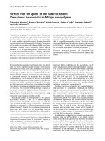
Subchondral bone immunostaining
To verify the production of MMP-13 and cathepsin K in human
OA subchondral bone, immunostaining for each of these two
proteases was performed. Data (n = 3) revealed that both pro-
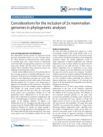
Figure 1
Representative immunohistochemical staining section for (a) metalloprotease-13 (MMP-13) and (b) cathepsin K in human osteoarthritis subchon-dral boneRepresentative immunohistochemical staining section for (a) metalloprotease-13 (MMP-13) and (b) cathepsin K in human osteoarthritis subchon-
dral bone. MMP-13 was detected in the osteoblasts (Ob) as well as in the osteoclasts (Oc). Cathepsin K was detected only in osteoclasts. Original
magnification, ×100.
Available online />Page 5 of 10
(page number not for citation purposes)
teases are produced and that MMP-13 was detected in both
osteoblasts and osteoclasts, whereas cathepsin K was
detected only in osteoclasts (Figure 1).
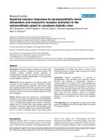
Effect of diacerein/rhein on metalloprotease-13
synthesis in osteoarthritis subchondral bone
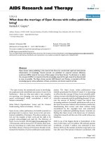
As illustrated in Figure 2, the synthesis of MMP-13 in subchon-
dral bone explants (n = 5 to 9) was significantly upregulated
by IL-1β. Diacerein and rhein reduced, in a dose-dependent
manner, the production of the IL-1β-induced MMP-13. The
effect reached statistical significance with the highest tested
dose (20 μg/mL).
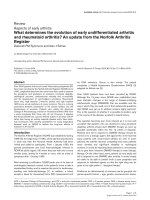
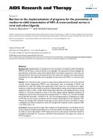
Effect of diacerein/rhein on intracellular signalling
pathways
To gain insight into the mechanisms of these drugs on the OA
subchondral bone osteoblasts, we further studied the effect of
the therapeutic concentration of these drugs, 20 μg/mL, on
the major intracellular signalling pathways pertinent to OA
pathology. On OA subchondral bone osteoblasts, data (n = 3
to 4) showed that, while IL-1β activated the ERK1/2 and p38
pathways (Figures 3a and 3b, respectively), diacerein and
rhein both significantly inhibited the phosphorylation levels of
ERK1/2 (Figure 3a) and both decreased the p38 phosphoryla-
tion with statistical significance reached for rhein. IL-1β also
markedly increased the SAPK/JNK (p46 and p54), particularly
the level of the p46 isoforms. Diacerein and rhein, however,
had no effect on the activation level of either kinase.
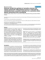
Effect of diacerein and rhein on metalloprotease-13 and
cathepsin K in osteoclasts
To better document and discriminate the effect of diacerein
and rhein on the different bone cell populations, further exper-
iments were performed on osteoclasts. To this end, a pre-oste-
oclastic murine cell line, Raw 264.7, was used. These cells,
upon stimulation by RANKL, differentiate into multinucleated
TRAP-positive osteoclasts [21,22]. As illustrated in Figure 4,
stimulation with IL-1β had no effect on the level of MMP-13
produced by Raw 264.7 cells (n = 8). Diacerein and rhein at
both concentrations (10 and 20 μg/mL) significantly inhibited
the MMP-13 level (Figure 4a). The intracellular level of cathe-
psin K was not stimulated by IL-1β (n = 8) (Figure 4b). Data
showed that both diacerein and rhein significantly decreased
the protease activity level in a dose-dependent manner.
Effect of diacerein and rhein on osteoclast
differentiation
Survival of differentiated osteoclasts
Cells were treated for 5 days with RANKL and then incubated
for 2 days together with RANKL in the absence or presence of
IL-1β and diacerein or rhein at 20 μg/mL (n = 8). At the end of
the incubation period, TRAP staining was performed and the
number of TRAP-positive and multinuclear cells was quanti-
fied. Data showed (Figure 5) that stimulation with IL-1β signif-
icantly increased the number of multinucleated differentiated
osteoclasts. Treatment with diacerein or rhein significantly
inhibited the IL-1β effect.
Differentiation/proliferation of osteoclasts
Cells were treated from the first day (before formation of differ-
entiated/mature osteoclasts) with RANKL in the absence or
presence of IL-1β and diacerein or rhein at 20 μg/mL (n = 6).
After the seventh day of incubation, cells were processed for
TRAP staining and multinuclear cells as well as the total
number of cells were quantified. As expected, there was a dif-
ferentiation process of Raw 264.7 cells under RANKL treat-
ment, which was associated with an increase in the rate of
osteoclast formation under IL-1β stimulation. Interestingly,
diacerein and rhein markedly and significantly inhibited osteo-
clast differentiation to a level that was even lower than the
basal level. Moreover, the drugs also significantly decreased
the proliferation rate of the Raw 264.7 cells (Figure 6b) as the
total cell number, after 7 days of culture, was significantly
lower under treatment with both diacerein and rhein.
Discussion
Diacerein and rhein have demonstrated positive effects on the
IL-1β system in cartilage, and recently a role in bone tissue
was suggested [19,23,24]. Based on the findings that joints
affected by OA demonstrate an increased bone remodelling
process, therapeutic strategies aimed at modifying the abnor-
mal metabolism of bone cells may be indicated for OA. We
therefore explored the effects of diacerein and rhein on OA
subchondral bone and osteoclasts to determine whether
Figure 2
Effect of diacerein and rhein on metalloprotease-13 (MMP-13) produc-tion in human osteoarthritis subchondral boneEffect of diacerein and rhein on metalloprotease-13 (MMP-13) produc-
tion in human osteoarthritis subchondral bone. Subchondral bone
explants were incubated for 5 days with or without interleukin-1-beta
(IL-1β) (5 ng/mL) and diacerein or rhein (10 or 20 μg/mL). Data are
expressed as fold changes compared with IL-1β-treated control, which
was assigned a value of 1. Statistical analysis was performed versus IL-
1β-treated control.
Arthritis Research & Therapy Vol 10 No 3 Boileau et al.
Page 6 of 10
(page number not for citation purposes)
these drugs could alter the abnormal bone remodelling proc-
ess in this tissue.
In bone, osteoblasts and osteoclasts contribute either alone or
in combination to the remodelling process, and the distur-
bance between the activities of these two cells is suggested
to be responsible for the development of an altered bone
metabolism. Such disturbance could be due to an upregula-
tion of the proteases, including MMP-13 and cathepsin K,
which are potent bone resorptive factors [25-30]. In an OA
dog model, the modulation of these proteases was shown to
be linked to subchondral bone structural changes [6]. In
humans, findings from the present study showed that cathep-
sin K was present quite selectively in subchondral bone oste-
oclasts, whereas MMP-13 was detected in the subchondral
bone osteoblasts as well as in osteoclasts. These findings
concur with the in situ localization of these proteases in an OA
dog model [6]. Moreover, as MMP-13 is known to work in con-
junction with cathepsin K in the induction of bone resorption,
their combined effect is likely to be very potent in inducing
resorption in the subchondral bone.
IL-1β, a pleiotropic cytokine highly involved during the OA
process, is well known to induce the expression of a large vari-
ety of pro-inflammatory molecules and cytokines as well as
several MMPs, including MMP-13 [26,29,31]. Our data
showed that diacerein and its active metabolite, rhein, both
inhibited the IL-1β-induced MMP-13 production in human OA
subchondral bone. In the same line of thought, a study per-
formed by Legendre and colleagues [32] recently demon-
Figure 3
Effect of diacerein and rhein on subchondral bone osteoblast intracellular mitogen-activated protein (MAP) kinase pathwaysEffect of diacerein and rhein on subchondral bone osteoblast intracellular mitogen-activated protein (MAP) kinase pathways. Subchondral bone
osteoblasts were pre-incubated for 2 hours with diacerein or rhein at 20 μg/mL and incubated for 30 minutes in the presence or absence of inter-
leukin-1-beta (IL-1β) (100 pg/mL). Levels of phosphorylated (a) extracellular signal-regulated kinase-1/2 (ERK1/2), (b) p38, and (c) stress-activated
protein kinase/c-jun N-terminal kinase (SAPK/JNK) (p46 and p54) MAP kinases were studied by Western blot and quantified by densitometry as
described in Materials and methods. Data are expressed as fold changes compared with IL-1β-treated control, which was assigned a value of 1. Sta-
tistical analysis was performed versus IL-1β-treated control.
Available online />Page 7 of 10
(page number not for citation purposes)
strated a similar inhibitory effect of rhein on MMP-13
production in articular chondrocytes. Hence, findings from
these studies support the beneficial effect of rhein on both the
subchondral bone and the cartilage.
The mechanisms by which these drugs exert their effect occur
through the downregulation of ERK1/2 and p38 MAP kinase
activation, but not that of SAPK/JNK. These findings also
agree with studies on other cell types demonstrating the criti-
cal role of ERK1/2 and p38 activation in the regulation of
MMP-13 as well as with data showing that rhein reduces the
IL-1β-induced ERK1/2 pathway in bovine chondrocytes
[32,33].
Subchondral bone immunohistochemical analysis showed
that both MMP-13 and cathepsin K were detected in mature
multinucleated osteoclasts. The role of MMP-13 in bone biol-
ogy is of major importance as, on the one hand, MMP-13
secretion from the osteoblasts could be responsible for
increasing type I collagen degradation and, on the other hand,
in osteoclasts it could contribute to an increased bone resorp-
tion process. Thus, in this tissue, diacerein and rhein could act
at two different levels, by limiting the extent of the type I colla-
gen degradation as well as the resorptive activity of the
subchondral bone. The role of cathepsin K in the remodelling
of this tissue has been well documented and a recent study
carried out in a dog OA model [6] reported that this enzyme
was not only involved in the subchondral bone but also likely
responsible for the resorption of the calcified cartilage. Thus,
in osteoclasts, the reduction in activity of these enzymes by the
drugs will impact the balance between bone resorption and
formation. Interestingly, our data showed that IL-1β on the
mature osteoclasts was without effect on the activity of either
MMP-13 or cathepsin K, but both drugs significantly
decreased their levels. Hence, the exact mechanism by which
these drugs act on these proteases in the osteoclasts needs
further investigation.
Figure 4
Effect of diacerein and rhein on the osteoclastic levels of (a) metallo-protease-13 (MMP-13) and (b) cathepsin KEffect of diacerein and rhein on the osteoclastic levels of (a) metallo-
protease-13 (MMP-13) and (b) cathepsin K. Determination was per-
formed in the conditioned medium for MMP-13 and on cell lysates for
cathepsin K. Raw 264.7 cells were incubated for 5 days with RANKL
(100 ng/mL), allowing the cells to differentiate into osteoclasts. After
this period, the cells were incubated for 2 days together with RANKL in
the presence or absence of interleukin-1-beta (IL-1β) (100 pg/mL) and
diacerein or rhein (10 or 20 μg/mL). Data are expressed as fold
changes compared with IL-1β-treated control, which was assigned a
value of 1. Statistical analysis was performed versus IL-1β-treated con-
trol. RANKL, receptor activator of nuclear factor-κB ligand.
Figure 5
Effect of diacerein and rhein on osteoclast survivalEffect of diacerein and rhein on osteoclast survival. Raw 264.7 cells
were incubated for 5 days with RANKL (100 ng/mL) and for an addi-
tional 2 days together with RANKL in the presence or absence of inter-
leukin-1-beta (IL-1β) (100 pg/mL) and diacerein or rhein (20 μg/mL).
The number of differentiated osteoclasts was determined by the tar-
trate-resistant acid phosphatase staining assay. Data are expressed as
fold changes compared with IL-1β-treated control, which was assigned
a value of 1. Statistical analysis was performed versus IL-1β-treated
control. RANKL, receptor activator of nuclear factor-κB ligand.
Arthritis Research & Therapy Vol 10 No 3 Boileau et al.
Page 8 of 10
(page number not for citation purposes)
In the context of the remodelling process, we then looked at
possible effects of these drugs on osteoclast differentiation
and survival processes. Our data showed that, indeed, these
drugs have a major role in controlling osteoclastogenesis. This
latter process is tightly controlled by some members of the
TNF superfamily [34]. In this particular system, RANKL, which
is synthesized by the osteoblastic lineage cells, is essential for
mediating bone resorption through the enhancement of oste-
oclast differentiation and proliferation. RANKL stimulates oste-
oclastogenesis and osteoclast function by binding to the cell
surface RANK located on osteoclast precursors and osteo-
clasts – the interaction necessary for the formation of osteo-
clasts, osteoclast survival, and bone resorption [35-37].
For our study, a murine cell line, Raw 264.7, was used to inves-
tigate osteoclast formation and survival capacity under diac-
erein/rhein treatment. These cells were chosen as they are in
a pre-osteoclast state and do not require any support (for
example, dentin) for osteoclast differentiation/formation, but
only RANKL treatment [21,22]. For the osteoclast survival
capacity, cells were pre-treated for 5 days with RANKL and
then the mature osteoclasts were treated with IL-1β. Data
showed, as expected, that the number of multinucleated
TRAP-positive osteoclasts was highly increased [38-41] and
that both drugs negatively modulated the survival capacity of
the mature osteoclasts. Diacerein reversed the IL-1β-
increased osteoclastogenesis, and rhein further decreased
the osteoclast survival below the basal level. The effect of rhein
on the basal level could be related to its activity on the apop-
totic mechanism of these cells and/or on the cells' membrane
functions. Hence, since mature osteoclasts are non-dividing
cells, the setup of an apoptotic mechanism is the only final end
stage of the differentiated osteoclasts. In this particular cellular
and molecular mechanism, caspase-3 has been shown to be
involved [42,43]. Therefore, treatment with rhein and/or diac-
erein could disturb the equilibrium by inducing pro-apoptotic
signals as well as caspase-3 activation, thereby accelerating
the subsequent apoptotic pathway occurring in the mature
osteoclast cells. Indeed, rhein has been found, in certain can-
cer cells, to induce apoptosis through the activation of cas-
pase-3 [44-46] and also to interact with the cell membrane,
resulting in an alteration of membrane-associated functions
[47,48].
Further findings showed that diacerein and rhein effectively
block not only the survival of mature osteoclasts but also the
differentiation and the proliferation processes of pre-osteo-
clasts into mature osteoclasts. In the presence of IL-1β, which
is a potent stimulator of osteoclastic bone resorption [38-41],
osteoclast differentiation was greatly induced. Treatment with
both diacerein and rhein significantly inhibited the IL-1β effect,
and rhein further reduced this differentiation below the basal
value. Complementary experiments (data not shown) revealed
that these drugs, in the presence of RANKL but without IL-1β,
also markedly decreased the differentiation process. These
effects could be related to a reduced proliferation rate as the
total cell number was significantly less under treatment with
diacerein and rhein than the control cells.
Although further studies are needed to fully elucidate the pre-
cise mechanism of action of diacerein/rhein on osteoclasts, it
could be related to their effect on PGE
2
, the levels of which
were shown to be increased by these drugs in many cell types
[16,49], including human subchondral bone osteoblasts [19].
Indeed, a previous study reported that high PGE
2
levels inhib-
ited bone resorption [50] and that human subchondral bone
osteoblasts expressing low levels of PGE
2
enhanced the for-
mation of osteoclasts from the Raw 264.7 cells, whereas
those expressing higher levels of PGE
2
did not. Although such
inhibition of high levels of PGE
2
on osteoclast formation could
take place indirectly, it could also act directly on the osteoclast
Figure 6
Effect of diacerein and rhein on osteoclast (a) proliferation/differentia-tion and (b) total cellsEffect of diacerein and rhein on osteoclast (a) proliferation/differentia-
tion and (b) total cells. Raw 264.7 cells were incubated for 7 days with
RANKL (100 ng/mL) in the presence or absence of interleukin-1-beta
(IL-1β) (100 pg/mL) and diacerein or rhein (20 μg/mL). The number of
differentiated osteoclasts was determined by the tartrate-resistant acid
phosphatase staining assay. Data are expressed as fold changes com-
pared with IL-1β-treated control, which was assigned a value of 1. Sta-
tistical analysis was performed versus IL-1β-treated control. RANKL,
receptor activator of nuclear factor-κB ligand.
Available online />Page 9 of 10
(page number not for citation purposes)
precursors. Indeed, Take and colleagues [51] recently demon-
strated the presence of a direct PGE
2
-induced inhibition of
osteoclast precursor formation, which occurs through the
interaction of PGE
2
with its specific receptors.
Conclusion
This study provides evidence that diacerein/rhein treatment
could impact the abnormal metabolism in OA subchondral
bone by reducing the altered resorptive activity in this tissue.
This study brings to light some new and interesting information
about the mechanisms by which diacerein/rhein could exert
protective effects on OA articular structural changes. How-
ever, these in vitro findings should be confirmed in vivo.
Competing interests
This study was supported by a grant from TRB Chemedica
International S.A. (Geneva, Switzerland). J-PP and JM-P have
received fees for their consultancy and lecturer services from
TRB Chemedica International S.A. The other authors declare
that they have no competing interests.
Authors' contributions
CB participated in study design, acquisition of data, analysis
and interpretation of data, manuscript preparation, and statis-
tical analysis. J-PP participated in study design, analysis and
interpretation of data, and manuscript preparation. JM-P par-
ticipated in study design, analysis and interpretation of data,
manuscript preparation, and statistical analysis. SKT partici-
pated in acquisition of data, analysis and interpretation of data,
and manuscript preparation. SC participated in acquisition of
data and manuscript preparation. All authors read and
approved the final manuscript.
Acknowledgements
The authors thank Virginia Wallis for her assistance in manuscript prep-
aration and François Mineau for his technical expertise. TRB Chemedica
International S.A. had no role in the study design, collection of data, anal-
ysis and interpretation of data, writing of the manuscript, or the decision
to submit the manuscript for publication.
References
1. Carlson CS, Loeser RF, Jayo MJ, Weaver DS, Adams MR, Jerome
CP: Osteoarthritis in cynomolgus macaques: a primate model
of naturally occurring disease. J Orthop Res 1994, 12:331-339.
2. Evans RG, Collins C, Miller P, Ponsford FM, Elson CJ: Radiologi-
cal scoring of osteoarthritis progression in STR/ORT mice.
Osteoarthritis Cartilage 1994, 2:103-109.
3. Carlson CS, Loeser RF, Purser CB, Gardin JF, Jerome CP: Oste-
oarthritis in cynomolgus macaques. III: Effects of age, gender,
and subchondral bone thickness on the severity of disease. J
Bone Miner Res 1996, 11:1209-1217.
4. Watson PJ, Hall LD, Malcolm A, Tyler JA: Degenerative joint dis-
ease in the guinea pig. Use of magnetic resonance imaging to
monitor progression of bone pathology. Arthritis Rheum 1996,
39:1327-1337.
5. Dedrick DK, Goldstein SA, Brandt KD, O'Connor BL, Goulet RW,
Albrecht M: A longitudinal study of subchondral plate and
trabecular bone in cruciate-deficient dogs with osteoarthritis
followed up for 54 months. Arthritis Rheum 1993,
36:1460-1467.
6. Pelletier JP, Boileau C, Brunet J, Boily M, Lajeunesse D, Reboul P,
Laufer S, Martel-Pelletier J: The inhibition of subchondral bone
resorption in the early phase of experimental dog osteoarthri-
tis by licofelone is associated with a reduction in the synthesis
of MMP-13 and cathepsin K. Bone 2004, 34:527-538.
7. Bettica P, Cline G, Hart DJ, Meyer J, Spector TD: Evidence for
increased bone resorption in patients with progressive knee
osteoarthritis: longitudinal results from the Chingford study.
Arthritis Rheum 2002, 46:3178-3184.
8. Messent EA, Ward RJ, Tonkin CJ, Buckland-Wright C: Tibial can-
cellous bone changes in patients with knee osteoarthritis. A
short-term longitudinal study using Fractal Signature Analysis.
Osteoarthritis Cartilage 2005, 13:463-470.
9. Hilal G, Martel-Pelletier J, Pelletier JP, Ranger P, Lajeunesse D:
Osteoblast-like cells from human subchondral osteoarthritic
bone demonstrate an altered phenotype in vitro : possible role
in subchondral bone sclerosis. Arthritis Rheum 1998,
41:891-899.
10. Massicotte F, Lajeunesse D, Benderdour M, Pelletier J-P, Hilal G,
Duval N, Martel-Pelletier J: Can altered production of interleukin
1β, interleukin-6, transforming growth factor-β and prostaglan-
din E
2
by isolated human subchondral osteoblasts identify two
subgroups of osteoarthritic patients? Osteoarthritis Cartilage
2002, 10:491-500.
11. Paredes Y, Massicotte F, Pelletier JP, Martel-Pelletier J, Laufer S,
Lajeunesse D: Study of the role of leukotriene B4 in abnormal
function of human subchondral osteoarthritis osteoblasts:
effects of cyclooxygenase and/or 5-lipoxygenase inhibition.
Arthritis Rheum 2002, 46:1804-1812.
12. Lajeunesse D, Martel-Pelletier J, Fernandes JC, Laufer S, Pelletier
JP: Treatment with licofelone prevents abnormal subchondral
bone cell metabolism in experimental dog osteoarthritis. Ann
Rheum Dis 2004, 63:78-83.
13. Couchourel D, Aubry I, Lavigne M, Martel-Pelletier J, Pelletier J-P,
Lajeunesse D: Abnormal mineralization of human osteoar-
thritic osteoblasts is linked to abnormal production of collagen
type 1. Arthritis Rheum 2006, 54:S572. (Abstract).
14. Martel-Pelletier J, Lajeunesse D, Reboul P, Pelletier JP: The role of
subchondral bone in osteoarthritis. In Osteoarthritis: A Com-
panion to Rheumatology 1st edition. Edited by: Sharma L, Beren-
baum F. Philadelphia: MosbyElsevier; 2007:15-32.
15. Martel-Pelletier J, Mineau F, Jolicoeur FC, Cloutier JM, Pelletier JP:
In vitro effects of diacerhein and rhein on IL-1 and TNF-α sys-
tems in human osteoarthritic tissues. J Rheumatol 1998,
25:753-762.
16. Pelletier JP, Mineau F, Fernandes JC, Duval N, Martel-Pelletier J:
Diacerhein and rhein reduce the interleukin 1 beta stimulated
inducible nitric oxide synthesis level and activity while stimu-
lating cyclooxygenase-2 synthesis in human osteoarthritic
chondrocytes. J Rheumatol 1998, 25:2417-2424.
17. Yaron M, Shirazi I, Yaron I: Anti-interleukin-1 effects of diacerein
and rhein in human osteoarthritic synovial tissue and cartilage
cultures. Osteoarthritis Cartilage 1999, 7:272-280.
18. Tamura T, Ohmori K: Rhein, an active metabolite of diacerein,
suppresses the interleukin-1alpha-induced proteoglycan deg-
radation in cultured rabbit articular chondrocytes. Jpn J Phar-
macol 2001, 85:101-104.
19. Pelletier JP, Lajeunesse D, Reboul P, Mineau F, Fernandes JC,
Sabouret P, Martel-Pelletier J: Diacerein reduces the excess
synthesis of bone remodeling factors by human osteoblast
cells from osteoarthritic subchondral bone. J Rheumatol 2001,
28:814-824.
20. Altman RD, Asch E, Bloch DA, Bole G, Borenstein D, Brandt KD,
Christy W, Cooke TD, Greenwald R, Hochberg M, Howell DS,
Kaplan D, Koopman W, Longley SI, Mankin HJ, McShane DJ,
Medsger TA Jr, Meehan R, Mikkelsen W, Moskowitz RW, Murphy
W, Rothschild B, Segal L, Sokoloff L, Wolfe F: Development of
criteria for the classification and reporting of osteoarthritis.
Classification of osteoarthritis of the knee. Arthritis Rheum
1986, 29:1039-1049.
21. Wittrant Y, Theoleyre S, Couillaud S, Dunstan C, Heymann D,
Redini F: Relevance of an in vitro osteoclastogenesis system to
study receptor activator of NF-kB ligand and osteoprotegerin
biological activities. Exp Cell Res 2004, 293:292-301.
22. Komarova SV, Pereverzev A, Shum JW, Sims SM, Dixon SJ: Con-
vergent signaling by acidosis and receptor activator of NF-
kappaB ligand (RANKL) on the calcium/calcineurin/NFAT
pathway in osteoclasts. Proc Natl Acad Sci USA 2005,
102:2643-2648.
Arthritis Research & Therapy Vol 10 No 3 Boileau et al.
Page 10 of 10
(page number not for citation purposes)
23. Hwa SY, Burkhardt D, Little C, Ghosh P: The effects of orally
administered diacerein on cartilage and subchondral bone in
an ovine model of osteoarthritis. J Rheumatol 2001,
28:825-834.
24. Tamura T, Shirai T, Kosaka N, Ohmori K, Takafumi N: Pharmaco-
logical studies of diacerein in animal models of inflammation,
arthritis and bone resorption. Eur J Pharmacol 2002,
448:81-87.
25. Hummel KM, Petrow PK, Franz JK, Muller-Ladner U, Aicher WK,
Gay RE, Bromme D, Gay S: Cysteine proteinase cathepsin K
mRNA is expressed in synovium of patients with rheumatoid
arthritis and is detected at sites of synovial bone destruction.
J Rheumatol 1998, 25:1887-1894.
26. Kusano K, Miyaura C, Inada M, Tamura T, Ito A, Nagase H, Kamoi
K, Suda T: Regulation of matrix metalloproteinases (MMP-2, -
3, -9, and -13) by interleukin-1 and interleukin-6 in mouse cal-
varia: association of MMP induction with bone resorption.
Endocrinology 1998, 139:1338-1345.
27. Yamaza T, Goto T, Kamiya T, Kobayashi Y, Sakai H, Tanaka T:
Study of immunoelectron microscopic localization of cathep-
sin K in osteoclasts and other bone cells in the mouse femur.
Bone 1998, 23:499-509.
28. Xia L, Kilb J, Wex H, Li Z, Lipyansky A, Breuil V, Stein L, Palmer JT,
Dempster DW, Bromme D: Localization of rat cathepsin K in
osteoclasts and resorption pits: inhibition of bone resorption
and cathepsin K-activity by peptidyl vinyl sulfones. Biol Chem
1999, 380:679-687.
29. Uchida M, Shima M, Shimoaka T, Fujieda A, Obara K, Suzuki H,
Nagai Y, Ikeda T, Yamato H, Kawaguchi H: Regulation of matrix
metalloproteinases (MMPs) and tissue inhibitors of metallo-
proteinases (TIMPs) by bone resorptive factors in osteoblastic
cells. J Cell Physiol 2000, 185:207-214.
30. Salminen HJ, Saamanen AM, Vankemmelbeke MN, Auho PK, Per-
ala MP, Vuorio EI: Differential expression patterns of matrix
metalloproteinases and their inhibitors during development of
osteoarthritis in a transgenic mouse model. Ann Rheum Dis
2002, 61:591-597.
31. Varghese S, Canalis E: Transcriptional regulation of colla-
genase-3 by interleukin-1 alpha in osteoblasts. J Cell Biochem
2003, 90:1007-1014.
32. Legendre F, Bogdanowicz P, Martin G, Domagala F, Leclercq S,
Pujol JP, Ficheux H: Rhein, a diacerhein-derived metabolite,
modulates the expression of matrix degrading enzymes and
the cell proliferation of articular chondrocytes by inhibiting
ERK and JNK-AP-1 dependent pathways. Clin Exp Rheumatol
2007, 25:546-555.
33. Martin G, Bogdanowicz P, Domagala F, Ficheux H, Pujol JP: Rhein
inhibits interleukin-1 beta-induced activation of MEK/ERK
pathway and DNA binding of NF-kappa B and AP-1 in chondro-
cytes cultured in hypoxia: a potential mechanism for its dis-
ease-modifying effect in osteoarthritis. Inflammation
2003,
27:233-246.
34. Khosla S: Minireview: the OPG/RANKL/RANK system. Endo-
crinology 2001, 142:5050-5055.
35. Darnay BG, Haridas V, Ni J, Moore PA, Aggarwal BB: Character-
ization of the intracellular domain of receptor activator of NF-
kappaB (RANK). Interaction with tumor necrosis factor recep-
tor-associated factors and activation of NF-kappaB and c-Jun
N-terminal kinase. J Biol Chem 1998, 273:20551-20555.
36. Gravallese EM, Goldring SR: Cellular mechanisms and the role
of cytokines in bone erosions in rheumatoid arthritis. Arthritis
Rheum 2000, 43:2143-2151.
37. Armstrong AP, Tometsko ME, Glaccum M, Sutherland CL, Cos-
man D, Dougall WC: A RANK/TRAF6-dependent signal trans-
duction pathway is essential for osteoclast cytoskeletal
organization and resorptive function. J Biol Chem 2002,
277:44347-44356.
38. Jimi E, Akiyama S, Tsurukai T, Okahashi N, Kobayashi K, Udagawa
N, Nishihara T, Takahashi N, Suda T: Osteoclast differentiation
factor acts as a multifunctional regulator in murine osteoclast
differentiation and function. J Immunol 1999, 163:434-442.
39. Jimi E, Nakamura I, Duong LT, Ikebe T, Takahashi N, Rodan GA,
Suda T: Interleukin 1 induces multinucleation and bone-
resorbing activity of osteoclasts in the absence of osteob-
lasts/stromal cells. Exp Cell Res 1999, 247:84-93.
40. Nakamura I, Kadono Y, Takayanagi H, Jimi E, Miyazaki T, Oda H,
Nakamura K, Tanaka S, Rodan GA, Duong le T: IL-1 regulates
cytoskeletal organization in osteoclasts via TNF receptor-
associated factor 6/c-Src complex. J Immunol 2002,
168:5103-5109.
41. Lee SK, Gardner AE, Kalinowski JF, Jastrzebski SL, Lorenzo JA:
RANKL-stimulated osteoclast-like cell formation in vitro is par-
tially dependent on endogenous interleukin-1 production.
Bone 2006, 38:678-685.
42. Okahashi N, Koide M, Jimi E, Suda T, Nishihara T: Caspases
(interleukin-1beta-converting enzyme family proteases) are
involved in the regulation of the survival of osteoclasts. Bone
1998, 23:33-41.
43. Piva R, Penolazzi L, Zennaro M, Bianchini E, Magri E, Borgatti M,
Lampronti I, Lambertini E, Tavanti E, Gambari R:
Induction of
apoptosis of osteoclasts by targeting transcription factors
with decoy molecules. Ann N Y Acad Sci 2006, 1091:509-516.
44. Huang YH, Zhen YS: [Rhein induces apoptosis in cancer cells
and shows synergy with mitomycin]. Yao Xue Xue Bao 2001,
36:334-338.
45. Ji ZQ, Huang CW, Liang CJ, Sun WW, Chen B, Tang PR: [Effects
of rhein on activity of caspase-3 in kidney and cell apoptosis
on the progression of renal injury in glomerulosclerosis].
Zhonghua Yi Xue Za Zhi 2005, 85:1836-1841.
46. Ip SW, Weng YS, Lin SY, Mei-Dueyang , Tang NY, Su CC, Chung
JG: The role of Ca
+2
on rhein-induced apoptosis in human cer-
vical cancer Ca Ski cells. Anticancer Res 2007, 27:379-389.
47. Delpino A, Paggi MG, Gentile PF, Castiglione S, Bruno T, Benass
M, Floridi A: Protein synthetic activity and adenylate energy
charge in Rhein-treated cultured human glioma cells. Cancer
Biochem Biophys 1992, 12:241-252.
48. Castiglione S, Fanciulli M, Bruno T, Evangelista M, Del Carlo C,
Paggi MG, Chersi A, Floridi A: Rhein inhibits glucose uptake in
Ehrlich ascites tumor cells by alteration of membrane-associ-
ated functions. Anticancer Drugs 1993, 4:407-414.
49. Franchi-Micheli S, Lavacchi L, Friedmann CA, Zilletti L: The influ-
ence of rhein on the biosynthesis of prostaglandin-like sub-
stances in vitro. J Pharm Pharmacol 1983, 35:262-264.
50. Kwan Tat S, Pelletier JP, Lajeunesse D, Fahmi H, Lavigne M, Mar-
tel-Pelletier J: The differential expression of osteoprotegerin
(OPG) and receptor activator of nuclear factor kappaB ligand
(RANKL) in human osteoarthritic subchondral bone osteob-
lasts is an indicator of the metabolic state of these disease
cells. Clin Exp Rheumatol 2008, 26:295-304.
51. Take I, Kobayashi Y, Yamamoto Y, Tsuboi H, Ochi T, Uematsu S,
Okafuji N, Kurihara S, Udagawa N, Takahashi N: Prostaglandin E
2
strongly inhibits human osteoclast formation. Endocrinology
2005, 146:5204-5214.