Báo cáo y học: "Fragmentation of decorin, biglycan, lumican and keratocan is elevated in degenerate human meniscus, knee and hip articular cartilages compared with age-matched macroscopically normal and control tissues" pptx
Bạn đang xem bản rút gọn của tài liệu. Xem và tải ngay bản đầy đủ của tài liệu tại đây (881.25 KB, 10 trang )
Open Access
Available online />Page 1 of 10
(page number not for citation purposes)
Vol 10 No 4
Research article
Fragmentation of decorin, biglycan, lumican and keratocan is
elevated in degenerate human meniscus, knee and hip articular
cartilages compared with age-matched macroscopically normal
and control tissues
James Melrose
1
, Emily S Fuller
1
, Peter J Roughley
2
, Margaret M Smith
1
, Briedgeen Kerr
3
,
Clare E Hughes
3
, Bruce Caterson
3
and Christopher B Little
1
1
Raymond Purves Research Laboratory, Institute of Bone & Joint Research, Kolling Institute of Medical Research, University of Sydney, Royal North
Shore Hospital, Reserve Road, St. Leonards, NSW 2065, Australia
2
Genetics Unit, 1529 Cedar, Rm 338, Shriners Hospital for Children, McGill University, Montreal, Quebec H3G 1A6, Canada
3
School of Molecular and Medical Biosciences, PO Box 911, University of Cardiff, Cardiff CF1 3US, UK
Corresponding author: James Melrose,
Received: 23 Apr 2008 Revisions requested: 10 Jun 2008 Revisions received: 18 Jun 2008 Accepted: 14 Jul 2008 Published: 14 Jul 2008
Arthritis Research & Therapy 2008, 10:R79 (doi:10.1186/ar2453)
This article is online at: />© 2008 Melrose et al.; licensee BioMed Central Ltd.
This is an open access article distributed under the terms of the Creative Commons Attribution License ( />),
which permits unrestricted use, distribution, and reproduction in any medium, provided the original work is properly cited.
Abstract
Introduction The small leucine-rich proteoglycans (SLRPs)
modulate tissue organization, cellular proliferation, matrix
adhesion, growth factor and cytokine responses, and sterically
protect the surface of collagen type I and II fibrils from
proteolysis. Catabolism of SLRPs has important consequences
for the integrity of articular cartilage and meniscus by interfering
with their tissue homeostatic functions.
Methods SLRPs were dissociatively extracted from articular
cartilage from total knee and hip replacements, menisci from
total knee replacements, macroscopically normal and fibrillated
knee articular cartilage from mature age-matched donors, and
normal young articular cartilage. The tissue extracts were
digested with chondroitinase ABC and keratanase-I before
identification of SLRP core protein species by Western blotting
using antibodies to the carboxyl-termini of the SLRPs.
Results Multiple core-protein species were detected for all of
the SLRPs (except fibromodulin) in the degenerate
osteoarthritic articular cartilage and menisci. Fibromodulin had
markedly less fragments detected with the carboxyl-terminal
antibody compared with other SLRPs. There were fewer SLRP
catabolites in osteoarthritic hip than in knee articular cartilage.
Fragmentation of all SLRPs in normal age-matched,
nonfibrillated knee articular cartilage was less than in fibrillated
articular cartilage from the same knee joint or total knee
replacement articular cartilage specimens of similar age. There
was little fragmentation of SLRPs in normal control knee
articular cartilage. Only decorin exhibited a consistent increase
in fragmentation in menisci in association with osteoarthritis.
There were no fragments of decorin, biglycan, lumican, or
keratocan that were unique to any tissue. A single fibromodulin
fragment was detected in osteoarthritic articular cartilage but
not meniscus. All SLRPs showed a modest age-related increase
in fragmentation in knee articular and meniscal cartilage but not
in other tissues.
Conclusion Enhanced fragmentation of SLRPs is evident in
degenerate articular cartilage and meniscus. Specific decorin
and fibromodulin core protein fragments in degenerate
meniscus and/or human articular cartilage may be of value as
biomarkers of disease. Once the enzymes responsible for their
generation have been identified, further research may identify
them as therapeutic targets.
Introduction
Musculoskeletal disorders that affect the knee and hip repre-
sent a major cause of disability and morbidity in Western soci-
eties, exert a severe socioeconomic impact on afflicted
individuals and are a heavy burden for health care resources
[1-6]. Disruption of collagen fibres in articular cartilage and
meniscus through the action of collagenolytic matrix metallo-
proteinases (MMPs) [7-9] and mechanical forces [10]
MMP = matrix metalloproteinase; OA = osteoarthritis; PAGE = polyacrylamide gel electrophoresis; SD = standard deviation; SLRP = small leucine-
rich proteoglycan; TBS = Tris-HCl 0.15 M NaCl (pH 7.2).
Arthritis Research & Therapy Vol 10 No 4 Melrose et al.
Page 2 of 10
(page number not for citation purposes)
represent a common end stage of musculoskeletal tissue dis-
ease. Numerous biosynthetic and catabolic events precede
pathological collagen breakdown. Identifying changes in the
extracellular matrix that not only precede collagen destruction
but also predispose and lead directly to disease progression
[11-13] may provide important targets for diagnosis and dis-
ease monitoring, and may facilitate early intervention strate-
gies when the likelihood of therapeutic repair is enhanced.
The small leucine-rich proteoglycans (SLRPs) – including big-
lycan, decorin, fibromodulin, lumican and keratocan – play
important linking, shape determining and matrix organizing
roles [14-16]. These roles are essential for the correct func-
tioning of musculoskeletal tissues such as the articular carti-
lages, which cover the ends of the long bones in the hip and
knee, and fibrocartilages of the meniscus [17,18] and interver-
tebral disc. These tissues provide weight-bearing and tensile
properties that are important for both joint articulation and the
flexibility and mechanical stability of the appendicular skeleton.
Menisci are semi-lunar fibrocartilages that lie on the superior
tibial surface and improve its congruency with the curved fem-
oral condylar surface. As such, the menisci are important sta-
bilizing and weight-bearing structures in the knee joint [18].
With the onset of osteoarthritis (OA), the extracellular matrix of
the menisci and articular cartilages undergo structural
changes that are detrimental to their normal weight-bearing
functional properties [18-22].
Direct evidence for the importance of the SLRPs in muscu-
loskeletal tissues has been demonstrated using knockout
mice. Although functional overlap between SLRP members is
evident, a major phenotype of biglycan, decorin, fibromodulin
and lumican single-knockout or double-knockout mice is age-
dependent tendon laxity, ectopic calcification and arthritis
[14,23-35]. We have recently shown that fragmentation of
fibromodulin and biglycan compared with areas of interverte-
bral disc undergoing remodelling in an ovine annular lesion
model of experimental disc degeneration [36].
The SLRPs have diverse functions in musculoskeletal tissues
as modulators of tissue organization, cellular proliferation,
matrix adhesion, and response to growth factors and cytokines
(for review [37]). Importantly, the physical presence of the
SLRPs on the surface of collagen type I and II fibrils can also
sterically hinder the access of MMPs to the fibril and retard
collagenolysis [11]. In light of their varied functions, catabo-
lism of SLRPs is likely to have important consequences for the
integrity of articular cartilage and meniscus by interfering with
their homeostatic functions as well as physically exposing the
collagen fibrils to enzymatic attack. To date, our knowledge
about the proteinases responsible for SLRP proteolysis in vivo
is very limited. Digests of purified or recombinant SLRPs have
identified them as potential substrates for a variety of enzymes
[38-42], but it is unclear whether the cleavages defined in vitro
reflect physiologically relevant processes that actually occur in
human tissue homeostasis or disease. Although changes with
ageing in SLRP content and expression in bone and joint tis-
sues have been well documented in humans [13,17,43-53],
studies identifying SLRP proteolytic fragments in diseased
human musculoskeletal tissues have thus far been restricted
to arthritic knee articular cartilage [54-56]. It is unknown
whether similar proteolysis of SLRPs occurs in degeneration/
disease of all musculoskeletal tissues or in articular cartilages
in all joints. The aim of this study was to evaluate and compare
biglycan, decorin, lumican, fibromodulin and keratocan frag-
mentation in normal and degenerate human articular cartilages
(hip versus knee) and meniscus.
Materials and methods
Tissues
This study was approved by the Human Research Ethics Com-
mittee of the Royal North Shore Hospital, St. Leonards, New
South Wales, Australia. All tissues, normally discarded at sur-
gery, were obtained with informed consent. Menisci (pooled
medial and lateral tissue), knee (pooled femoral and tibial) and
hip (femoral head) articular cartilage were obtained from
patients undergoing total knee and hip replacements. Age-
matched knee tissues (articular cartilage and meniscus) from
six human cadaveric donors aged 60 to 75 years were
obtained from The International Institute of Advancement in
Medicine (Jessup, PA, USA; a division of the Musculoskeletal
Foundation).
None of the donors had a history of OA or were on medication
for degenerative joint disease. No severe articular cartilage
erosion, osteophytosis or structural abnormalities were appar-
ent on visual inspection of the joints at dissection, other than
expected mild articular cartilage surface fibrillation in the
region of the tibial plateau not covered by the meniscus. Artic-
ular cartilage was sampled separately from macroscopically
'normal' and mildly surface fibrillated cartilage regions from the
60- to 75-year-old cadaveric donors; these are referred to in
this report as normal age-matched and fibrillated articular car-
tilage to distinguish these tissue samples from the more
degenerate articular cartilage sampled from the total knee
replacement femoral and tibial cartilages, which are referred to
as OA articular cartilage. Similarly menisci from the 'normal'
(non-OA) 60- to 75-year-old cadaveric donors were referred
to as 'normal' menisci to distinguish these from menisci sam-
pled from total knee replacement donors, which contained
degenerate OA articular cartilage; these latter tissues are
referred to as OA menisci because they contained degenerate
fibrillated and/or torn regions and macroscopically damaged
peripheral regions. Age-matched normal young knee articular
cartilage from two 29-year-old specimens was obtained with
ethical approval at the time of autopsy from the pathology
departments at Montreal General Hospital, Montreal, Quebec,
Canada.
Available online />Page 3 of 10
(page number not for citation purposes)
Antibodies
A number of affinity purified rabbit polyclonal antibodies to the
carboxyl-terminal peptide sequences of decorin, biglycan,
fibromodulin and lumican, and a monoclonal antibody to kera-
tocan core protein were used in this study [57]; details of
these are provided in Table 1.
Extraction of tissues
Tissues were cut into small pieces using scalpels and
extracted with 10 volumes of 4 M GuHCl 0.5 M sodium ace-
tate (pH 5.8) containing 10 mmol/l EDTA, 20 mmol/l benzami-
dine and 50 mmol/l 6-aminohexanoic acid using end-over-end
mixing for 48 hours at 4°C. The tissue residues were sepa-
rated from the extracts by centrifugation and discarded. An
aliquot of the 4 M GuHCl tissue extracts from each individual
was pooled to generate a representative extract of the differ-
ent tissues, and subjected to centrifugal diafiltration over a
100 kDa membrane and the diafiltrate (< 100 kDa) concen-
trated over a 5 kDa cut-off membrane. The 5 to 100 kDa frac-
tion so obtained was dialyzed against three changes of milliQ
(Millipore, N. Ryde, NSW, Australia) water and freeze dried.
The remaining tissue extracts from individual donors/tissues
were similarly dialyzed but were not fractionated by centrifugal
diafiltration.
Chondroitinase ABC and keratanase-I digestion of
tissue extracts
Freeze dried tissue extracts were re-dissolved (2 mg dry
weight/ml) overnight in 100 mmol/l Tris 0.03 M acetate buffer
(pH 6.5) at 4°C with constant end-over-end mixing, and aliq-
uots (0.5 ml) were digested with chondroitinase ABC (0.1 U)
and keratanase-I (0.05 U) overnight at 37°C.
Lithium dodecyl sulphate PAGE and detection of SLRP
fragments by Western blotting
Aliquots of the chondroitinase ABC, keratanase-I digested
samples (0.1 ml) were mixed with 4 × lithium dodecyl sulphate
PAGE application buffer (35 μl) and 500 mmol/l dithiothreitol
(15 μl). The samples were then heated at 70°C for 30 minutes,
cooled, and 25 μl aliquots were electrophoresed under reduc-
ing conditions on 10% NuPAGE Bis-Tris gels at 200 V con-
stant voltage for 50 minutes using NuPAGE MOPS (3- [N-
morpholino]-propanesulfonic acid) sodium dodecyl sulphate
running buffer. The gels were electroblotted to nitrocellulose
membranes (0.22 μm) using NuPAGE transfer buffer supple-
mented with 10% methanol at 30 V constant voltage for 1
hours. SeeBlue-2 prestained protein molecular weight stand-
ards (InVitrogen Australia, Mount Waverley, Vic, Australia)
were also electrophoresed for molecular weight calibration
and to assess the blotting transfer efficiency.
The blots were initially blocked for 3 hours with 5% bovine
serum albumin in 50 mmol/l Tris-HCl 0.15 M NaCl (pH 7.2;
TBS) and then rabbit affinity purified anti-carboxyl-terminal
antibodies (0.3–1 μg/ml) and anti-keratocan hybridoma condi-
tioned media (KER-1; 1/100 dilution) were added overnight in
2% bovine serum albumin in TBS. After a brief rinse in TBS,
goat anti-rabbit or anti-mouse IgG alkaline phosphatase conju-
gates (as appropriate) diluted in TBS (1/5,000 dilution) were
then added, and after a further 1 hour the blots were washed
in TBS (3 × 10 min). Then, NBT/BCIP (nitro-blue tetrazolium
chloride/5-bromo-4-chloro-3'-indolyphosphate) substrates
were added in alkaline phosphatase development buffer (0.1
M Tris-HCl [pH 9.5] containing 5 mmol/l MgCl2) for detection
of immune complexes. Colour development was allowed to
proceed for 20 minutes at room temperature and then the
blots were rinsed in milliQ distilled water and dried. Western
blots were repeated a minimum of three times, and the blots
presented are representative of these. Blots were also con-
ducted omitting primary antibody to check that no IgG species
were present in the tissue extracts that crossreacted with the
conjugated secondary detection antibodies; no false positive
bands were detected (data not shown).
Results
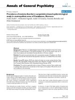
Female patients predominated in all donor groups/tissues
(60% to 70%) used in this study, which is consistent with the
higher incidence of OA in the ageing female population (Fig-
ure 1a). The knee and hip articular cartilage donor groups
ranged in age from 43 to 88 years (mean ± standard deviation
[SD]: 68.6 ± 10.5 years) and from 55 to 85 years (mean ± SD:
69.8 ± 7.4 years), respectively, and the meniscal group
ranged in age from 70 to 88 years (mean ± SD: 77.8 ± 5.4
years). The mean age of the meniscal donors used in this study
Table 1
Peptide sequences identified by the SLRP antibodies used
SLRP (antibody type) Peptide sequence identified Antibody
Decorin (polyclonal) (CGG)YVRSAIQLGNYK PR-84
Biglycan (polyclonal) (CGG)TDRLAIQFGNYKK PR-85
Fibromodulin (polyclonal) (CGG)LRLASLIEI PR 184
Lumican (polyclonal) (CGG)LRVANEVTLN PR-353
Keratocan (monoclonal) keratocan core protein (epitopes not identified) KER-1
KER, keratocan; SLRP, small leucine rich proteoglycan.
Arthritis Research & Therapy Vol 10 No 4 Melrose et al.
Page 4 of 10
(page number not for citation purposes)
was significantly older than all other sample groups (P < 0.006
for all analyses).
To try and compare all SLRPs in representative samples of dif-
ferent tissues, we pooled an aliquot of all 4 M GuHCl extracts
from like tissues. In these pooled samples we depleted aggre-
can species from samples destined for immunoblotting to
avoid possible interference with sample concentration and
electrophoretic separation of the SLRPs, by using centrifugal
diafiltration. These pooled tissue extracts exhibited significant
fragmentation of decorin, biglycan, lumican and keratocan in
all tissues examined in the study (Figure 1b), but importantly
the extent of fragmentation varied with SLRP, tissue type and
joint (knee versus hip). Fibromodulin was not as extensively
processed as the other SLRPs in the degenerate tissues ini-
tially examined in this study (Figure 1b). Meniscal extracts gen-
erally contained the most extensive range of SLRP fragments,
and hip articular cartilage the least extensive fragmentation
patterns. There was a marked difference between OA knee
and hip articular cartilage in the fragmentation of lumican and
keratocan but not biglycan, decorin or fibromodulin, despite
similar levels of intact core proteins of the SLRPs in the two
articular cartilages (Figure 1b).
To enable comparison of multiple samples of both OA and nor-
mal tissues and to avoid any potential losses in SLRP core pro-
tein fragments (which are interactive with some > 100 kDa
component in the extracts that apparently is resolved from the
SLRP fragments during electrophoresis), we repeated these
initial blotting experiments with individual tissue extracts that
were not subjected to centrifugal diafiltration (Figure 2). We
also examined extracts from age-matched macroscopically
normal knee articular cartilage and from areas of the same joint
displaying surface fibrillation, as well as extracts from normal
young nondegenerate articular cartilage (Figure 2). These
blots showed a similar range of SLRP fragments to those pre-
viously identified in the pooled tissue extracts (Figure 1b). In
contrast to the pooled extract, however, similar levels of SLRP
fragmentation were evident in meniscus and knee cartilage. In
the meniscus there was a consistent increase in fragmentation
of decorin but not the other SLRPs in OA versus age-matched
normal joints. In contrast, in knee articular cartilage all SLRPs
generally exhibited increased fragmentation in OA compared
with similarly aged normal joints. Furthermore, in surface-fibril-
lated compared with intact cartilage from the same non-OA
joints, there was a similar increase in fragmentation of all
SLRPs (Figure 2). SLRP fragmentation levels in the fibrillated
and OA knee articular cartilages were higher than the mature
age-matched macroscopically normal tissue or normal young
knee articular cartilage from two 29-year-old donors (Figure
2).
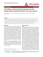
Six prominent decorin fragments (38,36, 25, 18, 16 and 14
kDa) were evident in the fibrillated and the degenerate carti-
lage specimens from the total knee replacement donors. Sim-
ilar fragments were also identified in the meniscal samples
from the total knee replacements, but these were largely unde-
tectable in the 29-year-old normal cartilage samples (Figure
2). Fragmentation of biglycan in meniscus and cartilage exhib-
ited a prominent triplet of 39 to 45 kDa and up to six variably
distributed smaller molecular weight core protein species (16
to 35 kDa). Little fragmentation of fibromodulin was apparent,
although one, almost full-length fibromodulin core protein frag-
ment (approximately 49 kDa) was evident in fibrillated and OA
cartilage but not meniscus. Lumican fragments in both menis-
cus and cartilage were of similar size, consisting of five catabo-
lites ranging from 15 to 38 kDa. Similarly, prominent 35 to 37
kDa full-length keratocan core proteins and four core protein
fragments (14 to 25 kDa) were evident, with a similar size dis-
tribution in meniscus and fibrillated and OA cartilage.
Figure 1
Assessment of SLRP fragmentationAssessment of SLRP fragmentation. Presented is an assessment of
small leucine-rich proteoglycan (SLRP) fragmentation in human menis-
cus (Men), knee and hip articular cartilage extracts by Western blotting.
(a) The age and sex distribution of the total knee and hip replacement
tissue donors used in this study. (b) Pooled tissue extracts were exam-
ined by Western blotting. Pooled 4 M GuHCl tissue extracts were frac-
tionated by centrifugal diafiltration and the 5 to 100 kDa fraction used.
All samples were pre-digested with chondroitinase ABC and keratan-
ase-I before electrophoresis. Sample loadings were normalized by load-
ing extracts corresponding to equivalent wet weights of tissue in each
lane for comparison.
Available online />Page 5 of 10
(page number not for citation purposes)
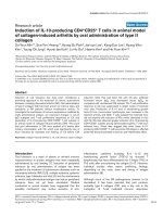
A third series of blots were undertaken on SLRP fragmentation
using six representative individual tissue samples from the
total knee replacement articular cartilage, hip replacement
articular cartilage and knee joint menisci from the total knee
replacement donors to determine whether there was an effect
of age (Figure 3). The SLRP fragmentation patterns obtained
were similar to those obtained earlier (Figures 1b and 2b). A
noteable trend toward increased abundance of fragments of
all SLRPs other than keratocan with age was evident in the
knee articular cartilage samples but not the meniscus or hip
cartilage. There were no fragments in any of the SLRPs that
were specifically associated with ageing in the knee articular
cartilage, but rather an increased staining of all fragments (Fig-
ure 3a–e). In the case of keratocan, the most notable change
with age in the knee articular cartilage was a decrease in the
intact core protein species (35 to 37 kDa; Figure 3e). As
noted previously (Figure 1), there was generally less staining
Figure 2
Identification of intact SLRP core proteins and fragments in knee articular cartilageIdentification of intact SLRP core proteins and fragments in knee articular cartilage. Presented is identification of intact small leucine-rich proteogly-
can (SLRP) core proteins and fragments in age-matched macroscopically normal (N), osteoarthritic (OA) or fibrillated (F) knee articular cartilage
(AC). We used affinity-purified anti-carboxyl-terminal SLRP antibodies (PR-84, PR-85, PR-184 and PR-353) and a monoclonal antibody to full-length
keratocan core protein (KER-1) to perform Western blotting of samples separated by 4% to 12% Bis-Tris lithium dodecyl sulphate PAGE and blot-
ted to nitrocellulose. All samples were pre-digested with chondroitinase ABC and keratanase-I before electrophoresis. The brackets above the lanes
indicate that macroscopically normal and fibrillated articular cartilage were sampled from the same individual nonarthritic joint for this comparison.
Extracts from an equivalent wet weight of tissue were loaded in each lane for comparison. The ages of tissue donors are indicated above each lane.
Arthritis Research & Therapy Vol 10 No 4 Melrose et al.
Page 6 of 10
(page number not for citation purposes)
of all SLRP fragments in age-matched extracts of hip com-
pared with knee OA articular cartilage.
Discussion
This study has shown that SLRPs undergo extensive proteoly-
sis in several diseased human and some normal age-matched
musculoskeletal tissues. We have extended previous studies
examining SLRP proteolysis that were restricted to arthritic
knee cartilage [54-56] by showing the presence and catabo-
lism of keratocan in this tissue. We have also compared and
shown differences in the degree of SLRP catabolism in OA hip
compared with knee cartilage. In addition we have, for the first
time, described SLRP catabolism in the meniscus from OA
and normal knees and in surface-fibrillated cartilage compared
with intact tissue from nonarthritic joints.
One of the limitations of the study is that, in order to achieve
sufficient tissue for analysis from individual OA joints, we gen-
erally pooled all available cartilage or meniscus for extraction.
Thus, it was not possible to correlate the degree of gross or
histological pathology with subsequent SLRP catabolism.
Post-extraction proteolysis of SLRPs could account for some
of the SLRP fragmentation observed in the present study, but
this seems unlikely as proteinase inhibitors and ultra-pure de-
ionized water were used in all steps. There was a dispropor-
tionate loss of SLRP catabolites in cartilage compared with
meniscal extracts that were subjected to diafiltration, which
may suggest interaction of SLRP fragments and removal with
the aggrecan present in much higher levels in cartilage. Finally,
we were only able to compare the molecular mass of the SLRP
catabolites, because to date the actual cleavage sites in the
core proteins and relevant neoepitope antibodies are not
available.
Interestingly, we found that not all SLRPs exhibited a similar
degree of fragmentation within the one tissue, and furthermore
there were distinct differences between tissues and even
between the same tissue from different joints (articular carti-
lage in knee versus hip). It is interesting to speculate that the
differences observed between hip and knee cartilage could be
associated with the generally more extensive pathology in hip
joints, such that the residual cartilage may be in a more
advanced stage of degeneration. It is also possible that some
cartilage repair occurs in late stage OA and is more prevalent
in the hip.
We did observe differences in SLRP proteolysis in the menis-
cus compared with the articular cartilage in OA joints,
although these were more subtle than expected, given the dis-
parity in cell type, matrix organization, matrix constituents (for
example, collagen types) and vascularity of the two tissues. In
all tissues examined in the present study the same molecular
mass fragments were found for all SLRPs, suggesting that
similar proteolytic events were responsible/occurring. When
SLRP catabolites were present, this was also true of normal
compared with arthritic joint tissues, again suggesting that the
elevated breakdown of SLRPs in disease is due to the upreg-
ulation of the same enzymes which are responsible for the
homeostatic turnover of these components in normal tissues.
The exception was fibromodulin, in which a 49 kDa fragment
was more evident in articular cartilage compared with menis-
cus. This suggests the presence of a specific proteolytic path-
way or organization of fibromodulin in articular cartilage.
Figure 3
Identification of SLRP core protein fragmentation in meniscus, knee and hip articular cartilageIdentification of SLRP core protein fragmentation in meniscus, knee and hip articular cartilage. Presented is identification of small leucine-rich prote-
oglycan (SLRP) core protein fragmentation in 4 M GuHCl extracts of meniscus, knee and hip articular cartilage of individual tissue specimens from
total knee or hip replacement patients. The ages (years) of each specimen are indicated at the top of each lane. Extracts from an equivalent wet
weight of tissue were loaded in each lane. The samples were pre-digested with chondroitinase ABC and keratanase-I prior to electrophoresis. Migra-
tion positions of Novex SeeBlue2 Protein standards are indicated on the left hand side of each segment.
Available online />Page 7 of 10
(page number not for citation purposes)
Furthermore, this catabolite may be a useful distinguishing
marker of degeneration in the two tissues. We were able to
demonstrate an age-related pattern of SLRP proteolysis in OA
knee articular cartilage but not meniscus (or hip). In the case
of meniscus this may be because a more limited and elderly
age range was available for comparison. It is unclear whether
the increased SLRP proteolysis in OA knee articular cartilage
is a true ageing phenomenon or whether joint disease was
also worse in the older patients. We were only able to access
a limited number of age-matched nonarthritic joints with a nar-
row age bracket (60 to 75 years) for comparison with OA, and
we did not observe an age-associated pattern of SLRP frag-
mentation in these samples.
There was also a difference between knee meniscus and artic-
ular cartilage when SLRP proteolysis in normal and OA sam-
ples were compared. There was a consistent pattern of
increased fragmentation of decorin, biglycan, fibromodulin,
keratocan and, to a lesser extent, lumican in articular cartilage
from age-matched OA compared with nonarthritic joints. This
pattern only held true for decorin but not the other SLRPs in
normal versus OA meniscus. This may be associated with the
greater degree of degeneration in the articular cartilage com-
pared with meniscus in OA joints. Alternatively, there may be
an elevated basal (normal) level of SLRP cleavage in the aged
meniscus, which would be consistent with the age-related
increase in expression of decorin in meniscus but not articular
cartilage previously reported [17]. It is difficult to explain why
only decorin exhibited consistent increased fragmentation in
meniscus in OA, but it may indicate differences between the
SLRPs in proteolytic pathways, regulation of synthesis, or
presentation/availability to enzymatic attack in the meniscus
compared with articular cartilage.
The apparent limited catabolism of fibromodulin in all tissues
may suggest that fibromodulin is more resistant to proteolysis
than the other SLRPs or that fibromodulin degradation prod-
ucts retaining the carboxyl-terminus may not be stably retained
in the tissue and are lost into the synovial fluid. Alternatively, it
could be an artefact of using an antibody to the extreme car-
boxyl-terminus of the core protein. Once the LRLASLIEI car-
boxyl-terminal peptide sequence identified by Ab PR-184 is
removed from the native fibromodulin core protein, it and any
subsequent catabolites are no longer detectable with this anti-
body. Thus, it is possible that fibromodulin may be more exten-
sively processed at the carboxyl-terminus in degenerate OA
connective tissues compared with other SLRPs, in which
extensive fragments were detected with antibodies to the
carboxyl-terminus.
Decorin and fibromodulin were the SLRPs with the most dis-
tinctly increased fragmentation in OA and fibrillated articular
cartilage from nondiseased joints compared with macroscopi-
cally normal articular cartilage from the age-matched donors.
This suggests that proteolysis of these two SLRPs may be par-
ticularly associated with pathology, and that age-related artic-
ular cartilage fibrillation in apparently normal joints may involve
similar proteolytic events as bona fide OA. Similar decorin
fragments were also evident in the meniscal extracts from total
knee replacement tissue donors but not the extracts of the
menisci from the age-matched normal tissue donors. This
coincident increase in decorin proteolysis in different tissues
in joint pathology suggests that humoral factors such as inter-
leukin-1 or tumour necrosis factor associated with disease
may stimulate degradation of this SLRP and may imply that dif-
ferent cytokine levels in the knee and hip account for the site
variations. Some of these decorin core protein species may
therefore represent useful diagnostic biomarkers of joint dis-
ease. Fibromodulin catabolism on the other hand was more
uniquely associated with articular cartilage and could be use-
ful for tissue discrimination. In the case of biglycan and lumi-
can in particular, despite an increase in fibrillated and OA
articular cartilage, detectable levels of some of the fragments
in macroscopically normal articular cartilage and meniscus
indicate that these fragments are also associated with the nor-
mal turnover of these tissues. This would probably limit their
utility as potential disease biomarkers.
A number of studies have examined the possible use of dis-
ease-associated protein fragments as biomarkers to evaluate
articular cartilage metabolism or disease progression in
spondyloarthritis and OA in humans and in animal models of
OA [58-67]. In the present study we only identified fragments
retained in the diseased tissues. However, with future determi-
nation of the specific cleavage site in the core protein that gen-
erate these catabolites, it will be possible to generate
antibodies that recognize both the specific amino- and car-
boxyl-termini resulting from such proteolysis. These antibodies
may permit discovery of potential biomarker peptides that are
released into body fluids.
Melching and coworkers [40] demonstrated that recombinant
aggrecanase-1 and aggrecanase-2 generated a major 27 kDa
carboxy-terminal biglycan fragment in vitro, with cleavage
being within the fifth leucine-rich domain. We also identified an
approximately 27 kDa biglycan fragment in extracts of degen-
erate meniscus, knee and hip articular articular cartilage, con-
sistent with ADAMTS (a disintegrin and metalloprotease
domain with thrombospondin type I motifs) cleavage. Decorin,
biglycan and fibromodulin can all also be degraded by MMP-
13 in vitro with fragments that recognized by the same anti-
bodies as used in the present study [41]. The 28 to 30 kDa
catabolites of decorin and biglycan we observed are consist-
ent with those generated by MMP-13. MMP-13 in vitro has
also been shown to degrade fibromodulin attached to collagen
with the generation of a 37 to 39 kDa carboxyl-terminal frag-
ment, but fibromodulin in free solution was not degraded [38].
We failed to detect any significant 37 to 39 kDa fibromodulin
catabolites expected from MMP-13 cleavage of this protein
using PR-184. Importantly, the majority of the naturally
Arthritis Research & Therapy Vol 10 No 4 Melrose et al.
Page 8 of 10
(page number not for citation purposes)
occurring SLRP catabolites identified in human tissues in the
present study do not correlate in size with fragments
generated by in vitro digests with specific proteinases. This
may indicate that enzymes other than those thus far studied in
vitro are responsible for SLRP catabolism in vivo or that the
cleavage sites and susceptibility may be different in situ as
opposed to solution-phase digests. It is important that in the
future the actual cleavages that occur in tissues are defined, in
order to enable the enzymes responsible to be identified and
potentially evaluated as targets for disease modification.
Conclusion
In general, an extensive array of SLRP core protein fragments
are present in degenerate knee articular cartilage and menis-
cus, but they were less prominent in degenerate hip articular
cartilage. Specific decorin and fibromodulin core protein frag-
ments, but not other SLRPs, were associated with the degen-
erate human meniscus and articular cartilage compared with
nondiseased tissue.
Fibromodulin core protein fragmentation was far less evident
than fragmentation of other members of the SLRP family. This
may be because of fibromodulin being relatively resistant to
proteolysis or, unlike other SLRPS studied, because the
extreme carboxyl-terminus of fibromodulin containing the anti-
body recognition site is rapidly and/or extensively processed.
The majority of the naturally occurring SLRP catabolites iden-
tified in human joint tissues in the present study do not corre-
late in size with fragments generated by in vitro digests with
specific proteinases. This may indicate that enzymes other
than those thus far studied in vitro are responsible for SLRP
catabolism in vivo or that the cleavage sites and susceptibility
may be different in situ as opposed to solution-phase digests.
Future work may demonstrate some of the aforementioned
SLRP core protein fragments as valuable biomarkers of joint
disease progression. Identification of the enzymes responsible
for their generation may also uncover useful targets for thera-
peutic intervention strategies for arthritic disorders.
Competing interests
The authors declare that they have no competing interests.
Authors' contributions
JM was responsible for the day to day running of the study,
experimental design and writing of the manuscript in conjunc-
tion with PJR and CBL. ESF undertook the collection of tis-
sues, Western blotting and other incidental duties required for
the day to day running of the project. PJR, CEH, BK and BC
were involved in the supply of antibodies, review of drafts of
the manuscript and technical support for antibody use in
Western blotting applications. MMS was involved in tissue
collection, manuscript revision. CBL provided intellectual over-
view and clinical relevance to the study.
Acknowledgements
The following surgeons from The Department of Orthopaedic and Trau-
matic Surgery, Royal North Shore Public and Private Hospitals, St.
Leonards, NSW Australia are thanked for providing surgical specimens
used in this study: A Ellis, M Coolican, D Parker, S Ruff, M Ryan, D
Papadimitriou and I Fairey. Ms Eileen Cole, Department of Orthopaedic
and Traumatic Surgery, is thanked for obtaining informed consent from
donor patients as part of the tissue procurement process. This study
was funded by NHMRC Project Grant 352562.
References
1. Brooks PM: The burden of musculoskeletal disease: a global
perspective. Clin Rheumatol 2006, 25:778-781.
2. Gupta S, Hawker GA, Laporte A, Croxford R, Coyte PC: The eco-
nomic burden of disabling hip and knee osteoarthritis (OA)
from the perspective of individuals living with this condition.
Rheumatology (Oxford) 2005, 44:1531-1537.
3. Leardini G, Vaccaro E: Osteoarthritis: socioeconomic
problems. Semin Arthritis Rheum 2005, 34:35-37.
4. Rabenda V, Manette C, Lemmens R, Mariani AM, Struvay N, Regin-
ster JY: Direct and indirect costs attributable to osteoarthritis
in active subjects. J Rheumatol 2006, 33:1152-1158.
5. Waal JM van der, Terwee CB, Windt DA van der, Bouter LM,
Dekker J: Health-related and overall quality of life of patients
with chronic hip and knee complaints in general practice. Qual
Life Res 2005, 14:795-803.
6. Woolf AD, Pfleger B: Burden of major musculoskeletal
conditions. Bull World Health Organ 2003, 81:646-656.
7. Flannery CR: Usurped SLRPs: novel arthritis biomarkers
exposed by catabolism of small leucine-rich proteoglycans?
Arthritis Res Ther 2006, 8:106.
8. Malemud CJ: Matrix metalloproteinases (MMPs) in health and
disease: an overview. Front Biosci 2006, 11:1696-1701.
9. Mort JS, Billington CJ: Articular cartilage and changes in arthri-
tis: matrix degradation. Arthritis Res 2001, 3:337-341.
10. Thibault M, Poole AR, Buschmann MD: Cyclic compression of
cartilage/bone explants in vitro leads to physical weakening,
mechanical breakdown of collagen and release of matrix
fragments. J Orthop Res 2002, 20:1265-1273.
11. Geng Y, McQuillan D, Roughley PJ: SLRP interaction can protect
collagen fibrils from cleavage by collagenases. Matrix Biol
2006, 25:484-491.
12. Iozzo RV: The biology of the small leucine rich repeat prote-
oglycans-funtional networks of interactive proteins.
J Biol
Chem 1999, 274:18843-18846.
13. Sztrolovics R, White RJ, Poole AR, Mort JS, Roughley PJ: Resist-
ance of small leucine-rich repeat proteoglycans to proteolytic
degradation during interleukin-1-stimulated cartilage
catabolism. Biochem J 1999, 339:571-577.
14. Liu CY, Birk DE, Hassell JR, Kane B, Kao WW: Keratocan-defi-
cient mice display alterations in corneal structure. J Biol Chem
2003, 278:21672-21677.
15. Scott JE: Elasticity in extracellular matrix 'shape modules' of
tendon, cartilage, etc. A sliding proteoglycan-filament model. J
Physiol 2003, 553:335-343.
16. Scott JE, Stockwell RA: Cartilage elasticity resides in shape
module decoran and aggrecan sumps of damping fluid: impli-
cations in osteoarthrosis. J Physiol 2006, 574:643-650.
17. McAlinden A, Dudhia J, Bolton MC, Lorenzo P, Heinegard D, Bay-
liss MT: Age-related changes in the synthesis and mRNA
expression of decorin and aggrecan in human meniscus and
articular cartilage. Osteoarthritis Cartilage 2001, 9:33-41.
18. McDevitt CA, Webber RJ: The ultrastructure and biochemistry
of meniscal cartilage. Clin Orthop Relat Res 1990:8-18.
19. Ghadially FN, Lalonde JM, Wedge JH: Ultrastructure of normal
and torn menisci of the human knee joint. J Anat 1983,
136:773-791.
20. Hellio Le Graverand MP, Vignon E, Otterness IG, Hart DA: Early
changes in lapine menisci during osteoarthritis development:
part I: cellular and matrix alterations. Osteoarthritis Cartilage
2001, 9:56-64.
21. Hough AJ Jr, Webber RJ: Pathology of the meniscus. Clin
Orthop Relat Res 1990:32-40.
Available online />Page 9 of 10
(page number not for citation purposes)
22. Noble J, Hamblen DL: The pathology of the degenerate menis-
cus lesion. J Bone Joint Surg Br 1975, 57:180-186.
23. Ameye L, Aria D, Jepsen K, Oldberg A, Xu T, Young MF: Abnormal
collagen fibrils in tendons of biglycan/fibromodulin-deficient
mice lead to gait impairment, ectopic ossification, and
osteoarthritis. FASEB J 2002, 16:673-680.
24. Ameye L, Young MF: Mice deficient in small leucine-rich prote-
oglycans: novel in vivo models for osteoporosis, osteoarthritis,
Ehlers-Danlos syndrome, muscular dystrophy, and corneal
diseases. Glycobiology 2002, 12:107R-116R.
25. Chakravarti S: Functions of lumican and fibromodulin: lessons
from knockout mice. Glycoconj J 2002, 19:287-293.
26. Chakravarti S, Paul J, Roberts L, Chervoneva I, Oldberg A, Birk DE:
Ocular and scleral alterations in gene-targeted lumican-fibro-
modulin double-null mice. Invest Ophthalmol Vis Sci 2003,
44:2422-2432.
27. Corsi A, Xu T, Chen XD, Boyde A, Liang J, Mankani M, Sommer B,
Iozzo RV, Eichstetter I, Robey PG, Bianco P, Young MF: Pheno-
typic effects of biglycan deficiency are linked to collagen fibril
abnormalities, are synergized by decorin deficiency, and
mimic Ehlers-Danlos-like changes in bone and other connec-
tive tissues. J Bone Miner Res 2002, 17:1180-1189.
28. Danielson KG, Baribault H, Holmes DF, Graham H, Kadler KE,
Iozzo RV: Targeted disruption of decorin leads to abnormal col-
lagen fibril morphology and skin fragility. J Cell Biol 1997,
136:729-743.
29. Ezura Y, Chakravarti S, Oldberg A, Chervoneva I, Birk DE: Differ-
ential expression of lumican and fibromodulin regulate colla-
gen fibrillogenesis in developing mouse tendons. J Cell Biol
2000, 151:779-788.
30. Jepsen KJ, Wu F, Peragallo JH, Paul J, Roberts L, Ezura Y, Oldberg
A, Birk DE, Chakravarti S: A syndrome of joint laxity and
impaired tendon integrity in lumican- and fibromodulin-defi-
cient mice. J Biol Chem 2002, 277:35532-35540.
31. Robinson PS, Huang TF, Kazam E, Iozzo RV, Birk DE, Soslowsky
LJ: Influence of decorin and biglycan on mechanical properties
of multiple tendons in knockout mice. J Biomech Eng 2005,
127:181-185.
32. Svensson L, Aszodi A, Reinholt FP, Fassler R, Heinegard D, Old-
berg A: Fibromodulin-null mice have abnormal collagen fibrils,
tissue organization, and altered lumican deposition in tendon.
J Biol Chem 1999, 274:9636-9647.
33. Wadhwa S, Embree M, Ameye L, Young MF: Mice deficient in
biglycan and fibromodulin as a model for temporomandibular
joint osteoarthritis. Cells Tissues Organs 2005, 181:136-143.
34. Wadhwa S, Embree MC, Kilts T, Young MF, Ameye LG: Acceler-
ated osteoarthritis in the temporomandibular joint of biglycan/
fibromodulin double-deficient mice. Osteoarthritis Cartilage
2005, 13:817-827.
35. Young MF, Bi Y, Ameye L, Chen XD: Biglycan knockout mice:
new models for musculoskeletal diseases. Glycoconj J 2002,
19:257-262.
36. Melrose J, Smith SM, Fuller ES, Young AA, Roughley PJ, Dart A,
Little CB: Biglycan and fibromodulin fragmentation correlates
with temporal and spatial annular remodelling in experimen-
tally injured ovine intervertebral discs. Eur Spine J 2007,
16:2193-2205.
37. Roughley PJ: The structure and function of cartilage
proteoglycans. Eur Cell Mater 2006, 12:92-101.
38. Heathfield TF, Onnerfjord P, Dahlberg L, Heinegard D: Cleavage
of fibromodulin in cartilage explants involves removal of the N-
terminal tyrosine sulfate-rich region by proteolysis at a site
that is sensitive to matrix metalloproteinase-13. J Biol Chem
2004, 279:6286-6295.
39. Imai K, Hiramatsu A, Fukushima D, Pierschbacher MD, Okada Y:
Degradation of decorin by matrix metalloproteinases: identifi-
cation of the cleavage sites, kinetic analyses and transforming
growth factor-beta1 release. Biochem J 1997, 322:809-814.
40. Melching LI, Fisher WD, Lee ER, Mort JS, Roughley PJ: The cleav-
age of biglycan by aggrecanases. Osteoarthritis Cartilage 2006,
14:1147-1154.
41. Monfort J, Tardif G, Reboul P, Mineau F, Roughley P, Pelletier JP,
Martel-Pelletier J: Degradation of small leucine-rich repeat pro-
teoglycans by matrix metalloprotease-13: identification of a
new biglycan cleavage site. Arthritis Res Ther 2006, 8:R26.
42. Kashiwagi M, Enghild JJ, Gendron C, Hughes C, Caterson B, Itoh
Y, Nagase H: Altered proteolytic activities of ADAMTS-4
expressed by C-terminal processing. J Biol Chem 2004,
279:
10109-10119.
43. Roughley PJ, Lee ER: Cartilage proteoglycans: structure and
potential functions. Microsc Res Tech 1994, 28:385-397.
44. Roughley PJ, Melching LI, Recklies AD: Changes in the expres-
sion of decorin and biglycan in human articular cartilage with
age and regulation by TGF-beta. Matrix Biol 1994, 14:51-59.
45. Roughley PJ, White RJ, Magny MC, Liu J, Pearce RH, Mort JS:
Non-proteoglycan forms of biglycan increase with age in
human articular cartilage. Biochem J 1993, 295:421-426.
46. Roughley PJ, White RJ, Mort JS: Presence of pro-forms of deco-
rin and biglycan in human articular cartilage. Biochem J 1996,
318:779-784.
47. Roughley PJ, White RJ, Cs-Szabo G, Mort JS: Changes with age
in the structure of fibromodulin in human articular cartilage.
Osteoarthritis Cartilage 1996, 4:153-161.
48. Alini M, Roughley PJ: Changes in leucine-rich repeat proteogly-
cans during maturation of the bovine growth plate. Matrix Biol
2001, 19:805-813.
49. Melching LI, Roughley PJ: Modulation of keratan sulfate synthe-
sis on lumican by the action of cytokines on human articular
chondrocytes. Matrix Biol 1999, 18:381-390.
50. Sztrolovics R, Alini M, Mort JS, Roughley PJ: Age-related
changes in fibromodulin and lumican in human intervertebral
discs. Spine 1999, 24:1765-1771.
51. Johnstone B, Markopoulos M, Neame P, Caterson B: Identifica-
tion and characterization of glycanated and non-glycanated
forms of biglycan and decorin in the human intervertebral disc.
Biochem J 1993, 292:661-666.
52. Lauder RM, Huckerby TN, Nieduszynski IA, Plaas AH: Age-related
changes in the structure of the keratan sulphate chains
attached to fibromodulin isolated from articular cartilage. Bio-
chem J 1998, 330:753-757.
53. Carrino DA, Onnerfjord P, Sandy JD, Cs-Szabo G, Scott PG, Sor-
rell JM, Heinegard D, Caplan AI: Age-related changes in the pro-
teoglycans of human skin. Specific cleavage of decorin to yield
a major catabolic fragment in adult skin. J Biol Chem 2003,
278:17566-17572.
54. Cs-Szabo G, Melching LI, Roughley PJ, Glant TT: Changes in
messenger RNA and protein levels of proteoglycans and link
protein in human osteoarthritic cartilage samples. Arthritis
Rheum 1997, 40:1037-1045.
55. Cs-Szabo G, Roughley PJ, Plaas AH, Glant TT: Large and small
proteoglycans of osteoarthritic and rheumatoid articular
cartilage. Arthritis Rheum 1995, 38:660-668.
56. Witsch-Prehm P, Miehlke R, Kresse H: Presence of small prote-
oglycan fragments in normal and arthritic human cartilage.
Arthritis Rheum 1992, 35:1042-1052.
57. Gealy EC, Kerr BC, Young RD, Tudor D, Hayes AJ, Hughes CE,
Caterson B, Quantock AJ, Ralphs JR: Differential expression of
the keratan sulphate proteoglycan, keratocan, during chick
corneal embryogenesis. Histochem Cell Biol 2007,
128:551-555.
58. Flannery CR: MMPs and ADAMTSs: functional studies. Front
Biosci 2006, 11:544-569.
59. Fujita Y, Hara Y, Nezu Y, Yamaguchi S, Schulz KS, Tagawa M:
Direct and indirect markers of cartilage metabolism in synovial
fluid obtained from dogs with hip dysplasia and correlation
with clinical and radiographic variables. Am J Vet Res 2005,
66:2028-2033.
60. Garnero P: Use of biochemical markers to study and follow
patients with osteoarthritis. Curr Rheumatol Rep 2006,
8:37-44.
61. Huebner JL, Kraus VB: Assessment of the utility of biomarkers
of osteoarthritis in the guinea pig. Osteoarthritis Cartilage
2006, 14:923-930.
62. Matyas JR, Atley L, Ionescu M, Eyre DR, Poole AR: Analysis of
cartilage biomarkers in the early phases of canine experimen-
tal osteoarthritis. Arthritis Rheum 2004, 50:543-552.
63. Mazieres B, Garnero P, Gueguen A, Abbal M, Berdah L, Lequesne
M, Nguyen M, Salles JP, Vignon E, Dougados M: Molecular mark-
ers of cartilage breakdown and synovitis at baseline as predic-
tors of structural progression of hip osteoarthritis. The
ECHODIAH Cohort. Ann Rheum Dis 2006, 65:354-359.
64. Na KS, Kim TH, Inman RD: Biomarkers in spondyloarthritis.
Curr Rheumatol Rep 2006, 8:283-286.
Arthritis Research & Therapy Vol 10 No 4 Melrose et al.
Page 10 of 10
(page number not for citation purposes)
65. Poole AR: Biologic markers and disc degeneration. J Bone
Joint Surg Am 2006, 88(suppl 2):72-75.
66. Schaller S, Henriksen K, Hoegh-Andersen P, Sondergaard BC,
Sumer EU, Tanko LB, Qvist P, Karsdal MA: In vitro, ex vivo, and in
vivo methodological approaches for studying therapeutic tar-
gets of osteoporosis and degenerative joint diseases: how
biomarkers can assist? Assay Drug Dev Technol 2005,
3:553-580.
67. Sharif M, Granell R, Johansen J, Clarke S, Elson C, Kirwan JR:
Serum cartilage oligomeric matrix protein and other biomar-
ker profiles in tibiofemoral and patellofemoral osteoarthritis of
the knee. Rheumatology (Oxford) 2006, 45:522-526.