Báo cáo y học: "Circulating immune complexes contain citrullinated fibrinogen in rheumatoid arthritis" ppt
Bạn đang xem bản rút gọn của tài liệu. Xem và tải ngay bản đầy đủ của tài liệu tại đây (1.04 MB, 13 trang )
Open Access
Available online />Page 1 of 13
(page number not for citation purposes)
Vol 10 No 4
Research article
Circulating immune complexes contain citrullinated fibrinogen in
rheumatoid arthritis
Xiaoyan Zhao
1,2
, Nwora Lance Okeke
1,2
, Orr Sharpe
1,2
, Franak M Batliwalla
3
, Annette T Lee
3
,
Peggy P Ho
4
, Beren H Tomooka
1,2
, Peter K Gregersen
3
and William H Robinson
1,2
1
GRECC, VA Palo Alto Health Care System, 3801 Miranda Avenue, Palo Alto, CA 94304, USA
2
Department of Medicine, Division of Immunology and Rheumatology, Stanford University School of Medicine, 269 Campus Drive West, Stanford,
CA 94305, USA
3
Robert S. Boas Center for Genomics and Human Genetics, Feinstein Institute for Medical Research, 350 Community Drive, Manhasset, NY 11030,
USA
4
Department of Neurology and Neurological Sciences, Stanford University School of Medicine, 269 Campus Drive West, Stanford, CA 94305, USA
Corresponding author: William H Robinson,
Received: 10 Jun 2008 Revisions requested: 1 Jul 2008 Revisions received: 22 Jul 2008 Accepted: 18 Aug 2008 Published: 18 Aug 2008
Arthritis Research & Therapy 2008, 10:R94 (doi:10.1186/ar2478)
This article is online at: />© 2008 Zhao et al.; licensee BioMed Central Ltd.
This is an open access article distributed under the terms of the Creative Commons Attribution License ( />),
which permits unrestricted use, distribution, and reproduction in any medium, provided the original work is properly cited.
Abstract
Introduction There is increasing evidence that autoantibodies
and immune complexes (ICs) contribute to synovitis in
rheumatoid arthritis (RA), yet the autoantigens incorporated in
ICs in RA remain incompletely characterised.
Methods We used the C1q protein to capture ICs from plasma
derived from human RA and control patients. Antibodies specific
for immunoglobulin were used to detect ICs, and fibrinogen
antibodies were used to detect fibrinogen-containing ICs. RA
and control plasma were separated by liquid chromatography,
and fractions then characterised by ELISA, immunoblotting and
mass spectrometry. Immunohistochemical staining was
performed on rheumatoid synovial tissue.
Results C1q-immunoassays demonstrated increased levels of
IgG (p = 0.01) and IgM (p = 0.0002) ICs in plasma derived from
RA patients possessing anti-cyclic citrullinated peptide (CCP+)
autoantibodies as compared with healthy controls. About one-
half of the anti-CCP+ RA possessed circulating ICs containing
fibrinogen (p = 0.0004). Fractionation of whole RA plasma
revealed citrullinated fibrinogen in the high molecular weight
fractions that contained ICs. Positive correlations were
observed between fibrinogen-containing ICs and anti-
citrullinated fibrinogen autoantibodies, anti-CCP antibody,
rheumatoid factor and certain clinical characteristics.
Immunohistochemical staining demonstrated co-localisation of
fibrinogen, immunoglobulin and complement component C3 in
RA pannus tissue. Mass spectrometry analysis of immune
complexes immunoprecipitated from RA pannus tissue lysates
demonstrated the presence of citrullinated fibrinogen.
Conclusion Circulating ICs containing citrullinated fibrinogen
are present in one-half of anti-CCP+ RA patients, and these ICs
co-localise with C3 in the rheumatoid synovium suggesting that
they contribute to synovitis in a subset of RA patients.
Introduction
Rheumatoid arthritis (RA) is a chronic autoimmune synovitis
affecting 0.6% of the world's population [1], yet the mecha-
nisms underlying the initiation and progression of RA are still
not completely understood. The presence of immune com-
plexes (ICs) in the blood and synovial fluid of patients with RA
is well described [2,3], and there is evidence they are involved
in the activation of the complement cascade in RA synovial tis-
BSA = bovine serum albumin; CCP = cyclic-citrullinated peptides; FPLC = fast protein liquid chromatography; GPI = glucose-6-phosphate isomer-
ase; HLA = human leucocyte antigen; HRP = horseradish peroxidase; IBD = inflammatory bowel disease; IC = immune complex; JRA = juvenile rheu-
matoid arthritis; MHC = major histocompatibility complex; PBS = phosphate buffered saline; PBST = phosphate buffered saline with 0.05% Tween-
20; PS = psoriasis; PsA = psoriatic arthritis; RA = rheumatoid arthritis; RF = rheumatoid factor; SLE = systemic lupus erythematosus.
Arthritis Research & Therapy Vol 10 No 4 Zhao et al.
Page 2 of 13
(page number not for citation purposes)
sue [4]. However, apart from rheumatoid factor (RF) [5] and
anti-collagen type II [6], the identity of the antigens involved in
ICs in RA remains obscure.
Studies suggest critical roles for protein citrullination, B cells
and autoantibodies in the pathogenesis of RA [7-10]. Citrulli-
nation is the post-translational conversion of arginine to citrul-
line, and in RA autoantibodies targeting cyclic citrullinated
peptide (CCP) provides a sensitivity of approximately 70%
and a specificity of 97% for the diagnosis of RA [7,11,12]. The
citrullinated α- and β-chains of fibrin have been identified as
potential targets of the autoantibody response in RA [13] and
citrullinated fibrinogen is detected in RA synovial fluid [14].
Korganow and colleagues identified ICs involving glucose-6-
phosphate isomerase (GPI) as mediating joint inflammation in
their spontaneous K/BxN model [15]. These mice produce
anti-GPI antibodies that form ICs that are deposited on articu-
lar surfaces and activate the alternative complement pathway
to cause synovitis. Although studies suggest that GPI is not a
specific autoantigen in RA [16], it is possible that the mecha-
nisms involved in anti-GPI antibody arthritis and IC arthritis are
relevant to a subset of human RA patients.
RA is characterised by excessive generation and breakdown
of fibrinogen [17]. The citrullinated α- and β-chains of fibrin
have also been identified as a potential target of the autoanti-
body response in RA [13,18] and citrullinated fibrinogen has
been identified in synovial fluid derived from RA patients [14].
Autoantibodies against citrullinated fibrinogen have been
described to provide diagnostic value in arthritis [18,19]. We
previously generated synovial microarrays containing more
than 500 proteins and peptides representing candidate
autoantigens in RA, including protein and overlapping pep-
tides representing native and citrullinated fibrinogen. Synovial
microarray analysis demonstrated targeting of citrullinated
fibrinogen in RA [20].
The methods described for the detection of ICs include chem-
ical precipitation methods from as far back as the 1960s [21]
and biological methods such as precipitation with Clq [22].
We adapted C1q capture immunoassays to utilise fibrinogen-
specific secondary antibodies to identify fibrinogen-containing
ICs, and applied these immunoassays to plasma samples
derived from RA and control patients.
In the present study, we further investigated the targets of the
autoantibody response and the antigens incorporated in ICs in
RA. We demonstrated that one-half of anti-CCP+ RA patients
possessed circulating (blood) ICs containing citrullinated
fibrinogen, and that fibrinogen, immunoglobulin and comple-
ment component C3 co-localize in pannus tissue derived from
RA patients. These data suggest that autoantibody targeting
of citrullinated fibrinogen results in the formation of fibrinogen-
containing ICs that characterise a subset of anti-CCP+ RA
patients and may contribute to synovitis in RA.
Materials and methods
Human samples
All RA and control plasma and joint samples were obtained
and studied with informed consent under Institutional Review
Board approved protocols. The plasma samples used came
from the Multiple Autoimmune Disease Genetics Consortium
[23] and the Stanford Arthritis Center, collected in EDTA
tubes (Table 1). The diagnosis of RA was made based on the
American College of Rheumatology 1987 criteria [24].
Mass spectrometry analysis
For in-gel digestion, protein spots were excised from the gel
and treated with trypsin overnight at 37°C. The tryptic pep-
tides were resolved by high-performance liquid chromatogra-
phy (HPLC) using a Zorbax 300SB-C18 nanocolumn (Agilent
Technologies, Palo Alto, CA, USA) packed with 3.5 μm parti-
cles (Agilent Technologies, Palo Alto, CA, USA) and eluted at
300 nL/minute with a 60 minute linear gradient from 0 to 95%
acetonitrile containing 0.1% formic acid. Separated peptides
were electrosprayed into an ion trap mass spectrometer (XCT
Plus, Agilent Technologies, Palo Alto, CA, USA). For ICs
immunoprecipitated from RA pannus tissue lysates, the pre-
cipitated complexes were directly digested with trypsin before
mass spectrometry analysis. Proteins were identified based on
raw MS/MS data compared with a SwissProt database using
Mascot (Matrix Science, UK) with valid peptide hits.
Detection of anti-citrullinated fibrinogen autoantibodies
Native fibrinogen (Calbiochem, San Diego, CA, USA) was cit-
rullinated in vitro with a peptidylarginine deiminase derived
from rabbit skeletal muscle (Sigma, St. Louis, MO, USA) using
protocols previously described [25]. Anti-citrullinated fibrino-
gen autoantibodies were assayed as previously described
[13,26,27]. Briefly, native fibrinogen or citrullinated fibrinogen
was coated on ELISA plates (MaxiSorp; Nunc, Rochester, NY,
USA) overnight at 4°C at a concentration of 20 μg/mL. Sub-
sequent incubations and washes were performed at room
temperature. The plates were blocked with 3% bovine serum
albumin (BSA) in phosphate buffered saline with 0.05%
Tween-20 (PBST) (Sigma, St. Louis, MO, USA) for one hour,
washed and incubated with centrifuged plasma (diluted 50-
fold) on a shaker for 1.5 hours. Anti-fibrinogen autoantibody
was detected using horseradish peroxidase (HRP)-conju-
gated secondary reagents specific for human IgG (γchain) or
IgM (μchain) specific antibodies diluted to 1:20,000.
Quantitation of immune complexes
ELISA plates were coated with 20 μg/mL C1q (Sigma, St.
Louis, MO, USA) in phosphate buffered saline (PBS) over-
night at 4°C. Subsequent incubations and washes were per-
formed at room temperature. The plates were blocked with 3%
BSA in PBST for one hour. After washing, plasma from RA
Available online />Page 3 of 13
(page number not for citation purposes)
patients or healthy controls were diluted to 1:50 in PBST and
incubated on a shaker for 1.5 hours. ICs were detected with
HRP-conjugated rabbit antiserum specific for human IgG or
IgM (Jackson Immunoresearch, West Grove, PA, USA).
Quantitation of fibrinogen-containing immune
complexes
ELISA plates coated with C1q were blocked with 3% BSA in
PBST for one hour. After washing, plasma from RA patients or
healthy controls was diluted to 1:10 and incubated on a
shaker for 1.5 hours. Fibrinogen contained within the captured
ICs was detected using a 1:4000 dilution of HRP-conjugated
rabbit anti-human fibrinogen antiserum (Dako, Carpinteria, CA,
USA).
Anti-CCP and RF (IgM) ELISA
The anti-CCP (Euro Diagnostica, Malmö, Sweden) and RF
ELISA kits (Alpha Diagnostic International, San Antonio, TX,
USA) were used according to the manufacturers' protocol,
except that plasma was used instead of serum. Anti-CCP and
RF values of the samples were expressed as IU/mL.
Fractionation of plasma samples
The plasma samples were filtered by a 0.45 μm cellulose ace-
tate membrane in a Spin-x centrifuge tube filter (Corning,
Corning, NY, USA) to remove cell debris and precipitates. A
volume of 150 μL of the filtered plasma sample was injected
into a fast protein liquid chromatography (FPLC) system (GE
Healthcare Bio-Sciences, Piscataway, NJ, USA) equipped
with a Superdex 200 10/300 gel filtration column (Amersham
Biosciences, Piscataway, NJ, USA). A mixture of protein
standard containing human fibrinogen, human albumin and
IgG was run in parallel to further identify different peaks. All liq-
uid chromatography runs were programmed at a flow rate of
0.4 mL/minute with PBS and fractions of 0.5 mL were col-
lected. To measure total protein content of the fractions, 20 μL
of each fraction was mixed with 100 μL of BCA buffer (Pierce
Biotechnology, Rockford, IL, USA) and the mixture was incu-
bated at 37°C for 30 minutes before the results were read at
562 nm on a spectraMAX190 instrument (Molecular Devices,
Sunnyvale, CA, USA). To measure IgG and fibrinogen ICs, 50
μL of each fraction was applied to the C1q ELISA described
above. To measure total IgG and fibrinogen content, the frac-
tions were first diluted 10-fold with PBS. Then 1 μL of the
dilutes was deposited onto a nitrocellulose membrane and left
to dry overnight. After blocking with 5% milk in PBST, HRP-
conjugated anti-human IgG or anti-human fibrinogen was
applied to the membranes. Detection was carried out with
SuperSignal West Pico Substrate (Pierce Biotechnology,
Rockford, IL, USA). The densitometry of exposed film were
measured with FluorChem imaging system (Alpha Innotech,
San Leandro, CA, USA).
Immunoblot
Plasma fractions were further separated with Precast Criterion
Tris-HCl gels (4 to 20% linear gradient; Bio-Rad, Hercules,
CA, USA), and separated proteins blotted onto nitrocellulose
membranes. After blocking with 3% BSA in PBS, sera from
RA patients or healthy controls were used to probe the mem-
branes. Bound antibodies were detected with HRP-conju-
gated anti-human IgG (Jackson Immunoresearch, West
Grove, PA, USA) using a SuperSignal kit (Pierce biotechnol-
ogy, Rockford, IL, USA) and chemiluminescence was imaged
with FluorChem imaging system (Alpha Innotech, San Lean-
dro, CA, USA). Immunoblot with anti-modified citrulline was
performed with an anti-citrulline detection kit (Upstate, Chi-
cago, IL, USA) according to the manufacturer's instructions
[13,27].
Immunohistochemistry
Slides were deparaffinised and hydrated with water. Endog-
enous peroxidase was inhibited with 3% hydrogen peroxide,
and non-specific staining blocked with DAKO Protein Block
Serum-Free (Dako, Carpinteria, CA, USA). Staining for com-
Table 1
Source and description of samples used in the study
Sample source Disease Number Female, no. (%) Age (range) anti-CCP positive, no. (%) RF positive, no. (%)
Dr. P. Gregersen, plasma set 1 RA 30 28 (93) 72.6 (51 to 89) 20 (67) 24 (80)
Healthy 10
Dr. P. Gregersen, plasma set 2 IBD 20 13 (65) 46.4 (23 to 82)
JRA 20 16 (80) 37.3 (10 to 71) 6 (30) 6 (30)
PsA 14 11 (79) 52.6 (23 to 75)
PS 20 10(50) 55.3(22 to 86)
RA 20 19(95) 59.0(35 to 89)
SLE 20 13 (65) 51.3 (29 to 67)
CCP, cyclic-citrullinated peptides; RF, rheumatoid factor; RA, rheumatoid arthritis; IBD, inflammatory bowel disease; JRA, juvenile rheumatoid
arthritis; PsA, psoriatic arthritis; PS, psoriasis; SLE, systemic lupus erythematosus.
Arthritis Research & Therapy Vol 10 No 4 Zhao et al.
Page 4 of 13
(page number not for citation purposes)
plement C3 was performed using a 1:2000 dilution of rabbit
polyclonal antibodies against human complement C3 (Dako,
Carpinteria, CA, USA). For fibrinogen and IgG staining, pre-
treatment of proteinase K (Dako, Carpinteria, CA, USA) was
used before the primary antibody incubation. Slides positive
for fibrinogen were immunohistochemically stained with a rab-
bit polyclonal antibody against human fibrinogen (Dako,
Carpinteria, CA, USA), at room temperature at a dilution of
1:1600 for 30 minutes. After incubation with primary antibody,
the tissue sections were sequentially incubated with Dako
Envision+ Rabbit System Labeled Polymer HRP (Dako,
Carpinteria, CA, USA) or biotinylated rabbit anti-goat antibod-
ies (Vector, Burlingame, CA, USA) followed by streptavidin
HRP (Dako, Carpinteria, CA, USA). Staining was developed
with Liquid DAB+ (Dako, Carpinteria, CA, USA) and counter-
stained with haematoxylin and eosin.
Statistics
All statistics were run using InStat™ software (GraphPad Soft-
ware Inc., San Diego, CA, USA). For quantitation of ICs and
autoantibodies to fibrinogen, unpaired t-tests with Welch cor-
rection were used.
Results
Identification of fibrinogen-containing circulating
immunecomplexes in RA
C1q binds aggregated immunoglobulin Fc regions and has
been used to capture and quantitate ICs [28]. We used C1q-
capture immunoassays and HRP-labelled secondary antibod-
ies specific for human IgG and IgM to quantitate circulating
ICs in plasma derived from anti-CCP+ RA, anti-CCP- RA and
healthy control patients (Figures 1a,b). Elevated circulating
IgG (p = 0.01; Figure 1a) and IgM (p = 0.0002; Figure 1b) ICs
were observed in anti-CCP+ RA patients when compared with
healthy controls. Most anti-CCP- RA patients did not possess
circulating ICs (Figures 1a,b).
To determine if circulating ICs containing fibrinogen are
present in RA, a fibrinogen-specific secondary antibody was
used after C1q capture. One-half of anti-CCP+ RA patients
possessed fibrinogen-containing ICs when compared with
healthy controls (p = 0.0004) and anti-CCP- RA patients (p =
0.0008) (Figure 1c). Anti-CCP+ RA patients showed elevated
titres of fibrinogen-containing ICs relative to anti-CCP- RA and
healthy controls with low standard deviations (Figure 1e).
To further demonstrate that the observed fibrinogen-contain-
ing ICs did not result from non-specific binding of fibrinogen
to immobilised C1q, we immobilised anti-C1q monoclonal
antibodies to capture the C1q-bound ICs followed by detec-
tion with anti-fibrinogen antibodies. Similar results were
obtained with anti-C1q monoclonal antibody capture as com-
pared with C1q capture of ICs, and yielded a R
2
value of 0.9
in a linear regression analysis of the two assays (data not
shown). Fibrinogen-containing ICs were also analysed from
freshly collected plasma samples from both anti-CCP+ RA
and anti-CCP- RA patients (within two hours of blood draw
using EDTA plasma collection tubes). Compared with the
same samples after a freeze-thaw cycle, no difference was
detected (Figure 1f).
To demonstrate that fibrinogen-containing ICs were specifi-
cally detected in RA compared with other autoimmune dis-
eases, these ICs were analysed from plasma samples
collected in a panel of healthy (n = 10), inflammatory bowel
disease (n = 20), juvenile RA (n = 20), psoriatic arthritis ([PsA]
n = 14), psoriasis (n = 20), systemic lupus erythematosus (n
= 20), and RA (n = 20) patients (Figure 1d). A subset of RA
and a small subset of juvenile RA patients exhibited elevated
circulating ICs containing fibrinogen, while patients with other
autoimmune diseases did not (Figure 1d). The subset of juve-
nile RA patients possessing circulating ICs containing fibrino-
gen also possessed anti-citrullinated fibrinogen antibodies, RF
and anti-CCP antibodies (Table 2). Chart reviews performed
on this subset of juvenile RA patients revealed that they exhib-
ited symmetrical polyarthritis (Table 2). These observations
suggest that this subset of 'juvenile RA' patients in fact have
adult RA, and is consistent with prior reports of 13% of juve-
nile RA patients exhibiting anti-CCP antibodies and clinical
features consistent with adult RA [29].
Liquid chromatographic separation demonstrates co-
fractionation of citrullinated fibrinogen with immune
complexes
To demonstrate that the fibrinogen-containing ICs in plasma
are physically distinct from free fibrinogen and free immu-
noglobulin, we used size exclusion chromatography as previ-
ously described [16]. Size exclusion chromatography was
applied to fractionate plasma derived from an RA patient with
fibrinogen-containing circulating ICs, an RA patient with circu-
lating ICs but not fibrinogen-containing circulating ICs, a PsA
patient and a healthy control (Figure 2a). Forty-five fractions
were generated of each patient's plasma and each fraction
was assayed for ICs, fibrinogen-containing ICs, total immu-
noglobulin, total fibrinogen and total protein.
ELISA analysis of the fractions containing IgG ICs showed two
peaks in the elution profile of both RA samples, but not in the
corresponding fractions from the PsA and healthy control sam-
ples (Figure 2a, green line). The first peak (RA1 and RA2,
green line) corresponded to the first three fractions after the
void volume, which had a molecular mass of 300 kD or higher,
and corresponded to the fractions in which ICs eluted. The
second peak (RA1 and RA2, green line) corresponded to free
IgG as compared with a chromatography run of standards
(STD, blue line). ELISA analysis of fibrinogen ICs on RA1
(which possessed fibrinogen ICs) showed a single peak (Fig-
ure 2a, RA1, red line) that was eluted in the same fractions at
the IgG IC peak (Figure 2a, RA1 and RA2, first peak of green
Available online />Page 5 of 13
(page number not for citation purposes)
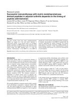
Figure 1
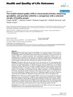
Fibrinogen-containing circulating immune complexes (ICs) in rheumatoid arthritis (RA)Fibrinogen-containing circulating immune complexes (ICs) in rheumatoid arthritis (RA). Circulating (a) IgG and (b) IgM ICs were detected in plasma
derived from healthy individuals and anti-cyclic-citrullinated peptides (CCP) – and anti-CCP+ RA patients. ELISA plates were coated with C1q, incu-
bated with 1:50 dilutions of plasma samples and horseradish peroxidase (HRP)-conjugated (a) anti-IgG or (b) anti-IgM secondary antibodies were
used to detect the immunoglobulin isotypes contained in ICs. (c, d) Circulating fibrinogen-containing ICs were detected using HRP-conjugated
fibrinogen-specific antisera as the secondary reagent. Statistical comparisons are based on an unpaired t-test with Welch correction. (e) Fibrinogen-
containing ICs were detected with different dilutions of RA patient samples and healthy controls. Error bars represent the standard deviation of
results from triplicate wells. (f) Fibrinogen-containing ICs were detected in fresh plasma and freeze-thawed plasma samples with no significant differ-
ences in values (data for anti-CCP status of RA5 is not available).
Arthritis Research & Therapy Vol 10 No 4 Zhao et al.
Page 6 of 13
(page number not for citation purposes)
line). Similar analysis on RA2 did not show a co-eluted peak
(RA2, red line).
To further determine that fibrinogen detected from IC fractions
was not a contamination from free fibrinogen in blood, free
fibrinogen from each RA1 fraction was quantitated by dot
assay (Figure 2a, right panel, pink line). The peak of free fibrin-
ogen was well separated from the peak of fibrinogen IC (Fig-
ure 2a, right panel, red line), as shown by the first two dotted
lines. PsA and healthy control patients did not possess circu-
lating ICs (PsA and healthy, green line). It is possible that fol-
lowing the collection of the plasma fractions, that the free IgG
fractions that contained high levels of IgG developed some
IgG aggregates that were then detected by the IgG IC assay.
These results demonstrate that the fibrinogen-containing cir-
culating ICs observed co-elute with the IgG ICs, and that the
fractions containing fibrinogen-ICs are distinct from those con-
taining free fibrinogen and free immunoglobulin.
To determine if the fibrinogen present in circulating ICs is cit-
rullinated, the FPLC fractions that contained fibrinogen ICs
were separated by sodium dodecyl sulfate polyacrylamide gel
electrophoresis (SDS-PAGE) and immunoblotted with anti-
modified citrulline antibody (Figure 2b). Citrullinated polypep-
tides that co-migrated with fibrinogen polypeptides were
detected only in the fractions derived from RA patients but not
in the corresponding fractions isolated from controls. The
band, indicated as fibrinogen beta chain, was further analysed
by mass spectrometry. Two distinct citrullinated peptides from
the fibrinogen beta chain were identified (Figure 2c).
Laboratory and clinical features associated with
fibrinogen-containing circulating immune complexes
We observed positive correlations between fibrinogen-con-
taining ICs with IgG and IgM ICs, anti-citrullinated fibrinogen
antibodies, anti-CCP antibodies, RF and certain clinical char-
acteristics (Figures 3a–h). Of anti-CCP+ RA patients, three-
quarters possess anti-citrullinated fibrinogen antibodies (Fig-
ures 3c, i) and one-half possess fibrinogen-containing circulat-
ing ICs (Figures 3d, i). All patients with fibrinogen-containing
circulating ICs possess RF, while more than one-half of RF+
patients did not possess fibrinogen-containing ICs (Figures
Table 2
Clinical and laboratory characteristics of the juvenile rheumatoid arthritis (JRA) patients characterised
Sample Clinical features and
rheumatoid factor status
Age
Onset
Age
History
Fibrinogen ICs (O.D.) Anti-cit. fibrinogen IgG (O.D.) anti-CCP
a
(IU/mL)
RF
b
(IU/mL)
JRA 1 Polyarthritis, RF- 2 2 0.18 0.11 21.3 11.7
JRA 4 Polyarthritis, RF+ 13 0.77 1.25 841.1 127.6
JRA 8 Polyarthritis, RF+ 3 16 0.53 0.43 483.2 262.6
JRA 13 Polyarthritis, RF+ 13 13 0.54 1.45 255.4 274.7
JRA 17 Systemic arthritis 0.15 0.10 24.8 24.9
JRA 22 Polyarthritis, RF- 15 15 0.13 0.09 22.7 27.1
JRA 27 Polyarthritis, RF- 1 2 0.10 0.08 20.3 7.6
JRA 31 Persistent oligoarthritis 0.16 0.13 21.5 7.3
JRA 32 Polyarthritis, RF- 5 5 0.12 0.37 26.7 6.3
JRA 41 Enthesitis-related arthritis 13 14 0.14 0.08 20.8 13.1
JRA 42 Systemic arthritis 12 12 0.17 0.08 21.8 7.1
JRA 44 Polyarthritis, RF- 9 9 0.19 0.09 20.9 10.1
JRA 49 Polyarthritis, RF+ 9 1.72 0.74 1275.4 295.2
JRA 51 Systemic arthritis 5 5 0.14 0.09 21.4 7.6
JRA 71 Polyarthritis, RF+ 10 0.19 0.21 360.9 186.1
JRA 79 Polyarthritis, RF- 4 4 0.20 0.10 22.1 11.7
JRA 88 Polyarthritis, RF+ 11 11 0.51 0.35 287.0 236.4
JRA 106 Extended oligoarthrits 1 3 0.15 0.12 22.0 10.0
JRA 110 Persistent oligoarthritis 0.15 0.10 24.9 14.6
JRA 112 Enthesitis related arthritis 0.14 0.12 22.5 15.9
a,b
Measured with commercial kits. JRA, juvenile rheumatoid arthritis; IC, immune complex; CCP, cyclic-citrullinated peptides; RF, rheumatoid
factor; O.D., optical density
Available online />Page 7 of 13
(page number not for citation purposes)
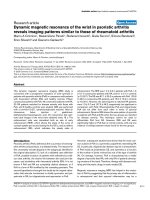
Figure 2
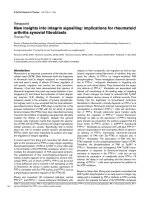
Citrullinated fibrinogen-containing immune complexes (ICs) are separated from rheumatoid arthritis (RA) plasmaCitrullinated fibrinogen-containing immune complexes (ICs) are separated from rheumatoid arthritis (RA) plasma. (a) Liquid chromatographic separa-
tion of RA plasma. Fast protein liquid chromatography (FPLC) was used to fractionate plasma derived from RA and control patients. Forty-five frac-
tions were collected from each plasma sample, and individual fractions were analysed for total protein, fibrinogen, IgG, IgG ICs and fibrinogen ICs
(FIC), and relative levels of each of these components are plotted. Plasma samples from two RA patients (RA1 and RA2), a psoriatic arthritis (PsA)
patients and a healthy control were characterised. The right panel presents individual traces from patient RA1, with the dashed lines indicating the
fractions containing the peak levels of ICs, free fibrinogen and free Ig. (b) Citrullinated fibrinogen was identified by anti-modified citrulline blot. (c) In-
gel trypsin digestion of the bands followed by mass spectrometry revealed two citrullinated peptides derived from beta chain of human fibrinogen.
Arthritis Research & Therapy Vol 10 No 4 Zhao et al.
Page 8 of 13
(page number not for citation purposes)
Figure 3
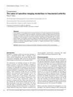
Fibrinogen-containing circulating immune complexes (ICs) are associated with anti-cyclic-citrullinated peptides (CCP) antibodies, rheumatoid factor (RF) and a disease duration of more than 10 yearsFibrinogen-containing circulating immune complexes (ICs) are associated with anti-cyclic-citrullinated peptides (CCP) antibodies, rheumatoid factor
(RF) and a disease duration of more than 10 years. Scatter plots are presented for the association of (a, b) fibrinogen ICs with IgG and IgM ICs; (c)
anti-citrullinated fibrinogen antibodies with anti-CCP antibodies; (d) fibrinogen ICs with anti-CCP antibodies; (e) RF; and (f) anti-citrullinated fibrino-
gen. Lines were drawn to mark the negative and positive measurements of each species. (g, h) Levels of fibrinogen ICs are also plotted in RA
patients with (g) more than 10 years disease duration and (h) smoking history. (i) Unsupervised hierarctical clustering [54] of 30 RA patients and lev-
els of fibrinogen-circulating ICs, anti-citrullinated fibrinogen antibodies, RF and anti-CCP are presented as a heatmap. Tree dendrograms represent
the statistical relatedness between patients.
Available online />Page 9 of 13
(page number not for citation purposes)
3e, i). Interestingly, fibrinogen-containing ICs were not
detected in a subset of the RA patients who possessed high
IgG and IgM plasma ICs, suggesting that circulating ICs con-
taining other antigens are present in this subset of RA patients
(Figures 3a,b). In RA patients, the presence of circulating ICs
containing fibrinogen was associated with a disease duration
of more than 10 years (p = 0.02; Figure 3g), and there were
trends towards associations with smoking (p = 0.1; Figure
3h).
Unsupervised hierarchical clustering of 30 RA patients based
on their anti-CCP antibody, RF, anti-citrullinated fibrinogen
antibody and fibrinogen-containing circulating IC levels dem-
onstrates statistical groupings (Figure 3i). The anti-CCP+ RF+
patients cluster together, and more than one-half of these
patients possess anti-citrullinated fibrinogen autoantibodies
and circulating ICs containing fibrinogen.
Immunohistochemistry demonstrates co-staining of
fibrinogen, complement component C3, and
immunoglobulin in pannus tissue derived from RA
patients
To further investigate the role of fibrinogen-containing ICs in
RA, we performed immunohistochemistry on remnant pannus
tissue derived from two anti-CCP+ RF+ RA patients. Pannus
tissue was obtained from RA patients at the time of knee
arthroplasty, fixed and sectioned, then consecutive sections
were stained with antibodies specific for complement compo-
nent C3, fibrinogen and immunoglobulin. Representative
results are presented from the analysis of consecutive sec-
tions of pannus derived from two independent patients. Immu-
nohistochemical staining demonstrates co-localisation of the
complement component C3, fibrinogen, and IgG in both RA
patients (Figures 4a,b).
RA synovial tissue was minced and the protein contents
extracted with tissue protein extraction buffer. Lysates were
immunoprecipitated with protein-G-sepharose to capture ICs
present in the rheumatoid synovial tissue. These ICs were
eluted from the protein-G beads, trypsinised and the trypsin
digests directly analysed by mass spectroscopy to demon-
strate the presence of citrullinated fibrinogen in ICs isolated
from RA pannus tissue (Figure 4c). These data suggest that
citrullinated-fibrinogen containing ICs either deposit or form in
synovial tissue in RA. The co-localisation of citrullinated fibrin-
ogen-containing ICs with complement component C3 in RA
pannus further suggests that they could activate the comple-
ment cascade to cause synovitis in RA.
Discussion
The presence of ICs in the blood and inflamed joints of
patients with RA was described decades ago [30], and sev-
eral recent findings have resulted in a resurgence of interest in
the role of autoantibodies and B cells in RA. These findings
include the facts that: anti-citrullinated protein autoantibodies
can predate the development of clinical arthritis and provide a
sensitivity of approximately 70% and a specificity of 97% for
the diagnosis of RA [7,11,12,31]; anti-CD20-mediated B cell
depletion provides efficacy in treating RA [9]; and the K/BxN
mouse model develops spontaneous arthritis mediated by
antibodies targeting the ubiquitous glycolytic enzyme GPI
[32]. Although ICs have been isolated from RA patient plasma
by means of polyethylene glycol precipitation [33] and C1q
affinity columns [22], the identity of the antigens incorporated
in these ICs is not well defined. In the present study we char-
acterise circulating and synovial tissue ICs, and demonstrate
the presence of circulating ICs containing fibrinogen in one-
half of anti-CCP+ RA patients (Figure 1). We used immunob-
lotting and mass spectroscopy to demonstrate that the fibrin-
ogen contained in these circulating ICs is citrullinated (Figures
2b,c), and that ICs isolated from RA pannus tissue also con-
tain citrullinated fibrinogen (Figure 4c). Finally, we demon-
strate co-localisation of complement component C3,
fibrinogen and immunoglobulin in RA pannus tissue (Figures
4a,b), suggesting that these complexes contribute to synovitis
in RA.
Although it is difficult to completely exclude the possibility that
the ICs detected are formed in vivo rather than ex vivo, Figure
1f provides reassurance that freezing and freeze-thaw are not
responsible for the observed ICs. Further, fibrinogen-contain-
ing ICs were not observed in plasma derived from patients
with a variety of other inflammatory arthritidies for which the
plasma was collected and stored alongside the anti-CCP+ RA
plasma in which fibrinogen ICs were demonstrated (Figures
1c,d). Although complement containing ICs usually bind to
erythrocytes and are transported to the liver for clearance, in
plasma derived from anti-CCP+ RA patients we found circu-
lating C1q-bound ICs that contain fibrinogen. Further, fibrino-
gen ICs were also detected by anti-C1q monoclonal antibody
capture and results were concordant with our results from
C1q capture of ICs (comparison of results yielded a R
2
value
of 0.9 in linear regression; data not shown).
There is growing evidence that fibrin could be an important
autoantigen in RA [13,18]. Consistent with previous findings
[34], autoantibody reactivity is only observed against citrulli-
nated fibrinogen, and not against its native form (data not
shown). Although the anti-citrullinated fibrinogen antibodies
observed in RA do not result in overt clinical haematological
manifestations, RA is characterised by extravascular coagula-
tion and the accumulation of fibrin in the arthritic joint [17,35].
It has been hypothesised that a local imbalance between
coagulation and fibrinolysis contributes to pathogenesis, and
it is possible that autoantibodies targeting citrullinated fibrino-
gen could contribute to this imbalance by altering the struc-
tural and/or functional properties of fibrinogen and/or fibrin.
Fibrin is one of the classical citrulline-modified proteins [36],
and the presence of citrullinated fibrinogen and/or fibrin has
Arthritis Research & Therapy Vol 10 No 4 Zhao et al.
Page 10 of 13
(page number not for citation purposes)
been demonstrated in the rheumatoid joint [14,37]. Neverthe-
less, citrullinated fibrinogen is generated in inflamed synovia
arising from a variety of inflammatory conditions [37]. Our
observation that ICs containing citrullinated fibrinogen are
present in the plasma of anti-CCP+ RA patients, but not in
plasma derived from anti-CCP- RA, anti-CCP-juvenile RA and
PsA patients (Figures 1 and 2), suggests a potential role for
citrullinated fibrinogen-containing circulating ICs in RA. Our
mass spectrometry analysis of the fibrinogen contained in cir-
culating ICs derived from anti-CCP+ RA patients demon-
strated a few citrullinated peptides from the α-chain of
fibrinogen, but these peptides were not included in Figure 2c
because of low Mascot scores. Our results are consistent with
several previous publications that describe citrullinated
epitopes derived from the beta, but not the alpha, chain of
fibrinogen [38,39]. Although trypsin has been described to be
incapable of cleaving C-terminal to citrulline residues [40], two
of the three citrullinated peptides identified contain a citrulline
at the C-terminus (Figures 2c and 4c). Using mass spectrom-
etry analysis, we have detected multiple citrullinated peptides
with C-terminal citrullines (as well as non-C terminal citrullines)
in tryspin digests of multiple different citrullinated proteins in
Figure 4
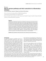
Synovial tissue immune complexes (ICs) contain citrullinated fibrinogenSynovial tissue immune complexes (ICs) contain citrullinated fibrinogen. (a, b) immunohistochemistry demonstrates co-localisation of fibrinogen,
complement component C3 and immunoglobulins in rheumatoid arthritis (RA) pannus tissue. Representative staining of synovium derived from two
separate cyclic-citrullinated peptides (CCP) + rheumatoid factor (RF) + RA patients are shown in (a) and (b). Immunohistochemistry was performed
on articular cartilage samples derived from RA patients. Samples were fixed, paraffin-embedded and sections stained with antisera specific for com-
plement component C3, fibrinogen and IgG, as well as with matched pre-immune sera. Horseradish peroxidase (HRP)-conjugated secondary anti-
bodies were utilised to detect primary antibody reactivity. These stainings demonstrate co-localisation of complement component C3, fibrinogen and
immunoglobulin staining at the surface of the articular cartilage sections. (c) Mass spectrometry analysis of ICs immunoprecipitated from RA synovial
tissue demonstrates the presence of citrullinated fibrinogen peptides.
Available online />Page 11 of 13
(page number not for citation purposes)
several experiments. In addition, citrullinated peptides with C-
terminal citrullines were also observed from multiple citrulli-
nated proteins that were sent to and analysed by an independ-
ent mass spectrometry core facility. The explanation for this
observation remains unclear, and it is possible that our results
are due to altered trypsin cleavage, which is polypeptide
sequence and/or trypsin reaction condition dependent. The
highly significant Mascot scores of our reported citrullinated
peptides (Figures 2c and 4c) support the validity of our results.
The excessive formation of fibrin in the rheumatoid joint in com-
bination with its citrullination and structural properties that
include repetitive antigenic motifs, could result in activation of
B cells specific for citrullinated fibrinogen via cross-linking of
surface immunoglobulin receptors. Citrullination of collagen
was demonstrated to increase its immunogenicity and arthri-
togenicity in a rat arthritis model [41]. Recently, immunisation
with citrullinated fibrinogen was described to induce arthritis in
human leucocyte antigen (HLA) DR4-IE expressing transgenic
mice, demonstrating the arthritogenic potential of citrullinated
fibrinogen in mice expression RA-associated major histocom-
patibility complex (MHC) class II molecules [42].
Fibrin and/or fibrinogen plays an important role in a variety of
inflammatory and immunological processes. Multiple cells,
including neutrophils and macrophages, express integrins and
other receptors that bind fibrin and/or fibrinogen [43]. Fibrino-
gen has also been hypothesised to serve as a structural scaf-
fold for the formation and growth of pannus [44]. Fibrinogen is
chemotactic for endothelial cells that are involved in angiogen-
esis [45], which is integral to the formation of pannus. Fibrin
deposits in RA synovial tissue are hypothesised to activate
synovial fibroblast proliferation and cytokine release, as well as
other inflammatory cell responses [17]. Fibrinogen has been
shown to stimulate macrophage chemokine secretion through
TLR-4 [46]. Further, it was recently demonstrated that RA-spe-
cific autoantibodies complexed to citrullinated fibrinogen stim-
ulate macrophages to produce TNF via engagement of FcγRIIa
[47].
Cantaert and colleagues suggested that the expression of cit-
rullinated proteins is essential but not sufficient for the devel-
opment of RA, and that generation of well-defined citrullinated
epitopes is likely to play a critical role [48]. In this context, our
results might suggest that the development of autoantibodies
targeting citrullinated epitopes specific to fibrinogen might
play an important role in the pathogenesis of RA. In further
support of a potential pathogenic role for citrullinated fibrino-
gen in RA, it was recently demonstrated that citrullinated fibrin-
ogen bound by autoantibodies present in RA patient sera
stimulate macrophage through FcγRIIa to secrete TNF [47].
It was unexpected to observe autoantibodies targeting citrulli-
nated fibrinogen along with fibrinogen-containing circulating
ICs in a subset of juvenile RA patients (Figure 1 and Table 2).
However, late-onset polyarticular juvenile RA is associated
with RF-positivity in about 5% of patients, and has been con-
sidered to be identical to adult RA. Further, a recent report
described 13% of polyarticular-onset juvenile RA patients
exhibiting anti-CCP antibodies [29]. Following the observation
of anti-citrullinated fibrinogen autoatibodies and fibrinogen-
containing circulating ICs in a subset of juvenileRA patients
(Table 2), we performed chart reviews with anti-CCP and RF
ELISA tests on these plasma samples. Of the six juvenile RA
patients exhibiting elevated levels of anti-citrullinated fibrino-
gen antibodies, all exhibited a symmetrical polyarthritis and
possessed RF antibodies. All but one of the six anti-CCP+ and
RF+ juvenile RA patients possessed high levels of fibrinogen
ICs. The age of disease onset of the anti-CCP+ and RF+ juve-
nile RA patients were 13, 11, 3, 13 and 9 years, and these
patients were relatively older than the other juvenile RA
patients included in this cohort. Interestingly, five out of six
anti-CCP+ and RF+ juvenile RA patients possessed fibrino-
gen-containing ICs, compared with only 50% of anti-CCP+
and RF+ adult RA patients. This observation suggests that
anti-fibrinogen autoimmunity and fibrinogen-containing ICs
play a significant role in this subset of juvenile RA patients.
Characterisation of larger cohorts of juvenile RA patients will
be necessary to validate and further investigate this observa-
tion.
Immunohistochemical analysis demonstrated co-localisation
of the staining for fibrinogen, complement component C3 and
immunoglobulin in serial sections derived from RA pannus tis-
sue (Figure 4). These results suggest that fibrinogen-contain-
ing ICs deposit on or form in synovial lining tissue, and activate
the complement cascade to cause inflammatory arthritis. In the
K/BxN model, arthritis is mediated by anti-GPI antibodies and
was demonstrated to depend on FcRγ and components of the
alternative complement pathway [49]. It has been speculated
that accumulation of ICs involving GPI may activate the alter-
native complement pathway to cause inflammatory arthritis
[16]. We hypothesise that autoantibodies targeting citrulli-
nated fibrinogen could result in IC-mediated arthritis based on
mechanisms analogous to those observed in the K/BxN model
[16] and via macrophage Fcγ
RIIa-mediated TNF production
[47].
Anti-citrullinated fibrinogen autoantibodies were detected in
three-quarters of anti-CCP+ RA patients (data not shown)
while fibrinogen containing ICs were found in one-half (Figures
1 and 3). These observations are consistent with RA being a
clinically and molecularly heterogeneous disease, as evi-
denced by differential expression of anti-citrulline antibodies
[7,11], variable responsiveness to anti-tumor necrosis factor
(TNF) therapy [50] and heterogeneity in the genetic back-
ground of patients which includes polymorphisms in the MHC
(major histocompatibility complex), TRAF1-C5 (encoding
tumor necrosis factor receptor-associated factor 1 and com-
plement component 5) [51], STAT4 (encoding signal trans-
Arthritis Research & Therapy Vol 10 No 4 Zhao et al.
Page 12 of 13
(page number not for citation purposes)
ducer and activator of transcription 4) [51,52] and PTPN22
(encoding protein tyrosine phosphatase, non-receptor type
22) [53] genes. CCP is derived from filaggrin, a protein
expressed by keratinocytes in the epidermis, and it is likely that
autoantibody reactivity against the CCPs derived from filag-
grin represents molecular cross reactivity. Our findings sug-
gest that the development of citrullinated fibrinogen-
containing ICs in RA synovial tissue activates the complement
cascade and contributes to synovitis in RA.
Conclusion
In summary, the data presented herein suggest that autoimmu-
nity targeting citrullinated fibrinogen and the development of
fibrinogen-containing ICs could contribute to synovitis in
approximately one-half of anti-CCP+ RA patients. These
results expand the possibility for the development of novel
diagnostics as well as for the development of specific thera-
pies for this subset of RA patients.
Competing interests
The authors declare that they have no competing interests.
Authors' contributions
X.Z. and W.H.R. conceived the studies, carried out the exper-
iments, analyzed the data, and wrote the manuscript. N.L.O.
and O.S. helped perform the mass spectrometry experiments.
F.M.B., A.T.L. and P.K.G. provided human samples and clinical
data, and contributed to interpretation of the data. P.P.H. and
B.H.T. contributed to data analysis.
Acknowledgements
We thank members of the Robinson laboratory and Elizabeth Chlipala
(Premier Laboratory) for their scientific input. This work was funded by a
T. Franklin Williams Scholars grant, an Arthritis Foundation Investigator
Award, NIH NHLBI contract N01 HV 28183, NIH NIAMS R21
AI069160, and Veterans Affairs Health Care System funding to W.H.R.
The mass spectrometry work was supported by the Stanford Digestive
Disease Center grant NIH P30 DK56339.
References
1. Firestein GS: Evolving concepts of rheumatoid arthritis. Nature
2003, 423:356-361.
2. Zubler RH, Nydegger U, Perrin LH, Fehr K, McCormick J, Lambert
PH, Miescher PA: Circulating and intra-articular immune com-
plexes in patients with rheumatoid arthritis. Correlation of
125I-Clq binding activity with clinical and biological features of
the disease. J Clin Invest 1976, 57:1308-1319.
3. Antes U, Heinz HP, Schultz D, Brackertz D, Loos M: C1q-bearing
immune complexes detected by a monoclonal antibody to
human C1q in rheumatoid arthritis sera and synovial fluids.
Rheumatol Int 1991, 10:245-250.
4. Low JM, Moore TL: A role for the complement system in rheu-
matoid arthritis. Curr Pharm Des 2005, 11:655-670.
5. Newkirk MM, Fournier MJ, Shiroky J: Rheumatoid factor avidity in
patients with rheumatoid arthritis: identification of pathogenic
RFs which correlate with disease parameters and with the
gal(0) glycoform of IgG. J Clin Immunol 1995, 15:250-257.
6. Steffen C, Ludwig H, Knapp W, Thumb N, Eberl R, Frank O, Freilin-
ger H: Collagen antibodies and collagen-anticollagen immune
complexes in rheumatoid arthritis. Z Rheumatol 1975,
34:391-399.
7. Schellekens GA, de Jong BA, Hoogen FH van den, Putte LB van
de, van Venrooij WJ: Citrulline is an essential constituent of
antigenic determinants recognized by rheumatoid arthritis-
specific autoantibodies. J Clin Invest 1998, 101:273-281.
8. Kuhn KA, Kulik L, Tomooka B, Braschler KJ, Arend WP, Robinson
WH, Holers VM: Antibodies against citrullinated proteins
enhance tissue injury in experimental autoimmune arthritis. J
Clin Invest 2006, 116:961-973.
9. Edwards JC, Szczepanski L, Szechinski J, Filipowicz-Sosnowska
A, Emery P, Close DR, Stevens RM, Shaw T: Efficacy of B-cell-
targeted therapy with rituximab in patients with rheumatoid
arthritis. N Engl J Med 2004, 350:2572-2581.
10. Agrawal S, Misra R, Aggarwal A: Autoantibodies in rheumatoid
arthritis: association with severity of disease in established
RA. Clin Rheumatol 2007, 26:201-204.
11. Schellekens GA, Visser H, de Jong BA, Hoogen FH van den,
Hazes JM, Breedveld FC, van Venrooij WJ: The diagnostic prop-
erties of rheumatoid arthritis antibodies recognizing a cyclic
citrullinated peptide. Arthritis Rheum 2000, 43:155-163.
12. Mimori T: Clinical significance of anti-CCP antibodies in rheu-
matoid arthritis. Intern Med 2005, 44:1122-1126.
13. Masson-Bessiere C, Sebbag M, Girbal-Neuhauser E, Nogueira L,
Vincent C, Senshu T, Serre G: The major synovial targets of the
rheumatoid arthritis-specific antifilaggrin autoantibodies are
deiminated forms of the alpha- and beta-chains of fibrin. J
Immunol 2001, 166:4177-4184.
14. Takizawa Y, Suzuki A, Sawada T, Ohsaka M, Inoue T, Yamada R,
Yamamoto K: Citrullinated fibrinogen detected as a soluble cit-
rullinated autoantigen in rheumatoid arthritis synovial fluids.
Ann Rheum Dis 2006, 65:1013-1020.
15. Korganow AS, Ji H, Mangialaio S, Duchatelle V, Pelanda R, Martin
T, Degott C, Kikutani H, Rajewsky K, Pasquali JL, Benoist C, Mathis
D: From systemic T cell self-reactivity to organ-specific
autoimmune disease via immunoglobulins. Immunity 1999,
10:451-461.
16. Matsumoto I, Maccioni M, Lee DM, Maurice M, Simmons B, Bren-
ner M, Mathis D, Benoist C: How antibodies to a ubiquitous
cytoplasmic enzyme may provoke joint-specific autoimmune
disease. Nat Immunol 2002, 3:360-365.
17. Sanchez-Pernaute O, Largo R, Calvo E, Alvarez-Soria MA, Egido J,
Herrero-Beaumont G: A fibrin based model for rheumatoid syn-
ovitis. Ann Rheum Dis 2003, 62:1135-1138.
18. Cruyssen B Vander, Cantaert T, Nogueira L, Clavel C, De Rycke L,
Dendoven A, Sebag M, Deforce D, Vincent C, Elewaut D, Serre G,
De Keyser F: Diagnostic value of anti-human citrullinated
fibrinogen ELISA and comparison with four other anti-citrulli-
nated protein assays. Arthritis Res Ther 2006, 8:R122.
19. Nielen MM, Horst AR van der, van Schaardenburg D, Horst-Bru-
insma IE van der, Stadt RJ van de, Aarden L, Dijkmans BA, Hamann
D: Antibodies to citrullinated human fibrinogen (ACF) have
diagnostic and prognostic value in early arthritis. Ann Rheum
Dis 2005, 64:1199-1204.
20. Hueber W, Kidd BA, Tomooka BH, Lee BJ, Bruce B, Fries JF,
Sønderstrup G, Monach P, Drijfhout JW, van Venrooij WJ, Utz PJ,
Genovese MC, Robinson WH: Antigen microarray profiling of
autoantibodies in rheumatoid arthritis.
Arthritis Rheum 2005,
52:2645-2655.
21. Kunkel HG, Muller-Eberhard HJ, Fudenberg HH, Tomasi TB:
Gamma globulin complexes in rheumatoid arthritis and cer-
tain other conditions. J Clin Invest 1961, 40:117-129.
22. Khalkhali-Ellis Z, Bulla GA, Schlesinger LS, Kirschmann DA, Moore
TL, Hendrix MJ: C1q-containing immune complexes purified
from sera of juvenile rheumatoid arthritis patients mediate IL-
8 production by human synoviocytes: role of C1q receptors. J
Immunol 1999, 163:4612-4620.
23. Criswell LA, Pfeiffer KA, Lum RF, Gonzales B, Novitzke J, Kern M,
Moser KL, Begovich AB, Carlton VE, Li W, Lee AT, Ortmann W,
Behrens TW, Gregersen PK: Analysis of families in the multiple
autoimmune disease genetics consortium (MADGC) collec-
tion: the PTPN22 620W allele associates with multiple autoim-
mune phenotypes. Am J Hum Genet 2005, 76:561-571.
24. Arnett FC, Edworthy SM, Bloch DA, McShane DJ, Fries JF, Cooper
NS, Healey LA, Kaplan SR, Liang MH, Luthra HS, Medsger TA,
Mitchell DM, Neustadt DH, Pinals RS, Schaller JG, Sharp JT,
Wilder RL, Hunder GG: The American Rheumatism Association
1987 revised criteria for the classification of rheumatoid arthri-
tis. Arthritis Rheum 1988, 31:315-324.
Available online />Page 13 of 13
(page number not for citation purposes)
25. Vossenaar ER, Despres N, Lapointe E, Heijden A van der, Lora M,
Senshu T, van Venrooij WJ, Menard HA: Rheumatoid arthritis
specific anti-Sa antibodies target citrullinated vimentin. Arthri-
tis Res Ther 2004, 6:R142-150.
26. Chapuy-Regaud S, Nogueira L, Clavel C, Sebbag M, Vincent C,
Serre G: IgG subclass distribution of the rheumatoid arthritis-
specific autoantibodies to citrullinated fibrin. Clin Exp Immunol
2005, 139:542-550.
27. Senshu T, Akiyama K, Kan S, Asaga H, Ishigami A, Manabe M:
Detection of deiminated proteins in rat skin: probing with a
monospecific antibody after modification of citrulline residues.
J Invest Dermatol 1995, 105:163-169.
28. Agnello V, Winchester RJ, Kunkel HG: Precipitin reactions of the
C1q component of complement with aggregated gamma-
globulin and immune complexes in gel diffusion. Immunology
1970, 19:909-919.
29. Ferucci ED, Majka DS, Parrish LA, Moroldo MB, Ryan M, Passo M,
Thompson SD, Deane KD, Rewers M, Arend WP, Glass DN, Nor-
ris JM, Holers VM: Antibodies against cyclic citrullinated pep-
tide are associated with HLA-DR4 in simplex and multiplex
polyarticular-onset juvenile rheumatoid arthritis. Arthritis
Rheum 2005, 52:239-246.
30. Zvaifler NJ: The immunopathology of joint inflammation in
rheumatoid arthritis. Adv Immunol 1973, 16:265-336.
31. Nielen MM, van Schaardenburg D, Reesink HW, Stadt RJ van de,
Horst-Bruinsma IE van der, de Koning MH, Habibuw MR, Vanden-
broucke JP, Dijkmans BA: Specific autoantibodies precede the
symptoms of rheumatoid arthritis: a study of serial measure-
ments in blood donors. Arthritis Rheum 2004, 50:380-386.
32. Matsumoto I, Staub A, Benoist C, Mathis D: Arthritis provoked by
linked T and B cell recognition of a glycolytic enzyme. Science
1999, 286:1732-1735.
33. Ferraccioli G, Karsh J, Osterland CK: Immunochemical analyses
of components of immune complexes in the sera of patients
with autoimmune diseases. J Rheumatol 1983, 10:881-888.
34. Hill JA, Al-Bishri J, Gladman DD, Cairns E, Bell DA: Serum
autoantibodies that bind citrullinated fibrinogen are frequently
found in patients with rheumatoid arthritis. J Rheumatol 2006,
33:2115-2119.
35. Busso N, Hamilton JA: Extravascular coagulation and the plas-
minogen activator/plasmin system in rheumatoid arthritis.
Arthritis Rheum 2002, 46:2268-2279.
36. Vossenaar ER, Zendman AJ, van Venrooij WJ, Pruijn GJ: PAD, a
growing family of citrullinating enzymes: genes, features and
involvement in disease. Bioessays 2003, 25:1106-1118.
37. Chapuy-Regaud S, Sebbag M, Baeten D, Clavel C, Foulquier C,
De Keyser F, Serre G: Fibrin deimination in synovial tissue is
not specific for rheumatoid arthritis but commonly occurs dur-
ing synovitides. J Immunol 2005, 174:5057-5064.
38. Tilleman K, Van Steendam K, Cantaert T, De Keyser F, Elewaut D,
Deforce D: Synovial detection and autoantibody reactivity of
processed citrullinated isoforms of vimentin in inflammatory
arthritides. Rheumatology (Oxford) 2008, 47:597-604.
39. Matsuo K, Xiang Y, Nakamura H, Masuko K, Yudoh K, Noyori K,
Nishioka K, Saito T, Kato T: Identification of novel citrullinated
autoantigens of synovium in rheumatoid arthritis using a pro-
teomic approach. Arthritis Res Ther 2006, 8:R175.
40. Kurokawa T, Hara S, Takahara H, Sugawara K, Ikenaka T: Conver-
sion of peanut trypsin-chymotrypsin inhibitor B-III to a chymo-
trypsin inhibitor by deimination of the P1 arginine residues in
two reactive sites. J Biochem 1987, 101:1361-1367.
41. Lundberg K, Nijenhuis S, Vossenaar ER, Palmblad K, van Venrooij
WJ, Klareskog L, Zendman AJ, Harris HE: Citrullinated proteins
have increased immunogenicity and arthritogenicity and their
presence in arthritic joints correlates with disease severity.
Arthritis Res Ther 2005, 7:R458-467.
42. Hill JA, Bell DA, Brintnell W, Yue D, Wehrli B, Jevnikar AM, Lee
DM, Hueber W, Robinson WH, Cairns E: Arthritis induced by
posttranslationally modified (citrullinated) fibrinogen in DR4-
IE transgenic mice. J Exp Med 2008, 205:967-979.
43. Wright SD, Weitz JI, Huang AJ, Levin SM, Silverstein SC, Loike JD:
Complement receptor type three (CD11b/CD18) of human
polymorphonuclear leukocytes recognizes fibrinogen. Proc
Natl Acad Sci USA 1988, 85:7734-7738.
44. Ishikawa H, Hirata S, Andoh Y, Kubo H, Nakagawa N, Nishibayashi
Y, Mizuno K: An immunohistochemical and immunoelectron
microscopic study of adhesion molecules in synovial pannus
formation in rheumatoid arthritis. Rheumatol Int 1996,
16:53-60.
45. Dejana E, Languino LR, Polentarutti N, Balconi G, Ryckewaert JJ,
Larrieu MJ, Donati MB, Mantovani A, Marguerie G: Interaction
between fibrinogen and cultured endothelial cells. Induction of
migration and specific binding. J Clin Invest 1985, 75:11-18.
46. Smiley ST, King JA, Hancock WW: Fibrinogen stimulates mac-
rophage chemokine secretion through toll-like receptor 4. J
Immunol 2001, 167:2887-2894.
47. Clavel C, Nogueira L, Laurent L, Iobagiu C, Vincent C, Sebbag M,
Serre G: Induction of macrophage secretion of tumor necrosis
factor alpha through Fcgamma receptor IIa engagement by
rheumatoid arthritis-specific autoantibodies to citrullinated
proteins complexed with fibrinogen. Arthritis Rheum 2008,
58:678-688.
48. Cantaert T, De Rycke L, Bongartz T, Matteson EL, Tak PP, Nicho-
las AP, Baeten D: Citrullinated proteins in rheumatoid arthritis:
crucial but not sufficient! Arthritis Rheum 2006, 54:3381-3389.
49. Ji H, Gauguier D, Ohmura K, Gonzalez A, Duchatelle V, Danoy P,
Garchon HJ, Degott C, Lathrop M, Benoist C, Mathis D: Genetic
influences on the end-stage effector phase of arthritis. J Exp
Med 2001, 194:321-330.
50. Moreland LW, Schiff MH, Baumgartner SW, Tindall EA, Fleis-
chmann RM, Bulpitt KJ, Weaver AL, Keystone EC, Furst DE,
Mease PJ, Ruderman EM, Horwitz DA, Arkfeld DG, Garrison L,
Burge DJ, Blosch CM, Lange ML, McDonnell ND, Weinblatt ME:
Etanercept therapy in rheumatoid arthritis. A randomized, con-
trolled trial. Ann Intern Med 1999, 130:478-486.
51. Plenge RM, Seielstad M, Padyukov L, Lee AT, Remmers EF, Ding
B, Liew A, Khalili H, Chandrasekaran A, Davies LR, Li W, Tan AK,
Bonnard C, Ong RT, Thalamuthu A, Pettersson S, Liu C, Tian C,
Chen WV, Carulli JP, Beckman EM, Altshuler D, Alfredsson L,
Criswell LA, Amos CI, Seldin MF, Kastner DL, Klareskog L,
Gregersen PK: TRAF1-C5 as a risk locus for rheumatoid arthri-
tis – a genomewide study. N Engl J Med 2007, 357:1199-1209.
52. Remmers EF, Plenge RM, Lee AT, Graham RR, Hom G, Behrens
TW, de Bakker PI, Le JM, Lee HS, Batliwalla F, Li W, Masters SL,
Booty MG, Carulli JP, Padyukov L, Alfredsson L, Klareskog L, Chen
WV, Amos CI, Criswell LA, Seldin MF, Kastner DL, Gregersen PK:
STAT4 and the risk of rheumatoid arthritis and systemic lupus
erythematosus. N Engl J Med 2007, 357:977-986.
53. Begovich AB, Carlton VE, Honigberg LA, Schrodi SJ, Chokkalin-
gam AP, Alexander HC, Ardlie KG, Huang Q, Smith AM, Spoerke
JM, Conn MT, Chang M, Chang SY, Saiki RK, Catanese JJ, Leong
DU, Garcia VE, McAllister LB, Jeffery DA, Lee AT, Batliwalla F,
Remmers E, Criswell LA, Seldin MF, Kastner DL, Amos CI, Sninsky
JJ, Gregersen PK: A missense single-nucleotide polymorphism
in a gene encoding a protein tyrosine phosphatase (PTPN22)
is associated with rheumatoid arthritis. Am J Hum Genet 2004,
75:330-337.
54. Eisen MB, Spellman PT, Brown PO, Botstein D: Cluster analysis
and display of genome-wide expression patterns. Proc Natl
Acad Sci USA 1998, 95:14863-14868.