Báo cáo y học: "The relationship between inflammation and new bone formation in patients with ankylosing spondylitis" docx
Bạn đang xem bản rút gọn của tài liệu. Xem và tải ngay bản đầy đủ của tài liệu tại đây (217.16 KB, 7 trang )
Open Access
Available online />Page 1 of 7
(page number not for citation purposes)
Vol 10 No 5
Research article
The relationship between inflammation and new bone formation
in patients with ankylosing spondylitis
Xenofon Baraliakos
1
, Joachim Listing
2
, Martin Rudwaleit
3
, Joachim Sieper
3
and Juergen Braun
1
1
Rheumazentrum Ruhrgebiet Herne, Ruhr-University Bochum, Landgrafenstr. 15, 44652 Herne, Germany
2
German Rheumatism Research Center, Charitéplatz 1, 10117 Berlin, Germany
3
Rheumatology, Charité, Campus Benjamin Franklin, Hindenburgdamm 30, 12200 Berlin, Germany
Corresponding author: Juergen Braun,
Received: 2 May 2008 Revisions requested: 4 Jul 2008 Revisions received: 23 Jul 2008 Accepted: 1 Sep 2008 Published: 1 Sep 2008
Arthritis Research & Therapy 2008, 10:R104 (doi:10.1186/ar2496)
This article is online at: />© 2008 Baraliakos et al.; licensee BioMed Central Ltd.
This is an open access article distributed under the terms of the Creative Commons Attribution License ( />),
which permits unrestricted use, distribution, and reproduction in any medium, provided the original work is properly cited.
Abstract
Introduction Spinal inflammation as detected by magnetic
resonance imaging and new bone formation as identified by
conventional radiographs are characteristic of ankylosing
spondylitis. Whether and how spondylitis and syndesmophyte
formation are linked are unclear. Our objective was to
investigate whether and how spinal inflammation are associated
with new bone formation in ankylosing spondylitis.
Methods Spinal magnetic resonance images and conventional
radiographs from 39 ankylosing spondylitis patients treated with
anti-tumour necrosis factor (anti-TNF) agents at baseline and
after 2 years were analysed for syndesmophyte formation at
vertebral edges with or without inflammatory lesions at baseline.
Results Overall, 922 vertebral edges at the cervical and lumbar
spine were analysed. At baseline, the proportion of vertebral
edges with and without inflammation (magnetic resonance
imaging) that showed structural changes (conventional
radiographs) was similar (in total, 16.6% of all vertebral edges in
71.4% of patients). From the perspective of syndesmophyte
formation (n = 26, 2.9%) after 2 years, there were more vertebral
edges without (62%) than with (38%) inflammation at baseline
(P = 0.03). From the perspective of spinal inflammation at
baseline (n = 153 vertebral edges), more syndesmophytes
developed at vertebral edges with (6.5%) than without (2.1%)
inflammation (P = 0.002, odds ratio 3.3, 95% confidence
interval 1.5 to 7.4). Inflammation persisted in 31% of the initially
inflamed vertebral edges (n = 132), and new lesions developed
in 8% of the vertebral edges without inflammation at baseline (n
= 410). From the perspective of spinal inflammation after 2
years (n = 72 vertebral edges), 5.6% of the vertebral edges
showed syndesmophyte development in contrast to 1.9% of the
vertebral edges with new syndesmophytes without inflammation
(P = 0.06).
Conclusions These findings obtained in patients treated with
anti-TNF agents suggest linkage and some dissociation of
inflammation and new bone formation in ankylosing spondylitis.
Although syndesmophytes were also found to develop at sites
where no inflammation had been seen by magnetic resonance
imaging at baseline, it was more likely that syndesmophytes
developed in inflamed vertebral edges. More effective
suppression of spinal inflammation may be required to inhibit
structural damage in ankylosing spondylitis.
Introduction
Ankylosing spondylitis (AS) is a frequent chronic inflammatory
rheumatic disease that already affects the axial skeleton at a
young age [1], starting in the sacroiliac joints and later spread-
ing to the spine [2]. Active inflammatory spinal lesions as
detected by magnetic resonance imaging (MRI) [3] and
chronic structural changes such as syndesmophytes as dem-
onstrated by conventional radiography [4] are characteristic of
AS and contribute to both decreased spinal mobility and func-
tional impairments of these affected patients [5]. Conventional
spinal x-rays are still the gold standard for assessment of struc-
tural changes in AS [6,7], whereas MRI techniques using
either short-tau-inversion-recovery (STIR) sequences [2,8] or
T1-post-gadolinium (T1-post-Gd) [9] are best for assessing
spinal inflammation.
For quantification of structural spinal changes in conventional
radiographs, the modified Stokes AS spinal score (mSASSS)
2yFU: 2-year follow-up; AS: ankylosing spondylitis; BL: baseline; CI: confidence interval; MRI: magnetic resonance imaging; OR: odds ratio; STIR:
short-tau-inversion-recovery; T1-post-Gd: T1-post-gadolinium; TNF: tumour necrosis factor; VE: vertebral edge.
Arthritis Research & Therapy Vol 10 No 5 Baraliakos et al.
Page 2 of 7
(page number not for citation purposes)
[10] is the best currently available scoring method based on
the OMERACT (Outcome Measures in Rheumatology) filter
[11]. For a sufficient sensitivity to change in depiction of struc-
tural spinal changes in AS when using conventional radio-
graphs, a minimal observation period of 2 years is required
[12]. Similarly, for assessment and quantification of inflamma-
tory spinal changes, the AS-spinal-MRI scoring system [13]
has shown good discriminatory capacity, validity, and sensitiv-
ity to change in MRI examinations for periods of between 6
weeks [14] and 2 years [15-17].
Tumour necrosis factor-alpha (TNF-α) plays a key proinflam-
matory role in AS [18,19] given that spinal inflammation was
shown to be associated with the presence of TNF-α mRNA
[18] and protein [20] in affected joint and bone structures.
Accordingly, inhibition of TNF-α was found to substantially
improve signs and symptoms of AS patients [21-23]. Similarly,
using MRI, a significant decrease of inflammatory lesions
already after 6 weeks of therapy [14] and ongoing improve-
ment of spinal inflammation for up to 2 years [15,16] of contin-
uous treatment have been reported. However, some
inflammatory lesions were still present even after this period
[15,17,24].
Chronic changes in the thoracic spine cannot be reliably
assessed by conventional x-rays but a valid quantification of
such lesions is possible in the cervical and the lumbar spine
[4]. Since MRI is able to visualise the entire spine, it is now
clear that the lower part of the thoracic spine is most fre-
quently involved in AS [3,17,25]. This is one possible reason
why so far it has not been possible to demonstrate major inhi-
bition of structural damage in AS patients on anti-TNF therapy
[26-28]. Nevertheless, a direct link between spinal inflamma-
tion and future radiographic progression has not been suffi-
ciently proven until now. Data from animal models have even
suggested that inflammation and new bone formation are
uncoupled [29,30]. In this study, we examined the relationship
of MRI-proven spinal inflammation at baseline (BL) with
respect to structural deterioration depicted by conventional
radiographs after 2 years in AS patients treated with anti-TNF-
α agents.
Materials and methods
Overall, conventional radiographs of 39 AS patients who were
diagnosed according to the modified New York criteria for
diagnosis of AS [31] were analysed. All patients had partici-
pated in clinical studies on anti-TNF therapy with infliximab (n
= 26) [21,32] or etanercept (n = 13) [24] for at least 2 years.
All patients whose images were analysed had already signed
informed consent forms for the radiographic images to be
taken and analysed, according to the ethics committees of the
participating centres which approved the original studies.
None of these patients received antiresorptive bone therapy
such as bisphosphonates or other drugs. Inclusion criteria
were the availability of complete sets of MRIs with STIR and
T1-post-Gd sequences and of conventional radiographs (see
below) at the time point of presentation (BL) and after 2 years
of follow-up (2yFU). All MRI and x-ray examinations were con-
ducted using the same standardised protocol, as recently
reported [4,13,24].
Quantification of inflammatory and chronic spinal
lesions
Depiction and quantification of inflammatory and chronic spi-
nal lesions were performed on the basis of vertebral edges
(VEs) in this study, in accordance with recent results
[4,33,34]. This method was the most specific and also the
most sensitive to change for the depiction of structural spinal
changes in patients with AS as compared with assessments
on the patient level or on the basis of change scores. For the
assessment of structural changes by conventional x-rays, com-
plete sets of radiographs of the cervical and the lumbar spine
in the lateral view at BL and 2yFU were taken. Because of the
known technical problems in the assessment of the thoracic
spine in standard x-rays [4], this part of the spine was not avail-
able for analysis. As recently proposed, we defined 'definite
radiographic damage' as the appearance of at least one syn-
desmophyte in each individual VE since this was the most reli-
able parameter to depict disease-related damage or change
between follow-ups in patients with AS [4]. Similarly, to assess
change over time, 'definite radiographic progression' was
defined as the development of new syndesmophytes or anky-
losis in individual VEs [4]. In comparison, for the assessment
of inflammatory changes by MRI, only the cervical and the lum-
bar spinal segments were analysed for spinal inflammation,
similar to the available x-rays. To be even more precise in the
relationship of inflammatory activity in MRI and new bone for-
mation of the same VEs in conventional radiographs, a VE in
MRI was defined as 'positive' for inflammation if the inflamma-
tory activity was present in the anterior half of the VE only. For
analysis of the relationship between spinal inflammation at BL
and radiographic progression after 2 years, all individual VEs
were examined by MRI for signs of inflammation at BL and the
same VEs were compared for development of new syndesmo-
phytes in conventional radiographs at BL and 2yFU.
Statistical analysis
The Fisher exact test was used for comparison between differ-
ent subgroups such as those with VEs and without spinal
inflammation or with and without definite radiographic damage
and progression. Furthermore, subgroups of VEs with signs of
inflammation as well as radiographic progression were
selected based on the definition of radiographic progression
as defined by four different subsamples. In each of those sub-
samples, two conditional probabilities were compared: first,
the likelihood of radiographic progression in VEs with signs of
inflammation and, second, the likelihood of radiographic pro-
gression in VEs without inflammation. The Wilcoxon test was
applied to compare both paired proportions across all VEs of
the subsamples.
Available online />Page 3 of 7
(page number not for citation purposes)
Results
Analysis of baseline data
Overall, 922 VEs of the cervical spine and lumbar spine of 39
AS patients were available for analysis at BL and at 2yFU.
Missing data are explained by incomplete radiographic image
sets since, for technical reasons, not all VEs could be captured
on some films [4]. Spinal inflammation at BL (STIR MRI
sequence) was present in 153/922 (16.6%) VEs, whereas no
signs of inflammation were seen in 769/922 (83.4%) VEs. At
least one vertebral body with signs of inflammation was found
in 28/39 (71.8%) patients. The BL data as assessed by T1-
post-Gd MRI showed similar results (Table 1). At BL, the VEs
with or without spinal inflammation in MRI showed similar pro-
portions of definite radiographic damage at BL (17.6% versus
15.6%, respectively; P > 0.05). Thus, there was no difference
at BL in the proportion of VEs showing structural changes
(syndesmophytes) in these subgroups.
Analysis of the 2-year follow-up data
Radiographic progression based on the development of new
syndesmophytes was seen in 26/922 VEs (2.8%) after 2
years. Of those, 10 VEs (38%) had initially shown signs of
inflammation as detected by MRI whereas the remaining 16
VEs (62%) had no such changes at BL (P = 0.006 between
VEs with and without BL inflammation) (Table 2). The analysis
based on the T1-post-Gd MRI sequences revealed similar
results (data not shown). In the prospective data analysis, def-
inite radiographic progression was found significantly more
often in VEs with MRI-proven inflammation at BL (10/153 VEs,
6.5%, 95% confidence interval [CI] 3.6% to 11.6%) than in
VEs without BL inflammation in MRI (16/769 VEs, 2.1%, 95%
CI 1.3% to 3.4%) (P = 0.006, odds ratio [OR] 3.3, 95% CI 1.5
to 7.4). This was similar for the T1-post-Gd MRI sequences
(Table 2 and Figure 1).
Relation of radiographic progression to persistent spinal
inflammation after 2 years
Follow-up MRIs after 2 years were available in 23/39 patients
Table 1
Baseline data on inflammation and occurrence of definite radiographic damage as assessed by both magnetic resonance imaging
sequences
MRI sequence Inflammation/radiographic damage Proportion (number) of vertebral edges at baseline P value
STIR Any inflammatory lesions 16.6% (153/922) -
No inflammatory lesions 83.4% (769/922)
Definite radiographic damage with inflammation 17.6% (27/153) 0.53
Definite radiographic damage without inflammation 15.6% (120/769)
T1-post-Gd Any inflammatory lesions 10.3% (95/922) -
No inflammatory lesions 89.7% (827/922)
Definite radiographic damage with inflammation 21.1% (20/95) 0.13
Definite radiographic damage without inflammation 15.1% (125/827)
There was no difference in the proportion of inflammatory lesions and occurrence or absence of syndesmophytes at baseline. MRI, magnetic
resonance imaging; STIR, short-tau-inversion-recovery; T1-post-Gd, T1-post-gadolinium.
Table 2
Proportion of vertebral edges showing development of new syndesmophytes at 2-year follow-up according to baseline status of
inflammation as assessed by both magnetic resonance imaging sequences
MRI sequence Inflammation status Proportion (number) of vertebral edges with development of new
syndesmophytes after 2 years
STIR Inflammation at baseline 6.5% (10/153) P = 0.006,
OR 3.3,
95% CI 1.5 to 7.4
No inflammation at baseline 2.1% (16/769)
T1-post-Gd Inflammation at baseline 6.3% (6/95) P = 0.043,
OR 2.7,
95% CI 1.1 to 7.0
No inflammation at baseline 2.4% (20/827)
CI, confidence interval; MRI, magnetic resonance imaging; OR, odds ratio; STIR, short-tau-inversion-recovery; T1-post-Gd, T1-post-gadolinium.
Arthritis Research & Therapy Vol 10 No 5 Baraliakos et al.
Page 4 of 7
(page number not for citation purposes)
(59%). In those, 542 VEs could be scored. There were 132
VEs with inflammation (STIR sequence) at BL (25%) and 410
VEs without (75%). After 2 years, there were 72 VEs with
inflammation (13%) and 410 VEs without (87%). In more
detail, there were 41/132 VEs (31%) with and 91/132 VEs
(69%) without persistent inflammation at follow-up, while 31/
410 VEs (8%) without BL inflammation showed (new) inflam-
matory lesions and 379/410 VEs (92%) remained without
such changes. Thus, after 2 years, there were still 72/542 of
the VEs (13.3%) showing inflammation. Development of syn-
desmophytes was found in 2/41 VEs (4.9%) and in 2/31 VEs
(6.5%) that showed inflammatory lesions at 2yFU. In contrast,
syndesmophytes were also developed in 4/91 VEs (4.4%) and
in 5/379 VEs (1.3%) that did not show inflammatory lesions at
2yFU. Thus, 4/72 VEs (5.6%) developed syndesmophytes on
the basis of inflammation at 2yFU in contrast to 9/470 VEs
(1.9%) that developed syndesmophytes not based on inflam-
mation at 2yFU (P = 0.06). The T1-post-Gd MRI data showed
similar results (data not shown).
Discussion
The results of the present study suggest that spinal inflamma-
tion and new bone formation are both uncoupled and linked in
AS since (a) the majority of syndesmophytes developed with-
out MRI evidence of spinal inflammation at BL and (b) the pro-
portion of VEs that developed syndesmophytes within 2 years
was threefold higher when spinal inflammation was present at
BL (compared with edges without BL inflammation). Since this
was observed in patients under anti-TNF-α treatment, these
data can be interpreted only on this basis. It will be important
to study whether this is also true for patients just treated with
nonsteroidal anti-inflammatory drugs or other agents. Never-
theless, the data may indicate that spinal inflammation was not
sufficiently suppressed by TNF blockers in this 2-year time
period.
In this study, the primary outcome was based on the analysis
of VEs because the patterns of spinal inflammation and the
development of syndesmophytes are likely to differ in and
between individuals. It was no surprise, therefore, that when
we did the analyses on an individual patient basis, no differ-
ences in the proportions of patients with and without spinal
inflammation with respect to the development of future syn-
desmophytes were found. This may also be explained by the
relatively small number of patients in this cohort. However,
since there clearly were patients who developed syndesmo-
phytes irrespective of BL inflammation, we do believe that it is
more useful to do the calculations on the basis of VEs rather
than on the patient level.
VEs that showed persistent inflammation seemed to be more
prone to develop new syndesmophytes after 2 years as com-
pared with those edges where inflammatory lesions disap-
peared after anti-TNF treatment. Indeed, in this study and in
others, it has been shown that spondylitis may still be present
after 2 years of anti-TNF therapy – even in patients with defi-
nite clinical improvement [15,24]. In addition, the analysis of
only the edges that were inflammation-free at 2yFU showed
that the tendency for the development of new
syndesmophytes was stronger for those edges that showed
inflammatory lesions at BL as compared with those edges that
had not been inflamed in either the BL or the 2yFU. Neverthe-
less, our findings are in line with previous data of ours [26,28]
and of other groups [35,27] showing that radiographic pro-
gression in AS is not or not completely inhibited by TNF
blockers.
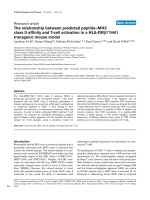
Figure 1
Formation of new syndesmophytes in the upper and lower edges of L1/L2 and L2/L3Formation of new syndesmophytes in the upper and lower edges of L1/L2 and L2/L3. (a) T1-pre-gadolinium (T1-pre-Gd) image. Spinal inflammation
in the same area is assessed by both magnetic resonance imaging (MRI) sequences: (b) T1-post-gadolinium (T1-post-Gd) and (c) short-tau-inver-
sion-recovery (STIR). Inflammation at baseline is seen as a 'spot' in the T1-post-Gd image only after application of gadolinium. The STIR image
shows signs of inflammatory activity in the same vertebral regions. Formation of new syndesmophytes in the upper and lower edges of L1/L2 and
L2/L3 is seen in conventional x-rays developing from (d) baseline to (e) 2 years later.
Available online />Page 5 of 7
(page number not for citation purposes)
Since the key feature of AS, much unlike rheumatoid arthritis
[36], is new bone formation rather than osteodestruction,
there is reason to consider different mechanisms for structural
change which appear on radiographs in these diseases. In AS,
uncoupling of spinal inflammation and new bone formation has
recently been suggested [37]. The data of our study show that
about 60% of all syndesmophytes that developed did not
show inflammation as detected by MRI. Since the sensitivity of
MRI to demonstrate spinal inflammation in AS is not precisely
known [9], the question of whether it was possible to really
detect all cases of spondylitis has to remain open and should
be studied further. Furthermore, it cannot be excluded that
inflammation has occurred at some point before and/or during
the study. In this study, new spondylitis lesions developed in
8% of the VEs investigated. Recent immunohistological data
showed low-grade spinal inflammation in biopsy specimens
obtained at spinal surgery of AS patients who had undergone
MRI before surgery, and no active inflammatory lesions had
been detected by appropriate MRI sequences [38]. Thus,
since we did not perform MRIs in between, we do not know
whether or for how long spinal inflammation may have
occurred in the patients included in this study.
This is the first study based on patient data on this issue –
even though we cannot exclude that the treatment of the
patients had an impact on the results. Indeed, there is some
evidence that blocking TNF-α may reverse the physiologic
inhibition of osteoblast function and stimulate osteoclast
resorption [39]. TNF and other proinflammatory cytokines are
known to promote bone formation by upregulating the expres-
sion of Dickkopf-1, a key target gene of TNF and an inhibitor
of osteophyte regulators [29]. Thus, by inhibiting TNF and
Dickkopf-1, TNF blockers may even block negative influences
on syndesmophyte formation after sufficient suppression of
inflammation [29]. The hypothesis that new bone formation in
AS is uncoupled from inflammation has been supported by
animal models showing that TNF inhibition did not affect joint
ankylosis [30].
Recent biomarker data generated from ASSERT (Ankylosing
Spondylitis Study for the Evaluation of Recombinant Infliximab
Therapy) [22,40] showed that previously low levels of osteo-
calcin and bone alkaline phosphatase were significantly
increased under infliximab therapy [41]. Furthermore, anti-TNF
therapy was shown to decrease osteoclast precursor cells
[42] and to increase bone mineral density [43] in AS patients.
Thus, there is evidence from patient-derived data that anti-TNF
agents increase bone mass. On the other hand, clinical expe-
rience may suggest that syndesmophytes grow especially at
locations where spondylitic lesions had occurred. One exam-
ple is the radiologic appearance of spondylitis anterior, the
well-known shiny corners or Romanus lesions [44]. Further-
more, it was already described some decades ago in histolog-
ical studies that inflammatory spinal lesions precede new bone
formation in AS patients [45]. Our study shows that the likeli-
hood that syndesmophytes developed was much higher for
VEs with MRI evidence of inflammation than for those without
(OR > 3). This suggests that there is some link between
inflammation and new bone formation, even though that may
not be a mandatory prerequisite for syndesmophyte develop-
ment. Furthermore, as indicated by the analysis of T1-post-Gd
sequences, which are more specific but not as sensitive as
STIR in the depiction of spinal inflammation in AS [9], forma-
tion of new syndesmophytes occurred in VEs with persistent
inflammation after 2 years (4.3%), whereas this was not the
case in edges without persistent inflammation.
The nature and the length of the time interval between inflam-
mation and new bone formation are unclear. Animal models
imply that new bone formation in AS is mainly due to 'response
to an inflammation-based bone-resorptive phase which serves
as a stress factor' and is followed by enchondral new bone for-
mation leading to bony bridges and vertebral fusion [37].
Although it is unclear whether and how such findings are rele-
vant for human disease, it is conceivable that there may be a
disease stage at which new bone formation occurs without
much actual inflammation; this, however, remains to be shown.
Thus, it seems possible that both hypotheses are true; this
implies that inflammation and new bone formation in AS are
not completely uncoupled in AS, as recently proposed [37],
but are at least partially linked.
While osteodestructive lesions in rheumatoid arthritis can
already be inhibited by anti-TNF-α therapy after 1 year [36],
inhibition of the osteoproliferation in patients with AS may
need longer treatment [28]. However, since it was shown that
the spinal inflammation is not completely inhibited by anti-TNF
therapy in this and other studies after 2 years [35,27], there
are also other factors [4] to be considered to explain this major
difference to response to therapy between these two dis-
eases. This includes the fact that only historical cohorts are
currently available for comparison in relevant studies [35,27].
In summary, in patients treated with anti-TNF-α, new bone for-
mation occurred almost threefold more often in regions with
MRI-proven spinal inflammation at BL, and, in the same cohort,
most of the newly developed syndesmophytes occurred in
VEs without evidence of inflammation at BL. These findings
suggest both a link and some dissociation of inflammation and
radiographic damage. There is no evidence for a major uncou-
pling of these characteristic features in AS. Thus, it seems still
possible that more effective suppression of spinal
inflammation may lead to a stronger inhibition of structural
damage in AS.
Conclusion
In patients treated with anti-TNF-α, new bone formation seems
to occur almost threefold more often in regions with MRI-
proven spinal inflammation at BL. But, similarly, some of the
newly developed syndesmophytes may also occur in VEs with-
Arthritis Research & Therapy Vol 10 No 5 Baraliakos et al.
Page 6 of 7
(page number not for citation purposes)
out evidence of inflammation at BL. These findings suggest
both a link and some dissociation of inflammation and radio-
graphic damage. There is no evidence for a major uncoupling
of these characteristic features in AS. It seems still possible
that more effective suppression of spinal inflammation may
lead to a stronger inhibition of structural damage in AS.
Competing interests
The authors declare that they have no competing interests.
Authors' contributions
XB helped to conceive of the idea for the study, prepared the
data and performed data analysis, and helped to write the
manuscript. JL analysed the data, performed the statistical
evaluation, and helped to write the manuscript. MR and JS
helped to write the manuscript. JB helped to conceive of the
idea for the study and to write the manuscript. All authors read
and approved the final manuscript.
References
1. Braun J, Bollow M, Remlinger G, Eggens U, Rudwaleit M, Distler
A, Sieper J: Prevalence of spondylarthropathies in HLA-B27
positive and negative blood donors. Arthritis Rheum 1998,
41:58-67.
2. Braun J, Sieper J: The sacroiliac joint in the
spondyloarthropathies. Curr Opin Rheumatol 1996, 8:275-287.
3. Baraliakos X, Landewe R, Hermann KG, Listing J, Golder W,
Brandt J, Rudwaleit M, Bollow M, Sieper J, Heijde D van der, Braun
J: Inflammation in ankylosing spondylitis: a systematic
description of the extent and frequency of acute spinal
changes using magnetic resonance imaging. Ann Rheum Dis
2005, 64:730-734.
4. Baraliakos X, Listing J, Rudwaleit M, Haibel H, Brandt J, Sieper J,
Braun J: Progression of radiographic damage in patients with
ankylosing spondylitis: defining the central role of
syndesmophytes. Ann Rheum Dis 2007, 66:910-915.
5. Wanders A, Landewe R, Dougados M, Mielants H, Linden S van
der, Heijde D van der: Association between radiographic dam-
age of the spine and spinal mobility for individual patients with
ankylosing spondylitis: can assessment of spinal mobility be a
proxy for radiographic evaluation? Ann Rheum Dis 2005,
64:988-994.
6. Braun J, Heijde D van der: Imaging and scoring in ankylosing
spondylitis. Best Pract Res Clin Rheumatol 2002, 16:573-604.
7. Heuft-Dorenbosch L, Landewe R, Weijers R, Wanders A, Houben
H, Linden S van der, Heijde D van der: Combining information
obtained from magnetic resonance imaging and conventional
radiographs to detect sacroiliitis in patients with recent onset
inflammatory back pain. Ann Rheum Dis 2006, 65:804-808.
8. Braun J, Bollow M, Sieper J: Radiologic diagnosis and pathology
of the spondyloarthropathies. Rheum Dis Clin North Am 1998,
24:697-735.
9. Baraliakos X, Hermann KG, Landewe R, Listing J, Golder W,
Brandt J, Rudwaleit M, Bollow M, Sieper J, Heijde D van der, Braun
J: Assessment of acute spinal inflammation in patients with
ankylosing spondylitis by magnetic resonance imaging (MRI):
a comparison between contrast enhanced T1 and short-tau
inversion recovery (STIR) sequences. Ann Rheum Dis 2005,
64:1141-1144.
10. Creemers MC, Franssen MJ, van't Hof MA, Gribnau FW, Putte LB
van de, van Riel PL: Assessment of outcome in ankylosing
spondylitis: an extended radiographic scoring system. Ann
Rheum Dis 2005, 64:
127-129.
11. Wanders AJ, Landewe RB, Spoorenberg A, Dougados M, Linden
S van der, Mielants H, Tempel H van der, Heijde DM van der: What
is the most appropriate radiologic scoring method for ankylos-
ing spondylitis? A comparison of the available methods based
on the Outcome Measures in Rheumatology Clinical Trials
filter. Arthritis Rheum 2004, 50:2622-2632.
12. Spoorenberg A, de Vlam K, Linden S van der, Dougados M, Mie-
lants H, Tempel H van de, Heijde D van der: Radiological scoring
methods in ankylosing spondylitis. Reliability and change over
1 and 2 years. J Rheumatol 2004, 31:125-132.
13. Braun J, Baraliakos X, Golder W, Brandt J, Rudwaleit M, Listing J,
Bollow M, Sieper J, Heijde D Van Der: Magnetic resonance imag-
ing examinations of the spine in patients with ankylosing
spondylitis, before and after successful therapy with inflixi-
mab: evaluation of a new scoring system. Arthritis Rheum
2003, 48:1126-1136.
14. Rudwaleit M, Baraliakos X, Listing J, Brandt J, Sieper J, Braun J:
Magnetic resonance imaging of the spine and the sacroiliac
joints in ankylosing spondylitis before and during therapy with
etanercept. Ann Rheum Dis 2005, 64:1305-1310.
15. Sieper J, Baraliakos X, Listing J, Brandt J, Haibel H, Rudwaleit M,
Braun J: Persistent reduction of spinal inflammation as
assessed by magnetic resonance imaging in patients with
ankylosing spondylitis after 2 yrs of treatment with the anti-
tumour necrosis factor agent infliximab. Rheumatology
(Oxford) 2005, 44:1525-1530.
16. Braun J, Landewe R, Hermann KG, Han J, Yan S, Williamson P,
Heijde D van der: Major reduction in spinal inflammation in
patients with ankylosing spondylitis after treatment with inflix-
imab: results of a multicenter, randomized, double-blind, pla-
cebo-controlled magnetic resonance imaging study. Arthritis
Rheum 2006, 54:1646-1652.
17. Baraliakos X, Davis J, Tsuji W, Braun J: Magnetic resonance
imaging examinations of the spine in patients with ankylosing
spondylitis before and after therapy with the tumor necrosis
factor alpha receptor fusion protein etanercept. Arthritis
Rheum 2005, 52:1216-1223.
18. Braun J, Bollow M, Neure L, Seipelt E, Seyrekbasan F, Herbst H,
Eggens U, Distler A, Sieper J: Use of immunohistologic and in
situ hybridization techniques in the examination of sacroiliac
joint biopsy specimens from patients with ankylosing
spondylitis. Arthritis Rheum 1995, 38:499-505.
19. François RJ, Neure L, Sieper J, Braun J: Immunohistological
examination of open sacroiliac biopsies of patients with anky-
losing spondylitis: detection of tumour necrosis factor {alpha}
in two patients with early disease and transforming growth
factor {beta} in three more advanced cases. Ann Rheum Dis
2006, 65:713-720.
20. Lories RJ, Derese I, Luyten FP: Modulation of bone morphoge-
netic protein signaling inhibits the onset and progression of
ankylosing enthesitis. J Clin Invest 2005, 115:1571-1579.
21. Braun J, Brandt J, Listing J, Zink A, Alten R, Golder W, Gromnica-
Ihle E, Kellner H, Krause A, Schneider M, Sorensen H, Zeidler H,
Thriene W, Sieper J: Treatment of active ankylosing spondylitis
with infliximab: a randomised controlled multicentre trial. Lan-
cet 2002, 359:1187-1193.
22. Braun J, Deodhar A, Dijkmans B, Geusens P, Sieper J, Williamson
P, Xu W, Visvanathan S , Baker D , Goldstein N, Heijde D van der:
Efficacy and safety of infliximab in patients with ankylosing
spondylitis over a two-year period. Ann Rheum Dis 2008,
59:1270-1278.
23. Davis JC, Heijde DM van der, Braun J, Dougados M, Cush J, Clegg
D, Inman RD, Kivitz A, Zhou L, Solinger A, Tsuji W: Sustained
durability and tolerability of etanercept in ankylosing spondyli-
tis for 96 weeks. Ann Rheum Dis 2005, 64:1557-1562.
24. Baraliakos X, Brandt J, Listing J, Haibel H, Sorensen H, Rudwaleit
M, Sieper J, Braun J: Outcome of patients with active ankylosing
spondylitis after two years of therapy with etanercept: Clinical
and magnetic resonance imaging data. Arthritis Rheum 2005,
53:856-863.
25. Braun J, Baraliakos X, Golder W, Hermann KG, Listing J, Brandt J,
Rudwaleit M, Zuehlsdorf S, Bollow M, Sieper J, Heijde D van der:
Analysing chronic spinal changes in ankylosing spondylitis: a
systematic comparison of conventional x rays with magnetic
resonance imaging using established and new scoring
systems. Ann Rheum Dis 2004, 63:1046-1055.
26. Baraliakos X, Listing J, Rudwaleit M, Brandt J, Sieper J, Braun J:
Radiographic progression in patients with ankylosing spond-
ylitis after 2 years of treatment with the tumour necrosis factor
alpha antibody infliximab. Ann Rheum Dis 2005,
64:1462-1466.
27. Heijde D van der, Landewe R, Einstein S, Ory P, Vosse D, Ni L, Lin
SL, Tsuji W, Davis JC Jr: Radiographic progression of ankylos-
Available online />Page 7 of 7
(page number not for citation purposes)
ing spondylitis after up to two years of treatment with
etanercept. Arthritis Rheum 2008, 58:1324-1331.
28. Baraliakos X, Listing J, Brandt J, Haibel H, Rudwaleit M, Sieper J,
Braun J: Radiographic progression in patients with ankylosing
spondylitis after 4 yrs of treatment with the anti-TNF-alpha
antibody infliximab. Rheumatology (Oxford) 2007,
46:1450-1453.
29. Diarra D, Stolina M, Polzer K, Zwerina J, Ominsky MS, Dwyer D,
Korb A, Smolen J, Hoffmann M, Scheinecker C, Heide D van der,
Landewe R, Lacey D, Richards WG, Schett G: Dickkopf-1 is a
master regulator of joint remodeling. Nat Med 2007,
13:156-163.
30. Lories RJ, Derese I, de Bari C, Luyten FP: Evidence for uncou-
pling of inflammation and joint remodeling in a mouse model
of spondylarthritis. Arthritis Rheum 2007, 56:489-497.
31. Linden S van der, Valkenburg HA, Cats A: Evaluation of diagnos-
tic criteria for ankylosing spondylitis. A proposal for modifica-
tion of the New York criteria. Arthritis Rheum 1984,
27:361-368.
32. Braun J, Brandt J, Listing J, Zink A, Alten R, Burmester G, Golder
W, Gromnica-Ihle E, Kellner H, Schneider M, Sorensen H, Zeidler
H, Reddig J, Sieper J: Long-term efficacy and safety of inflixi-
mab in the treatment of ankylosing spondylitis: an open,
observational, extension study of a three-month, randomized,
placebo-controlled trial. Arthritis Rheum 2003, 48:2224-2233.
33. Braun J, Baraliakos X, Zelder C, Seemayer C, Gay R, Boehm H,
Gay S, Neidhart M: Clinical and histopathological findings in
patients with ankylosing spondylitis before and after surgical
treatment for axis correction and erection of the spine. Ann
Rheum Dis 2008.
34. Baraliakos X, Listing J, Recke A von der, Braun J: Radiographic
progression in ankylosing spondylitis (AS) – the natural
course. A retrospective cohort study. Oral presentation given at:
Annual Scientific Meeting of the American College of Rheumatol-
ogy; 6–11 November 2007; Boston, MA .
35. Heide D Van der, Landewe R, Deodhar A, Baker D, Han J, Xu W,
Williamson P, Houben H, Baraliakos X, Braun J: Radiographic
progression in patients with ankylosing spondylitis after 2
years of treatment not inhibited with infliximab. Ann Rheum
Dis 2007, 66(Suppl II):85.
36. Smolen JS, Heijde DM Van Der, St Clair EW, Emery P, Bathon JM,
Keystone E, Maini RN, Kalden JR, Schiff M, Baker D, Han C, Han
J, Bala M: Predictors of joint damage in patients with early
rheumatoid arthritis treated with high-dose methotrexate with
or without concomitant infliximab: results from the ASPIRE
trial. Arthritis Rheum 2006, 54:702-710.
37. Schett G, Landewe R, Heijde D van der: Tumour necrosis factor
blockers and structural remodelling in ankylosing spondylitis:
what is reality and what is fiction? Ann Rheum Dis 2007,
66:709-711.
38. Appel H, Loddenkemper C, Grozdanovic Z, Ebhardt H, Dreimann
M, Hempfing A, Stein H, Metz-Stavenhagen P, Rudwaleit M,
Sieper J: Correlation of histopathological findings and mag-
netic resonance imaging in the spine of patients with ankylos-
ing spondylitis. Arthritis Res Ther 2006, 8:R143.
39. Sieper J, Appel H, Braun J, Rudwaleit M: Critical appraisal of
assessment of structural damage in ankylosing spondylitis:
implications for treatment outcomes. Arthritis Rheum 2008,
58:649-656.
40. Heijde D van der, Dijkmans B, Geusens P, Sieper J, DeWoody K,
Williamson P, Braun J, Ankylosing Spondylitis Study for the Evalu-
ation of Recombinant Infliximab Therapy Study Group: Efficacy
and safety of infliximab in patients with ankylosing spondylitis:
results of a randomized, placebo-controlled trial (ASSERT).
Arthritis Rheum 2005, 52:582-591.
41. Visvanathan S, Wagner C, Marini JC, Baker D, Gathany T, Han J,
Heijde D van der, Braun J: Inflammatory biomarkers, disease
activity, and spinal disease measures in patients with ankylos-
ing spondylitis after treatment with infliximab. Ann Rheum Dis
2008, 67:511-517.
42. Gengenbacher M, Sebald HJ, Villiger PM, Hofstetter W, Seitz M:
Infliximab inhibits bone resorption by circulating osteoclast
precursor cells in patients with rheumatoid arthritis and anky-
losing spondylitis. Ann Rheum Dis 2008, 67:620-624.
43. Allali F, Breban M, Porcher R, Maillefert JF, Dougados M, Roux C:
Increase in bone mineral density of patients with spondyloar-
thropathy treated with anti-tumour necrosis factor alpha. Ann
Rheum Dis 2003, 62:347-349.
44. Romanus R, Yden S: Destructive and ossifying spondylitic
changes in rheumatoid ankylosing spondylitis. Acta Orthop
Scand 1952, 22:88-99.
45. Engfeldt B, Romanus R, Yden S: Histological studies of pelvo-
spondylitis ossificans (ankylosing spondylitis) correlated with
clinical and radiological findings. Ann Rheum Dis 1954,
13:219-228.