Báo cáo y học: "4-Hydroxynonenal induces apoptosis in human osteoarthritic chondrocytes: the protective role of glutathione-S-transferase" doc
Bạn đang xem bản rút gọn của tài liệu. Xem và tải ngay bản đầy đủ của tài liệu tại đây (1.56 MB, 11 trang )
Open Access
Available online />Page 1 of 11
(page number not for citation purposes)
Vol 10 No 5
Research article
4-Hydroxynonenal induces apoptosis in human osteoarthritic
chondrocytes: the protective role of glutathione-S-transferase
France Vaillancourt, Hassan Fahmi, Qin Shi, Patrick Lavigne, Pierre Ranger, Julio C Fernandes and
Mohamed Benderdour
Orthopaedic Research Laboratory, Hôpital du Sacré-Cæur de Montréal, Department of Surgery, University of Montreal, 5400 Gouin Blvd. West,
Montreal, QC, H4J 1C5, Canada
Corresponding author: Mohamed Benderdour,
Received: 23 Jun 2008 Revisions requested: 15 Jul 2008 Revisions received: 16 Aug 2008 Accepted: 9 Sep 2008 Published: 9 Sep 2008
Arthritis Research & Therapy 2008, 10:R107 (doi:10.1186/ar2503)
This article is online at: />© 2008 Vaillancourt et al.; licensee BioMed Central Ltd.
This is an open access article distributed under the terms of the Creative Commons Attribution License ( />),
which permits unrestricted use, distribution, and reproduction in any medium, provided the original work is properly cited.
Abstract
Introduction 4-Hydroxynonenal (HNE) is one of the most
abundant and reactive aldehydes of lipid peroxidation products
and exerts various effects on intracellular and extracellular
signalling cascades. We have previously shown that HNE at low
concentrations could be considered as an important mediator of
catabolic and inflammatory processes in osteoarthritis (OA). In
the present study, we focused on characterizing the signalling
cascade induced by high HNE concentration involved in cell
death in human OA chondrocytes.
Methods Markers of apoptosis were quantified with commercial
kits. Protein levels were evaluated by Western blotting.
Glutathione (GSH) and ATP levels were measured with
commercial kits. Glucose uptake was assessed by 2-deoxy-D-
[
3
H]-glucose. The role of GSH-S-transferase A4-4 (GSTA4-4)
in controlling HNE-induced chondrocyte apoptosis was
investigated by chondrocyte transfection with small interfering
RNA (siRNA) or with the expression vector of GSTA4-4.
Results Our data showed that HNE at concentrations of up to
10 μM did not alter cell viability but was cytotoxic at
concentrations of greater than or equal to 20 μM. HNE-induced
chondrocyte death exhibited several classical hallmarks of
apoptosis, including caspase activation, cytochrome c and
apoptosis-induced factor release from mitochondria, poly (ADP-
ribose) polymerase cleavage, Bcl-2 downregulation, Bax
upregulation, and DNA fragmentation. Our study of signalling
pathways revealed that HNE suppressed pro-survival Akt kinase
activity but, in contrast, induced Fas/CD95 and p53 expression
in chondrocytes. All of these effects were inhibited by an
antioxidant, N-acetyl-cysteine. Analysis of cellular energy and
redox status showed that HNE induced ATP, NADPH, and GSH
depletion and inhibited glucose uptake and citric acid cycle
activity. GSTA4-4 ablation by the siRNA method augmented
HNE cytotoxicity, but, conversely, its overexpression efficiently
protected chondrocytes from HNE-induced cell death.
Conclusion Our study provides novel insights into the potential
mechanisms of cell death in OA cartilage and suggests the
potential role of HNE in OA pathophysiology. GSTA4-4
expression is critically important for cellular defence against
oxidative stress-induced cell death in OA cartilage, possibly by
HNE elimination.
Introduction
Osteoarthritis (OA) is a degenerative disease characterized by
the loss and abnormal remodelling of cartilage extracellular
matrix (ECM) [1,2]. Changes in matrix quality stem from the
failure of chondrocytes to maintain a balance between protein
synthesis and degradation. Chondrocytes are the only cell
type found in mature cartilage, and their death may contribute
to metabolic and structural changes in OA cartilage. Depend-
AIF: apoptosis-inducing factor; ATF/CRE: activating transcription factor/cAMP response element; Col II: type II collagen; COX-2: cyclooxygenase-2;
DMEM: Dulbecco's modified Eagle's medium; ECM: extracellular matrix; EDTA: ethylenediaminetetraacetic acid; ELISA: enzyme-linked immunosorb-
ent assay; FBS: fetal bovine serum; GSH: glutathione; GSSG: oxidized glutathione; GSTA4-4: glutathione-S-transferase A4-4; HNE: 4-hydroxynon-
enal; iNOS: inducible nitric oxide synthase; LPO: lipid peroxidation; MMP-13: matrix metalloproteinase-13; mNADP
+
-ICDH: mitochondrial NADP
+
-
dependent isocitrate dehydrogenase; MTT: 3-(4,5-dimethylthiazol-2-yl)-2,5-diphenyltetrazolium bromide; NAC: N-acetyl-cysteine; NF-κB: nuclear fac-
tor-kappa B; NO: nitric oxide; OA: osteoarthritis; PARP: poly (ADP-ribose) polymerase; PBS: phosphate-buffered saline; ROS: reactive oxygen spe-
cies; siRNA: small interfering RNA.
Arthritis Research & Therapy Vol 10 No 5 Vaillancourt et al.
Page 2 of 11
(page number not for citation purposes)
ing upon the region being examined, cartilage may be devoid
of chondrocytes, presumably as a result of cell death, or con-
tain clusters of chondrocytes that have undergone division,
possibly in response to ECM depletion [2]. The superficial
zones of OA cartilage contain empty lacunae, fragmented
chondrocytes, and nuclear condensation [3]. Several studies
have revealed increased apoptotic cell death in lesional areas
compared with non-lesional areas in cartilage from the same
OA patient, while apoptotic cells are rarely seen in normal car-
tilage [4]. In an experimental mouse model of OA, it has been
shown that chondrocyte apoptosis in articular cartilage is cor-
related with age and disease severity [5]. In addition to cells
with ultrastructural features of apoptosis, human OA cartilage
contains cells that appear necrotic. Since cartilage does not
have mononuclear phagocytes and is avascular, dead cells or
apoptotic bodies are not removed but remain in the lacunae,
where they disintegrate and release their contents. Ultrastruc-
tural evidence suggests that disintegration of chondrocytes in
articular cartilage may lead to the formation of membrane-
enclosed structures resembling matrix vesicles [3,6]. These
structures, which are remnants of dead cells, may in fact be
apoptotic bodies and may contribute to matrix mineralization
or degradation in OA.
Oxidative stress has been at the centre of the pathophysiolog-
ical scene of OA disease over the past 30 years. Alterations in
mitochondrial redox metabolism and respiratory functions may
elicit the increased production of reactive oxygen species
(ROS) in chondrocytes. Besides causing degradation [7] or
inhibiting the synthesis of cartilage matrix [8], ROS may induce
chondrocyte apoptosis. Nitric oxide (NO) has long been con-
sidered as the primary inducer of chondrocyte apoptosis medi-
ated by caspase-3 and tyrosine kinase activation [9]. However,
it has become clear that NO by itself cannot initiate apoptosis
and that the concomitant production of superoxide anion is
required [10], indicating a role for peroxinitrite in this process.
In addition, there is a significant correlation between the level
of NO generation and the prevalence of apoptotic cells in car-
tilage tissue during experimentally induced OA in rabbits [11].
Aldehydes are produced from ROS- and NO-induced lipid
peroxidation (LPO) of membrane polyunsaturated fatty acids.
Similar to free radicals, aldehydes are electrophiles that bind
to nucleophilic groups of proteins, (amino)phospholipids, and
nucleic acids, but their relatively longer half-life makes them
candidates for the propagation of damage to neighbouring
cells. Among the aldehydes, 4-hydroxy-2-alkenals, such as 4-
hydroxynonenal (HNE), are considered to be the most reactive
species because of their α, β-double bond [12]. This aldehyde
is highly reactive with a variety of biomolecules, such as pro-
teins, lipids, and nucleic acids, contributing to the pathogene-
sis of human chronic diseases [13]. Like ROS, HNE can also
induce, in many cell types of different origins, various biologi-
cal effects, such as alterations in cell proliferation, cell cycle
procession, and apoptosis [14,15]. Studies have shown that
antioxidant agents such as N-acetyl-cysteine (NAC) or glutath-
ione-S-transferase A4-4 (GSTA4-4) overexpression suppress
HNE production and inhibit the apoptotic process in several
cell lines induced by this aldehyde [16,17]. In a recent study,
we observed that the level of HNE protein adducts is higher in
OA synovial fluid compared with normal subjects [18]. Moreo-
ver, we have demonstrated that, in OA cartilage, HNE can
induce transcriptional as well as post-translational modifica-
tions of type II collagen (Col II) and matrix metalloproteinase-
13 (MMP-13), resulting in cartilage ECM degradation. Addi-
tionally, HNE can selectively induce cyclooxygenase-2 (COX-
2) expression via ATF/CRE (activating transcription factor/
cAMP response element) activation and inhibit the inducible
form of NO synthase (iNOS) via nuclear factor-kappa B (NF-
κB) inactivation in human chondrocytes [19]. The objective of
this study, which expands on our previous works, was to inves-
tigate the mechanism of HNE-induced apoptotic cell death in
human OA chondrocytes. In addition, we evaluated the role of
NAC treatment and GSTA4-4 overexpression in the protection
against chondrocyte apoptosis induced by HNE.
Materials and methods
Specimen selection and chondrocyte culture
Cartilage specimens were obtained from OA patients who
underwent total knee arthroplasty (64 ± 9 years, mean ±
standard error, n = 34). Diagnoses were established accord-
ing to American College of Rheumatology criteria [20]. OA
cartilage (femoral condyles and tibial plateaus) was obtained
under aseptic conditions and carefully dissected from the
underlying bone in each specimen. This project and the
informed consent form were approved by the institutional Eth-
ics Committee Board of the Hôpital du Sacré-Coeur de Mon-
tréal. OA chondrocytes were released from cartilage explants
as described previously [18]. Isolated chondrocytes were
seeded at high density in culture flasks until confluence in Dul-
becco's modified Eagle's medium (DMEM) supplemented
with 10% heat-inactivated fetal bovine serum (FBS) and 100
U/mL penicillin/100 mg/mL streptomycin (Invitrogen Canada
Inc, Burlington, ON, Canada). First-passage chondrocytes
were seeded in culture plates at 10
5
cells/cm
2
and incubated
for 48 hours in the above medium. Before the experiments, the
medium was replaced by fresh medium containing 2% FBS
and treated as indicated in the experimental protocols.
Cell viability
HNE-induced chondrocyte cytotoxicity was evaluated by MTT
(3-[4,5-dimethylthiazol-2-yl]-2,5-diphenyltetrazolium bromide)
assay [21]. Tests were performed in 96-well plates. Briefly,
chondrocytes were incubated for 16 hours with increasing
concentrations (0 to 30 μM) of HNE (Cayman Chemical Com-
pany, Ann Arbor, MI, USA) or with 30 μM HNE for increasing
times of incubation in the presence or absence of 200 μM
NAC (Sigma-Aldrich, Oakville, ON, Canada). To explore the
signalling cascade in HNE-induced cell death, cells were incu-
bated for 1 hour with the inhibitor of poly (ADP-ribose)
Available online />Page 3 of 11
(page number not for citation purposes)
polymerase-1 (PARP-1), 5-iodo-6-amino-1,2-benzopyrone, at
50 and 100 μM (INH
2
BP; Sigma-Aldrich) or with anti-Fas/
CD95 antibody at 20 μg/mL, followed by another incubation
for 16 hours with 30 μM HNE. Then, the cells were incubated
with 0.5 mg/mL MTT for 15 minutes at 37°C. Thereafter, 100
μL of solubilization solution (0.04 M HCl-isopropanol) was
added. The amount of MTT formazan product was quantified
by measuring of optical density at 570 nm with a microplate
reader (BioTek Instruments, Winooski, VT, USA).
Nuclear morphology study for apoptosis
Apoptotic nuclear morphology was assessed by Hoechst
33258 incorporation (Sigma-Aldrich). Briefly, chondrocytes
(10
5
cells/cm
2
) were treated with or without 30 μM HNE for
16 hours. The cells were fixed with 4% paraformaldehyde at
room temperature for 15 minutes and then washed and
stained with 10 μg/mL Hoechst 33258 in phosphate-buffered
saline (PBS) at room temperature for 10 minutes. Hoechst-
stained cells were analyzed by fluorescence microscopy.
Measurement of caspase activities
Enzymatic caspase-8, -9, and -3 activities were measured with
commercial kits. Chondrocytes (10
5
cells/cm
2
) were treated
with 30 μM HNE for increasing incubation times (0 to 16
hours). To measure caspase-8 and -9 activities, the cells were
washed with PBS and resuspended in 100 μL of lysis buffer
(R&D Systems, Minneapolis, MN, USA), left on ice for 10 min-
utes, and centrifuged. Protein concentration of the superna-
tants was measured according to the bicinchoninic acid
method (Pierce, Rockford, IL, USA). Total proteins (50 μg)
were reacted with 200 μM IETD-pNA or LEHD-pNA substrate
in the presence of 100 μL of reaction buffer. To quantitate cas-
pase-3 activity, the cells were washed with PBS and lysed in
100 μL of lysis buffer (Sigma-Aldrich), left on ice for 15 min-
utes, and centrifuged. Total proteins (5 μg) were reacted with
200 μM DEVD-pNA substrate in the presence of 100 μL of
reaction buffer. After 16 hours of incubation at 37°C, p-
nitroanilide release was measured at 405 nm for caspase-3, -
8, and -9.
Quantitation of Bcl-2
Chondrocytes (10
5
cells/cm
2
) were treated with 30 μM HNE
for increasing incubation times (0 to 16 hours). The protein
expression of the antiapoptotic Bcl-2 was assayed in cell
extracts with a Bcl-2 enzyme-linked immunosorbent assay
(ELISA) kit (catalogue number QIA23; Calbiochem, now part
of EMD Biosciences, Inc., San Diego, CA, USA) according to
the manufacturer's instructions. The Bcl-2 level was expressed
in units per milligram of protein.
Measurement of DNA fragmentation
Cytoplasmic histone-associated DNA fragments were quanti-
tated with a Cell Death Detection ELISA
PLUS
kit (Roche
Applied Science, Laval, QC, Canada) according to the manu-
facturer's recommendations. Briefly, chondrocytes (2 × 10
6
cells) were treated for 16 hours with increasing HNE concen-
trations (0 to 30 μM) with or without 200 μM NAC. After incu-
bation, the cells were lysed with lysis buffer for 30 minutes and
centrifuged at 200 g for 10 minutes. The supernatant and a
mixture of anti-histone-biotin and anti-DNA-peroxidase were
added to streptavidin-coated microplates and incubated for 2
hours at room temperature. After adding the substrate,
absorbance was measured at 405 nm.
Quantitation of cytochrome c release in cytosolic
fractions
To measure cytochrome c release, chondrocytes (1 × 10
6
cells) were treated for 16 hours with increasing HNE concen-
trations (0 to 30 μM) with or without 200 μM NAC, washed
with PBS, and resuspended in 1 mL of buffer (220 mM man-
nitol, 70 mM sucrose, 10 mM Hepes KOH, pH 7.4, 10 mM
ethylenediaminetetraacetic acid [EDTA], 10% glycerol). The
cell suspension was incubated on ice for 15 minutes and cen-
trifuged at 10,000 g for 15 minutes. The pellet containing the
mitochondria was resuspended in 200 μL of the above buffer
and served to assess citric acid activity as described below.
Cytochrome c level was measured in cytosolic fractions
(supernatants) with a Cytochrome c ELISA kit (EMD Bio-
sciences, Inc.) according to the manufacturer's directions.
Cytochrome c level was expressed in nanograms per milligram
of protein.
Citric acid cycle activity
Mitochondrial NADP
+
-dependent isocitrate dehydrogenase
(mNADP
+
-ICDH) activity was assessed in mitochondrial frac-
tions prepared, as described above, from chondrocytes
treated with 30 μM HNE for 16 hours. The assay was per-
formed in the presence of 5 mM isocitrate, 1 mM NADP, and
2 mM MgCl
2
. Activities were expressed in units per milligram
of protein, where 1 unit was defined as the amount of enzyme
catalyzing the conversion of 1 μmol substrate per minute at
37°C.
Quantitation of ATP level
ATP level was assessed in cellular extracts from chondrocytes
treated with 30 μM HNE for 16 hours with an ATP Assay kit
from EMD Biosciences, Inc. The results were expressed as
picomoles per milligram of proteins.
Quantification of reduced glutathione and oxidized
glutathione levels
Chondrocytes (2 × 10
6
cells) were incubated for increasing
time periods (0 to 16 hours) with 30 μM HNE. The cells were
washed with PBS and centrifuged at 800 g for 5 minutes. Pel-
lets were resuspended in buffer (0.4 M 2-[N-morpholino]
ethanesulphonic acid, 0.1 M phosphate, 2 mM EDTA) and
centrifuged at 10,000 g for 15 minutes. Glutathione (GSH)
and oxidized GSH (GSSG) levels were quantified with a Glu-
tathione Assay Kit (Cayman Chemical Company) according to
Arthritis Research & Therapy Vol 10 No 5 Vaillancourt et al.
Page 4 of 11
(page number not for citation purposes)
the manufacturer's directions. Values were expressed as the
GSSG/[GSSG+GSH] ratio.
Western blot analysis
Chondrocytes were incubated for increasing time periods (0
to 16 hours) with 30 μM HNE or with increasing concentra-
tions of HNE (0 to 30 μM) for 16 hours with or without 200 μM
NAC. Cellular protein extract (20 μg) or nuclear protein (5 μg),
isolated as previously described [19], was subjected to dis-
continuous 4% to 12% SDS-PAGE as previously described
[18]. The primary antibodies used were rabbit anti-human
PARP, rabbit anti-human apoptosis-inducing factor (AIF), rab-
bit anti-human Bax, rabbit anti-human caspase-3, -9, anti-Akt
(EMD Biosciences, Inc.), rabbit anti-human caspase-8, anti-
p53, and mouse anti-human Fas/CD95 (Santa Cruz Biotech-
nology, Santa Cruz, CA, USA), and anti-GSTA4-4 (Abnova,
Taipei, Taiwan). After serial washes, primary antibodies were
detected by goat anti-rabbit IgG or goat anti-mouse IgG con-
jugated to horseradish peroxidase (Jackson ImmunoResearch
Laboratories, Inc., West Grove, PA, USA). Specific signals
were visualized with enhanced chemiluminescence detection
kits (Pierce).
Plasmids and transient GSTA4-4 transfection
GSTA4-4 small interfering RNA (siRNA) and randomly
sequenced siRNA as negative controls were purchased from
Ambion (Austin, TX, USA). Wild-type and mutant GSTA4-4
expression plasmids were generously provided by Dr Sanjay
Awasthi [17] (University of North Texas Health Science
Center, Fort Worth, TX, USA). Subconfluent chondrocytes
were transiently transfected by Lipofectamine 2000™ reagent
(Invitrogen Canada Inc) according to the manufacturer's pro-
tocol. Briefly, transfections were conducted for 6 hours with
DNA lipofectamine complexes containing 10 μL of lipo-
fectamine reagent, 100 nM GSTA4-4 siRNA (a mixture of
three siRNAs targeting the GSTA4-4 gene) or randomly
sequenced siRNA, or 2 μg of DNA plasmid and 0.5 μg of
pCMV-β-gal (as a control of transfection efficiency). After
washing, experiments were performed in 2% FBS fresh
medium supplemented or not supplemented with 30 μM HNE.
Then, cell viability and GSTA4-4 expression were analyzed by
the MTT method and Western blotting, respectively, as
described previously. β-gal level was measured with ELISA
kits from Roche Diagnostics Canada (Laval, QC, Canada).
Glucose uptake
Chondrocytes were cultured for 16 hours in 24-well plates at
5 × 10
5
cells per well in 2% FBS/DMEM in the presence or
absence of 30 μM HNE. The culture media were replaced by
2% FBS/glucose-free DMEM containing 10 μCi/mL 2-deoxy-
D-[
3
H]-glucose. Then, plates were incubated for 20 minutes at
37°C. Subsequently, the media were aspirated and the cells
were washed three times with cold PBS. They were then lysed
with 400 μL/well of cell death lysis buffer (EMD Biosciences,
Inc.) for 15 minutes. Volumes of 300 μL of cell lysates were
transferred to scintillation vials, and radioactivity was meas-
ured by scintillation counting. The data are expressed as
counts per minute (cpm) per milligram of proteins.
Statistical analysis
Results were expressed as the mean ± standard error of the
mean of eight specimens, and assays were performed in three
independent experiments. Statistical analysis was performed
using the two-tailed paired Student t test, and a difference of
less than or equal to 0.05 was considered significant.
Results
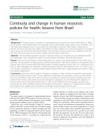
HNE caused cell death in human osteoarthritis
chondrocytes
To investigate the effect of HNE on chondrocyte apoptosis,
we first documented HNE cytotoxicity by MTT assay. After 16
hours of incubation, up to 10 μM HNE did not alter cell viabil-
ity, but 20 and 30 μM HNE was cytotoxic and significantly
decreased cell viability by approximately 50% and 52%,
respectively. Furthermore, pre-treatment with 200 μM NAC for
1 hour before adding 30 μM HNE (Figure 1a) completely pre-
vented HNE-induced cell death. However, the addition of anti-
Fas/CD95 (20 μg/mL) (Figure 1b) or the PARP inhibitor
INH
2
BP (50 and 100 μM) (Figure 1c) 1 hour before incubation
with 30 μM HNE partially prevented HNE-induced cell death.
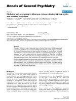
Next, Hoechst 33342 staining was undertaken to examine the
morphological changes occurring to chondrocytes. Upon
exposure to 30 μM HNE, numerous chondrocytes exhibited
characteristics typical of apoptosis with highly condensed
nuclei (Figure 2). In contrast, very few apoptotic cells were
observed in untreated cells.
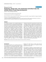
HNE induced caspases activation
Caspases play important roles in the terminal execution of
apoptosis induced by various stimuli. Caspase-3, -8, and -9
activities were measured with commercial kits, and their pro-
tein cleavage was detected by Western blot analysis. Com-
pared with untreated cells, HNE incubation resulted in a time-
dependent increase of caspase-8 activity (Figure 3a). HNE
significantly induced caspase-9 activity after 2, 4, and 8 hours
of incubation and significantly induced caspase-3 activity after
4 and 8 hours of incubation. At 16 hours, both caspase-3 and
-9 activities were reduced to the control level (Figure 3b, c). At
the protein level, 20 and 30 μM HNE decreased pro-caspase-
8, -9, and -3 levels after 16 hours of incubation, probably via
the cleavage process of the pro-caspase (Figure 3d). In con-
trast, the addition of 200 μM NAC prevented the HNE-
induced caspase activation.
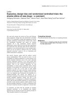
HNE affected Bcl-2 and Bax expression and induced
cytochrome c release from mitochondria
The Bcl-2 family is involved in apoptosis by regulating mem-
brane permeability and induces cytochrome c release from
mitochondria into the cytosol [22-24]. To investigate the
effects of HNE on Bcl-2 and Bax expression, cells were
Available online />Page 5 of 11
(page number not for citation purposes)
treated with 30 μM HNE for increasing incubation times (0 to
16 hours) and then ELISA and Western blot experiments were
performed. The level of the anti-apoptotic protein Bcl-2 was
significantly decreased after 4 hours of incubation with 30 μM
HNE (Figure 4a). In contrast, HNE at this concentration
increased the apoptotic protein Bax after 4 and 8 hours of
incubation and remained elevated at 16 hours of incubation
(Figure 4b). We then analyzed the effect of HNE on cyto-
Figure 1
4-Hydroxynonenal (HNE)-induced cell death4-Hydroxynonenal (HNE)-induced cell death. (a) Chondrocytes were
pre-incubated for 1 hour with or without 200 μM N-acetyl-cysteine
(NAC) followed by another incubation for 16 hours with increasing con-
centrations of HNE (0 to 30 μM). Chondrocytes were pre-incubated for
1 hour with or without (b) 20 μg/mL anti-Fas/CD95 antibody or (c) 50
and 100 μM INH
2
BP followed by another incubation for 16 hours with
or without 30 μM HNE. Cell viability was evaluated by MTT assay. Data
are mean ± standard error of the mean (n = 8). Statistics: Student
unpaired t test; ***P < 0.001 (30 μM HNE versus untreated cells),
#
P <
0.05 (30 μM HNE+inhibitor versus 30 μM HNE).
Figure 2
Nuclear morphology study for apoptosisNuclear morphology study for apoptosis. Chondrocytes were incu-
bated for 16 hours with or without 30 μM 4-hydroxynonenal (HNE),
stained with Hoechst 33258, and then analyzed by fluorescence micro-
scopy.
Arthritis Research & Therapy Vol 10 No 5 Vaillancourt et al.
Page 6 of 11
(page number not for citation purposes)
chrome c release from mitochondria to the cytosol. As shown
in Figure 4c, the cytochrome c level in cytosolic fractions sig-
nificantly increased in chondrocytes treated with 20 or 30 μM
HNE for 16 hours (Figure 4c). However, the pre-incubation of
chondrocytes with 200 μM NAC prevented HNE-induced
DNA fragmentation, PARP activation, and AIF translocation to
the nucleus.
Figure 3
Caspase-8, -9, and -3 activation by 4-hydroxynonenal (HNE)Caspase-8, -9, and -3 activation by 4-hydroxynonenal (HNE). Chondro-
cytes were treated with 30 μM HNE for the indicated times, and enzy-
matic activities of caspase-8 (a), caspase-9 (b), or caspase-3 (c) were
determined with commercial kits. (d) Chondrocytes were pre-incubated
for 1 hour with or without 200 μM N-acetyl-cysteine (NAC) followed by
another incubation for 16 hours with increasing concentrations of HNE
(0 to 30 μM). Pro-caspase-8, pro-caspase-9, and pro-caspase-3 were
analyzed by Western blot. Data are mean ± standard error of the mean
(n = 8) and expressed as a percentage of untreated cells. Statistics:
Student unpaired t test; *P < 0.05, **P < 0.01, ***P < 0.001.
Figure 4
4-Hydroxynonenal (HNE) modified Bcl-2/Bax protein expression and induced cytochrome c release from mitochondria4-Hydroxynonenal (HNE) modified Bcl-2/Bax protein expression and
induced cytochrome c release from mitochondria. Chondrocytes were
treated with 30 μM HNE for the indicated times and then Bcl-2 (a) and
Bax (b) protein levels were determined by commercial kit and Western
blot, respectively. (c) Chondrocytes were pre-incubated with or without
200 μM N-acetyl-cysteine (NAC) for 1 hour followed by another incu-
bation for 16 hours in the presence of increasing concentrations of
HNE (0 to 30 μM). Cytochrome c level was assessed in cytosolic frac-
tions with a kit. *P < 0.05, **P < 0.01, ***P < 0.001.
Available online />Page 7 of 11
(page number not for citation purposes)
HNE induced DNA fragmentation, PARP cleavage, and
apoptosis-inducing factor translocation to the nucleus
Nuclear damage is very important in cell death. Therefore, we
further studied the role of HNE in DNA fragmentation, PARP
activation, and AIF translocation to the nucleus. Chondrocytes
were exposed to HNE in the presence or absence of 200 μM
NAC. The extent of nuclear DNA fragmentation was measured
quantitatively by ELISA. As shown in Figure 5a, the level of
cytoplasmic histone-associated DNA fragments was
increased when DNAs were extracted from chondrocytes after
exposure to 20 and 30 μM HNE for 16 hours compared with
the control. In contrast, cells pre-treated with 200 μM NAC
were protected against HNE-induced DNA fragmentation.
PARP activation and AIF translocation to the nuclei during
apoptosis are implicated on a large scale in DNA fragmenta-
tion and peripheral chromatin condensation. As shown in Fig-
ure 5b, 30 μM HNE induced PARP cleavage and AIF
translocation in the nuclei after 4 hours of incubation. Moreo-
ver, pre-treatment with 200 μM NAC blocked PARP cleavage
and AIF translocation.
HNE induced Fas/CD95 and p53 expression but inhibited
Akt
To study the molecular mechanisms involved in the induction
of cell death by HNE, we examined Akt activity as well as Fas/
CD95 and p53 protein expression in chondrocytes after differ-
ent incubation times. Western blot analysis indicated that 30
μM HNE elicited Fas/CD95 and p53 protein expression but,
in contrast, reduced survival signalling, including the phospho-
rylated form of Akt (Figure 6). Total Akt was unchanged.
HNE altered redox status and energy metabolism
In the next series of experiments, we determined whether the
redox status and energy metabolism of chondrocytes could be
implicated in part in HNE-induced cell death. As illustrated in
Figure 7, 30 μM HNE decreased the reduced form of GSH
and the reducing equivalent, NADPH, needed for its regener-
ation (Figure 7a), after 16 hours of incubation. We additionally
established that HNE, at this concentration, evoked a signifi-
cant diminution of energy depletion through inhibition of
mNADP
+
-ICDH activity (Figure 7b), glucose uptake (Figure
7c), and intracellular ATP synthesis (Figure 7d).
HNE-induced cell death is controlled by GSTA4-4
expression
GSTA4-4 is a known aldehyde-detoxifying enzyme as has
been shown by previous studies [16,17]. To assess the func-
tional consequences of GSTA4-4 inhibition versus overex-
pression in chondrocytes, the cytotoxicity of 30 μM HNE was
evaluated by MTT cytotoxicity assay. First, the ablation of
GSTA4-4 with GSTA4-4 siRNA in isolated chondrocytes aug-
mented the HNE-induced cell mortality as measured by MTT
assay at 4, 8, and 16 hours of incubation (Figure 8a). These
results indicate that GSTA4-4 offers a significant protection
Figure 5
4-Hydroxynonenal (HNE) induced DNA fragmentation, poly (ADP-ribose) polymerase (PARP) cleavage, and apoptosis-inducing factor (AIF) translocation to the nucleus4-Hydroxynonenal (HNE) induced DNA fragmentation, poly (ADP-
ribose) polymerase (PARP) cleavage, and apoptosis-inducing factor
(AIF) translocation to the nucleus. Chondrocytes were pre-incubated
for 1 hour with or without 200 μM N-acetyl-cysteine (NAC) and then
incubated for another 16 hours with 30 μM HNE or with increasing
concentrations of HNE (0 to 30 μM). (a) The cytoplasmic histone-asso-
ciated DNA fragments were quantified with a kit. (b) Chondrocytes
were pre-incubated for 1 hour with or without 200 μM NAC followed by
another incubation with 30 μM HNE at different incubation times.
PARP cleavage and AIF translocation in nuclear fractions were ana-
lyzed by Western blot. Data are mean ± standard error of the mean and
expressed as a percentage of untreated cells. Statistics: Student
unpaired t test; *P < 0.05, **P < 0.01.
Figure 6
4-Hydroxynonenal (HNE) induced Fas/CD95 and p53 protein expres-sion and reduced Akt phosphorylation4-Hydroxynonenal (HNE) induced Fas/CD95 and p53 protein expres-
sion and reduced Akt phosphorylation. Chondrocytes were incubated
with 30 μM HNE for increasing incubation times. Total cell lysates or
nuclear extracts (~20 μg) were subjected to Western analysis (n = 8)
using antibodies anti-Fas/CD95, anti-p53, anti-phospho Akt, and anti-
total Akt.
Arthritis Research & Therapy Vol 10 No 5 Vaillancourt et al.
Page 8 of 11
(page number not for citation purposes)
Figure 7
4-Hydroxynonenal (HNE) altered redox status and energy metabolism in osteoarthritis chondrocytes4-Hydroxynonenal (HNE) altered redox status and energy metabolism
in osteoarthritis chondrocytes. Chondrocytes were treated for 16 hours
with 30 μM HNE, and cellular extracts were subjected to different anal-
ysis to determine (a) GSSG/(GSSG+GSH) and NADP/
(NADPH+NADP) ratios, (b) mNADP
+
-ICDH activity, (c) glucose
uptake, and (d) ATP level. Data are mean ± standard error of the mean
and expressed as a percentage of untreated cells. Statistics: Student
unpaired t test; **P < 0.01, ***P < 0.001. GSH, glutathione; GSSG,
oxidized glutathione; mNADP
+
-ICDH, mitochondrial NADP
+
-dependent
isocitrate dehydrogenase.
Figure 8
Glutathione-S-transferase A4-4 (GSTA4-4) controlled the 4-hydrox-ynonenal (HNE) cytotoxicityGlutathione-S-transferase A4-4 (GSTA4-4) controlled the 4-hydrox-
ynonenal (HNE) cytotoxicity. Chondrocytes were transfected with (a)
small interfering RNA (siRNA) GSTA4-4 or with (c) wild-type or mutant
GSTA4-4 expression plasmids, and then cell viability was determined
by MTT assay. (b) and (d) GSTA4-4 protein expression was evaluated
respectively in cellular extracts of transfected chondrocytes with siRNA
or expression plasmids of GSTA4-4 by Western blotting. Data are
mean ± standard error of the mean and expressed as a percentage of
untreated cells. Statistics: Student unpaired t test; *P < 0.05, **P <
0.01, ***P < 0.001. CTL, control; MTT, 3-(4,5-dimethylthiazol-2-yl)-2,5-
diphenyltetrazolium bromide; mut, mutant; wt, wild-type.
Available online />Page 9 of 11
(page number not for citation purposes)
against HNE-induced DNA damage in chondrocyte cells and
that siRNA ablation of this enzyme augments the HNE-
induced cell death. The increase in cell mortality in transfected
chondrocytes with GSTA4-4 siRNA would be attributed to the
inhibition of GSTA4-4 expression by more than 80% as com-
pared with control siRNA (Figure 8b). Second, we tested
whether the increased HNE-metabolizing capacity conferred
on these cells by transfection of GSTA4-4 expression vectors
could reverse the cytotoxic effects of HNE. Our data showed
that GSTA4-4 overexpression provided cell resistance to
direct HNE cytotoxicity (Figure 8c). Western blot analysis of
cell extracts with the polyclonal antibody against GSTA4-4
revealed a strong band in cellular extracts of chondrocytes
transfected with GSTA4-4 and a weak signal in untransfected
cells (Figure 8d).
Discussion
There is growing evidence that HNE, generated during the
LPO process, is an efficient cell signalling molecule and con-
sidered as a key mediator of oxidative stress-induced patho-
physiological effects. In fact, by modulating the expression of
different genes, HNE exhibits a wide array of biological activi-
ties, including signal transduction, gene expression, and mod-
ulation of cell proliferation [13]. In particular, the relevance of
HNE to joint biology and pathology is now becoming clearer.
In our previous study, we reported, for the first time, that HNE
was significantly increased in synovial fluids of OA patients
and in OA chondrocytes treated with ROS donors [18]. We
demonstrated that 10 μM HNE affects Col II and MMP-13 at
the transcriptional and post-translational levels, indicating that
HNE could play a role in cartilage degradation in OA. In a sec-
ond study, we identified two distinct mechanisms of HNE
action in OA chondrocytes, which included upregulation of
COX-2 via ATF/CRE activation and iNOS inhibition via NF-κB
inactivation [19]. In the present study, we evaluated the poten-
tial role of HNE at high concentrations in the apoptotic proc-
ess in OA chondrocytes. With the ultimate goal of clarifying
this role, we documented the ability of HNE to modulate mark-
ers of apoptosis, redox status, and energy metabolism of
chondrocytes. Then, we investigated the potential role of the
HNE-metabolizing enzyme, GSTA4-4, in the control of
chondrocyte death. To date, no data have demonstrated the
effect of HNE on apoptotic signalling in OA.
In the present study, we reported morphological and biochem-
ical evidence implicating HNE in the induction of chondrocyte
apoptosis in a dose- and time-dependent manner, and this
effect could be inhibited by NAC addition. First, we observed
that HNE up to 10 μM did not alter cell viability but equal or
greater than to 20 μM, HNE was cytotoxic and significantly
decreased cell viability (up to 50%) and induced chromatin
condensation, compared with untreated cells. Pre-treatment
of cells with anti-Fas/CD95 or PARP inhibitor partially reduced
cell mortality, suggesting a role for Fas/CD95 and PARP in
HNE-inducing cell death. Second, in investigating the classi-
cal markers of apoptosis, we obtained data showing that HNE
induced caspase-8, -9, and -3 activities and cleavage, AIF and
cytochrome c release from mitochondria, PARP activation,
and DNA fragmentation. These effects were prevented by
NAC. A major question arising with regard to apoptosis in gen-
eral is whether the apoptotic response of chondrocytes to
HNE requires upregulation of pro-apoptotic protein synthesis
or whether it relies on pre-existing apoptotic machinery. Our
data show that the apoptotic response of chondrocytes to
HNE is associated with decreased Bcl-2 expression and
increased Bax expression. This suggests that chondrocytes
need to modulate the synthesis of at least several anti- and
pro-apototic factors to be able to undergo apoptosis when
stimulated by HNE. It is noteworthy that HNE is capable of
inducing apoptosis in several cell types, including hepatic
cells, murine alveolar macrophages, RAW 264.7 cells, neu-
rons as well as colonic cancer cells [15,25,26]. It is well doc-
umented that these cells upregulate pro-apoptotic factors (for
example, caspases, PARP, Bax, and Bcl-2) to undergo apop-
tosis in response to HNE. The DNA fragmentation evoked by
HNE required PARP activation and AIF translocation in the
nucleus [27,28]. Moreover, our data disclosed that HNE-
induced apoptosis in chondrocytes needed the induction of
p53 and Fas/CD95 protein expression in a time-dependent
manner. These data are in concordance with those in the liter-
ature suggesting that HNE can induce apoptosis in various
cells through the death receptor Fas/CD95-mediated extrinsic
pathway as well as through the p53-dependent intrinsic path-
way [29,30]. The induction of Fas/CD95 by HNE was subse-
quently followed by the activation of JNKs, caspase 3, and the
onset of apoptosis in human lens epithelial cells [29]. How-
ever, the induction of p53 by HNE remains unclear. In RAW
264.7 cells, Haynes and colleagues [25] reported that HNE-
induced apoptosis was not associated with p53 accumula-
tion. This suggests that the apoptosis process elicited by HNE
could vary in different cell types due to changes in the availa-
bility or sensitivity of specific regulatory pathways that are acti-
vated by the aldehyde.
Investigation of the molecular mechanisms of HNE-induced
apoptosis in OA chondrocytes showed that HNE reduced ser-
ine/theonine kinase Akt activity. It has also been established
that HNE-induced apoptosis leads to the inhibition of Akt
phosphorylation. Akt activation results in the phosphorylation
of numerous other proteins involved in the regulation of glu-
cose metabolism, cell proliferation, apoptosis, cell migration,
and gene expression [31]. HNE is definitely able to interfere
with Akt activity, but so far the available findings appear, at
least in part, to be contradictory. In fact, Liu and colleagues
[32] reported that HNE (20 μ
M) induced apoptosis in human
T-cell leukemia Jurkat cells through impairment of the Akt-
mediated cell survival pathway.
The next set of experiments was focused on analyzing redox
status and cell metabolism in apoptotic chondrocytes. Signifi-
Arthritis Research & Therapy Vol 10 No 5 Vaillancourt et al.
Page 10 of 11
(page number not for citation purposes)
cant inhibition of cellular GSH pools, mNADP
+
-ICDH activity,
glucose uptake, and ATP level was observed. Liu and col-
leagues [27] actually showed that exogenously added HNE
quickly reduced cellular GSH levels in human T-lymphoma Jur-
kat cells. Parallelling the change in GSH levels, GSSG levels
decreased, suggesting that HNE is directly reacting with GSH
for consumption rather than acting as a source of pro-oxidants
to simply promote GSH/GSSG exchange. It is, however, also
possible that HNE decreased the GSH pool through inhibition
of GSH synthesis. In any case, pre-treatment of cells with anti-
oxidants, such as cysteine, NAC, and dithiothreitol, inhibited
the action of HNE to reduce the GSH/GSSG pool, supporting
the view that SH group-reactive HNE activity is primarily impor-
tant for the observed event. On the other hand, the inhibition
of mNADP
+
-ICDH, an important regulator of the citric acid
cycle, by HNE supports the role of this aldehyde in the altera-
tion of energy metabolism in different cell types as reported by
the literature data. Our previous study revealed that mNADP
+
-
ICDH was considered as a potential target for HNE binding
[33]. This key enzyme in cellular defence against oxidative
damage supplies NADPH in the mitochondria needed for the
regeneration of mitochondrial GSH [34]. In the present study,
we further demonstrated HNE cytotoxicity in chondrocytes
presumably via ATP depletion caused by the inhibition of mito-
chondrial respiratory enzymes and glucose uptake. Glucose
serves as the major energy substrate for articular chondro-
cytes and as the main precursor for the synthesis of ECM gly-
cosaminoglycans in cartilage [35,36]. The inhibition of
proteoglycan synthesis in OA chondrocytes by HNE (data not
shown) could be attributed to glucose uptake blockage
observed in the present work. ATP is the main energy source
for cellular proliferation, differentiation, and apoptosis [37,38].
Kanwar and colleagues [39] demonstrated that cellular ATP
depletion markedly decreased the synthesis of sulfated prote-
oglycans by a mechanism that was normalized by increasing
cellular ATP levels.
To determine whether HNE-metabolizing enzymes played a
role in chondrocyte apoptosis, we conducted additional exper-
iments to show that the changes in GSTA4-4 expression
could have an impact in HNE-induced cell death. In chondro-
cytes, GSTA4-4 ablation with GSTA4-4 siRNA augmented
the cytotoxic effect of HNE as determined by MTT assay. In
contrast, the overexpression of this enzyme in chondrocytes
offered significant protection against HNE-induced cell cyto-
toxicity. These data concur with a previous report indicating
that accelerated HNE metabolism in HL-60 and K562 human
erythroleukemia cells overexpressing GSTA4-4 blocks/delays
apoptosis triggered by HNE [16,17]. Increased HNE-metabo-
lizing capacity in GSTA4-4-transfected cells appeared to
result in an increased rate of proliferation as well as decreased
cell death by preventing PARP and caspase activation. Trans-
fection of GSTA4-4 also protected against cytotoxic H
2
O
2
cytotoxicity to a similar degree, an effect that could be
ascribed either to the direct enzymatic detoxification of H
2
O
2
through the GSH-peroxidase activity of GSTA4-4 or to
increased metabolism via GSH conjugation of HNE formed as
a consequence of H
2
O
2
exposure.
Conclusion
In this study, we identified, for the first time, a novel mechanism
linking oxidative stress to apoptosis signalling in OA chondro-
cytes through the action of HNE, an LPO end product. Our
data suggest that the increased level of HNE in articular tissue
may contribute to OA development via its ability to alter cellular
viability and the metabolic activity of chondrocytes. In the light
of previous data on decreased GSTA4-4 activity in patients
with OA, particular interest should be addressed to the patho-
physiological role of this enzyme in OA development.
Competing interests
The authors declare that they have no competing interests.
Authors' contributions
FV performed the experimental study, contributed to the prep-
aration of the manuscript, and undertook the statistical analy-
sis. HF, PL, and JCF evaluated and interpreted the data and
assisted with the preparation of the manuscript. QS assisted
in the experiments and in the isolation of chondrocytes from
human cartilage. PR provided cartilage specimens and partic-
ipated to the study design. MB designed the study, supervised
the project, evaluated and interpreted the data, and prepared
the manuscript. All authors read and approved the final manu-
script.
Acknowledgements
The authors thank Dr Sanjay Awasthi for the generous gift of the
GSTA4-4 expression plasmids. This study was supported by the Arthri-
tis Society and Fonds de la recherche en santé du Québec (FRSQ)
(grant 5330). MB is a research scholar at the FRSQ.
References
1. Poole AR: An introduction to the pathophysiology of osteoar-
thritis. Front Biosci 1999, 4:D662-D670.
2. Sandell LJ, Aigner T: Articular cartilage and changes in arthritis.
An introduction: cell biology of osteoarthritis. Arthritis Res
2001, 3:107-113.
3. Blanco FJ, Guitian R, Vazquez-Martul E, de Toro FJ, Galdo F: Oste-
oarthritis chondrocytes die by apoptosis. A possible pathway
for osteoarthritis pathology. Arthritis Rheum 1998, 41:284-289.
4. Kim HA, Lee YJ, Seong SC, Choe KW, Song YW: Apoptotic
chondrocyte death in human osteoarthritis. J Rheumatol 2000,
27:455-462.
5. Mistry D, Oue Y, Chambers MG, Kayser MV, Mason RM:
Chondrocyte death during murine osteoarthritis. Osteoarthritis
Cartilage 2004, 12:131-141.
6. Hashimoto S, Ochs RL, Komiya S, Lotz M: Linkage of chondro-
cyte apoptosis and cartilage degradation in human osteoar-
thritis. Arthritis Rheum 1998, 41:1632-1638.
7. Murrell GA, Jang D, Williams RJ: Nitric oxide activates metallo-
protease enzymes in articular cartilage. Biochem Biophys Res
Commun 1995, 206:15-21.
8. Taskiran D, Stefanovic-Racic M, Georgescu H, Evans C: Nitric
oxide mediates suppression of cartilage proteoglycan synthe-
sis by interleukin-1. Biochem Biophys Res Commun 1994,
200:142-148.
9. Blanco FJ, Ochs RL, Schwarz H, Lotz M: Chondrocyte apoptosis
induced by nitric oxide. Am J Pathol 1995, 146:75-85.
Available online />Page 11 of 11
(page number not for citation purposes)
10. Del CM Jr, Loeser RF: Nitric oxide-mediated chondrocyte cell
death requires the generation of additional reactive oxygen
species. Arthritis Rheum 2002, 46:394-403.
11. Hashimoto S, Takahashi K, Amiel D, Coutts RD, Lotz M: Chondro-
cyte apoptosis and nitric oxide production during experimen-
tally induced osteoarthritis. Arthritis Rheum 1998,
41:1266-1274.
12. Esterbauer H, Schaur RJ, Zollner H: Chemistry and biochemistry
of 4-hydroxynonenal, malonaldehyde and related aldehydes.
Free Radic Biol Med 1991, 11:81-128.
13. Poli G, Schaur RJ, Siems WG, Leonarduzzi G: 4-Hydroxynone-
nal: a membrane lipid oxidation product of medicinal interest.
Med Res Rev 2008, 28:569-631.
14. Barrera G, Pizzimenti S, Laurora S, Briatore F, Toaldo C, Dianzani
MU: 4-hydroxynonenal and cell cycle. Biofactors 2005,
24:151-157.
15. Cerbone A, Toaldo C, Laurora S, Briatore F, Pizzimenti S, Dianzani
MU, Ferretti C, Barrera G: 4-Hydroxynonenal and PPARgamma
ligands affect proliferation, differentiation, and apoptosis in
colon cancer cells. Free Radic Biol Med 2007, 42:1661-1670.
16. Cheng JZ, Singhal SS, Saini M, Singhal J, Piper JT, Van Kuijk FJ,
Zimniak P, Awasthi YC, Awasthi S: Effects of mGST A4 transfec-
tion on 4-hydroxynonenal-mediated apoptosis and differentia-
tion of K562 human erythroleukemia cells. Arch Biochem
Biophys 1999, 372:29-36.
17. Cheng JZ, Singhal SS, Sharma A, Saini M, Yang Y, Awasthi S, Zim-
niak P, Awasthi YC: Transfection of mGSTA4 in HL-60 cells pro-
tects against 4-hydroxynonenal-induced apoptosis by
inhibiting JNK-mediated signaling. Arch Biochem Biophys
2001, 392:197-207.
18. Morquette B, Shi Q, Lavigne P, Ranger P, Fernandes JC, Bender-
dour M: Production of lipid peroxidation products in osteoar-
thritic tissues: new evidence linking 4-hydroxynonenal to
cartilage degradation. Arthritis Rheum 2006, 54:271-281.
19. Vaillancourt F, Morquette B, Shi Q, Fahmi H, Lavigne P, Di Battista
JA, Fernandes JC, Benderdour M: Differential regulation of
cyclooxygenase-2 and inducible nitric oxide synthase by 4-
hydroxynonenal in human osteoarthritic chondrocytes
through ATF-2/CREB-1 transactivation and concomitant inhi-
bition of NF-kappaB signaling cascade. J Cell Biochem 2007,
100:1217-1231.
20. Altman R, Asch E, Bloch D, Bole G, Borenstein D, Brandt K,
Christy W, Cooke TD, Greenwald R, Hochberg M: Development
of criteria for the classification and reporting of osteoarthritis.
Classification of osteoarthritis of the knee. Diagnostic and
Therapeutic Criteria Committee of the American Rheumatism
Association. Arthritis Rheum 1986, 29:1039-1049.
21. Mosmann T: Rapid colorimetric assay for cellular growth and
survival: application to proliferation and cytotoxicity assays. J
Immunol Methods 1983, 65:55-63.
22. Cory S, Adams JM: The Bcl2 family: regulators of the cellular
life-or-death switch. Nat Rev Cancer 2002, 2:647-656.
23. Rosse T, Olivier R, Monney L, Rager M, Conus S, Fellay I, Jansen
B, Borner C: Bcl-2 prolongs cell survival after Bax-induced
release of cytochrome c. Nature 1998, 391:496-499.
24. Kluck RM, Bossy-Wetzel E, Green DR, Newmeyer DD: The
release of cytochrome c from mitochondria: a primary site for
Bcl-2 regulation of apoptosis. Science 1997, 275:1132-1136.
25. Haynes RL, Brune B, Townsend AJ: Apoptosis in RAW 264.7
cells exposed to 4-hydroxy-2-nonenal: dependence on cyto-
chrome C release but not p53 accumulation. Free Radic Biol
Med 2001, 30:884-894.
26. Peng ZF, Koh CH, Li QT, Manikandan J, Melendez AJ, Tang SY,
Halliwell B, Cheung NS: Deciphering the mechanism of HNE-
induced apoptosis in cultured murine cortical neurons: tran-
scriptional responses and cellular pathways. Neuropharmacol-
ogy 2007, 53:687-698.
27. Liu W, Kato M, Akhand AA, Hayakawa A, Suzuki H, Miyata T,
Kurokawa K, Hotta Y, Ishikawa N, Nakashima I: 4-hydroxynonenal
induces a cellular redox status-related activation of the cas-
pase cascade for apoptotic cell death. J Cell Sci 2000,
113:635-641.
28. Ramachandran V, Perez A, Chen J, Senthil D, Schenker S, Hend-
erson GI: In utero ethanol exposure causes mitochondrial dys-
function, which can result in apoptotic cell death in fetal brain:
a potential role for 4-hydroxynonenal. Alcohol Clin Exp Res
2001, 25:862-871.
29. Li J, Sharma R, Patrick B, Sharma A, Jeyabal PV, Reddy PM, Saini
MK, Dwivedi S, Dhanani S, Ansari NH, Zimniak P, Awasthi S,
Awasthi YC: Regulation of CD95 (Fas) expression and Fas-
mediated apoptotic signaling in HLE B-3 cells by 4-hydrox-
ynonenal. Biochemistry 2006, 45:12253-12264.
30. Awasthi YC, Sharma R, Sharma A, Yadav S, Singhal SS, Chaudary
P, Awasthi S: Self-regulatory role of 4-hydroxynonenal in sign-
aling for stress-induced programmed cell death. Free Radic
Biol Med 2008, 45:111-118.
31. Brazil DP, Hemmings BA: Ten years of protein kinase B signal-
ling: a hard Akt to follow. Trends Biochem Sci 2001,
26:657-664.
32. Liu W, Akhand AA, Takeda K, Kawamoto Y, Itoigawa M, Kato M,
Suzuki H, Ishikawa N, Nakashima I: Protein phosphatase 2A-
linked and -unlinked caspase-dependent pathways for down-
regulation of Akt kinase triggered by 4-hydroxynonenal. Cell
Death Differ 2003, 10:772-781.
33. Benderdour M, Charron G, Deblois D, Comte B, Des Rosiers C:
Cardiac mitochondrial NADP
+
-isocitrate dehydrogenase is
inactivated through 4-hydroxynonenal adduct formation: an
event that precedes hypertrophy development. J Biol Chem
2003, 278:45154-45159.
34. Jo SH, Son MK, Koh HJ, Lee SM, Song IH, Kim YO, Lee YS, Jeong
KS, Kim WB, Park JW, Song BJ, Huh TL: Control of mitochon-
drial redox balance and cellular defense against oxidative
damage by mitochondrial NADP
+
-dependent isocitrate dehy-
drogenase. J Biol Chem 2001, 276:16168-16176.
35. Wang J, Zhou J, Bondy CA: Igf1 promotes longitudinal bone
growth by insulin-like actions augmenting chondrocyte hyper-
trophy. FASEB J 1999, 13:1985-1990.
36. Mobasheri A, Vannucci SJ, Bondy CA, Carter SD, Innes JF,
Arteaga MF, Trujillo E, Ferraz I, Shakibaei M, Martin-Vasallo P: Glu-
cose transport and metabolism in chondrocytes: a key to
understanding chondrogenesis, skeletal development and
cartilage degradation in osteoarthritis. Histol Histopathol 2002,
17:1239-1267.
37. Agresti C, Meomartini ME, Amadio S, Ambrosini E, Volonte C,
Aloisi F, Visentin S: ATP regulates oligodendrocyte progenitor
migration, proliferation, and differentiation: involvement of
metabotropic P2 receptors. Brain Res Brain Res Rev 2005,
48:157-165.
38. Bernardi P, Scorrano L, Colonna R, Petronilli V, Di Lisa F: Mito-
chondria and cell death. Mechanistic aspects and methodo-
logical issues. Eur J Biochem 1999, 264:687-701.
39. Kanwar Y, Yoshinaga Y, Liu Z, Wallner E, Carone F: Biosynthetic
regulation of proteoglycans by aldohexoses and ATP. Proc
Natl Acad Sci USA 1992, 89:8621-8625.