Báo cáo y học: "Role of STAT4 polymorphisms in systemic lupus erythematosus in a Japanese population: a case-control association study of the STAT1-STAT4 region" pot
Bạn đang xem bản rút gọn của tài liệu. Xem và tải ngay bản đầy đủ của tài liệu tại đây (223.61 KB, 9 trang )
Open Access
Available online />Page 1 of 9
(page number not for citation purposes)
Vol 10 No 5
Research article
Role of STAT4 polymorphisms in systemic lupus erythematosus in
a Japanese population: a case-control association study of the
STAT1-STAT4 region
Aya Kawasaki
1
, Ikue Ito
1
, Koki Hikami
1
, Jun Ohashi
1
, Taichi Hayashi
2
, Daisuke Goto
2
,
Isao Matsumoto
2
, Satoshi Ito
2
, Akito Tsutsumi
2,3
, Minori Koga
4
, Tadao Arinami
4
,
Robert R Graham
5
, Geoffrey Hom
5
, Yoshinari Takasaki
6
, Hiroshi Hashimoto
6
,
Timothy W Behrens
5
, Takayuki Sumida
2
and Naoyuki Tsuchiya
1
1
Molecular and Genetic Epidemiology Laboratory, Doctoral Program in Life System Medical Sciences, Graduate School of Comprehensive Human
Sciences, University of Tsukuba, 1-1-1 Tennodai, Tsukuba 305-8575, Japan
2
Division of Clinical Immunology, Doctoral Program in Clinical Sciences, Graduate School of Comprehensive Human Science, University of Tsukuba,
1-1-1 Tennodai, Tsukuba 305-8575, Japan
3
Department of Medicine, Takikawa Municipal Hospital, 2-2-34 Omachi, Takikawa 073-0033, Japan
4
Department of Medical Genetics, Doctoral Program in Life System Medical Sciences, Graduate School of Comprehensive Human Sciences,
University of Tsukuba, 1-1-1 Tennodai, Tsukuba 305-8575, Japan
5
Genentech, Inc., 1 DNA Way, South San Francisco, CA 94080, USA
6
Division of Rheumatology, Department of Internal Medicine, 2-1-1 Hongo, Bunkyo-ku, Tokyo 113-8421, Japan
Corresponding author: Naoyuki Tsuchiya,
Received: 15 Aug 2008 Revisions requested: 5 Sep 2008 Revisions received: 16 Sep 2008 Accepted: 19 Sep 2008 Published: 19 Sep 2008
Arthritis Research & Therapy 2008, 10:R113 (doi:10.1186/ar2516)
This article is online at: />© 2008 Kawasaki et al.; licensee BioMed Central Ltd.
This is an open access article distributed under the terms of the Creative Commons Attribution License ( />),
which permits unrestricted use, distribution, and reproduction in any medium, provided the original work is properly cited.
Abstract
Introduction Recent studies identified STAT4 (signal
transducers and activators of transcription-4) as a susceptibility
gene for systemic lupus erythematosus (SLE). STAT1 is
encoded adjacently to STAT4 on 2q32.2-q32.3, upregulated in
peripheral blood mononuclear cells from SLE patients, and
functionally relevant to SLE. This study was conducted to test
whether STAT4 is associated with SLE in a Japanese
population also, to identify the risk haplotype, and to examine the
potential genetic contribution of STAT1. To accomplish these
aims, we carried out a comprehensive association analysis of 52
tag single nucleotide polymorphisms (SNPs) encompassing the
STAT1-STAT4 region.
Methods In the first screening, 52 tag SNPs were selected
based on HapMap Phase II JPT (Japanese in Tokyo, Japan) data,
and case-control association analysis was carried out on 105
Japanese female patients with SLE and 102 female controls. For
associated SNPs, additional cases and controls were
genotyped and association was analyzed using 308 SLE
patients and 306 controls. Estimation of haplotype frequencies
and an association study using the permutation test were
performed with Haploview version 4.0 software. Population
attributable risk percentage was estimated to compare the
epidemiological significance of the risk genotype among
populations.
Results In the first screening, rs7574865, rs11889341, and
rs10168266 in STAT4 were most significantly associated (P <
0.01). Significant association was not observed for STAT1.
Subsequent association studies of the three SNPs using 308
SLE patients and 306 controls confirmed a strong association
of the rs7574865T allele (SLE patients: 46.3%, controls:
33.5%, P = 4.9 × 10
-6
, odds ratio 1.71) as well as TTT
haplotype (rs10168266/rs11889341/rs7574865) (P = 1.5 ×
10
-6
). The association was stronger in subgroups of SLE with
nephritis and anti-double-stranded DNA antibodies. Population
attributable risk percentage was estimated to be higher in the
Japanese population (40.2%) than in Americans of European
descent (19.5%).
Conclusions The same STAT4 risk allele is associated with
SLE in Caucasian and Japanese populations. Evidence for a role
of STAT1 in genetic susceptibility to SLE was not detected. The
contribution of STAT4 for the genetic background of SLE may
be greater in the Japanese population than in Americans of
European descent.
anti-dsDNA: anti-double-stranded DNA; CI: confidence interval; IFN: interferon; IL: interleukin; IRF5: interferon regulatory factor-5; JPT: Japanese in
Tokyo, Japan; LD: linkage disequilibrium; OR: odds ratio; PAR%: population attributable risk percentage; RR: relative risk; SLE: systemic lupus ery-
thematosus; SNP: single nucleotide polymorphism; STAT: signal transducers and activators of transcription.
Arthritis Research & Therapy Vol 10 No 5 Kawasaki et al.
Page 2 of 9
(page number not for citation purposes)
Introduction
Systemic lupus erythematosus (SLE) is a complex disease
characterized by autoantibody production and involvement of
multiple organs, including kidneys. Both genetic and environ-
mental factors contribute to the development of SLE [1]. Until
now, several genes have been reported to be associated with
SLE, of which interferon regulatory factor-5 (IRF5) has been
identified as a susceptibility gene common to multiple popula-
tions [2-6]. Recently, association of STAT4 (signal transduc-
ers and activators of transcription-4) haplotype tagged by
rs7574865T with SLE was demonstrated in Caucasians [7].
Subsequently, two genome-wide association studies [8,9], a
study focused on the STAT4 region in Caucasians [10], and
replication studies in Colombians [11] and a Japanese popu-
lation [12] have confirmed the association. In addition, an
association of STAT4 with SLE phenotypes such as anti-dou-
ble-stranded DNA (anti-dsDNA) autoantibodies, renal disor-
der, and age at diagnosis was reported [10,13]. An
association of rs7574865 with other autoimmune diseases
such as rheumatoid arthritis and primary Sjögren syndrome
has also been demonstrated [7,11,12,14]. The STAT4 gene
encodes a transcription factor belonging to the STAT family
expressed in lymphocytes, macrophages, and dendritic cells.
STAT4 is essential for interleukin (IL)-12 signaling and induces
interferon-gamma (IFNγ) production and Th1 differentiation
[15]. STAT4 is also activated by type I IFNs (IFNα/β) [16].
Moreover, the requirement of STAT4 in IL-23-induced IL-17
production has been suggested [17]. Two isoforms of STAT4,
STAT4α and STAT4β, are known [18]. Expression of STAT4β,
lacking the transactivation domain, did not appear to be
affected by the STAT4 single nucleotide polymorphisms
(SNPs) [13]. STAT1, another member of the STAT family, is
activated by type I IFNs and IFNγ and plays an important role
in immune responses [19]. STAT1 has been reported to be
upregulated in peripheral blood mononuclear cells from SLE
patients and in kidneys of lupus mice with nephritis [20,21],
suggesting that STAT1 may play a role in the pathogenesis of
SLE. A possible role of SNPs in the STAT1-STAT4 region
other than the haplotype tagged by rs7574865T has recently
been excluded in Caucasians [10]. However, in view of sub-
stantial differences in disease-associated alleles among popu-
lations [2], such analysis should be performed in each
population. In this study, we carried out a comprehensive
association analysis of the STAT1-STAT4 region with SLE in
a Japanese population by scanning 52 tag SNPs of the region
encompassing STAT1 and STAT4.
Materials and methods
Patients and healthy controls
Patients and controls were recruited at Juntendo University,
the University of Tsukuba, and the University of Tokyo. All
patients and healthy controls were unrelated Japanese per-
sons living in the central part of Japan. Three hundred eight
SLE patients (18 males and 290 females, average age 41.4 ±
13.5 years) and 306 healthy individuals (119 males and 187
females, average age 32.6 ± 9.8 years) were studied. Diagno-
sis of SLE and classification of the patients into clinical sub-
sets were carried out according to the American College of
Rheumatology criteria for SLE [22]. There was no overlap in
cases or controls between this study and the recently reported
study in a Japanese population [12]. These studies were
reviewed and approved by the research ethics committees of
the University of Tsukuba, the University of Tokyo, and Jun-
tendo University. Informed consent was obtained from all
study participants.
Association study
Fifty-two tag SNPs in the STAT1-STAT4 region were selected
with an r
2
threshold of 0.9 based on the HapMap Phase II JPT
(Japanese in Tokyo, Japan) data. These tag SNPs captured
127 SNPs with a minor allele frequency of greater than or
equal to 0.05. First screening was performed in 105 Japanese
female SLE patients and 102 female healthy controls using the
GoldenGate SNP genotyping assay (Illumina, Inc., San Diego,
CA, USA). For the three SNPs that exhibited significant asso-
ciation (P < 0.01), additional samples were genotyped using
the TaqMan SNP Genotyping Assay (Applied Biosystems,
Foster City, CA, USA), and association was examined in 308
SLE patients and 306 healthy individuals.
Statistical analysis
Association of each SNP was analyzed by chi-square test.
Because of the replicative nature of this study, correction for
multiple testing was not performed, and unadjusted P values
are shown. Haplotype frequency estimation and association
analysis using the permutation test were performed with Hap-
loview version 4.0 software (Broad Institute of MIT and Har-
vard, Cambridge, MA, USA). In the haplotype analysis, the
genotype data for rs10168266, rs11889341, and rs7574865
were used and these SNPs were assumed to compose a sin-
gle haplotype block. In the permutation test, only frequencies
of haplotypes in this block were compared (that is, the 'Haplo-
types in Blocks Only' option was used). Ten million permuta-
tions were performed. To test the significance of each SNP
conditional on the genotypes of other SNPs, logistic regres-
sion analysis was performed under the additive model for the
minor allele. Assuming a polymorphic site with two alleles A
and a, genotypes were encoded as 0 = aa, 1 = Aa, and 2 =
AA. Population attributable risk percentage (PAR%) for the
risk genotype (rs7574865T/T and T/G) was estimated by the
formula
PAR% = Pe (RR - 1)/(Pe [RR - 1] + 1),
Available online />Page 3 of 9
(page number not for citation purposes)
where Pe represents the risk genotype frequency in the popu-
lation and RR represents relative risk of the risk genotype [23].
Given the low prevalence of SLE, Pe can be estimated based
on the genotype frequencies in healthy controls and RR can
be approximated by odds ratio (OR) for the risk genotypes.
Results and Discussion
The STAT4 gene is located on 2q32.2-q32.3 adjacently to
STAT1 gene, and the region encompassing STAT1 and
STAT4 spans approximately 180 kilobase pairs. In the first
screening, 52 tag SNPs in the STAT1-STAT4 region, selected
with an r
2
threshold of 0.9 based on the HapMap Phase II JPT
data, were genotyped in 105 Japanese female SLE patients
and 102 female healthy controls, and allele frequencies were
compared between SLE patients and controls. A linkage dise-
quilibrium (LD) plot and the results of the association study in
the STAT1-STAT4 region are shown in Figure 1. Pairwise r
2
values between 52 tag SNPs were calculated using genotyp-
ing data from 102 healthy individuals.
Among the tag SNPs, rs10168266C>T, rs11889341C>T,
and rs7574865G>T were most significantly associated with
SLE in the first screening (P < 0.01). Allele frequencies of
rs10168266T, rs11889341T, and rs7574865T were signifi-
cantly increased in SLE compared with healthy controls (Table
1 and Figure 1). These SNPs were located in the introns of
STAT4 and in LD with each other. In contrast, significant asso-
ciation was not detected for SNPs in the STAT1 region (P >
0.05).
To confirm the association detected in the first screening,
additional patients and controls were genotyped for the three
SNPs using the TaqMan SNP Genotyping Assay, and associ-
ation was examined in 308 SLE patients and 306 healthy con-
trols in total (Table 2). Significant deviation from Hardy-
Weinberg equilibrium was not detected in healthy controls (P
> 0.05). The rs7574865T allele, previously shown to be asso-
ciated with SLE in Caucasians, was significantly increased in
SLE patients (46.3%) compared with controls (33.5%, P =
4.9 × 10
-6
, OR 1.71). The association was compatible with the
dominant model, under which the OR was 2.19 (T/T + G/T
versus G/G).
The SNPs rs11889341 and rs10168266 were in LD with
rs7574865 (r
2
: 0.57 to 0.78, D': 0.91 to 0.97) and were also
significantly associated with SLE (allele frequency: P = 6.6 ×
10
-6
and P = 6.3 × 10
-6
, respectively). Haplotype analysis
revealed that the haplotype carrying rs10168266T,
rs11889341T, and rs7574865T was significantly increased
(SLE: 36.8%, control: 24.3%, P = 1.5 × 10
-6
) whereas the
haplotype carrying 10168266C, rs11889341C, and
rs7574865G was significantly decreased in SLE (SLE:
52.7%, control: 65.0%, P = 1.0 × 10
-5
). Logistic regression
analysis demonstrated that the association of each SNP lost
statistical significance when adjusted for genotype of the other
SNPs (Table 3). Thus, due to the strong LD, it was impossible
to identify a single causative SNP among the three.
We next tested whether STAT4 rs7574865 was associated
with phenotypes of SLE such as presence of nephritis, anti-
dsDNA antibodies, and early age of onset (less than 20 years)
as STAT4 genotype has been shown to be more strongly
associated with subgroups of SLE with these phenotypes [10]
(Table 4). Association of rs7574865 was observed both in
SLE patients with nephritis (P = 1.0 × 10
-5
, OR = 1.85) and
in those without nephritis (P = 0.0031, OR = 1.55). The asso-
ciation was stronger in SLE patients with nephritis, although
the difference between SLE with and without nephritis (case-
only analysis) did not reach statistical significance. Similarly,
rs7574865T was significantly increased in SLE patients with
anti-dsDNA antibodies compared with healthy controls,
whereas association was not detected in SLE patients without
anti-dsDNA antibodies. The frequency of rs7574865T was
slightly higher in the patients with an age of onset of less than
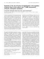
Figure 1
Linkage disequilibrium plot of the STAT1-STAT4 region in a Japanese population and first screening of 52 tag single nucleotide polymor-phisms (SNPs)Linkage disequilibrium plot of the STAT1-STAT4 region in a Japanese
population and first screening of 52 tag single nucleotide polymor-
phisms (SNPs). In the upper panel, P values for differences in allele fre-
quencies were calculated by chi-square test using two-by-two
contingency tables. The -log P value for each SNP is shown. In the
lower panel, r
2
values calculated using Haploview version 4.0 software
based on data from 102 healthy individuals are shown. The location
and direction of transcription of STAT1 and STAT4 are indicated by
arrows. SNPs rs10168266, rs11889341, and rs7574865 belong to
the same haplotype block.
Arthritis Research & Therapy Vol 10 No 5 Kawasaki et al.
Page 4 of 9
(page number not for citation purposes)
Table 1
Minor allele frequencies and P values for 52 tag single nucleotide polymorphisms in the STAT1-STAT4 region in the first screening
Minor allele frequency
SNP Chromosomal position
a
Minor allele SLE patients (n = 105) Controls (n = 102) P value
rs3771300 191543841 C 0.305 0.309 0.929
rs7575823 191544163 A 0.167 0.147 0.584
rs16824035 191545879 A 0.057 0.074 0.500
rs1914408 191548221 A 0.271 0.314 0.344
rs2066804 191550004 A 0.471 0.480 0.855
rs2280235 191552075 A 0.486 0.471 0.758
rs3755312 191554236 C 0.181 0.176 0.905
rs2280234 191558344 G 0.162 0.186 0.513
rs2280232 191559011 C 0.143 0.123 0.543
rs11887698 191563119 G 0.327 0.304 0.629
rs7562024 191563766 G 0.090 0.108 0.554
rs11904548 191567235 A 0.162 0.137 0.482
rs12693591 191568747 A 0.257 0.235 0.606
rs16833155 191569622 A 0.043 0.054 0.600
rs2066805 191571146 G 0.038 0.054 0.442
rs11677408 191574860 A 0.129 0.108 0.514
rs2030171 191577408 G 0.329 0.309 0.666
rs11693463 191578156 G 0.195 0.196 0.983
rs11885069 191578869 A 0.162 0.137 0.482
rs10199181 191581798 T 0.267 0.265 0.964
rs2066802 191582912 G 0.257 0.255 0.956
rs13029532 191584146 C 0.082 0.103 0.457
rs3024904 191603447 A 0.112 0.141 0.400
rs3024936 191603621 C 0.024 0.055 0.112
rs1517351 191604290 C 0.490 0.464 0.602
rs3024896 191604961 A 0.448 0.412 0.461
rs925847 191605785 A 0.538 0.490 0.330
rs3024886 191608694 A 0.457 0.417 0.407
rs6715106 191621279 G 0.067 0.083 0.520
rs16833215 191622044 G 0.495 0.441 0.270
rs1400654 191623918 T 0.066 0.083 0.524
rs3024861 191632851 T 0.471 0.397 0.127
rs1517352 191639709 A 0.481 0.397 0.086
rs10168266 191644049 A 0.400 0.245 7.6 × 10
-4
rs7594501 191646845 A 0.114 0.152 0.250
rs16833239 191648505 A 0.110 0.152 0.200
rs7601754 191648696 G 0.129 0.178 0.162
Available online />Page 5 of 9
(page number not for citation purposes)
20 years as compared with greater than or equal to 20 years,
although the difference was not statistically significant. These
tendencies are consistent with those reported in Caucasians
[10]. These interpretations were not affected when the signif-
icance level was corrected for the number of comparisons
(three phenotypes).
To evaluate the epidemiological significance of STAT4 poly-
morphism in the genetic background of SLE in the Japanese
population, we estimated the PAR% in Japanese persons and
Caucasians using our present data and previously reported
data [8,11,12] (Table 5). Because the frequency and OR of
the risk genotype of rs7574865 were greater in the Japanese
population than those of North Americans of European
descent [8], PAR% in the Japanese population (40.2%) was
much higher than that of the latter (19.5%). A similarly high
PAR% was observed in two of the three Japanese case-con-
trol series reported by Kobayashi and colleagues [12] and in
Colombians [11]. Because PAR% may be affected by the dif-
ference in the method of ascertainment of each study, this
comparison may not be completely valid. Nevertheless, these
observations suggested that the contribution of STAT4 for
SLE is greater in the Japanese population as compared with
the Americans of European descent.
At this point, molecular mechanisms that account for the asso-
ciation of STAT4 intron SNPs with SLE remain unclear. Stud-
ies with lupus model mice lacking Stat4 showed conflicting
results. Stat4 deficiency reduced nephritis and autoantibody
production in B6.NZM.Sle1.Sle2.Sle3 mice [24]. In contrast,
Stat4-deficient NZM (New Zealand mixed) mice developed
accelerated nephritis and increased mortality in the absence
of high levels of autoantibodies [25]. STAT4 has been shown
to be involved in the induction of IFNγ, differentiation of Th1
and Th17 cells, and signal transduction from type I IFN recep-
tors [15]. Th1 cytokines, especially IFNγ, have been shown to
play a role in the pathogenesis of lupus nephritis [26].
Recently, T cells from SLE patients were shown to produce
excessive amounts of IFNγ upon stimulation [27]. These
observations may implicate the role of STAT4 SNPs in IFNγ
production.
The role of type I IFNs in SLE has been established [1]. Ele-
vated serum type I IFN levels and expression of IFN-inducible
genes in peripheral mononuclear cells were reported in SLE
[28,29]. The association of IRF5, which induces type I IFNs,
with SLE has been established [2-6]. STAT4 is activated by
type I IFN as well as IL-12 signals and produces IFNγ [15].
Thus, STAT4 may also contribute to SLE as a component of
the type I IFN signal pathway. Furthermore, STAT4 has been
reported to transduce IL-12 signals to induce IFNγ production
in B cells [30].
It is interesting to note that significant association of STAT4
was not observed in SLE patients without anti-dsDNA anti-
bodies (Table 4). It would have been interesting to examine the
effect of the genotype on the levels, rather than presence or
absence, of anti-dsDNA antibody However, because the anti-
body levels fluctuate in association with disease activity and
treatment, association with the genotype should be examined
rs11889341 191651987 A 0.443 0.299 0.003
rs16833249 191656517 G 0.567 0.480 0.079
rs6434435 191662109 A 0.099 0.141 0.192
rs7574865 191672878 A 0.471 0.324 0.002
rs12463658 191673589 C 0.581 0.471 0.025
rs6752770 191681808 G 0.205 0.245 0.326
rs1551443 191704763 A 0.238 0.206 0.431
rs2356350 191710783 G 0.510 0.407 0.036
rs10189819 191716994 G 0.133 0.118 0.630
rs7596818 191717555 A 0.320 0.295 0.580
rs11685878 191717700 A 0.429 0.431 0.954
rs12991409 191717762 G 0.100 0.113 0.674
rs12327969 191719016 G 0.390 0.402 0.811
rs12988825 191722509 C 0.119 0.132 0.683
rs7572482 191723317 G 0.490 0.461 0.545
a
Chromosomal positions are shown according to the National Center for Biotechnology Information (Bethesda, MD, USA) reference assembly.
SLE, systemic lupus erythematosus; SNP, single nucleotide polymorphism; STAT, signal transducers and activators of transcription.
Table 1 (Continued)
Minor allele frequencies and P values for 52 tag single nucleotide polymorphisms in the STAT1-STAT4 region in the first screening
Arthritis Research & Therapy Vol 10 No 5 Kawasaki et al.
Page 6 of 9
(page number not for citation purposes)
Table 2
Association of STAT4 single nucleotide polymorphisms rs10168266, rs11889341, and rs7574865 with systemic lupus erythematosus
SLE patients (n = 308) Healthy controls (n = 306) P value Odds ratio 95% CI
Number Percentage Number Percentage
rs10168266
Genotype frequency
C/C 118 38.3 166 54.2
C/T 147 47.7 122 39.9 7.5 × 10
-5a
1.91 1.39–2.63
a
T/T 43 14.0 18 5.9
Allele frequency
T 233 37.8 158 25.8 6.3 × 10
-6
1.75 1.37–2.23
rs11889341
Genotype frequency
C/C 99 32.1 153 50.0
C/T 161 52.3 126 41.2 6.9 × 10
-6a
2.11 1.52–2.92
a
T/T 48 15.6 27 8.8
Allele frequency
T 257 41.7 180 29.4 6.6 × 10
-6
1.72 1.36–2.17
rs7574865
Genotype frequency
G/G 80 26.0 133 43.5
G/T 171 55.5 141 46.1 5.3 × 10
-6a
2.19 1.56–3.07
a
T/T 57 18.5 32 10.5
Allele frequency
T 285 46.3 205 33.5 4.9 × 10
-6
1.71 1.36–2.15
rs10168266/rs11889341/rs7574865
Haplotype frequency
CCG 52.7 65.0 1.0 × 10
-5b
TTT 36.8 24.3 1.5 × 10
-6b
CCT 4.9 5.1 NS
b
CTT 4.6 4.1 NS
b
a
P values, odds ratios, and 95% confidence intervals (CIs) were calculated under the dominant model for the minor allele.
b
P values were calculated by permutation
test using Haploview version 4.0 software. Ten million permutations were performed. NS, not significant; SLE, systemic lupus erythematosus; STAT, signal transducers
and activators of transcription.
Table 3
Logistic regression analysis of the systemic lupus erythematosus-associated single nucleotide polymorphisms in STAT4
P adjusted for
SNP P value rs10168266 rs11889341 rs7574865
rs10168266 4.9 × 10
-6
NA 0.272 0.146
rs11889341 4.7 × 10
-6
0.251 NA 0.388
rs7574865 2.1 × 10
-6
0.052 0.130 NA
NA, not applicable; SNP, single nucleotide polymorphism; STAT, signal transducers and activators of transcription.
Available online />Page 7 of 9
(page number not for citation purposes)
using the lifetime highest anti-dsDNA antibody level of each
patient. Such data were not available for this study, and we
hope that we can address this issue in the future.
Most of these observations imply that STAT4 risk genotype
may be associated with an elevated expression level and/or
function of STAT4 protein. A recent study reported that the
STAT4 risk allele was associated with overexpression of
STAT4 in osteoblasts but not in B cells [13]. To address the
significance of such findings, it will be necessary to examine
the effect of this genotype on the expression levels and splic-
ing isoforms in T and B cells.
Conclusion
Through comprehensive association analysis of the STAT1-
STAT4 region with SLE in the Japanese population, we dem-
onstrated that the same STAT4 risk allele in Caucasians was
strongly associated with susceptibility to SLE in the Japanese
population. In contrast, evidence for an association of STAT1
SNPs was not observed. The contribution of STAT4 SNPs to
Table 4
Association of STAT4 rs7574865 with characteristics of systemic lupus erythematosus such as nephritis, age of onset, and anti-
double-stranded-DNA antibodies
T allele P value Odds ratio (95% CI)
Number Frequency
Case subgroup versus healthy controls
Nephritis
Present (n = 165) 159 48.2% 1.0 × 10
-5
1.85 (1.41–2.42)
Absent (n = 138) 121 43.8% 0.0031 1.55 (1.16–2.07)
Anti-double-stranded DNA antibodies
Present (n = 130) 125 48.1% 4.9 × 10
-5
1.84 (1.37–2.47)
Absent (n = 34) 24 35.3% NS 1.08 (0.64–1.83)
Age of onset
<20 years (n = 86) 83 48.3% 3.9 × 10
-4
1.85 (1.32–2.60)
≥20 years (n = 198) 180 45.5% 1.4 × 10
-4
1.65 (1.28–2.14)
Healthy controls (n = 306) 205 33.5%
Case-only (present versus absent or <20 versus ≥ 20 years)
Nephritis NS 1.19 (0.86–1.64)
Anti-double-stranded DNA antibodies NS 1.70 (0.98–2.95)
Age of onset NS 1.12 (0.78–1.60)
Systemic lupus erythematosus (SLE) patients were stratified into subgroups according to the presence or absence of nephritis, anti-double-stranded DNA (anti-
dsDNA) antibodies, and age of onset (<20 or ≥ 20 years). Allele frequencies were compared between each SLE subgroup and healthy controls as well as between
SLE subgroups (case-only analysis, nephritis present versus absent, anti-dsDNA antibodies present versus absent, and age of onset <20 versus ≥ 20 years). CI,
confidence interval; NS, not significant; STAT, signal transducers and activators of transcription.
Table 5
Population attributable risk percentage of STAT4 rs7574865 under the dominant model
Population [reference] Frequency of (T/T+T/G) Odds ratio PAR%
Japanese (this study) 56.5% 2.19 40.2%
Japanese (TWMU) [
12] 52.3% 1.81 29.7%
Japanese (RIKEN) [12] 51.7% 1.51 20.8%
Japanese (Tokushima/Fukuoka) [
12] 51.9% 2.07 35.8%
Americans of European descent [
8] 41.2% 1.59 19.5%
Colombians [
11] 51.7% 1.87 31.0%
PAR%, population attributable risk percentage; RIKEN, The Institute of Physical and Chemical Research, Wako, Japan; STAT, signal transducers and activators of
transcription; TWMU, Tokyo Women's Medical University, Tokyo, Japan.
Arthritis Research & Therapy Vol 10 No 5 Kawasaki et al.
Page 8 of 9
(page number not for citation purposes)
the genetic background of SLE may be greater in the Japa-
nese population than in Americans of European descent.
Competing interests
RRG, GH, and TWB are employees of and hold stocks or
shares in Genentech, Inc. (South San Francisco, CA, USA).
The other authors declare that they have no competing
interests.
Authors' contributions
AK participated in the study design, carried out all genotyping
and statistical analyses, and wrote the manuscript. II, KH, MK,
and TA participated in the first screening using Illumina Gold-
enGate assay (with AK), including tag SNP selection, geno-
typing, and statistical analysis. JO carried out statistical
analysis with AK and helped in the manuscript preparation. TH,
DG, IM, SI, AT, YT, HH, and TS recruited Japanese patients
with SLE and collected clinical information. RRG and GH pro-
vided Caucasian data. NT conceived of the study, together
with TWB, and participated in its design and coordination,
recruited patients and controls, and helped in the manuscript
preparation. All authors read and approved the final
manuscript.
Acknowledgements
This work was supported by KAKENHI (Grant-in-Aid for Scientific
Research) (B) from the Japan Society for the Promotion of Science;
KAKENHI on the Priority Area 'Applied Genomics' from the Ministry of
Education, Culture, Sports, Science and Technology of Japan; and
grants from the Ministry of Health, Labour and Welfare of Japan; the
Japan Rheumatism Foundation; and the Naito Foundation.
References
1. Kyogoku C, Tsuchiya N: A compass that points to lupus: genetic
studies on type I interferon pathway. Genes Immun 2007,
8:445-455.
2. Kawasaki A, Kyogoku C, Ohashi J, Miyashita R, Hikami K, Kusaoi
M, Tokunaga K, Takasaki Y, Hashimoto H, Behrens TW, Tsuchiya
N: Association of IRF5 polymorphisms with systemic lupus
erythematosus in a Japanese population: support for a crucial
role of intron 1 polymorphisms. Arthritis Rheum 2008,
58:826-834.
3. Sigurdsson S, Nordmark G, Göring HH, Lindroos K, Wiman AC,
Sturfelt G, Jönsen A, Rantapää-Dahlqvist S, Möller B, Kere J,
Koskenmies S, Widén E, Eloranta ML, Julkunen H, Kristjansdottir
H, Steinsson K, Alm G, Rönnblom L, Syvänen AC: Polymor-
phisms in the tyrosine kinase 2 and interferon regulatory fac-
tor 5 genes are associated with systemic lupus
erythematosus. Am J Hum Genet 2005, 76:528-537.
4. Graham RR, Kyogoku C, Sigurdsson S, Vlasova IA, Davies LR,
Baechler EC, Plenge RM, Koeuth T, Ortmann WA, Hom G, Bauer
JW, Gillett C, Burtt N, Cunninghame Graham DS, Onofrio R, Petri
M, Gunnarsson I, Svenungsson E, Rönnblom L, Nordmark G,
Gregersen PK, Moser K, Gaffney PM, Criswell LA, Vyse TJ,
Syvänen AC, Bohjanen PR, Daly MJ, Behrens TW, Altshuler D:
Three functional variants of IFN regulatory factor 5 (IRF5)
define risk and protective haplotypes for human lupus. Proc
Natl Acad Sci USA 2007, 104:6758-6763.
5. Ferreiro-Neira I, Calaza M, Alonso-Perez E, Marchini M, Scorza R,
Sebastiani GD, Blanco FJ, Rego I, Pullmann R Jr, Pullmann R,
Kallenberg CG, Bijl M, Skopouli FN, Mavromati M, Migliaresi S,
Barizzone N, Ruzickova S, Dostal C, Schmidt RE, Witte T, Papas-
teriades C, Kappou-Rigatou I, Endreffy E, Kovacs A, Ordi-Ros J,
Balada E, Carreira P, Gomez-Reino JJ, Gonzalez A: Opposed
independent effects and epistasis in the complex association
of IRF5 to SLE. Genes Immun 2007, 8:429-438.
6. Kelly JA, Kelley JM, Kaufman KM, Kilpatrick J, Bruner GR, Merrill JT,
James JA, Frank SG, Reams E, Brown EE, Gibson AW, Marion
MC, Langefeld CD, Li QZ, Karp DR, Wakeland EK, Petri M, Ram-
sey-Goldman R, Reveille JD, Vilá LM, Alarcón GS, Kimberly RP,
Harley JB, Edberg JC: Interferon regulatory factor-5 is geneti-
cally associated with systemic lupus erythematosus in African
Americans. Genes Immun 2008, 9:187-194.
7. Remmers EF, Plenge RM, Lee AT, Graham RR, Hom G, Behrens
TW, de Bakker PI, Le JM, Lee HS, Batliwalla F, Li W, Masters SL,
Booty MG, Carulli JP, Padyukov L, Alfredsson L, Klareskog L, Chen
WV, Amos CI, Criswell LA, Seldin MF, Kastner DL, Gregersen PK:
STAT4 and the risk of rheumatoid arthritis and systemic lupus
erythematosus. N Engl J Med 2007, 357:
977-986.
8. Hom G, Graham RR, Modrek B, Taylor KE, Ortmann W, Garnier S,
Lee AT, Chung SA, Ferreira RC, Pant PV, Ballinger DG, Kosoy R,
Demirci FY, Kamboh MI, Kao AH, Tian C, Gunnarsson I, Bengtsson
AA, Rantapää-Dahlqvist S, Petri M, Manzi S, Seldin MF, Rönnblom
L, Syvänen AC, Criswell LA, Gregersen PK, Behrens TW: Associ-
ation of systemic lupus erythematosus with C8orf13-BLK and
ITGAM-ITGAX. N Engl J Med 2008, 358:900-909.
9. International Consortium for Systemic Lupus Erythematosus
Genetics (SLEGEN), Harley JB, Alarcón-Riquelme ME, Criswell
LA, Jacob CO, Kimberly RP, Moser KL, Tsao BP, Vyse TJ,
Langefeld CD: Genome-wide association scan in women with
systemic lupus erythematosus identifies susceptibility vari-
ants in ITGAM, PXK, KIAA1542 and other loci. Nat Genet 2008,
40:204-210.
10. Taylor KE, Remmers EF, Lee AT, Ortmann WA, Plenge RM, Tian C,
Chung SA, Nititham J, Hom G, Kao AH, Demirci FY, Kamboh MI,
Petri M, Manzi S, Kastner DL, Seldin MF, Gregersen PK, Behrens
TW, Criswell LA: Specificity of the STAT4 genetic association
for severe disease manifestations of systemic lupus
erythematosus. PLoS Genet 2008, 4:e1000084.
11. Palomino-Morales RJ, Rojas-Villarraga A, González CI, Ramírez G,
Anaya JM, Martín J: STAT4 but not TRAF1/C5 variants influence
the risk of developing rheumatoid arthritis and systemic lupus
erythematosus in Colombians. Genes Immun 2008,
9:379-382.
12. Kobayashi S, Ikari K, Kaneko H, Kochi Y, Yamamoto K, Shimane K,
Nakamura Y, Toyama Y, Mochizuki T, Tsukahara S, Kawaguchi Y,
Terai C, Hara M, Tomatsu T, Yamanaka H, Horiuchi T, Tao K, Yas-
utomo K, Hamada D, Yasui N, Inoue H, Itakura M, Okamoto H,
Kamatani N, Momohara S: Association of STAT4 with suscepti-
bility to rheumatoid arthritis and systemic lupus erythemato-
sus in the Japanese population. Arthritis Rheum 2008,
58:1940-1946.
13. Sigurdsson S, Nordmark G, Garnier S, Grundberg E, Kwan T, Nils-
son O, Eloranta M-L, Gunnarsson I, Svenungsson E, Sturfelt G,
Bengtsson AA, Jönsen A, Truedsson L, Rantapää-Dahlqvist S,
Eriksson C, Alm G, Göring HHH, Pastinen T, Syvänen A-C, Rönn-
blom L: A common STAT4 risk haplotype for systemic lupus
erythematosus is over-expressed, correlates with anti-dsDNA
production and shows additive effects with two IRF5 risk
alleles. Hum Mol Genet 2008, 17:2868-2876.
14. Korman BD, Alba MI, Le JM, Alevizos I, Smith JA, Nikolov NP, Kast-
ner DL, Remmers EF, Illei GG: Variant form of STAT4 is associ-
ated with primary Sjögren's syndrome. Genes Immun 2008,
9:267-270.
15. Watford WT, Hissong BD, Bream JH, Kanno Y, Muul L, O'Shea JJ:
Signaling by IL-12 and IL-23 and the immunoregulatory roles
of STAT4. Immunol Rev 2004, 202:139-156.
16. Nguyen KB, Watford WT, Salomon R, Hofmann SR, Pien GC,
Morinobu A, Gadina M, O'Shea JJ, Biron CA: Critical role for
STAT4 activation by type 1 interferons in the interferon-
gamma response to viral infection. Science 2002,
297:2063-2066.
17. Mathur AN, Chang HC, Zisoulis DG, Stritesky GL, Yu Q, O'Malley
JT, Kapur R, Levy DE, Kansas GS, Kaplan MH: Stat3 and Stat4
direct development of IL-17-secreting Th cells. J Immunol
2007, 178:4901-4907.
18. Hoey T, Zhang S, Schmidt N, Yu Q, Ramchandani S, Xu X, Naeger
LK, Sun YL, Kaplan MH: Distinct requirements for the naturally
occurring splice forms Stat4α and Stat4β in IL-12 responses.
EMBO J 2003, 22:4237-4248.
Available online />Page 9 of 9
(page number not for citation purposes)
19. Takeda K, Akira S: STAT family of transcription factors in
cytokine-mediated biological responses. Cytokine Growth
Factor Rev 2000, 11:199-207.
20. Baechler EC, Gregersen PK, Behrens TW: The emerging role of
interferon in human systemic lupus erythematosus. Curr Opin
Immunol 2004, 16:801-807.
21. Dong J, Wang QX, Zhou CY, Ma XF, Zhang YC: Activation of the
STAT1 signalling pathway in lupus nephritis in MRL/lpr mice.
Lupus 2007, 16:101-109.
22. Hochberg MC: Updating the American College of Rheumatol-
ogy revised criteria for the classification of systemic lupus
erythematosus. Arthritis Rheum 1997, 40:1725.
23. Schildkraut JM: Examining complex genetic interactions. In
Approach to Gene Mapping in Complex Human Diseases Edited
by: Haines JL, Pericak-Vance MA. New York, NY: Wiley-Liss;
1998:379-410.
24. Xu Z, Duan B, Croker BP, Morel L: STAT4 deficiency reduces
autoantibody production and glomerulonephritis in a mouse
model of lupus. Clin Immunol 2006, 120:189-198.
25. Jacob CO, Zang S, Li L, Ciobanu V, Quismorio F, Mizutani A, Satoh
M, Koss M: Pivotal role of Stat4 and Stat6 in the pathogenesis
of the lupus-like disease in the New Zealand mixed 2328 mice.
J Immunol 2003, 171:1564-1571.
26. Akahoshi M, Nakashima H, Tanaka Y, Kohsaka T, Nagano S,
Ohgami E, Arinobu Y, Yamaoka K, Niiro H, Shinozaki M, Hirakata
H, Horiuchi T, Otsuka T, Niho Y: Th1/Th2 balance of peripheral
T helper cells in systemic lupus erythematosus. Arthritis
Rheum 1999, 42:1644-1648.
27. Harigai M, Kawamoto M, Hara M, Kubota T, Kamatani N, Miyasaka
N: Excessive production of IFN-γ in patients with systemic
lupus erythematosus and its contribution to induction of B
lymphocyte stimulator/B cell-activating factor/TNF ligand
superfamily-13B. J Immunol 2008, 181:2211-2219.
28. Crow MK: Interferon-α: a new target for therapy in systemic
lupus erythematosus? Arthritis Rheum 2003, 48:2396-2401.
29. Baechler EC, Batliwalla FM, Karypis G, Gaffney PM, Ortmann WA,
Espe KJ, Shark KB, Grande WJ, Hughes KM, Kapur V, Gregersen
PK, Behrens TW: Interferon-inducible gene expression signa-
ture in peripheral blood cells of patients with severe lupus.
Proc Natl Acad Sci USA 2003, 100:2610-2615.
30. Durali D, de Goër de Herve M-G, Giron-Michel J, Azzarone B, Del-
fraissy J-F, Taoufik Y: In human B cells, IL-12 triggers a cascade
of molecular events similar to Th1 commitment. Blood 2003,
102:4084-4089.