Báo cáo y học: "Myeloid dendritic cells display downregulation of C-type lectin receptors and aberrant lectin uptake in systemic lupus erythematosus" pdf
Bạn đang xem bản rút gọn của tài liệu. Xem và tải ngay bản đầy đủ của tài liệu tại đây (566.06 KB, 10 trang )
Open Access
Available online />Page 1 of 10
(page number not for citation purposes)
Vol 10 No 5
Research article
Myeloid dendritic cells display downregulation of C-type lectin
receptors and aberrant lectin uptake in systemic lupus
erythematosus
Seetha U Monrad, Kristine Rea, Seth Thacker and Mariana J Kaplan
Division of Rheumatology, Department of Internal Medicine, University of Michigan Medical School, 1150 West Medical Center Drive, 5520 MSRBI,
Ann Arbor, MI 48109, USA
Corresponding author: Mariana J Kaplan,
Received: 6 Aug 2008 Revisions requested: 16 Sep 2008 Revisions received: 18 Sep 2008 Accepted: 23 Sep 2008 Published: 23 Sep 2008
Arthritis Research & Therapy 2008, 10:R114 (doi:10.1186/ar2517)
This article is online at: />© 2008 Monrad et al.; licensee BioMed Central Ltd.
This is an open access article distributed under the terms of the Creative Commons Attribution License ( />),
which permits unrestricted use, distribution, and reproduction in any medium, provided the original work is properly cited.
Abstract
Introduction There is a growing body of evidence implicating
aberrant dendritic cell function as a crucial component in the
immunopathogenesis of systemic lupus erythematosus. The
purpose of the present study was to characterize the phagocytic
capacity and expression of receptors involved in pathogen
recognition and self-nonself discrimination on myeloid dendritic
cells from patients with lupus.
Methods Unstimulated or stimulated monocyte-derived
dendritic cells were obtained from lupus patients and healthy
control individuals, and expression of C-type lectin receptors
(mannose receptor and dendritic cell-specific intercellular
adhesion molecule-grabbing nonintegrin), complement-receptor
3 and Fcγ receptors was determined by flow cytometry. Dextran
uptake by lupus and control dendritic cells was also assessed
by flow cytometry. Serum IFNγ was quantified by ELISA, and
uptake of microbial products was measured using fluorescently
labeled zymosan.
Results When compared with dendritic cells from healthy
control individuals, unstimulated and stimulated lupus dendritic
cells displayed significantly decreased dextran uptake and
mannose receptor and dendritic cell-specific intercellular
adhesion molecule-grabbing nonintegrin expression. Decreased
expression of the mannose receptor was associated with high
serum IFNγ levels, but not with maturation status or medications.
Diminished dextran uptake and mannose receptor expression
correlated with lupus disease activity. There were no differences
between control and lupus dendritic cells in the expression of
other pattern recognition receptors or in the capacity to uptake
zymosan particles
Conclusions Lupus dendritic cells have diminished endocytic
capacity, which correlates with decreased mannose receptor
expression. While this phenomenon appears primarily intrinsic
to dendritic cells, modulation by serum factors such as IFNγ
could play a role. These abnormalities may be relevant to the
aberrant immune homeostasis and the increased susceptibility
to infections described in lupus.
Introduction
Systemic lupus erythematosus (SLE) is an autoimmune dis-
ease with protean clinical manifestations, typically character-
ized by the presence of autoantibodies to nuclear components
and by the deposition of immune complexes in various tissues.
While many cell types have been implicated as pathogenic in
this disease, a growing body of literature demonstrates the
potential role that dendritic cells (DCs) may play in the devel-
opment and perpetuation of disease in SLE (reviewed in [1]).
DCs regulate both innate and adaptive immune effector cells,
and have powerful and widespread effects on all aspects of
the immune system. Breakdown of DC regulation can lead to
loss of tolerance at multiple levels, and can thereby promote
autoimmune responses. Additionally, plasmacytoid DCs are
the primary cellular producers of type I interferons – cytokines
strongly implicated in SLE immunopathogenesis [2].
BSA: bovine serum albumin; CR3: type 3 complement receptor; DC: dendritic cell; DC-SIGN: dendritic cell-specific intercellular adhesion molecule-
grabbing nonintegrin; ELISA: enzyme-linked immunosorbent assay; Fc: crystallizable fragment; FD: FITC-dextran; FITC: Fluorescein isothiocyanate;
IFN: interferon; IL: interleukin; CTLR: C-type lectin receptor; mAb: monoclonal antibody; moDC: monocyte-derived dendritic cell; MR: mannose recep-
tor; PBS: phosphate-buffered saline; SLE: systemic lupus erythematosus; TNF: tumor necrosis factor.
Arthritis Research & Therapy Vol 10 No 5 Monrad et al.
Page 2 of 10
(page number not for citation purposes)
Myeloid DCs reside in an inactive, highly phagocytic state at
sites of potential antigen exposure. Uptake of harmless envi-
ronmental or self-antigens (often products of normal cellular
senescence, apoptosis or necrosis) results in low-level migra-
tion to regional lymph nodes, where antigen presentation
induces tolerance or anergy in resident lymphocytes. Uptake
of pathogenic antigens in the presence of other stimulatory
signals induces DC maturation, manifested by downregulation
of phagocytic receptors and upregulation of antigen-presenta-
tion machinery, and migration to lymphoid tissues to trigger
secondary specific immune responses. DCs are therefore cru-
cial for generating and maintaining peripheral tolerance, a key
component in the prevention of autoimmunity, as well as stim-
ulating immune responses in appropriate settings [3].
Abnormal DC function could result in aberrant uptake and
presentation of harmless self-antigen, triggering inappropriate
immune responses to self, a hallmark of SLE. It could also lead
to inadequate response to truly pathogenic stimuli, with result-
ant inability to properly combat infections. This also is of poten-
tial relevance in lupus, as individuals with this disease have
significant morbidity/mortality from infections. Whether the
poor outcomes after infection are secondary to intrinsic abnor-
malities in immune function seen in this disease or to the use
of immunosuppressive medications, however, is unclear [4,5].
A crucial aspect of normal DC function is to discriminate
between harmless self-antigens and potentially harmful foreign
antigens. To this end, DCs express a number of pattern recog-
nition receptors, which recognize specific molecular patterns
exhibited on a variety of cell types and pathogens. Among
these are the C-type lectin receptors (CTLRs). The CTLRs
comprise a family of evolutionarily conserved proteins contain-
ing one or more C-type lectin domains, and may bind carbohy-
drate moieties in a calcium-dependent manner [6]. CTLRs can
recognize pathogen-associated molecular patterns expressed
on microbes, as well as ligands expressed on apoptotic and
malignant endogenous cells. Additionally, they can interact
with other pattern recognition receptors such as Toll-like
receptors. DCs express a number of different membrane-
bound CTLRs, which can function as pathogenic antigen-rec-
ognition and antigen-uptake receptors, internalizing and
processing for efficient presentation to effector cells. CTLRs
can also recognize endogenous glycoproteins and can bind
cellular adhesion molecules, thus having roles in homeostatic
clearance and migration (reviewed in [7-9]).
One DC-associated CTLR is the mannose receptor (MR),
CD206. This type I transmembrane protein is expressed by
both macrophages and DCs, and has numerous ligands
including bacterial cell wall components [10] and endogenous
glycoproteins (lysosomal hydrolases) [11]. The MR internal-
izes antigens to early endosomes before recycling back to the
surface. Antigens are subsequently processed for presenta-
tion on Major Histocompatibility Complex (MHC) molecules as
well as (in the case of the Mycobacterium tuberculosis lipoara-
binomannan component [12]) on CD1b.
Another CTLR expressed exclusively by human myeloid DCs
is the DC-specific intercellular adhesion molecule-grabbing
nonintegrin (DC-SIGN), CD209. A type II transmembrane pro-
tein, DC-SIGN binds intercellular adhesion molecule 2 (on
endothelial cells) and intercellular adhesion molecule 3 (on
leukocytes), thereby regulating DC migration and T-cell inter-
actions [13,14]. DC-SIGN also is involved in the transport of
HIV-1 for subsequent transinfection of CD4
+
T cells [15].
Other DC-associated CTRLs include DEC-205 (CD205) and
DC-associated C-type lectin-1 (Dectin-1), an important binder
of β-glucan.
DCs express other uptake receptors involved in pathogen rec-
ognition and self-nonself discrimination [16]. Type III comple-
ment receptor (CR3), CD11b/CD18, is a β
2
-integrin that
serves both as an adhesion molecule and a myeloid phago-
cytic receptor for complement-opsonized particles [17]. Fcγ
receptor I (CD64), Fcγ receptor II (CD32) and Fcγ receptor III
(CD16) are present on different subsets of human DCs. In
addition to binding immunoglobulin-opsonized particles, liga-
tion of Fcγ receptor II by nucleic acid-containing immune com-
plexes can trigger IFNα production by plasmacytoid DCs
[18,19]. Recent genome-wide association studies in lupus
patients have identified single nucleotide polymorphisms in or
near ITGAM and FCGR2A (the genes for CR3 and Fcγ recep-
tor II, respectively) [20], supporting a potential role for variants
of these genes in lupus susceptibility.
Our group has previously demonstrated that monocyte-
derived DCs (moDCs) from human SLE patients display an
activated phenotype, characterized by accelerated differentia-
tion, increased baseline maturation, augmented synthesis of
proinflammatory cytokines, and increased ability to promote
increased proliferation and activation of allogeneic control T
cells [21]. In the present study, we investigated the endocytic
capacity and surface expression of different pattern recogni-
tion receptors in SLE moDCs.
Materials and methods
Patient selection
The study was approved by the University of Michigan Medical
Institutional Review Board and the research was in compli-
ance with the Helsinki Declaration. Written informed consent
was obtained for all patients.
Patients fulfilling the American College of Rheumatology crite-
ria for SLE [22,23] were recruited during routine outpatient
rheumatology clinic visits as well as during inpatient admis-
sions at the University of Michigan. Patients were excluded if
they had undergone or were undergoing treatment for concur-
rent malignancy or they had significant clinical overlap with
Available online />Page 3 of 10
(page number not for citation purposes)
another autoimmune condition. Healthy control individuals
were obtained by advertisement.
The SLE activity was assessed by the SLE Disease Activity
Index [24]. Patient cells and control cells were cultured and
analyzed in parallel. Information regarding the demographics,
disease activity, and use of medications is presented in Table
1. Only two patients had evidence of active lupus nephritis and
one patient had active lupus cerebritis. The majority of SLE
clinical manifestations were cutaneous, arthritic or
hematologic.
Reagents
Human recombinant IL-4, TNFα, and IFNγ were purchased
from PeproTech (Rocky Hill, NJ, USA). Human granulocyte-
macrophage colony-stimulating factor was either purchased
(recombinant) from Invitrogen (Carlsbad, CA, USA) or kindly
donated from Berlex (Montville, NJ, USA).
The culture media for DCs included X-vivo 15 serum-free
media (BioWhittaker, Walkersville, MD, USA), RPMI1640,
fetal calf serum, L-glutamine and penicillin/streptomycin/
amphotericin B (Gibco/Invitrogen, Carlsbad, CA, USA).
Lipopolysaccharide (O26:B6), D-mannose, and FITC-dextran
(FD; 40 kDa) were purchased from Sigma (St Louis, MO,
USA).
Anti-human mAbs and their appropriate isotype controls con-
jugated to FITC, Phycoerythrin, allophycocyanin, and
CyChrome were purchased from BD Biosciences (San Jose,
CA, USA), from Ancell (Bayport, MN, USA), and from Bioleg-
end (San Diego, CA, USA). These mAbs include anti-CD11c,
CD11b, CD14, CD16, CD32, CD64, CD209, CD206, CD40,
CD80, CD83, CD86, and class 2. Unlabeled zymosan A and
zymosan A fluorescent BioParticles were purchased from
Molecular Probes/Invitrogen (Carlsbad, CA, USA).
Generation and stimulation of monocyte-derived
dendritic cells
The moDCs were obtained as previously described [21].
Human peripheral blood mononuclear cells were isolated from
whole blood by standard density gradient centrifugation on
Ficoll-Hypaque Plus (Amersham Biosciences, Sweden) and
were resuspended at 6 × 10
6
cells/ml in RPMI 1640 with anti-
biotics, L-glutamine and 10% fetal bovine serum. Cells were
transferred to tissue culture plates, and monocytes were
allowed to adhere to the plastic surface for 1 hour at 37°C.
Nonadherent cells were removed by washing with PBS, and
monocytes were further cultured for 5 days in DC growth
Table 1
Demographic and clinical characteristics of systemic lupus erythematosus patients
Characteristic Systemic lupus erythematosus patients Control individuals
Number 63 31
Female (%) 85.7 67.8
Age, mean (range) (years) 40.6 (21 to 67) 31.9 (23 to 54)
Systemic Lupus Erythematosus Disease Activity Index (mean) 4.2 ± 0.4
Systemic Lupus Erythematosus Disease Activity Index >2 (%) 57.2
Elevated dsDNA antibodies (%) 58.7
Decreased C3 and/or C4 (%) 31.7
Medications (%)
Antimalarials 73.0
Azathioprine 4.8
Cyclophosphamide 4.8
Methotrexate 4.8
Mycophenolate 30.2
Prednisone (%)
None 36.5
<30 mg/day 52.4
>30 mg/day 11.1
No medications (%) 14
Arthritis Research & Therapy Vol 10 No 5 Monrad et al.
Page 4 of 10
(page number not for citation purposes)
medium (serum-free X-vivo-15 containing antibiotics, 50 ng/ml
granulocyte-macrophage colony-stimulating factor and 5 ng/
ml IL-4). At days 5 to 7, cells were harvested for immediate
analysis, or stimulated with 1 μg/ml LPS and 100 ng/ml TNFα
for an additional 48 hours prior to harvest.
FITC-dextran uptake
Harvested moDCs were washed and resuspended in RPMI/
antibiotics/10% fetal bovine serum with or without D-mannose
(100 mg/ml). Cells were preincubated for 15 minutes at either
4°C or 37°C, followed by incubation for 1 hour with FD (1 mg/
ml). The uptake reaction was terminated by washing three
times with ice-cold PBS, followed by staining as described
below.
Zymosan uptake
Fluorescently labeled and unlabeled zymosan was reconsti-
tuted in 2 mM sodium azide/PBS (Sigma, St. Louis, MO, USA)
as per the manufacturer's protocol to a concentration of 20
mg/ml each. As preliminary experiments revealed that fluores-
cein-labeled zymosan particles saturated the FITC channel of
the flow cytometer and were not fully quenchable by acidic
trypan blue, fluorescein zymosan was diluted 1:100 in unla-
beled zymosan. Immature moDCs were harvested and prein-
cubated as above, followed by addition of the diluted zymosan
mixture to achieve a ratio of 1 DC:10 particles zymosan (25 μl
zymosan mixture per 1 million DCs). Incubation, washing,
processing, and flow cytometry was performed as for the FD
experiments.
Immunofluorescence staining and flow cytometry
DCs were washed with PBS/0.2% BSA, and Fc receptors
were blocked by incubating cells for 20 minutes with 50%
control human plasma. DCs were then incubated for 30 min-
utes at 4°C with 0.06 to 0.15 μg/ml fluorochrome-conjugated
mAbs or appropriate isotype-matched control antibodies fol-
lowing the manufacturer's directions. Cells were then washed
three times with PBS/0.2% BSA, fixed in 2% w/v paraformal-
dehyde, and analyzed on the FACSCalibur (BD Biosciences)
and EPICS XL flow cytometers (Beckman Coulter, Fullerton,
CA).
Data analysis was performed using WinMDI 2.8 software.
Stained cells were gated by side-scatter and forward-scatter
characteristics and were further identified by surface markers.
The results were expressed as the percentage of cells staining
positive for different markers as well as by mean channel fluo-
rescence. The cutoff point for positive staining was above the
level of the control isotype antibodies.
IFNγ measurement
Plasma was collected from patient samples during peripheral
blood mononuclear cell isolation and frozen at -80°C until use.
IFNγ plasma levels were determined by ELISA using Ready-
Set-Go kits with precoated plates (eBioscience, San Diego,
CA, USA) as per the manufacturer's protocol.
Drug treatment
Monocytes were cultured to induce DC differentiation in the
presence or absence of graded concentrations of indometh-
acin (0.01 to 1 μg/ml), hydroxychloroquine (0.02 to 2 μg/ml),
hydrocortisone (0.01 to 1 μM), 6-mercaptopurine (0.01 to 1
μM) and mycophenolate-mofetil (0.04 to 4 μg/ml) (all obtained
from Sigma-Aldrich) or vehicle, as described previously [21].
Statistical analysis
Data are expressed as the mean ± standard error of the mean.
P values were calculated using two-tailed Student's t-tests. All
correlations were calculated using the Spearman's rank corre-
lation test.
Results
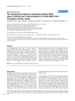
SLE dendritic cells exhibit diminished FITC-dextran
uptake
We first demonstrated that moDCs from SLE patients have
impaired endocytic capacity. Healthy control moDCs exhibited
low basal FD uptake at 4°C, which significantly increased
when cells were incubated at 37°C. Lupus moDCs, however,
exhibited decreased uptake of FD, both before (percentage
uptake: control (n = 20), 83.1 ± 3.2 versus SLE (n = 47), 63.9
± 3.9; P = 0.003; Figure 1a,c) and after stimulation with LPS
and TNFα (percentage uptake: control (n = 13), 83.1 ± 5.8
and SLE (n = 30), 64.6 ± 6.5; P = 0.05; Figure 1b). FD uptake
was blunted by preincubation of cells with D-mannose (Figure
1d), confirming that a mannose-dependent uptake mechanism
is involved.
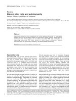
SLE dendritic cells have decreased surface mannose
receptor expression, which correlates with FITC-dextran
uptake
As the MR is the major receptor responsible for FD uptake
inhibited by mannose, we then assessed MR expression in
SLE moDCs and in control moDCs (Figure 2a). Both lupus
and control moDCs expressed surface MR, which significantly
downregulated upon stimulation/maturation with LPS and
TNFα (P = 0.03 for lupus DCs, P = 0.007 for control). Imma-
ture moDCs from lupus patients, however, displayed signifi-
cantly less MR when compared with control moDCs
(percentage expression: control (n = 29), 73.6 ± 2.7 and SLE
(n = 49), 59.2 ± 3.5; P = 0.0002). This difference was not sig-
nificant after DC stimulation. Linking levels of MR to C-type
lectin uptake, there was a positive correlation between MR
expression and FD uptake in both unstimulated (r = 0.64) and
stimulated (r = 0.80) lupus moDCs (P < 0.0001; Figure 2b).
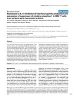
Decreased mannose receptor expression correlates with
circulating IFNγ
We next investigated potential factors contributing to the
observed aberrant phenotype in DCs from SLE patients. To
Available online />Page 5 of 10
(page number not for citation purposes)
determine whether the DC maturation status could account for
the diminished FD uptake and MR expression, the association
with expression of the maturation marker CD86 was examined
(Figure 3a). Whereas in unstimulated control DCs there was a
significant negative correlation between CD86 expression and
Figure 1
Lupus monocyte-derived dendritic cells display decreased FITC-dextran uptakeLupus monocyte-derived dendritic cells display decreased FITC-dextran uptake. (a) Unstimulated dendritic cells (DCs) (*P = 0.003). (b)
Lipopolysaccharide/TNFα-stimulated DCs (**P = 0.05). Results are expressed as the mean ± standard error of the mean. (c) Representative histo-
gram demonstrating impaired FITC-dextran (FD) uptake by unstimulated systemic lupus erythematosus (SLE) DCs. (d) Representative histogram
showing blunted FD uptake by unstimulated DCs after D-mannose preincubation. Line colors: dark blue = control, 37°C; red = SLE, 37°C; light blue
= control, 4°C; light green = SLE, 4°C; black = control + D-mannose, 37°C; dark green = SLE + D-mannose, 37°C.
Figure 2
Unstimulated lupus monocyte-derived dendritic cells, mannose receptor expression, and FITC-dextran uptakeUnstimulated lupus monocyte-derived dendritic cells, mannose receptor expression, and FITC-dextran uptake. Unstimulated lupus mono-
cyte-derived dendritic cells display decreased mannose receptor (MR) expression, which correlates with FITC-dextran (FD) uptake. (a) Both groups
significantly downregulate MR upon lipopolysaccharide/TNF stimulation (*P = 0.007, **P = 0.03, ***P = 0.0002). Results are expressed as the
mean ± standard error of the mean. (b) Positive correlation between MR expression and FD uptake. This was observed in unstimulated lupus DCs
(*r = 0.64, P < 0.0001) and stimulated lupus DCs (**r = 0.80, P < 0.0001).
Arthritis Research & Therapy Vol 10 No 5 Monrad et al.
Page 6 of 10
(page number not for citation purposes)
FD uptake (r = -0.76, P = 0.03) and there was a near-signifi-
cant negative correlation with MR expression (r = -0.46, P =
0.08), this was not found in unstimulated lupus DCs (r = -0.23,
P = 0.33 for FD uptake; r = -0.33, P = 0.46 for MR expres-
sion). Similarly, no correlation was found with other maturation
markers, including CD40, CD80, CD83 and class II Major His-
tocompatibility Complex (data not shown).
We also examined whether medications commonly used to
treat lupus could account for decreased FD uptake or MR
expression in this disease. There was no correlation of these
variables with the prednisone dosage (Figure 3c) or with the
use of nonsteroidal anti-inflammatory drugs, hydroxychloro-
quine, azathioprine, or mycophenolate (data not shown). Addi-
tionally, healthy control moDCs cultured in the presence of
graded doses of the above medications did not exhibit
decreased FD uptake or MR expression when compared with
autologous untreated DCs (data not shown).
Overall, these results indicate that the abnormal phenotype
and function of this CTLR are not secondary to a drug factor
or DC maturation status.
IFNγ downregulates transcription and surface expression of
the MR, and elevated levels of this cytokine have been
described in SLE [25]. To assess whether CTLR abnormalities
were secondary, at least in part, to this cytokine, plasma levels
of IFNγ were quantified. Indeed, SLE individuals with IFNγ con-
centration >100 ng/ml had significantly lower moDC MR
expression than those with lower levels (percentage expres-
sion: <100 ng/ml (n = 12), 75.2 ± 5.4 and >100 ng/ml (n =
3), 48.1 ± 5.6; P = 0.02; Figure 3b).
Figure 3
Mannose receptor expression in systemic lupus erythematosusMannose receptor expression in systemic lupus erythematosus. Mannose receptor (MR) expression is not correlated with CD86 expression or
prednisone use, but is associated with high serum IFNγ in systemic lupus erythematosus (SLE) patients. (a) Correlation between CD86 expression
and either FITC-dextran (FD) uptake or MR expression. In control dendritic cells (DCs) there is significant or near-significant negative correlation
(*FD uptake: r = -0.76, P = 0.03; **MR expression: r = -0.46, P = 0.08), whereas there is no correlation in lupus DCs (FD uptake: r = -0.23, P =
0.46; MR expression: r = -0.33, P = 0.33). (b) Patients with higher levels of circulating IFNγ display lower expression of MR on autologous DCs.
Graph displays patients with IFNγ levels >100 ng/ml (n = 3) compared with patients with lower plasma levels (n = 12; *P = 0.03). Results are
expressed as the mean ± standard error of the mean. (c) Prednisone dose does not correlate with MR expression or FD uptake. Graph shows the
distribution of MR expression (black diamonds) and FD uptake (clear squares) relative to the prednisone dose.
Available online />Page 7 of 10
(page number not for citation purposes)
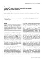
Decreased mannose receptor expression and endocytic
capacity correlates with lupus disease activity
In both unstimulated and stimulated lupus moDCs, the MR
expression negatively correlated with SLE Disease Activity
Index scores (unstimulated, r = -0.36, P = 0.006; stimulated, r
= -0.48, P = 0.002; Figure 4a) and with serum anti-dsDNA tit-
ers (unstimulated, r = -0.35, P = 0.01; stimulated, r = -0.33, P
= 0.04; Figure 4b). Similar negative correlations were
observed between the FD uptake and SLE Disease Activity
Index scores (unstimulated, r = -0. 34, P = 0.02; stimulated, r
= -0.55, P = 0.001; Figure 4c) or anti-dsDNA (unstimulated, r
= -0.29, P = 0.05; stimulated, r = -0.49, P = 0.004; Figure 4d).
Lupus dendritic cells display decreased DC-SIGN, but
present normal CR3 and Fcγ receptor expression
To determine whether this endocytic defect was restricted to
the MR or whether other receptors were also aberrantly
expressed, the surface expression of other receptors involved
in antigen uptake was evaluated. There were no differences in
CR3 and Fcγ receptor I, Fcγ receptor II or Fcγ receptor III
expression between lupus DCs and control DCs (data not
shown).
The CTLR DC-SIGN, however, was also downregulated in
SLE moDCs – both before and after stimulation (unstimulated
percentage expression: control (n = 30), 71.3 ± 3.5 and SLE
(n = 52), 53.2 ± 3.7; P = 0.005; stimulated percentage
expression: control (n = 21), 64.7 ± 6.0 and SLE (n = 39),
48.6 ± 4.5; P = 0.03; Figure 5). Unlike the MR, the DC-SIGN
expression did not correlate with FD uptake, either in control
DCs or lupus DCs (data not shown). Control DCs and SLE
DCs showed a strong correlation between MR and DC-SIGN
expression (control, r = 0.75 for unstimulated cells and r =
0.62 for stimulated cells, P < 0.005; SLE, r = 0.32, P = 0.03).
Additionally, only stimulated DCs exhibited a correlation
between DC-SIGN expression and the SLE Disease Activity
Index score (r = -0.35, P = 0.04).
Lupus dendritic cells have normal uptake of zymosan A
particles
As zymosan A can be taken up by the MR [26], we determined
whether lupus moDCs displayed diminished zymosan uptake.
There was no significant difference in zymosan uptake
between control individuals and lupus patients, either as deter-
mined by the mean fluorescence intensity or by the percent-
age of fluorescein positivity (percentage positivity: control,
Figure 4
Mannose receptor expression correlation with disease activity in monocyte-derived dendritic cellsMannose receptor expression correlation with disease activity in monocyte-derived dendritic cells. Disease activity and levels of anti-dsDNA
antibody correlate with lower levels of mannose receptor (MR) and aberrant dextran uptake by lupus monocyte-derived dendritic cells. (a) Correla-
tion between MR expression and systemic lupus erythematosus Disease Activity Index (SLEDAI) scores (*r = -0.36, P = 0.006; **r = -0.48, P =
0.002) in systemic lupus erythematosus (SLE) patients. (b) Correlation between anti-dsDNA antibodies and MR expression (*r = -0.35, P = 0.01; **r
= -0.33, P = 0.04). (c) Correlation between lupus dendritic cell FITC-dextran (FD) uptake and SLEDAI scores (*r = -0. 34, P = 0.02; **r = -0.55, P
= 0.001). (d) Correlation between anti-dsDNA antibody titers and FD uptake (*r = -0.29, P = 0.05; **r = -0.49, P = 0.004).
Arthritis Research & Therapy Vol 10 No 5 Monrad et al.
Page 8 of 10
(page number not for citation purposes)
40.6 ± 5.3 and SLE, 42.0 ± 5.15; P = 0.85; mean fluores-
cence intensity: control, 286.2 ± 86.5 and SLE, 295.6 ± 39.4;
P = 0.90).
Discussion
A growing body of literature is defining the spectrum of abnor-
malities associated with DCs in SLE. Lupus DCs exhibit an
aberrantly activated and mature phenotype [21,27]. As DC
maturation is associated with increased migratory capacity,
this phenotype may account for the decreased numbers of cir-
culating DCs detected in the blood of SLE patients [28,29] as
well as for the increased numbers found in affected organs
[30,31]. DC maturation also results in downregulation of anti-
gen uptake machinery and diminished phagocytic capacity.
Our findings of decreased FD uptake by moDCs from SLE
patients are therefore consistent with an overactivated pheno-
type. This impaired uptake capability, however, is not exclu-
sively a function of maturation status, as FD uptake did not
correlate with expression of maturation markers in SLE DCs,
whereas it did in control DCs. There thus appears to be a lec-
tin phagocytosis abnormality by lupus DCs that is independent
of the maturation status.
FD uptake by moDCs has been shown to occur primarily via
the MR, although fluid phase pinocytosis also contributes [32].
We demonstrated that SLE moDCs exhibit decreased expres-
sion of MR compared with control DCs. As expected,
decreased MR expression correlated with low FD uptake.
Additionally, these deficits appear to be associated with active
disease activity. Whether low MR expression and associated
diminished lectin uptake capacity are pathogenic in active
lupus and/or the result of other systemic abnormalities present
during disease activity is unclear and warrants further
investigation.
We also found downregulation of an additional CTLR, DC-
SIGN, indicating a more global defect in expression of mem-
bers of this family. Interestingly, we detected no decrease in
surface expression of CR3 or any of the Fcγ receptors. This is
not necessarily surprising; although common variants of these
genes alter lupus susceptibility in large population studies
[20], specific quantitative or functional receptor deficits asso-
ciated with these allelic variants have yet to be identified.
A number of exogenous factors of potential relevance in lupus
can affect MR expression. In particular, medications used to
treat SLE could contribute to the phenotypic differences
observed in circulating DCs and monocytes. MoDCs cultured
in the presence of dexamethasone exhibit upregulated MR
expression, a more globally immature phenotype, and higher
endocytic activity [33]. We might therefore expect steroid
treatment to result in increased MR expression. No association
between steroid use and MR expression could be detected,
however, either by analysis of patient steroid use or with in
vitro treatment of control DCs. Additionally, exposure to other
immunosuppressive agents could not account for the down-
regulation observed in CTLRs.
IFNγ downregulates transcription [34] and surface expression
[35] of the MR. As peripheral blood IFNγ levels are elevated in
SLE patients and have been shown to correlate with nephritis
[25], this could be of potential relevance. Indeed, we did doc-
ument lower MR expression on DCs from patients with high
serum levels of IFNγ. Therefore, while clearly there exists an
intrinsic deficit in receptor expression by lupus DCs, IFNγ may
contribute to aberrant MR expression in the subset of patients
with high serum levels.
The functional consequences of decreased MR expression in
SLE DCs are unclear, particularly with regards to susceptibility
to infection. In vitro transfection studies have found the MR to
be sufficient for uptake of various pathogens such as Candida
sp., Pneumocystis, and others [36,37]. Studies with MR
knockout mice, however, reveal no evidence of increased pre-
disposition towards infections such as Pneumocystis [38],
Candida albicans [39], and Leishmania sp. [40] – although a
recent study has found hastened mortality from cryptococcal
infections [41]. This may be due to considerable redundancy
in receptor-mediated uptake of pathogens, with various other
receptors able to perform similar phagocytic functions as the
MR. We were unable to demonstrate any significant differ-
ences between lupus DCs and control DCs in ability to uptake
zymosan, an MR ligand – probably for that reason. Decreased
MR expression in combination with the other receptor deficits
and immunologic aberrancies seen in SLE, however, could still
contribute to the overall increased susceptibility of patients to
assorted infections.
Figure 5
Dendritic cell-specific intercellular adhesion molecule-grabbing nonin-tegrin expression in systemic lupus erythematosus dendritic cellsDendritic cell-specific intercellular adhesion molecule-grabbing
nonintegrin expression in systemic lupus erythematosus dendritic
cells. Dendritic cell-specific intercellular adhesion molecule-grabbing
nonintegrin (DC-SIGN) expression is downregulated in unstimulated
and stimulated systemic lupus erythematosus (SLE) monocyte-derived
dendritic cells. Results represent the mean ± standard error of the
mean of 30 control individuals and 52 SLE patients (*P = 0.005, **P =
0.03).
Available online />Page 9 of 10
(page number not for citation purposes)
MR deficiency results in increased circulating lysosomal
hydrolases, which indicates that these molecules may be nec-
essary for certain aspects of glycoprotein homeostasis [11].
Surface glycoprotein rearrangement is an important step in
normal cellular apoptosis/necrosis [42]. Dysregulated apopto-
sis has been strongly correlated with the development and
perpetuation of autoimmunity in SLE [43]. Additionally, anti-
bodies against glycoproteins have pathologic relevance in
SLE [44]. Aberrant glycoprotein processing could therefore
have implications in lupus pathogenesis, and future studies
will assess this possibility.
Conclusion
We have demonstrated that monocyte-derived DCs from
patients with SLE have diminished phagocytic capacity asso-
ciated with decreased expression of specific CTLRs. This is an
important addition to our understanding of the many pivotal
roles DCs play in lupus immunopathogenesis. Decreased
phagocytosis of apoptotic material and other normally harm-
less self-antigens could result in an autoimmunity-promoting
milieu with loss of tolerance, inappropriate autoantigen pres-
entation and, ultimately, the serologic and clinical manifesta-
tions characteristic of SLE. Additionally, while individual
receptors may not be exclusively responsible for clearance of
individual pathogens, aberrant phagocytic machinery and
uptake capacity could still contribute to inadequate responses
to harmful pathogens.
Competing interests
The authors declare that they have no competing interests.
Authors' contributions
SUM, KR and ST performed all experiments and analyzed the
data. SUM drafted the manuscript. MJK conceived and
designed the study and helped to draft the manuscript. All
authors read and approved the final document.
Acknowledgements
The authors wish to thank Emily Lewis B.Sc. and Jennifer Johnson B.Sc.
for obtaining patient blood samples; Taejah Vemuri, Marc Anderson and
Amanda Bradke B.Sc. for help with blood processing and cell culture;
Emily Somers Ph.D., Sc.M. for assistance with statistical analysis; and
Michael Denny Ph.D. for helpful discussions. The present work was sup-
ported by Public Health Service Grants AR050554 and AR048235, as
well as by the Anthony S. Gramer Fund in Inflammation Research and by
the Research and Education Foundation/American College of Rheuma-
tology. The research was also supported (in part) by the National Insti-
tutes of Health through the University of Michigan's Cancer Center
Support Grant (P30 CA46592), the Rheumatic Diseases Core Center
Grant (P30 AR48310) and training grants T32 AR 07080 and T32
A107413.
References
1. Monrad S, Desch K, Kaplan M: Role of dendritic cells in the
pathogenesis of systemic lupus erythematosus. Future
Rheumatol 2008, 3:269-280.
2. Banchereau J, Pascual V: Type I interferon in systemic lupus
erythematosus and other autoimmune diseases. Immunity
2006, 25:383-392.
3. Ueno H, Klechevsky E, Morita R, Aspord C, Cao T, Matsui T, Di
Pucchio T, Connolly J, Fay JW, Pascual V, Palucka AK,
Banchereau J: Dendritic cell subsets in health and disease.
Immunol Rev 2007, 219:118-142.
4. Zandman-Goddard G, Shoenfeld Y: Infections and SLE. Autoim-
munity 2005, 38:473-485.
5. Falagas ME, Manta KG, Betsi GI, Pappas G: Infection-related
morbidity and mortality in patients with connective tissue dis-
eases: a systematic review. Clin Rheumatol 2007, 26:663-670.
6. Zelensky AN, Gready JE: The C-type lectin-like domain
superfamily. FEBS J 2005, 272:6179-6217.
7. Figdor CG, van Kooyk Y, Adema GJ: C-type lectin receptors on
dendritic cells and Langerhans cells. Nat Rev Immunol 2002,
2:77-84.
8. Geijtenbeek TB, van Vliet SJ, Engering A, t Hart BA, van Kooyk Y:
Self- and nonself-recognition by C-type lectins on dendritic
cells. Annu Rev Immunol 2004, 22:33-54.
9. Gijzen K, Cambi A, Torensma R, Figdor CG: C-type lectins on
dendritic cells and their interaction with pathogen-derived and
endogenous glycoconjugates. Curr Protein Pept Sci 2006,
7:283-294.
10. Zamze S, Martinez-Pomares L, Jones H, Taylor PR, Stillion RJ, Gor-
don S, Wong SY: Recognition of bacterial capsular polysac-
charides and lipopolysaccharides by the macrophage
mannose receptor. J Biol Chem 2002, 277:41613-41623.
11. Lee SJ, Evers S, Roeder D, Parlow AF, Risteli J, Risteli L, Lee YC,
Feizi T, Langen H, Nussenzweig MC: Mannose receptor-medi-
ated regulation of serum glycoprotein homeostasis. Science
2002, 295:1898-1901.
12. Prigozy TI, Sieling PA, Clemens D, Stewart PL, Behar SM, Porcelli
SA, Brenner MB, Modlin RL, Kronenberg M: The mannose recep-
tor delivers lipoglycan antigens to endosomes for presenta-
tion to T cells by CD1b molecules. Immunity 1997, 6:187-197.
13. Geijtenbeek TB, Krooshoop DJ, Bleijs DA, van Vliet SJ, van Duijn-
hoven GC, Grabovsky V, Alon R, Figdor CG, van Kooyk Y: DC-
SIGN-ICAM-2 interaction mediates dendritic cell trafficking.
Nat Immunol 2000, 1:353-357.
14. Geijtenbeek TB, Torensma R, van Vliet SJ, van Duijnhoven GC,
Adema GJ, van Kooyk Y, Figdor CG: Identification of DC-SIGN,
a novel dendritic cell-specific ICAM-3 receptor that supports
primary immune responses. Cell 2000, 100:575-585.
15. Geijtenbeek TB, Kwon DS, Torensma R, van Vliet SJ, van Duijn-
hoven GC, Middel J, Cornelissen IL, Nottet HS, KewalRamani VN,
Littman DR, Figdor CG, van Kooyk Y: DC-SIGN, a dendritic cell-
specific HIV-1-binding protein that enhances trans-infection of
T cells. Cell 2000, 100:587-597.
16. Bajtay Z, Csomor E, Sandor N, Erdei A: Expression and role of
Fc- and complement-receptors on human dendritic cells.
Immunol Lett 2006, 104:46-52.
17. Ross GD: Regulation of the adhesion versus cytotoxic func-
tions of the Mac-1/CR3/αMβ
2
-integrin glycoprotein. Crit Rev
Immunol 2000, 20:197-222.
18. Bave U, Magnusson M, Eloranta ML, Perers A, Alm GV, Ronnblom
L: Fc gamma RIIa is expressed on natural IFN-alpha-produc-
ing cells (plasmacytoid dendritic cells) and is required for the
IFN-alpha production induced by apoptotic cells combined
with lupus IgG. J Immunol 2003, 171:3296-3302.
19. Means TK, Latz E, Hayashi F, Murali MR, Golenbock DT, Luster
AD: Human lupus autoantibody-DNA complexes activate DCs
through cooperation of CD32 and TLR9. J Clin Invest 2005,
115:407-417.
20. Harley JB, Alarcon-Riquelme ME, Criswell LA, Jacob CO, Kimberly
RP, Moser KL, Tsao BP, Vyse TJ, Langefeld CD, Nath SK,
Guthridge JM, Cobb BL, Mirel DB, Marion MC, Williams AH,
Divers J, Wang W, Frank SG, Namjou B, Gabriel SB, Lee AT,
Gregersen PK, Behrens TW, Taylor KE, Fernando M, Zidovetzki R,
Gaffney PM, Edberg JC, Rioux JD, Ojwang JO, et al.: Genome-
wide association scan in women with systemic lupus ery-
thematosus identifies susceptibility variants in ITGAM, PXK,
KIAA1542 and other loci. Nat Genet 2008, 40:204-210.
21. Ding D, Mehta H, McCune WJ, Kaplan MJ: Aberrant phenotype
and function of myeloid dendritic cells in systemic lupus
erythematosus. J Immunol 2006, 177:5878-5889.
Arthritis Research & Therapy Vol 10 No 5 Monrad et al.
Page 10 of 10
(page number not for citation purposes)
22. Tan EM, Cohen AS, Fries JF, Masi AT, McShane DJ, Rothfield NF,
Schaller JG, Talal N, Winchester RJ: The 1982 revised criteria for
the classification of systemic lupus erythematosus. Arthritis
Rheum 1982, 25:1271-1277.
23. Hochberg MC: Updating the American College of Rheumatol-
ogy revised criteria for the classification of systemic lupus
erythematosus. Arthritis Rheum 1997, 40:1725.
24. Bombardier C, Gladman DD, Urowitz MB, Caron D, Chang CH:
Derivation of the SLEDAI. A disease activity index for lupus
patients. The Committee on Prognosis Studies in SLE. Arthritis
Rheum 1992, 35:630-640.
25. Masutani K, Akahoshi M, Tsuruya K, Tokumoto M, Ninomiya T, Koh-
saka T, Fukuda K, Kanai H, Nakashima H, Otsuka T, Hirakata H:
Predominance of Th1 immune response in diffuse prolifera-
tive lupus nephritis. Arthritis Rheum 2001, 44:2097-2106.
26. Speert DP, Silverstein SC: Phagocytosis of unopsonized
zymosan by human monocyte-derived macrophages: matura-
tion and inhibition by mannan. J Leukoc Biol 1985, 38:655-658.
27. Decker P, Kotter I, Klein R, Berner B, Rammensee HG: Monocyte-
derived dendritic cells over-express CD86 in patients with sys-
temic lupus erythematosus. Rheumatology (Oxford) 2006,
45:1087-1095.
28. Gill MA, Blanco P, Arce E, Pascual V, Banchereau J, Palucka AK:
Blood dendritic cells and DC-poietins in systemic lupus
erythematosus. Hum Immunol 2002, 63:1172-1180.
29. Migita K, Miyashita T, Maeda Y, Kimura H, Nakamura M, Yatsuhashi
H, Ishibashi H, Eguchi K: Reduced blood BDCA-2
+
(lymphoid)
and CD11c
+
(myeloid) dendritic cells in systemic lupus
erythematosus. Clin Exp Immunol 2005, 142:84-91.
30. Tucci M, Quatraro C, Lombardi L, Pellegrino C, Dammacco F, Sil-
vestris F: Glomerular accumulation of plasmacytoid dendritic
cells in active lupus nephritis: role of interleukin-18. Arthritis
Rheum 2008, 58:251-262.
31. Blomberg S, Eloranta ML, Cederblad B, Nordlin K, Alm GV, Ron-
nblom L: Presence of cutaneous interferon-alpha producing
cells in patients with systemic lupus erythematosus. Lupus
2001, 10:484-490.
32. Sallusto F, Cella M, Danieli C, Lanzavecchia A: Dendritic cells use
macropinocytosis and the mannose receptor to concentrate
macromolecules in the major histocompatibility complex
class II compartment: downregulation by cytokines and bacte-
rial products. J Exp Med 1995, 182:389-400.
33. Piemonti L, Monti P, Allavena P, Sironi M, Soldini L, Leone BE,
Socci C, Di Carlo V: Glucocorticoids affect human dendritic cell
differentiation and maturation. J Immunol 1999,
162:6473-6481.
34. Harris N, Super M, Rits M, Chang G, Ezekowitz RA: Characteriza-
tion of the murine macrophage mannose receptor: demon-
stration that the downregulation of receptor expression
mediated by interferon-gamma occurs at the level of
transcription. Blood 1992, 80:2363-2373.
35. Marodi L, Schreiber S, Anderson DC, MacDermott RP, Korchak
HM, Johnston RB Jr: Enhancement of macrophage
candidacidal activity by interferon-gamma. Increased phago-
cytosis, killing, and calcium signal mediated by a decreased
number of mannose receptors. J Clin Invest 1993,
91:2596-2601.
36. Ezekowitz RA, Sastry K, Bailly P, Warner A: Molecular character-
ization of the human macrophage mannose receptor: demon-
stration of multiple carbohydrate recognition-like domains
and phagocytosis of yeasts in Cos-1 cells. J Exp Med 1990,
172:1785-1794.
37. Ezekowitz RA, Williams DJ, Koziel H, Armstrong MY, Warner A,
Richards FF, Rose RM: Uptake of Pneumocystis carinii medi-
ated by the macrophage mannose receptor. Nature 1991,
351:155-158.
38. Swain SD, Lee SJ, Nussenzweig MC, Harmsen AG: Absence of
the macrophage mannose receptor in mice does not increase
susceptibility to Pneumocystis carinii infection in vivo
. Infect
Immun 2003, 71:6213-6221.
39. Lee SJ, Zheng NY, Clavijo M, Nussenzweig MC: Normal host
defense during systemic candidiasis in mannose receptor-
deficient mice. Infect Immun 2003, 71:437-445.
40. Akilov OE, Kasuboski RE, Carter CR, McDowell MA: The role of
mannose receptor during experimental leishmaniasis. J Leu-
koc Biol 2007, 81:1188-1196.
41. Dan JM, Kelly RM, Lee CK, Levitz SM: Role of the mannose
receptor in a murine model of Cryptococcus neoformans
infection. Infect Immun 2008, 76:2362-2367.
42. Gaipl US, Sheriff A, Franz S, Munoz LE, Voll RE, Kalden JR, Her-
rmann M: Inefficient clearance of dying cells and autoreactivity.
Curr Top Microbiol Immunol 2006, 305:161-176.
43. Kaplan MJ: Apoptosis in systemic lupus erythematosus. Clin
Immunol 2004, 112:210-218.
44. Marai I, Tincani A, Balestrieri G, Shoenfeld Y: Anticardiolipin and
anti-beta-2-glycoprotein I antibodies. Autoimmunity 2005,
38:33-38.