Báo cáo y học: "Glomerular matrix metalloproteinases and their regulators in the pathogenesis of lupus nephritis" pdf
Bạn đang xem bản rút gọn của tài liệu. Xem và tải ngay bản đầy đủ của tài liệu tại đây (402.55 KB, 8 trang )
Page 1 of 8
(page number not for citation purposes)
Available online />Abstract
Lupus nephritis is a major contributor to morbidity and mortality in
systemic lupus erythematosus, but little is known about the
pathogenic processes that underlie the progressive decay in renal
function. A common finding in lupus nephritis is thickening of
glomerular basement membranes associated with immune complex
deposition. It has been speculated that alterations in the synthesis
or degradation of membrane components might contribute to such
changes, and thereby to initiation and progression of nephritis
through facilitation of immune complex deposition. Matrix metallo-
proteinases (MMPs) are enzymes that are intimately involved in the
turnover of major glomerular basement membrane constituents,
including collagen IV and laminins. Alterations in the expression
and activity of MMPs have been described in a number of renal
diseases, suggesting their relevance to the pathogenesis of various
glomerulopathies. The same is true for their natural inhibitors, the
tissue inhibitor of metalloproteinase family. Recent data from our
group have identified an increase in proteolytic activity within the
glomerulus coinciding with the development of proteinuria in the
(NXB×NZW)F
1
mouse model of systemic lupus erythematosus.
Here we review current understanding of MMP/tissue inhibitor of
metalloproteinase function within the kidney, and discuss their
possible involvement in the development and progression of lupus
nephritis.
Introduction
Systemic lupus erythematosus (SLE) is a complex auto-
immune disease that is characterized by chronic inflammatory
processes involving autoimmunity against multiple organ-
specific and ubiquitous self-antigens. One commonly affected
organ is the kidney, with the appearance of lupus nephritis
ranging in severity from mild proteinuria to overt nephrotic
syndrome progressing to end-stage renal disease. Although
the molecular mechanisms that underlie the pathogenesis of
nephritis remain largely obscure, disturbances in apoptotic
signalling, phagocytosis and complement function have all
been proposed as factors involved in initiation of auto-
immunity and progression of the disease [1,2].
Expansion and/or disruption of the intraglomerular extra-
cellular matrix is a well recognized phenomenon occurring
during the development of lupus nephritis that may have an
impact on renal immune complex deposition. Little is known,
however, about the structure and composition of the expanded
regions or the mediators of such changes. Increased or altered
synthesis of extracellular matrix (ECM) constituents and/or
their decreased breakdown could potentially play a role,
although the contribution made by each of these factors
remains unknown.
Another common finding in lupus nephropathy is the appear-
ance of electron dense structures (EDSs) within mesangium
or intimately linked to the glomerular capillary membranes, as
seen on electron micrographs. These structures contain
immune complexes with autoantibodies and chromatin
fragments [3,4], and a recent study [5] has demonstrated a
considerable affinity of nucleosomes toward the major matrix
constituents laminin and collagen IV. It is therefore possible
that alterations in the composition of the glomerular ECM may
affect its interaction with immune complexes, thus facilitating
their deposition and subsequent damage to glomerular struc-
tures. Indeed, qualitative as well as quantitative alterations in
the makeup of the extracellular membranes of the glomerulus
in lupus nephritis have already been described [6,7]. Candidate
mediators of such changes include enzymes and signalling
substances involved in maintaining the delicate balance
between synthesis and breakdown of the proteins and
proteoglycans that make up the ECM.
Although some studies have provided evidence of increased
levels of expression of collagens and laminins, less is known
about the kinetics of breakdown of these proteins. Turnover
of ECM proteins is largely achieved through the action of
matrix metalloproteinases (MMPs), which represent a major
class of matrix-degrading proteinases. Thus, from its effect on
Review
Glomerular matrix metalloproteinases and their regulators in the
pathogenesis of lupus nephritis
Anders Tveita, Ole Petter Rekvig and Svetlana N Zykova
Department of Biochemistry, Institute of Medical Biology, Medical Faculty, University of Tromsø, N-9037 Tromsø, Norway
Corresponding author: Anders Aune Tveita,
Published: 1 December 2008 Arthritis Research & Therapy 2008, 10:229 (doi:10.1186/ar2532)
This article is online at />© 2008 BioMed Central Ltd
ADAM = a disintegrin and metalloproteinase; ECM = extracellular matrix; EDS = electron dense structure; MMP = matrix metalloproteinase; PLZF =
promyelocytic leukaemia zinc finger protein; SLE = systemic lupus erythematosus; TIMP = tissue inhibitor of metalloproteinase.
Page 2 of 8
(page number not for citation purposes)
Arthritis Research & Therapy Vol 10 No 6 Tveita et al.
capillary membranes and mesangial matrix composition, a
putative role emerges for altered glomerular MMP activity in
lupus nephritis. Exploring this possibility, however, is compli-
cated by the many levels of regulation of proteinase activity.
Also, there is an emerging appreciation of considerable
functional divergence of both MMPs and their regulators,
particularly the tissue inhibitors of metalloproteinase (TIMPs).
In this review we outline some of the current knowledge on
MMP expression and regulation within the kidney in lupus
nephritis, including clues gained from studies in other renal
inflammatory diseases.
Matrix metalloproteinases
MMPs are a group of Zn
2+
-dependent proteins that are found
in the extracellular milieu of various tissues. Based on
sequence homology and substrate specificities, the MMPs can
be classified into several subgroups including collagenases,
gelatinases, stromelysins, matrilysins and the membrane-type
metalloproteinases. There is considerable overlap in substrate
specificities, and the MMPs appear to be involved in
degradation of abundant ECM components, including laminins,
collagens and fibronectin, but also in the release and turnover
of cytokines and cell surface receptors of adjacent cells [8].
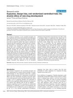
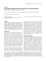
MMP-2 (gelatinase A) and MMP-9 (gelatinase B) constitute
the gelatinases (Figure 1). On account of their propensity to
cleave the major glomerular basement membrane component
collagen IV, they have been particularly implicated in a variety
of acute and chronic kidney diseases, including both immune
and non-immune glomerulopathies, and are therefore the
main focus of this review.
The gelatinases cleave a number of substrates, including
native forms of collagens I, IV, V, VII, X and XI, elastin, laminin,
fibronectin, myelin and the core protein of proteoglycans. (A
comprehensive list of substrates for the various MMPs can be
found in the Overall Lab Web Site [9].) Another metallo-
proteinase that is notable for its affinity for collagen IV is
MMP-7 (matrilysin 1) [10]. Produced in both the tubular and
glomerular compartment, it was recently described to be
involved in several types of renal diseases with glomerular
involvement, including diabetic nephropathy and X-linked
Alport syndrome [11,12]. In addition to collagen IV, MMP-7 is
a major factor in the turnover of tenascin (an oligomeric glyco-
protein that is important for the functioning of the glomerular
filtration barrier) [13] and other basement membrane
components, such as laminin, entactin and proteoglycans, as
well as in activation of several proinflammatory mediators,
including MMP-2 and MMP-9 [14,15]. The relevance of
MMP-7 in SLE has not yet been evaluated, but it remains an
interesting candidate mediator of changes in membrane
composition in lupus nephritis.
MMP-2 is constitutively expressed in mesangial cells, with
some contribution made by the podocytes and little or no
expression in glomerular endothelial cells [16,17]. The
expression is dramatically increased in various glomerulo-
pathies, probably as a result of proinflammatory signalling
[18,19]. MMP-9 is present at negligible levels in normal
kidney glomeruli, but it is induced during the course of several
renal inflammatory diseases, with mesangial cells and
infiltrating neutrophils being the main sources [20]. Recent
data from our laboratory [21] indicate an increase in glomerular
proteolytic activity at around the onset of proteinuria in a
model of lupus nephritis in (NZB×NZW)F
1
mice, with MMP-9
being a major contributor.
Tissue inhibitors of metalloproteinases
The many roles of MMPs in vivo require a complex network of
modulation of enzymatic activity. Key regulators are the
TIMPs, of which four subtypes are currently known. The
TIMPs form 1:1 complexes with metalloproteinases, with only
modest variations in affinity toward the different MMPs [22].
The central role played by TIMPs in the regulation of MMP
activity has led to the hypothesis that shifts in the balance
between these two families of proteins could distort the
kinetics of membrane turnover and cause pathological
changes in membrane composition [23,24].
When considering the relevance of TIMP expression in renal
disease, it is important to note that several members of the
TIMP family appear to have functions of pathophysiological
significance that is not directly related to ECM homeostasis/
MMP regulation. One such facet is the apparent involvement
in both pro-apoptotic and anti-apoptotic signalling pathways
[25-27]. Various disturbances in apoptosis and the clearance
of dead cells have been proposed to form a source of
autoreactivity in SLE, which raises the possibility that TIMPs
are involved in regulating apoptotic cell death in the context
of autoimmune disorders.
A full account of the field of TIMP biology is beyond the
scope of this review, and we limit the discussion to
apoptosis-related aspects of their functioning. Whereas the
molecular basis for TIMP-mediated signalling is still poorly
understood, an emerging view is that they interact extensively
with cell surface proteins, thus imposing modulation of
various downstream signalling pathways.
Cell culture studies have reported anti-apoptotic effects of
TIMP-1, some of which rely on its MMP-inhibitory function,
whereas others appear independent of interaction with
MMPs [28-30]. A recent in vitro study conducted in HeLa
cervical cancer cells identified promyelocytic leukaemia zinc
finger protein (PLZF) - a well known transcriptional repressor -
to be a potential binding partner for TIMP-1 [31]. It was
shown that the addition/over-expression of TIMP-1 reduced
the percentage of apoptotic cells in this system in a PLZF-
dependant manner. PLZF is expressed in myeloid cells,
ovaries and, at low level, in kidney and lung tissues. The
interaction between TIMP-1 and PLZF is reported to occur
Page 3 of 8
(page number not for citation purposes)
by direct interaction between the two proteins within the
nucleus [31].
The concept of TIMP-1 translocating into the nucleus remains
controversial [32,33], and the functions of TIMP in this
location in vivo remain to be identified. However, as dis-
cussed below, several recent studies have reported that
MMPs are present within the nucleus [34], offering new
perspectives on the biological roles played MMPs and
possibly TIMPs as well. For TIMP-2 and TIMP-4, reports have
been partly contradictory, with both pro-apoptotic and anti-
apoptotic effects being described, and further studies are
awaited to characterize their putative roles in apoptosis and
cell viability. TIMP-3 appears to have pro-apoptotic proper-
ties, attributed to the inhibition of both the MMP and ADAM
(a disintegrin and metalloproteinase) families of matrix
metalloproteinases [35]. One possible basis for these
functions is the fact that certain ADAMs and MMPs are
involved in the shedding of a number of cell surface receptors
that are involved in pro-apoptotic and anti-apoptotic signalling
pathways, including tumour necrosis factor receptors [36]
and Fas receptor [37]. Studies conducted in tumor cells have
shown that over-expression of TIMP-3 causes stimulation of
Fas/Fas ligand signalling [38] and results in increased
apoptotic activity. Involvement of TIMPs in the regulation of
death pathways including Fas/Fas ligand is an interesting
finding, because alterations in Fas function appear to be
relevant to autoantibody production and possibly to the
development of nephritis [39].
Challenges and pitfalls in assays of matrix
metalloproteinase activity
Studies of tissue MMPs are complicated by the complexity of
the regulatory network that governs their activity in vivo. A
MMP-9 knockout mouse exhibited no or modest structural/
functional abnormalities, both on a healthy background and in
a model of Alport syndrome [40], which could be explained
by redundancy of the system based on observations of
compensatory upregulation of other MMPs, including MMP-2
[41]. Caution is therefore required when interpreting studies
that are limited to one or a few MMPs, and such findings also
suggest that therapeutic utilization of broad-spectrum
inhibitors of MMP activity might be a more desirable strategy
than more targeted ones, at least in some settings.
Increased gene expression and protein levels of the MMPs
are often found to be accompanied by increases in the levels
of one or more of the TIMPs. It is therefore not obvious what
can be the net result of these opposing stimuli in terms of
ECM turnover. In situ zymography is a technique that allows
localization of active proteinases (including MMPs) within the
tissues, providing valuable information about the net result of
Available online />Figure 1
Schematic structure of MMP-2 and MMP-9. The catalytic site contains three essential zinc ion binding sites. At the zymogen stage, a cysteine
residue within the prodomain interacts with zinc to prevent substrate binding. The haemopexin domain mediates interaction with enzyme
substrates. Specific to the gelatinases is the fibronectin-like domain, which further facilitates substrate binding. MMP, matrix metalloproteinase.
MMP regulation. It is done by incubating tissue sections with
a fluorescence-marked substrate, which gives a direct visual
impression of local proteinase activity [42].
Matrix metalloproteinase activity in lupus
nephritis and related diseases
Data on metalloproteinase activity in lupus nephritis is limited
to few reports of altered gene expression patterns in murine
and human kidneys [19,20,43]. There have also been reports
of increased circulating levels of several MMPs, notably
MMP-9, in sera from lupus patients [44-47]. Of note, circula-
ting MMP-9 levels have been found to be inversely correlated
to levels of antibodies against double-stranded DNA, which is
commonly used as a marker of SLE disease activity [48]. No
such correlations were observed for MMP-2 or MMP-3
[44,48,49]. The source(s) of serum MMPs probably includes
circulating leucocytes, especially neutrophils and monocytes,
whereas the contribution from the tissues is uncertain. Serum
MMP measurements thus may be of limited value in elucidating
their potential roles in end organ disease, and the organ-
focused studies suggest different roles for the MMPs within the
various tissue spaces. Nevertheless, increased circulating
levels of MMP-9 have been described in SLE patients with
evidence of neuropsychiatric manifestations [50]. Also, a
recent study [51] identified increased MMP-9 activity in
cerebrospinal fluid from SLE patients, with significantly higher
levels in patients with evidence of central nervous system
involvement. There are reports indicating a central role for
MMPs in increasing permeability of the blood-brain barrier in
inflammatory settings [52], which could be of relevance in SLE.
Matrix metalloproteinases and
glomerulopathies
Owing to overlapping morphological and clinical presentations
of various kinds of inflammatory diseases of the glomerulus,
the results of studies of the involvement of MMP activity in
other renal pathologies should provide valuable guidance in
elucidating the role of these enzymes in lupus nephritis.
Although one might speculate that increased MMP activity is
not detrimental, but rather represents a favourable compen-
satory response to aberrant matrix synthesis, this appears
unlikely considering the favourable outcome of MMP
inhibition/knockout strategies in other glomerulopathies [53].
Much of the data currently available come from work in
models of antibody-induced nephropathies, such as anti-Thy
1.1 nephritis [54] and passive Heymann’s nephritis [55], and
from non-immune models such as ischemia/reperfusion renal
scarring induced by ureteral ligation [56,57], all of which
trigger inflammatory responses [58] that lead to progressive
tubulointerstitial fibrosis and glomerulosclerosis. A common
theme in these studies is a marked increase in either one or
both of the gelatinases (MMP-2 or MMP-9) [18,19,59,60].
Often an increase in TIMP-1 is observed within the glomeruli,
which (as mentioned above) complicates the interpretation of
results. The finding that gelatinase levels are increased in a
situation of ECM accumulation might appear paradoxical,
because this would be expected to increase collagen break-
down. A simple explanation is that the increased expression
is a compensatory response to an increase in the synthesis of
matrix components. Although there are reports indicating that
collagen IV increases early in glomerulonephritis [6], others have
found collagen IV expression to appear relatively stable [61].
Preliminary data from our laboratory support the latter in the
case of (NZB×NZW)F
1
and MRL/lpr lupus-prone mice
(Tveita A, unpublished data). As stated above, the relative
contribution of increased synthesis and decreased degrada-
tion of collagen IV to ECM accumulation remains undeter-
mined. Studies showing that MMP inhibition attenuates ECM
accumulation in rat allograft nephropathy [62], anti-Thy 1.1
nephritis [63] and other experimental inflammatory renal
diseases would suggest that matrix degradation plays at least
some role in this process. As discussed below, there are also
indications that the MMPs confer proliferative stimuli upon
mesangial cells, providing another factor that might explain an
increase in MMP activity in the face of nephritis and matrix
proliferation [64].
Metalloproteinases and mesangial cell
proliferation
The mesangium appears to play a central role in the
development of glomerulonephritis. The contribution made by
mesangial cells to inflammatory diseases of the kidneys is
thought to be twofold: increased proliferation and pro-
inflammatory response; and synthesis of matrix components,
causing ECM accumulation. Mesangial cell culture experi-
ments have implicated MMP-2 as a possible regulator of both
of the above factors. Indeed, inhibition by both pharmaco-
logical and ribozyme-mediated approaches have shown
reduction in MMP-2 activity to be associated with trans-
formation of actively proliferating mesangial cells to a state of
quiescence by induction of G
0
/G
1
cell cycle arrest [65].
Treatment of cultured rat mesangial cell with a relatively
unspecific MMP inhibitor caused a decrease in proliferative
activity of up to 75% and evidence of decreased levels of
activation [63]. In addition, MMP inhibitor experiments also
demonstrated a significant increase in the number of apop-
totic cells both in anti-Thy 1.1 nephritis and in cultured
mesangial cells, probably mediated through a caspase-
independent pathway [27]. A new aspect of function of MMPs
has emerged from the reports of MMPs being present within
mammalian cell nuclei [66,67]. It was recently shown that
MMP-3 can translocate to the nucleus in vitro, where it was
reported to exert pro-apoptotic functions mediated through its
catalytic domain [34]. The same report showed evidence of
intranuclear MMP-2 and MMP-3 on human liver sections.
Cryptic epitopes and immune complex
deposition
Studies in multiple sclerosis and rheumatoid arthritis have
demonstrated that cleavage of particular collagen fragments
Arthritis Research & Therapy Vol 10 No 6 Tveita et al.
Page 4 of 8
(page number not for citation purposes)
by MMPs leads to the exposure of highly immunoreactive
epitopes [68,69]. These findings led to the proposition of a
model for the generation of autoantibodies, termed the
‘remnant epitope generate autoimmunity’ (REGA) model
[69,70]. Briefly, the underlying concept is that in an inflam-
matory context, a local increase in proteolytic activity
generates a large number of substrate fragments for
presentation by activated antigen-presenting cells, including
exposed cryptic antigen epitopes. This leads to both quanti-
tative and qualitative changes in the local antigen repertoire.
Although highly speculative, one could envision a situation in
which dysregulation of MMP activity leads to a quantitative
increase in the exposure of such cryptic epitopes. Further-
more, qualitative alterations in ECM composition could lead
to cleavage of substrates not normally found in this location,
causing the appearance of novel epitopes within the matrix. In
the face of a persisting inflammatory process, such as
evolving lupus nephritis, quantitative and qualitative changes
in antigen repertoire might conceivably increase production
of autoantibodies against matrix structures. Alternatively,
alterations in glomerular membrane composition could favour
the deposition of immune complex-associated structures
such as nucleosomes, thus accelerating the formation of
EDS-like structures within the membranes.
In light of our recent findings of increased MMP activity and
qualitative changes in collagen IV expression within glomeruli
of lupus-prone mice during the development of nephritis, this
scenario provides an attractive model to explain the
relationship between immune complex deposits and renal
dysfunction. In this framework, immune complexes propagate
proinflammatory stimuli to resident and infiltrating cells, either
directly or through complement activation, triggering an
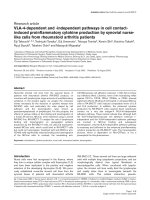
increase in MMP production and activity (Figure 2). In turn,
MMPs mediate changes in glomerular basement membrane
structure, favouring immune complex deposition and
compromising the physical integrity of the membrane.
Central to the pathogenesis of lupus is continuous activation
and proliferation of B-lymphocytes and T-lymphocytes with
specificity for self-structures such as exposed chromatin
fragments. Such structures may also serve as renal targets
for the induced autoimmunity. Encounters with these antigens
initiate proinflammatory signalling cascades, recruiting effector
cells of the innate immune system, including monocytes/
macrophages and neutrophils. As part of this inflammatory
process, several MMPs and TIMPs are secreted by activated
infiltrating cells and by cells intrinsic to the inflamed site,
facilitating penetration into the tissue and structural remodel-
ing as part of the healing process [71]. An inflammatory
reaction invariably causes local cellular decay, serving as a
potential reservoir for exposed self-structures, including
nuclear antigens. Chromatin structures derived from dead
cells are found deposited as EDSs in glomerular membranes
in lupus kidneys, where they co-localize with deposited
autoantibodies [4]. Ingestion of chromatin-containing immune
complexes by infiltrating macrophages could conceivably
upregulate MMP secretion through activation of the Toll-like
receptor 9 signalling pathway [72,73]. Persistently increased
glomerular MMP activity could therefore be the result of an
inflammatory process that is maintained by retained necrotic
or apoptotic cellular debris. In turn, excessive matrix degrada-
tion by the MMPs would facilitate the deposition of immune
complexes by compromising the integrity of the glomerular
membranes. In this manner, the combined presence of
autoreactive lymphocytes and an inflammatory process that
exposes the inciting autoantigens allows the translation of a
latent systemic autoreactivity to a focused end organ
Available online />Page 5 of 8
(page number not for citation purposes)
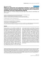
Figure 2
Conceptual framework for progression of lupus nephritis. An
inflammatory reaction is brought about by complement- or Fc-mediated
responses to autoantibodies in deposited immune complexes or locally
exposed danger signals (such as necrotic chromatin; see text),
triggering release of MMPs from intrinsic and infiltrating cells.
Increased proteolytic degradation of the membrane exposes matrix
components, facilitating binding of autoantibodies to capillary and
mesangial antigens. This maintains the inflammatory reaction and
continued stimulation of matrix degradation, leading to disruption of
glomerular membrane barriers and progression toward end-stage renal
failure. MMP, matrix metalloproteinase.
inflammatory disease. Increased MMP activity forms part of a
spectrum of changes at the site of inflammation that ensures
continued engagement of the innate immune system and
progression of local tissue damage.
Conclusion
MMP inhibitory strategies have been tested in animal models
of a number of chronic inflammatory diseases, including
chronic obstructive pulmonary diseases, inflammatory bowel
disease, rheumatoid arthritis and atherosclerosis. The
progress of various such trials was recently reviewed by Hu
and coworkers [53]. For glomerulonephritis, MMP inhibition
has exhibited promising results in rat anti-Thy 1.1 nephritis
[63], but several key parameters in such a strategy remain ill
defined, including the target MMP(s), timing and duration of
intervention, specificity, dosage and delivery system. A more
rigorous understanding of the spectrum of in vivo
biochemical roles played by MMPs/TIMPs might be a
prerequisite for the development and success of such
targeted experimental and pharmacological interventions.
Our knowledge about the role played by MMPs within the
context of lupus nephritis remains sparse and inconclusive.
Studies in murine lupus-prone strains are underway and will
hopefully shed light on this. The evidence that MMPs and
TIMPs might be involved in the regulation of apoptosis
provides further cause to look more closely into the matter of
MMP activity, because disturbances in the clearance of
apoptotic material is thought to be among the central
elements in the development of lupus [74,75]. Also, by
forming a part of the Toll-like receptor mediated response to
danger signals such as necrotic chromatin, increased MMP
activity could be an important factor in initiating end organ
manifestations of an autoimmune response.
Identifying the signalling pathways that are involved in
inducing the observed alterations in MMP expression may
contribute to our understanding of the initiation of kidney
damage in lupus nephritis. Hopefully, this might pave the road
to therapeutic strategies directed at preventing the develop-
ment of glomerulonephritis and kidney failure in lupus patients.
Competing interests
The authors declare that they have no competing interests.
Acknowledgements
We thank Dr Jan-Olof Winberg for critical review of the manuscript.
References
1. Mortensen ES, Fenton KA, Rekvig OP: Lupus nephritis: the
central role of nucleosomes revealed. Am J Pathol 2008, 172:
275-283.
2. Berden JH, Grootscholten C, Jurgen WC, van der Vlag J: Lupus
nephritis: a nucleosome waste disposal defect? J Nephrol
2002, 15(suppl 6):S1-S10.
3. Kalaaji M, Sturfelt G, Mjelle JE, Nossent H, Rekvig OP: Critical
comparative analyses of anti-alpha-actinin and glomerulus-
bound antibodies in human and murine lupus nephritis. Arthri-
tis Rheum 2006, 54:914-926.
4. Kalaaji M, Fenton KA, Mortensen ES, Olsen R, Sturfelt G, Alm P,
Rekvig OP: Glomerular apoptotic nucleosomes are central
target structures for nephritogenic antibodies in human SLE
nephritis. Kidney Int 2007, 71:664-672.
5. Mjelle JE, Rekvig OP, Fenton KA: Nucleosomes possess high
affinity for glomerular laminin and collagen IV and bind
nephritogenic antibodies in murine lupus-like nephritis. Ann
Rheum Dis 2007, 66:1661-1668.
6. Bergijk EC, Van Alderwegen IE, Baelde HJ, de Heer E, Funabiki K,
Miyai H, Killen PD, Kalluri RK, Bruijn JA: Differential expression
of collagen IV isoforms in experimental glomerulosclerosis. J
Pathol 1998, 184:307-315.
7. Peutz-Kootstra CJ, Hansen K, De Heer E, Abrass CK, Bruijn JA:
Differential expression of laminin chains and anti-laminin
autoantibodies in experimental lupus nephritis. J Pathol 2000,
192:404-412.
8. Somerville RP, Oblander SA, Apte SS: Matrix metallopro-
teinases: old dogs with new tricks. Genome Biol 2003, 4:216.
9. The Overall Lab Web Page []
10. Imai K, Yokohama Y, Nakanishi I, Ohuchi E, Fujii Y, Nakai N,
Okada Y: Matrix metalloproteinase 7 (matrilysin) from human
rectal carcinoma cells. Activation of the precursor, interaction
with other matrix metalloproteinases and enzymic properties.
J Biol Chem 1995, 270:6691-6697.
11. McLennan SV, Kelly DJ, Schache M, Waltham M, Dy V, Langham
RG, Yue DK, Gilbert RE: Advanced glycation end products
decrease mesangial cell MMP-7: a role in matrix accumulation
in diabetic nephropathy? Kidney Int 2007, 72:481-488.
12. Rao VH, Lees GE, Kashtan CE, Delimont DC, Singh R, Meehan
DT, Bhattacharya G, Berridge BR, Cosgrove D: Dysregulation of
renal MMP-3 and MMP-7 in canine X-linked Alport syndrome.
Pediatr Nephrol 2005, 20:732-739.
13. Mignatti P: Extracellular matrix remodeling by metallopro-
teinases and plasminogen activators. Kidney Int Suppl 1995,
49:S12-S14.
14. Haro H, Crawford HC, Fingleton B, Shinomiya K, Spengler DM,
Matrisian LM: Matrix metalloproteinase-7-dependent release
of tumor necrosis factor-alpha in a model of herniated disc
resorption. J Clin Invest 2000, 105:143-150.
15. Wang FQ, So J, Reierstad S, Fishman DA:
Matrilysin (MMP-7)
promotes invasion of ovarian cancer cells by activation of
progelatinase. Int J Cancer 2005, 114:19-31.
16. Martin J, Knowlden J, Davies M, Williams JD: Identification and
independent regulation of human mesangial cell metallopro-
teinases. Kidney Int 1994, 46:877-885.
17. Lenz O, Striker LJ, Jacot TA, Elliot SJ, Killen PD, Striker GE:
Glomerular endothelial cells synthesize collagens but little
gelatinase A and B. J Am Soc Nephrol 1998, 9:2040-2047.
18. Camp TM, Smiley LM, Hayden MR, Tyagi SC: Mechanism of
matrix accumulation and glomerulosclerosis in sponta-
neously hypertensive rats. J Hypertens 2003, 21:1719-1727.
19. Zhang ZG, Liu XG, Chen GP, Zhang XR, Guo MY: Significance
of MMP-2 and TIMP-2 mRNA expressions on glomerular cells
in the development of glomerulosclerosis. Chin Med Sci J
2004, 19:84-88.
20. Urushihara M, Kagami S, Kuhara T, Tamaki T, Kuroda Y:
Glomerular distribution and gelatinolytic activity of matrix
metalloproteinases in human glomerulonephritis. Nephrol Dial
Transplant 2002, 17:1189-1196.
21. Tveita A, Rekvig OP, Zykova SN: Increased glomerular matrix
metalloproteinase activity in murine lupus nephritis. Kidney Int
2008, 74:1150-1158.
22. Murphy G, Willenbrock F: Tissue inhibitors of matrix metalloen-
dopeptidases. Methods Enzymol 1995, 248:496-510.
23. Zaoui P, Barro C, Maynard C, Descotes JL, Maurizi-Balzan J, Cor-
donnier DJ, Morel F: Inter-regulated balance between gelati-
nases and tissue inhibitor (TIMP-1) in isolated human
glomeruli. Ren Fail 1998, 20:201-209.
24. Han SY, Jee YH, Han KH, Kang YS, Kim HK, Han JY, Kim YS,
Cha DR: An imbalance between matrix metalloproteinase-2
and tissue inhibitor of matrix metalloproteinase-2 contributes
to the development of early diabetic nephropathy. Nephrol
Dial Transplant 2006, 21:2406-2416.
25. Chromek M, Tullus K, Lundahl J, Brauner A: Tissue inhibitor of
metalloproteinase 1 activates normal human granulocytes,
protects them from apoptosis, and blocks their transmigra-
tion during inflammation. Infect Immun 2004, 72:82-88.
Arthritis Research & Therapy Vol 10 No 6 Tveita et al.
Page 6 of 8
(page number not for citation purposes)
26. Chirco R, Liu XW, Jung KK, Kim HR: Novel functions of TIMPs in
cell signaling. Cancer Metastasis Rev 2006, 25:99-113.
27. Daniel C, Duffield J, Brunner T, Steinmann-Niggli K, Lods N, Marti
HP: Matrix metalloproteinase inhibitors cause cell cycle arrest
and apoptosis in glomerular mesangial cells. J Pharmacol Exp
Ther 2001, 297:57-68.
28. Liu XW, Taube ME, Jung KK, Dong Z, Lee YJ, Roshy S, Sloane
BF, Fridman R, Kim HR: Tissue inhibitor of metalloproteinase-1
protects human breast epithelial cells from extrinsic cell
death: a potential oncogenic activity of tissue inhibitor of met-
alloproteinase-1. Cancer Res 2005, 65:898-906.
29. Liu XW, Bernardo MM, Fridman R, Kim HR: Tissue inhibitor of
metalloproteinase-1 protects human breast epithelial cells
against intrinsic apoptotic cell death via the focal adhesion
kinase/phosphatidylinositol 3-kinase and MAPK signaling
pathway. J Biol Chem 2003, 278:40364-40372.
30. Guedez L, Stetler-Stevenson WG, Wolff L, Wang J, Fukushima P,
Mansoor A, Stetler-Stevenson M: In vitro suppression of pro-
grammed cell death of B cells by tissue inhibitor of metallo-
proteinases-1. J Clin Invest 1998, 102:2002-2010.
31. Rho SB, Chung BM, Lee JH: TIMP-1 regulates cell proliferation
by interacting with the ninth zinc finger domain of PLZF. J Cell
Biochem 2007, 101:57-67.
32. Ritter LM, Garfield SH, Thorgeirsson UP: Tissue inhibitor of
metalloproteinases-1 (TIMP-1) binds to the cell surface and
translocates to the nucleus of human MCF-7 breast carci-
noma cells. Biochem Biophysic Res Commun 1999, 257:494-
499.
33. Hockenbery DM: MMPs in unusual places. Am J Pathol 2006,
169:1101-1103.
34. Si-Tayeb K, Monvoisin A, Mazzocco C, Lepreux S, Decossas M,
Cubel G, Taras D, Blanc JF, Robinson DR, Rosenbaum J: Matrix
metalloproteinase 3 is present in the cell nucleus and is
involved in apoptosis. Am J Pathol 2006, 169:1390-1401.
35. Baker AH, Zaltsman AB, George SJ, Newby AC: Divergent
effects of tissue inhibitor of metalloproteinase-1, -2, or -3
overexpression on rat vascular smooth muscle cell invasion,
proliferation, and death in vitro. TIMP-3 promotes apoptosis. J
Clin Invest 1998, 101:1478-1487.
36. Smith MR, Kung H, Durum SK, Colburn NH, Sun Y: TIMP-3
induces cell death by stabilizing TNF-alpha receptors on the
surface of human colon carcinoma cells. Cytokine 1997, 9:
770-780.
37. Ahonen M, Poukkula M, Baker AH, Kashiwagi M, Nagase H, Eriks-
son JE, Kahari VM: Tissue inhibitor of metalloproteinases-3
induces apoptosis in melanoma cells by stabilization of death
receptors. Oncogene 2003, 22:2121-2134.
38. Finan KM, Hodge G, Reynolds AM, Hodge S, Holmes MD, Baker
AH, Reynolds PN: In vitro susceptibility to the pro-apoptotic
effects of TIMP-3 gene delivery translates to greater in vivo
efficacy versus gene delivery for TIMPs-1 or -2. Lung Cancer
2006, 53:273-284.
39. Nakajima A, Hirai H, Kayagaki N, Yoshino S, Hirose S, Yagita H,
Okumura K: Treatment of lupus in NZB/W F1 mice with mono-
clonal antibody against Fas ligand. J Autoimmun 2000, 14:151-
157.
40. Andrews KL, Betsuyaku T, Rogers S, Shipley JM, Senior RM,
Miner JH: Gelatinase B (MMP-9) is not essential in the normal
kidney and does not influence progression of renal disease in
a mouse model of Alport syndrome. Am J Pathol 2000, 157:
303-311.
41. Zeisberg M, Khurana M, Rao VH, Cosgrove D, Rougier JP,
Werner MC, Shield CF 3rd, Werb Z, Kalluri R: Stage-specific
action of matrix metalloproteinases influences progressive
hereditary kidney disease. PLoS Med 2006, 3:e100.
42. Galis ZS, Sukhova GK, Libby P: Microscopic localization of
active proteases by in situ zymography: detection of matrix
metalloproteinase activity in vascular tissue. FASEB J 1995,
9:974-980.
43. Nakamura T, Ebihara I, Osada S, Takahashi T, Yamamoto M,
Tomino Y, Koide H: Gene expression of metalloproteinases
and their inhibitor in renal tissue of New Zealand black/white
F1 mice. Clin Sci (Lond) 1993, 85:295-301.
44. Faber-Elmann A, Sthoeger Z, Tcherniack A, Dayan M, Mozes E:
Activity of matrix metalloproteinase-9 is elevated in sera of
patients with systemic lupus erythematosus. Clin Exp
Immunol 2002, 127:393-398.
45. Keyszer G, Lambiri I, Nagel R, Keysser C, Keysser M, Gromnica-
Ihle E, Franz J, Burmester GR, Jung K: Circulating levels of
matrix metalloproteinases MMP-3 and MMP-1, tissue inhibitor
of metalloproteinases 1 (TIMP-1), and MMP-1/TIMP-1
complex in rheumatic disease. Correlation with clinical activity
of rheumatoid arthritis versus other surrogate markers. J
Rheumatol 1999, 26:251-258.
46. Kotajima L, Aotsuka S, Fujimani M, Okawa-Takatsuji M, Kinoshita
M, Sumiya M, Obata K: Increased levels of matrix metallopro-
teinase-3 in sera from patients with active lupus nephritis.
Clin Exp Rheumatol 1998, 16:409-415.
47. Zucker S, Hymowitz M, Conner C, Zarrabi HM, Hurewitz AN,
Matrisian L, Boyd D, Nicolson G, Montana S: Measurement of
matrix metalloproteinases and tissue inhibitors of metallopro-
teinases in blood and tissues. Clinical and experimental appli-
cations. Ann N Y Acad Sci 1999, 878:212-227.
48. Makowski GS, Ramsby ML: Concentrations of circulating
matrix metalloproteinase 9 inversely correlate with autoim-
mune antibodies to double stranded DNA: implications for
monitoring disease activity in systemic lupus erythematosus.
Mol Pathol 2003, 56:244-247.
49. Zucker S, Mian N, Drews M, Conner C, Davidson A, Miller F,
Birembaut P, Nawrocki B, Docherty AJ, Greenwald RA, Grimson
R, Barland P: Increased serum stromelysin-1 levels in sys-
temic lupus erythematosus: lack of correlation with disease
activity. J Rheumatol 1999, 26:78-80.
50. Ainiala H, Hietaharju A, Dastidar P, Loukkola J, Lehtimaki T, Peltola
J, Korpela M, Heinonen T, Nikkari ST: Increased serum matrix
metalloproteinase 9 levels in systemic lupus erythematosus
patients with neuropsychiatric manifestations and brain mag-
netic resonance imaging abnormalities. Arthritis Rheum 2004,
50:858-865.
51. Trysberg E, Blennow K, Zachrisson O, Tarkowski A: Intrathecal
levels of matrix metalloproteinases in systemic lupus erythe-
matosus with central nervous system engagement. Arthritis
Res Ther 2004, 6:R551-R556.
52. Mun-Bryce S, Rosenberg GA: Gelatinase B modulates selec-
tive opening of the blood-brain barrier during inflammation.
Am J Physiol 1998, 274:R1203-R1211.
53. Hu J, Van den Steen PE, Sang QX, Opdenakker G: Matrix metal-
loproteinase inhibitors as therapy for inflammatory and vas-
cular diseases. Nat Rev Drug Discov 2007, 6:480-498.
54. Bagchus WM, Hoedemaeker PJ, Rozing J, Bakker WW:
Glomerulonephritis induced by monoclonal anti-Thy 1.1 anti-
bodies. A sequential histological and ultrastructural study in
the rat. Lab Invest 1986, 55:680-687.
55. Heymann W, Hackel DB, Harwood S, Wilson SG, Hunter JL: Pro-
duction of nephrotic syndrome in rats by Freund’s adjuvants
and rat kidney suspensions. Proc Soc Exp Biol Med 1959, 100:
660-664.
56. Gonzalez-Avila G, Iturria C, Vadillo-Ortega F, Ovalle C, Montano
M: Changes in matrix metalloproteinases during the evolution
of interstitial renal fibrosis in a rat experimental model. Patho-
biology 1998, 66:196-204.
57. Iimura O, Takahashi H, Yashiro T, Madoiwa S, Sakata Y, Asano Y,
Kusano E: Effect of ureteral obstruction on matrix metallopro-
teinase-2 in rat renal cortex. Clin Exp Nephrol 2004, 8:223-229.
58. Zoja C, Abbate M, Remuzzi G: Progression of chronic kidney
disease: insights from animal models. Curr Opin Nephrol
Hypertens 2006, 15:250-257.
59. Harendza S, Schneider A, Helmchen U, Stahl RA: Extracellular
matrix deposition and cell proliferation in a model of chronic
glomerulonephritis in the rat. Nephrol Dial Transplant 1999,
14:2873-2879.
60. Hayashi K, Horikoshi S, Osada S, Shofuda K, Shirato I, Tomino Y:
Macrophage-derived MT1-MMP and increased MMP-2 activity
are associated with glomerular damage in crescentic
glomerulonephritis. J Pathol 2000, 191:299-305.
61. Tomita M, Koike H, Han GD, Shimizu F, Kawachi H: Decreased
collagen-degrading activity could be a marker of prolonged
mesangial matrix expansion. Clin Exp Nephrol 2004, 8:17-26.
62. Lutz J, Yao Y, Song E, Antus B, Hamar P, Liu S, Heemann U: Inhi-
bition of matrix metalloproteinases during chronic allograft
nephropathy in rats. Transplantation 2005, 79:655-661.
63. Steinmann-Niggli K, Ziswiler R, Kung M, Marti HP: Inhibition of
matrix metalloproteinases attenuates anti-Thy1.1 nephritis. J
Am Soc Nephrol 1998, 9:397-407.
Available online />Page 7 of 8
(page number not for citation purposes)
64. Marti HP: The role of matrix metalloproteinases in the activa-
tion of mesangial cells. Transplant Immunol 2002, 9:97-100.
65. Turck J, Pollock AS, Lee LK, Marti HP, Lovett DH: Matrix metallo-
proteinase 2 (gelatinase A) regulates glomerular mesangial
cell proliferation and differentiation. J Biol Chem 1996, 271:
15074-15083.
66. Kwan JA, Schulze CJ, Wang W, Leon H, Sariahmetoglu M, Sung
M, Sawicka J, Sims DE, Sawicki G, Schulz R: Matrix metallopro-
teinase-2 (MMP-2) is present in the nucleus of cardiac
myocytes and is capable of cleaving poly (ADP-ribose) poly-
merase (PARP) in vitro. FASEB J 2004, 18:690-692.
67. Wang W, Schulze CJ, Suarez-Pinzon WL, Dyck JR, Sawicki G,
Schulz R: Intracellular action of matrix metalloproteinase-2
accounts for acute myocardial ischemia and reperfusion
injury. Circulation 2002, 106:1543-1549.
68. Nelissen I, Martens E, Van den Steen PE, Proost P, Ronsse I,
Opdenakker G: Gelatinase B/matrix metalloproteinase-9
cleaves interferon-beta and is a target for immunotherapy.
Brain 2003, 126:1371-1381.
69. Opdenakker G, Dillen C, Fiten P, Martens E, Van Aelst I, Van den
Steen PE, Nelissen I, Starckx S, Descamps FJ, Hu J, Piccard H,
Van Damme J, Wormald MR, Rudd PM, Dwek RA: Remnant epi-
topes, autoimmunity and glycosylation. Biochim Biophys Acta
2006, 1760:610-615.
70. Opdenakker G, Van den Steen PE, Van Damme J: Gelatinase B:
a tuner and amplifier of immune functions. Trends Immunol
2001, 22:571-579.
71. Gill SE, Parks WC: Metalloproteinases and their inhibitors:
regulators of wound healing. Int J Biochem Cell Biol 2008, 40:
1334-1347.
72. Lim EJ, Lee SH, Lee JG, Kim JR, Yun SS, Baek SH, Lee C: Toll-
like receptor 9 dependent activation of MAPK and NF-kB is
required for the CpG ODN-induced matrix metalloproteinase-
9 expression. Exp Mol Med 2007, 39:239-245.
73. Merrell MA, Ilvesaro JM, Lehtonen N, Sorsa T, Gehrs B, Rosenthal
E, Chen D, Shackley B, Harris KW, Selander KS: Toll-like recep-
tor 9 agonists promote cellular invasion by increasing matrix
metalloproteinase activity. Mol Cancer Res 2006, 4:437-447.
74. Gaipl US, Voll RE, Sheriff A, Franz S, Kalden JR, Herrmann M:
Impaired clearance of dying cells in systemic lupus erythe-
matosus. Autoimmun Rev 2005, 4:189-194.
75. Lorenz HM, Herrmann M, Winkler T, Gaipl U, Kalden JR: Role of
apoptosis in autoimmunity. Apoptosis 2000, 5:443-449.
Arthritis Research & Therapy Vol 10 No 6 Tveita et al.
Page 8 of 8
(page number not for citation purposes)