Báo cáo y học: "Limited reliability of the indirect immunofluorescence technique for the detection of anti-Rib-P antibodies" pps
Bạn đang xem bản rút gọn của tài liệu. Xem và tải ngay bản đầy đủ của tài liệu tại đây (448.2 KB, 10 trang )
Open Access
Available online />Page 1 of 10
(page number not for citation purposes)
Vol 10 No 6
Research article
Limited reliability of the indirect immunofluorescence technique
for the detection of anti-Rib-P antibodies
Michael Mahler
1
, Jennifer T Ngo
2
, Johannes Schulte-Pelkum
1
, Tanja Luettich
3
and Marvin J Fritzler
2
1
Dr. Fooke Laboratorien GmbH, Mainstraße 85 D-41469, Neuss, Germany
2
Department of Biochemistry and Molecular Biology, University of Calgary, 3330 Hospital Drive NW, Calgary, AB T2N 4N1, Canada
3
Mikrogen GmbH, Floriansbogen 2-4 82061, Neuried, Germany
Corresponding author: Marvin J Fritzler,
Received: 5 Aug 2008 Revisions requested: 19 Sep 2008 Revisions received: 26 Oct 2008 Accepted: 11 Nov 2008 Published: 11 Nov 2008
Arthritis Research & Therapy 2008, 10:R131 (doi:10.1186/ar2548)
This article is online at: />© 2008 Mahler et al.; licensee BioMed Central Ltd.
This is an open access article distributed under the terms of the Creative Commons Attribution License ( />),
which permits unrestricted use, distribution, and reproduction in any medium, provided the original work is properly cited.
Abstract
Introduction Autoantibodies to the ribosomal P proteins
represent a highly specific marker for the diagnosis of systemic
lupus erythematosus, where they have been associated with
certain clinical manifestations. Historically, autoantibodies
against ribosomal P proteins have been detected by indirect
immunofluorescence, immunodiffusion, immunoblot, and other
immunoassays. More recently, enzyme-linked immunosorbent
assays and line and addressable laser bead immunoassays have
become more widely used. The primary goal of this study was to
determine the sensitivity of indirect immunofluorescence using
conventional HEp-2 substrates in the detection of sera with
ribosomal P antibodies as detected by other immunoassays.
Methods Anti-ribosomal P-positive sera (n = 345) as detected
by an addressable laser bead immunoassay were collected
between 2003 and 2007 and analysed by indirect
immunofluorescence. Furthermore, 51 anti-ribosomal P-positive
samples from an unselected systemic lupus erythematosus
cohort (n = 100) and the Centers for Disease Control and
Prevention (CDC) anti-nuclear antibody (ANA) reference sera
were tested for anti-ribosomal P reactivity.
Results In the cohort of 345 anti-ribosomal P-positive samples
identified by addressable laser bead immunoassay, a low
sensitivity (<30%) of indirect immunofluorescence on HEp-2
cell substrates was observed. Although the degree of sensitivity
varied among different manufacturers, all immunofluorescence
substrates exhibited limited sensitivity and false-negative results
were not restricted to samples with low anti-ribosomal P titers.
Even the anti-ribosomal P reactivity of CDC ANA reference
serum number 12 was not clearly predictable by indirect
immunofluorescence. Comparison of five different methods for
the detection of anti-ribosomal P found moderate qualitative
agreements.
Conclusions Based on our data, we conclude that indirect
immunofluorescence on HEp-2 cells is not a reliable screening
test for the prediction of ribosomal P antibodies. As this method
is widely used as a first-line screening test for anti-nuclear and
other autoantibodies, special considerations for the detection of
ribosomal P antibodies are needed. As with many other
autoantibodies, further effort is required for the standardisation
of ribosomal P immunoassays.
Introduction
Although more than 25 years have passed since their first
description as a highly specific biomarker for systemic lupus
erythematosus (SLE) [1], autoantibodies (aab) to the ribo-
somal P proteins (referred to as Rib-P) have not achieved the
attention or clinical utility that anti-Sm, anti-dsDNA (anti-dou-
ble-stranded DNA), or anti-cardiolipin antibodies have. This
might be attributed to the limited reliability of indirect immuno-
fluorescence (IIF) assays for the detection of these aab, the
lack of access to international reference serum samples, and
the misunderstanding of their clinical relevance. The variation
in the observed frequency of anti-Rib-P in SLE (approximately
aab: autoantibodies; ACR: American College of Rheumatology; ALBIA: addressable laser bead immunoassay; ANA: anti-nuclear antibody; AP: alka-
line phosphatase; C22: synthetic peptide comprising the 22 C-terminal amino acids; CDC: Centers for Disease Control and Prevention; CHB: casein
hydrolysate buffer; CNS: central nervous system; CSP: cytoplasmic staining pattern; DID: double immunodiffusion; dsDNA: double-stranded DNA;
ELISA: enzyme-linked immunosorbent assay; ENA: anti-extractable nuclear antigen; IB: immunoblot; IIF: indirect immunofluorescence; LIA: line immu-
noassay; MFU: median fluorescence units; NPSLE: neuropsychiatric systemic lupus erythematosus; NSPA: neuronal surface P antigen; PBS: phos-
phate-buffered saline; Rib-P: ribosomal P protein; RT: room temperature; SARD: systemic autoimmune rheumatic disorders; SLE: systemic lupus
erythematosus.
Arthritis Research & Therapy Vol 10 No 6 Mahler et al.
Page 2 of 10
(page number not for citation purposes)
10% to 40%) may be related to a number of factors but is
largely dependent on patient selection and the test system
used to detect the aab [2-4].
The Rib-P autoantigen consists of three protein components
of the 60S ribosomal subunit which have been designated P0
(38 kDa), P1 (19 kDa), and P2 (17 kDa) [2]. A pentameric
complex composed of one copy of P0 and two copies each of
P1 and P2 interacts with the 28S rRNA molecule to form a
GTPase domain, which is active during the elongation step of
protein translation [2]. Historically, aab against these Rib-P
and related antigens were detected by IIF [5], double
immunodiffusion (DID), immunoblot (IB) [6-8], radioimmuno-
assay [9], and counter-immunoelectrophoresis. More recently,
enzyme-linked immunosorbent assays (ELISAs) [3,10-14], line
immunoassays (LIAs) [15], and addressable laser bead
immunoassays (ALBIAs) [13] have achieved increasingly
widespread use in clinical and research laboratories. Of note,
several ELISA systems designed for research studies as well
as clinical diagnostic applications have been produced and
evaluated [3,7,12-14,16,17]. The Rib-P antigens used in
these assays included purified native proteins, recombinant
polypeptides, a synthetic peptide comprising the 22 C-
terminal amino acids (C22), and a multiple-peptide construct
[2,7,13,17,18]. Recently, two studies have shown that ELISAs
with a mixture of the three Rib-P antigens yielded high
sensitivity and specificity [3,14].
When human sera were tested by IIF on HEp-2 cell substrates,
it was reported that anti-Rib-P antibodies produce a cytoplas-
mic staining pattern (CSP) that corresponded to the cellular
location of the ribosomal P autoantigen [5]. Now that a variety
of relatively sensitive techniques (that is, ELISA and ALBIA)
are used in clinical laboratories, what is less well studied is the
sensitivity or specificity of IIF as a screening test for the detec-
tion of Rib-P aab in relation to the sensitive confirmation
assays. The objectives of this study were to analyse the sensi-
tivity of IIF using conventional HEp-2 cells substrates for the
detection of anti-Rib-P antibodies and to compare different
state-of-the-art diagnostic technologies for the detection of
anti-Rib-P antibodies.
Materials and methods
Sera
Three hundred forty-five serum samples that had a positive
anti-Rib-P test as detected by an ALBIA (QuantaPlex ENA8;
INOVA, San Diego, CA, USA) between 2003 and 2007 in the
Mitogen Advanced Diagnostics Laboratory (Calgary, AB, Can-
ada) were retrospectively analysed for aab by IIF on a HEp-2
substrate kit (HEp-2000; ImmunoConcepts, Sacramento, CA,
USA) that included fluorescein-conjugated goat antibodies to
human IgG (H+L). IIF patterns were read at serum dilutions of
1:160 and 1:640 on a Zeiss Axioskop 2 plus (Carl Zeiss, Jena,
Germany) fitted with a 100-watt USHIO super-high-pressure
mercury lamp (Ushio, Steinhöring, Germany) by two experi-
enced technologists with more than 5 years of experience who
had no knowledge of the ALBIA results.
Of the 345 anti-Rib-P-positive samples, 51 were randomly
selected and tested for anti-Rib-P antibodies by ELISA (syn-
thetic peptide; Dr. Fooke Laboratorien GmbH, Neuss, Ger-
many), LIA (recomLine ENA/ANA IgG with recombinant P0;
Mikrogen GmbH, Neuried, Germany), and EliA
®
ribosomal P
based on recombinant P0, P1, and P2 (Phadia, Freiburg, Ger-
many), retested by IIF on HEp-2 cells, and analysed by IB as
described by Mahler and colleagues [13], with some modifica-
tions (see below). Low (20 to 49 median fluorescence units,
MFU), medium (50 to 100 MFU), and highly (>100 MFU) reac-
tive samples (15 each) were selected based on the ALBIA
results and analysed on HEp-2 slide kits from three different
suppliers: INOVA, Euroimmun (Lübeck, Germany), and Immu-
noConcepts. All tests were carried out according to the man-
ufacturers' instructions for use. In addition, a clinically defined
cohort of SLE patients (n = 100) who met the American Col-
lege of Rheumatology (ACR) classification criteria [19] and
the international Centers for Disease Control and Prevention
(CDC) anti-nuclear antibody (ANA) reference sera [20,21]
was assayed for anti-Rib-P antibodies by all methods. This
study was approved by the Conjoint Biomedical Ethics Review
Board at the University of Calgary.
Immunoblot
An in-house IB with nuclear and cytoplasmic extracts from
HeLa cells was employed for determination of Rib-P antibod-
ies. In brief, nuclear and cytoplasmic extracts from HeLa cells
were separated by SDS gel electrophoresis on a 13.5% poly-
acrylamide gel followed by transfer on nitrocellulose. After the
nitrocellulose strips were blocked with casein hydrolysate
buffer (CHB) (1% casein hydrolysate in phosphate-buffered
saline [PBS]/Tween) for 30 minutes at room temperature (RT),
they were incubated with serum samples diluted 1:100 in
CHB for 1 hour at RT. After washings (3 × 5 minutes) with
PBS/Tween, the strips were incubated 1 hour at RT with an
alkaline phosphatase (AP)-conjugated anti-human IgG from
Sigma-Aldrich (St. Louis, MO, USA) (A-3187), which was
diluted 1:10,000 in CHB. The strips were washed again with
PBS/Tween (3 × 5 minutes) and then equilibrated with AP
buffer (pH 9.5) for 5 minutes and finally developed with the AP
enzyme substrate NBT/BCIP (nitro blue tetrazolium/5-bromo-
4-chloro-3-indolylphosphate). Development of the blots was
stopped after 7 minutes by aspiration of the substrate followed
by equilibration with distilled H
2
O. The IB strips were sub-
jected to visual evaluation using a panel of reference strips,
among them strips containing nuclear and cytoplasmic
extracts that showed the characteristic Rib-P band pattern
(37, 19, and 17 kDa). For evaluation of the blots, a subjective
semi-quantitative scale was used: -, negative; (+), equivocal;
+, weak positive; ++, moderate/strong positive; and +++, very
strong positive. A serum was considered to be anti-Rib-P-pos-
Available online />Page 3 of 10
(page number not for citation purposes)
itive when at least one of the three characteristic Rib-P bands
was observed on both strips (nuclear and cytoplasmic).
Statistical evaluation
The data were statistically evaluated using the Analyse-it soft-
ware (version 1.62; Analyse-it Software, Ltd., Leeds, UK). Chi-
square, Spearman correlation, and Cohen kappa agreement
tests were carried out to analyse the agreement between por-
tions, and P values of less than 0.05 were considered signifi-
cant. Differences with P values of less than 0.05 were
considered significant.
Results
Anti-Rib-P reactivity in approximately 20,000
consecutive samples
Over an audit period between 2003 and 2007 in a clinical
diagnostic laboratory (Mitogen Advanced Diagnostics Labora-
tory), 345 of approximately 20,000 (approximately 2%) serum
samples with anti-Rib-P reactivity were identified by ALBIA.
Forty-five of these 345 samples (13%) were monospecific for
anti-Rib-P antibodies in the context of other aab (chromatin,
SS-A 60, Ro52, SS-B/La, Sm, U1-RNP, topoisomerase I, and
Jo-1) detected by ALBIA. Only 35 of 345 (10.1%) of anti-Rib-
P-positive sera displayed an IIF CSP consistent with the pres-
ence of anti-Rib-P antibodies as described in other studies [5].
The proportion of samples that had the typical IIF CSP
increased when only moderate- and high-titer anti-Rib-P-posi-
tive samples were considered. Twenty-six of 145 (17.9%)
moderate- and high-titer anti-Rib-P and 18 of 82 (22.0%) high-
titer samples showed CSP. Eight of 345 (2.3%) anti-Rib-P-
positive samples did not have any IIF staining pattern. Six of
those samples had low titers and two high titers of anti-Rib-P
antibodies by ALBIA.
Confirmation of anti-Rib-P reactivity in 51 samples by
other methods
When 51 samples with a positive anti-Rib-P test result by
ALBIA were tested for anti-Rib-P reactivity by other assays, 13
of 51 (25.5%) samples were confirmed by LIA (recombinant
P0), 27 of 51 (52.9%) by ELISA (synthetic peptide), 21 of 51
(39.6%) by EliA
®
Rib-P, and 20 of 51 (39.2%) by IB. Of 20
samples with anti-Rib-P reactivity by IB, only 8 (40.0%)
showed the IIF CSP that is considered to indicate the pres-
ence of anti-Rib-P antibodies (kappa = 0.19, P = 0.1514). The
results of all methods showed no statistically significant agree-
ment with an IIF CSP. When only medium and highly reactive
(>1,000 MFU) samples as detected by ALBIA were consid-
ered, the percentage of confirmed results significantly
increased (Table 1). The agreement between the individual
methods and the IB was found at 0.57 (P < 0.0001) (ELISA),
0.71 (P < 0.0001), and 0.96 (P < 0.0001) according to the
kappa method. Repeat analysis of these 51 sera by IIF on
HEp-2 cells revealed CSP in 14 (27.5%) of the sera. The per-
centage of confirmed results did not increase significantly in
medium- and/or high-titer samples for LIA and ELISA but did
increase slightly for IIF.
When a serum sample that was 'monospecific' for anti-Rib-P
antibodies (no antibodies to dsDNA, Sm, U1-RNP, SS-A/Ro,
and so on) was tested by IIF, it was noted that there was vari-
ation in the staining pattern produced on the HEp-2 substrates
from three different manufacturers (Figure 1). We expanded
this study to ensure that the limited sensitivity of IIF for the
detection of anti-Rib-P antibodies is not restricted to slides of
a certain manufacturer. Fifteen sera having low, medium, or
high titers of anti-Rib-P as determined by ALBIA were ana-
lysed on HEp-2 slides from the three different suppliers (Table
2) and this analysis indicated two main observations. First,
there is inter-manufacturer difference in the display of the typ-
Table 1
Sensitivity of LIA, ELISA, EliA
®
Rib-P, IB, and IIF versus ALBIA
All Medium
a
+ high MFU Only high
b
MFU
n = 51 n = 27 n = 20
LIA Positive + borderline, number (percentage) 19 (37.3) 9 (33.3) 7 (35.0)
Positive, number (percentage) 13 (25.5) 8 (29.6) 7 (35.0)
ELISA Positive + borderline, number (percentage) 36 (70.6) 21 (77.8) 14 (70.0)
Positive, number (percentage) 27 (52.9) 17 (63.0) 12 (60.0)
EliA
®
Positive + borderline, number (percentage) 22 (43.1) 12 (44.4) 8 (40.0)
Positive, number (percentage) 21 (41.2) 11 (40.7) 8 (40.0)
IIF (CSP) Positive, number (percentage) 14 (27.5) 11 (40.7) 8 (40.0)
IB At least one Rib-P band, number (percentage) 20 (39.2) 10 (37.0) 7 (35.0)
a
Medium 50 to 100 median fluorescence units (MFU);
b
high 100 MFU. ALBIA, addressable laser bead immunoassay; CSP, cytoplasmic staining
pattern; ELISA, enzyme-linked immunosorbent assay; IB, immunoblot; IIF, indirect immunofluorescence; LIA, line immunoassay; Rib-P, ribosomal P
protein.
Arthritis Research & Therapy Vol 10 No 6 Mahler et al.
Page 4 of 10
(page number not for citation purposes)
ical CSP IIF patterns when medium or highly reactive sera
were analysed and none of the manufacturers' HEp-2 sub-
strates had a typical CSP pattern in sera with low levels of anti-
Rib-P. Second, anti-Rib-P sera characterised by higher titer
(MFU) tended to have a higher frequency of the typical CSP
pattern, irrespective of manufacturer. Nevertheless, even on
HEp-2 substrates from the manufacturer with the apparently
highest frequency of CSP, less than 50% had the typical CSP
Rib-P staining pattern.
Autoantibody profile of 51 anti-Rib-P-positive samples
To determine whether anti-Rib-P antibodies are commonly
associated with other aab, the profiles of 51 anti-Rib-P-posi-
tive samples were established by LIA and ALBIA (Table 3).
The highest prevalence of other concurrent aab in the Rib-P
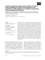
Figure 1
Indirect immunofluorescence staining pattern of anti-Rib-P-positive samplesIndirect immunofluorescence staining pattern of anti-Rib-P-positive samples. One anti-Rib-P-positive serum that did not have autoantibodies to other
known antigens (a-c) and the Centers for Disease Control and Prevention (CDC) anti-nuclear antibody reference serum number 12 (d-f) were
tested at dilutions of 1:500 and 1:100, respectively, on slides from three different suppliers. Significant differences were observed in patterns of
staining for the monospecific anti-Rib-P sera (a-c) on HEp-2 substrates from INOVA (San Diego, CA, USA) (a), ImmunoConcepts (Sacramento, CA,
USA) (b), and Euroimmun (Lübeck, Germany) (c). Furthermore, the indirect immunofluorescence of the high-titer CDC anti-Rib-P reference serum
produced only weak cytoplasmic staining on HEp-2 substrates from the same manufacturers (d-f). Rib-P, ribosomal P protein.
Table 2
Presence of Rib-P-like cytoplasmic indirect immunofluorescence pattern on HEp-2 substrates from different manufacturers
Manufacturer Low
a
MFU, number
(percentage)
Medium
b
MFU, number
(percentage)
High
c
MFU, number
(percentage)
All, number (percentage)
n = 15 n = 15 n = 15 n = 45
INOVA (San Diego, CA,
USA)
0 3 (20.0) 7 (46.6) 10 (22.2)
ImmunoConcepts
(Sacramento, CA, USA)
0 1 (6.7) 5 (33.0) 6 (13.3)
Euroimmun (Lübeck,
Germany)
0 0 3 (20.0) 3 (6.7)
a
Low = 20 to 49 median fluorescence units (MFU);
b
medium = 50 to 100 MFU;
c
high 100 MFU. Rib-P, ribosomal P protein.
Available online />Page 5 of 10
(page number not for citation purposes)
sera were anti-histone and anti-dsDNA by LIA and anti-chro-
matin by ALBIA, and the lowest prevalence was found for Jo-
1 by LIA and for Jo-1 and SS-B by ALBIA. Anti-Rib-P antibod-
ies were present in 33.3% of the dsDNA-negative sera and in
72.5% of the dsDNA-negative or borderline samples.
Anti-Rib-P reactivity in a systemic lupus erythematosus
cohort and controls
Anti-Rib-P reactivity was then analysed in a cohort of 100 SLE
patients by ALBIA, ELISA, IIF, and LIA. Sensitivity ranged from
11% (ALBIA) to 28% (ELISA), depending on the cutoff used.
The quantitative agreement between the results of ALBIA and
ELISA was 0.20 (confidence interval 0.01 to 0.38, two-tailed
P < 0.0443) according to Spearman (Figure 2). The qualitative
agreement between the different methods varied between
Table 3
Prevalence of autoantibodies in anti-Rib-P-positive samples (n = 51)
Positive + borderline, number (percentage) Positive, number (percentage)
Mikrogen ENA line assay
a
Histone 38 (74.5) 18 (35.3)
dsDNA 34 (66.7) 14 (27.5)
SmB 23 (45.1) 13 (25.5)
Ro52 22 (43.1) 17 (33.3)
SmD 21 (41.2) 6 (11.8)
SS-A/Ro60 21 (41.2) 13 (25.5)
Rib-P 19 (37.3) 13 (25.5)
RNPA 11 (21.6) 8 (15.7)
RNPC 8 (15.7) 5 (9.8)
SS-B/La 7 (13.7) 7 (13.7)
PCNA 6 (11.8) 3 (5.9)
RNP68 4 (7.8) 3 (5.9)
CENPB 3 (5.9) 2 (3.9)
Topoisomerase-I 2 (3.9) 1 (2.0)
Jo-1 1 (2.0) 1 (2.0)
ELISA
Rib-P 36 (70.6) 27 (52.9)
ALBIA
Rib-P 51 (100.0) 27 (52.9)
Chromatin 27 (52.9) 22 (43.1)
Ro52 13 (25.5) 12 (23.5)
SS-A/Ro60 11 (21.6) 8 (15.7)
RNP 8 (15.7) 2 (3.9)
Topoisomerase I 8 (15.7) 4 (7.8)
Sm 8 (15.7) 5 (9.8)
Jo-1 1 (2.0) 0 (0.0)
SS-B/La 1 (2.0) 1 (2.0)
a
Mikrogen GmbH (Neuried, Germany). ALBIA, addressable laser bead immunoassay; ELISA, enzyme-linked immunosorbent assay; ENA, anti-
extractable nuclear antigen; Rib-P, ribosomal P protein.
Arthritis Research & Therapy Vol 10 No 6 Mahler et al.
Page 6 of 10
(page number not for citation purposes)
0.23 and 0.43 (kappa) (Table 4). To ensure that the high sen-
sitivity of the ELISA was not accompanied by loss of specifi-
city, we tested a panel of disease controls (n = 100), which
showed 100% specificity. When anti-Rib-P reactivity was ana-
lysed in the context of anti-dsDNA antibodies as measured by
LIA, it was observed that 4 of 27 (14.8%) dsDNA-negative
SLE samples had anti-Rib-P reactivity by ALBIA, 3 (11.1%) by
ELISA, and 1 (3.7%) by LIA. Four out of 66 (6.1%) dsDNA-
negative or borderline samples were anti-Rib-P-positive by
ALBIA, 11 of 66 (16.6%) by ELISA, and 5 of 66 (7.6%) by LIA.
Anti-Rib-P reactivity in Centers for Disease Control and
Prevention anti-nuclear antibody reference sera
Anti-Rib-P reactivity was determined in the sera of the CDC
ANA reference serum panel by ELISA, LIA, ALBIA, and IIF on
HEp-2 cells. When the CDC Rib-P reference (ANA #12) sam-
ple was tested at a dilution of 1:100 by IIF on commercial HEp-
2 cell substrates, there was weak cytoplasmic staining on
slides from all three manufacturers (Figure 1). However, when
ALBIA, LIA, and ELISA methods of detecting anti-Rib-P anti-
bodies were used, this reference serum showed strong reac-
tivity (Table 5).
Discussion
Although most studies have found that anti-Rib-P antibodies
are highly specific for SLE, they have yet to achieve the clinical
impact that anti-Sm or anti-dsDNA antibodies have [17,18].
The reported prevalence of anti-Rib-P aab in SLE ranges from
10% to 40% [2,3], a variation that may be related to several
factors but appears to be significantly dependent on patient
selection, the antigens used, and the diagnostic platform
employed [2,3,11]. In a recent international multi-centre study
of 947 SLE patients and 1,113 controls, the high disease spe-
cificity (99.3%) of anti-Rib-P antibodies was confirmed [3]. Of
interest, in one patient initially diagnosed with rheumatoid
arthritis, anti-Rib-P antibodies predicted the later clinical con-
version into definite SLE [3,17]. Based on studies showing the
high positive predictive value of anti-Rib-P, it has been pro-
posed that, akin to anti-Sm and anti-dsDNA, anti-Rib-P may be
considered for inclusion as a criterion for the classification of
SLE [17,18].
The use of anti-Rib-P antibodies is often based on the under-
standing that they are a serological marker for neuropsychiat-
ric SLE (NPSLE). Thus, in a clinical setting in which there is
Figure 2
Correlation of addressable laser bead immunoassay (ALBIA) and enzyme-linked immunosorbent assay (ELISA)Correlation of addressable laser bead immunoassay (ALBIA) and enzyme-linked immunosorbent assay (ELISA). A correlation diagram was gener-
ated and the agreement was calculated according to Spearman, showing moderate agreement between the two assays. CI, confidence interval; DF,
degrees of freedom; LU, luminescence units; RU, relative units.
Available online />Page 7 of 10
(page number not for citation purposes)
some doubt as to the etiology of neuropsychiatric events (for
example, disease flare or pathogenesis), it is thought that the
detection of anti-Rib-P can assist in the differentiation of
NPSLE from psychoses and neurological features from other
causes. Despite substantial investigation, the relationship
between anti-Rib-P and organic central nervous system (CNS)
involvement and/or NPSLE remains controversial [17,22-24].
A recent meta-analysis has shown low sensitivity and specifi-
city and thus limited diagnostic value of anti-Rib-P for NPSLE
[25]. These discrepancies have been attributed to methodo-
logical differences in aab detection, the difference in the crite-
ria used to clinically define and identify various disease
features, the demographics and/or the make-up of the patient
cohorts, and the analysis of results [17].
Two recent investigations have provided new insights in the
putative association between anti-Rib-P aab and CNS mani-
festations. Matus and colleagues [26] used human anti-Rib-P
antibodies affinity-purified via an 11mer peptide derived from
the C-terminal part of the cognate protein to demonstrate
cross-reactivity with a new neuronal surface P antigen
(NSPA). In that study, the affinity-purified anti-Rib-P antibodies
caused rapid and sustained increase in calcium influx in neu-
rons followed by apoptotic cell death. The epitope on NSPA
responsible for the cross-reactivity has been speculated to
consist of the Rib-P epitope core GFGLFD and acidic struc-
tures constituting a conformational epitope. In another recent
investigation, anti-ribosomal P antibodies have been shown to
be associated with CNS disease only at the time of diagnosis
[27], an important observation that potentially explains previ-
ous controversies. In a mouse model, Katzav and colleagues
[28] were able to induce depression-like behaviour via intrac-
erebroventricular injection of affinity-purified anti-Rib-P anti-
bodies. Thus, the detection of anti-Rib-P antibodies might be
important not only for the diagnosis of SLE but also for the
evaluation of CNS complications, particularly at disease onset
and diagnosis.
Difference in assay performance
The observed differences in sensitivity of the anti-Rib-P anti-
body assays in the present study are in agreement with find-
ings from previous studies [2,7,13]. Apart from the
methodological differences, two types of antigens were used
in our study. The ELISA and ALBIA are based on a synthetic
peptide (C22), whereas recombinant antigens were used in
the LIA (P0) and EliA
®
Rib-P (P0, P1, and P2). Although
epitopes outside the major antigenic region (C22) are recog-
nised by anti-Rib-P antibodies on ribosomal P0, the LIA had
lower sensitivity when compared with other methods. This
might be explained by the relatively higher epitope concentra-
tion of the C22 peptide in the ELISA and ALBIA.
Table 4
Agreement between different methods in a cohort of 100
patients with systemic lupus erythematosus
ELISA (>1.5 RU) versus ALBIA (>350 LU)
ELISA + ELISA -
ALBIA + 8 3 11
ALBIA - 21 68 89
29 71
ALBIA (>350 LU) versus LIA
LIA + LIA -
ALBIA + 5 6 11
ALBIA - 5 84 89
10 90
ELISA (>1.5 RU) versus LIA
ELISA + ELISA -
LIA + 10 0 10
LIA - 19 71 90
29 71
ELISA (>1.5 RU) versus IIF (CSP)
ELISA + ELISA -
CSP + 7 4 11
CSP - 22 67 89
29 71
ALBIA (>350 LU) versus IIF (CSP)
ALBIA + ALBIA -
CSP + 3 8 11
CSP - 8 81 89
11 89
LIA versus IIF (CSP)
LIA + LIA -
CSP + 4 7 11
CSP - 6 83 89
10 90
Kappa agreements
ALBIA (350 LU) LIA IIF
ELISA 0.29 0.43 0.23
LIA 0.38 - -
ALBIA, addressable laser bead immunoassay; CSP, cytoplasmic staining
pattern; ELISA, enzyme-linked immunosorbent assay; IIF, indirect
immunofluorescence; LIA, line immunoassay; LU, luminescence units; RU,
relative units.
Arthritis Research & Therapy Vol 10 No 6 Mahler et al.
Page 8 of 10
(page number not for citation purposes)
Limited sensitivity of indirect immunofluorescence and
inter-manufacturer variation of patterns
The limited sensitivity of IIF for the detection of anti-Rib-P anti-
bodies reported in this study might be attributed to a combina-
tion of different factors. These factors may include a limited
antigen concentration in the cytoplasm, limited epitope expo-
sure of the antigens in the 60S subunit of the ribosome, inter-
ference or masking by other aab that produce a variety of
staining patterns, and a variety of cell preparation and fixation
protocols used by different manufacturers. In one of the semi-
nal studies of ribosome antibodies in SLE, it was noted that
only 1% had the accompanying CSP on human organ sec-
tions [29]. This observation is concordant with our findings as
approximately 10% to 20% of unselected SLE patients have
anti-Rib-P antibodies and only a small proportion of these
(approximately 10% to 30%) have a CSP on HEp-2 cells.
Although Rib-P antibodies have been known in some detail for
more than 20 years, it may have been that the limited sensitivity
of IIF for the detection of anti-Rib-P antibodies was the reason
these aab were not included in the classification criteria of SLE
[30]. It is not surprising that higher frequencies of anti-Rib-P
are now seen in SLE cohorts that correspond to the emer-
gence of more sensitive assays such as ELISA and ALBIA
[2,17]. Nevertheless, there is a prevailing notion that anti-Rib-
P antibodies are commonly detected by IIF on HEp-2 sub-
strates. Two case reports provide an interesting perspective
on this topic [31,32]. Paller and colleagues [31] reported an
18-year-old, female, ANA-negative patient with anti-Rib-P anti-
bodies that were detected by the relatively insensitive DID
technique. In the second case report, published by Sugisaki
and colleagues [32], a female ANA-negative patient with anti-
phospholipid syndrome, lupus nephritis, and anti-Rib-P aab
was described. Both reports emphasise the clinical impor-
tance of detecting anti-Rib-P antibodies in ANA-negative SLE
patients.
Our data suggest that anti-Rib-P are not commonly manifest
as an IIF CSP, although this was somewhat dependent on the
manufacturer of the HEp-2 kits. The percentage of ALBIA Rib-
P-positive sera that were reported to show a CSP was signif-
icantly higher in the small group of samples (n = 51) selected
for further analysis compared with the routine cohort of 345
samples. In the initial cohort, staining patterns were read in the
routine laboratory without special attention to the CSP. In con-
trast, the smaller cohort was retrospectively analysed with the
focus on anti-Rib-P aab reactivity known to the investigator. In
this context, it is important to mention that limitations in the
sensitivity of the IIF test are not restricted to the detection of
anti-Rib-P aab but have been observed for other aab (that is,
SSA/Ro, SSB, and Jo1) [33-35]. In a previous study, the sen-
sitivity of IIF as compared with an LIA as the primary assay for
the detection of extractable nuclear antigen (ENA) antibodies
was only 72% [33], and a significant portion of the IIF-nega-
tive/LIA-positive patients had clinical evidence of systemic
autoimmune rheumatic disorders (SARD). Therefore, the
authors concluded that, akin to IIF on HEp-2 cells, confirma-
tion assays such as LIA should be performed when there is a
clinical suspicion of SARD.
Impact of anti-Rib-P antibodies on the diagnosis of
systemic lupus erythematosus
Additionally, in a previous international study, 52 of 143
(36.3%) anti-Rib-P-positive samples have been reported as
anti-dsDNA-negative [3]. This observation is in keeping with
Table 5
Centers for Disease Control and Prevention reference standards tested for anti-Rib-P antibodies
Sample Specificity ELISA, RU
a
Interpretation ALBIA, LU
b
Interpretation LIA Interpretation
CDC1 Anti-dsDNA 1.0 Borderline 143 Negative 0 Negative
CDC2 Anti-SS-B/La 0.3 Negative 97 Negative 0 Negative
CDC3 Speckled ANA 0.4 Negative 80 Negative 0 Negative
CDC4 U1-RNP 0.6 Negative 157 Negative 0 Negative
CDC5 Sm 0.6 Negative 89 Negative 0 Negative
CDC6 Nucleolar; anti-fibrillarin 0.3 Negative 210 Negative 0 Negative
CDC7 SS-A/Ro60 0.8 Negative 149 Negative 0 Negative
CDC8 Centromere 0.5 Negative 119 Negative 0 Negative
CDC9 Topoisomerase-I 0.9 Negative 166 Negative 0 Negative
CDC10 Jo-1 0.3 Negative 75 Negative 0 Negative
CDC11 PM/Scl 0.5 Negative 126 Negative 0 Negative
CDC12 Rib-P 6.6 Positive 11,664 Positive 3 Positive
a
Cutoff = 1.5 relative units (RU);
b
cutoff = 350 luminescence units (LU). ALBIA, addressable laser bead immunoassay; CDC, Centers for Disease
Control and Prevention; dsDNA, double-stranded DNA; ELISA, enzyme-linked immunosorbent assay; LIA, line immunoassay; Rib-P, ribosomal P
protein.
Available online />Page 9 of 10
(page number not for citation purposes)
the findings of the present study, in which a significant portion
of anti-Rib-P-positive samples show no anti-dsDNA reactivity.
In patients who have anti-Rib-P antibodies but no antibodies to
dsDNA or Sm, confirmatory serology may be missing and this
in turn could result in an unfortunate delay of diagnosis and
treatment of such patients. Accordingly, we recommend that
the serological ACR criteria for the classification of SLE be
reconsidered and revised to include anti-Rib-P.
Conclusion
Based on our findings, we conclude that routine screening for
aab by IIF on HEp-2 cell substrates has low sensitivity and,
thus, limited reliability for the detection of anti-Rib-P in a rou-
tine clinical laboratory setting. Up to 90% of samples with anti-
Rib-P reactivity, depending on the anti-Rib-P titer determined
by ALBIA, were not accompanied by a CSP IIF staining pattern
on commercially prepared HEp-2 cells. Although the differ-
ence between other anti-Rib-P assays (such as ELISA, IB, LIA,
or EliA
®
) and the prevalence of IIF CSP was less pronounced,
a significant portion of samples demonstrated no evidence of
anti-Rib-P reactivity in IIF. Furthermore, we conclude that, as
for many other aab, the standardisation of assays requires fur-
ther effort and attention.
Competing interests
MM and JS-P are employed at Dr. Fooke Laboratorien GmbH,
which sells the Rib-P ELISA. TL is an employee of Mikrogen
GmbH, which manufactures diagnostic assays for autoim-
mune diseases. MJF is the director of Mitogen Advanced Diag-
nostics Laboratory, which provides diagnostic testing
services. JN declares that she has no competing interests.
Authors' contributions
MM developed the Rib-P ELISA, contributed to the study
design, evaluated the data, and participated in the preparation
of the manuscript. MJF contributed to the study design, pro-
vided laboratory resources, and participated in the preparation
and revisions of the manuscript. JS-P participated in data anal-
ysis and the preparation of the manuscript. TL helped in the
evaluation of data. JTN performed Rib-P assays and contrib-
uted to the data analysis. All authors read and approved the
final manuscript.
Acknowledgements
We thank Melanie Petschinka (Dr. Fooke Laboratorien GmbH), Mark
Fritzler, and Jane Yang (University of Calgary) for technical assistance.
MJF holds the Arthritis Society Chair at the University of Calgary. This
research was supported by a grant (MOP-38034) from the Canadian
Institutes of Health Research.
References
1. Francoeur AM, Peebles CL, Heckman KJ, Lee JC, Tan EM: Identi-
fication of ribosomal protein autoantigens. J Immunol 1985,
135:2378-2384.
2. Gerli R, Caponi L: Anti-ribosomal P protein antibodies. Autoim-
munity 2005, 38:85-92.
3. Mahler M, Kessenbrock K, Szmyrka M, Takasaki Y, Garcia-De La
Torre I, Shoenfeld Y, Hiepe F, Shun-le C, von Mühlen CA, Locht H,
Höpfl P, Wiik A, Reeves W, Fritzler MJ: International multicenter
evaluation of autoantibodies to ribosomal P proteins. Clin
Vaccine Immunol 2006, 13:77-83.
4. Kiss E, Shoenfeld Y: Are anti-ribosomal P protein antibodies
relevant in systemic lupus erythematosus? Clin Rev Allergy
Immunol 2007, 32:37-46.
5. Miyachi K, Tan EM: Antibodies reacting with ribosomal ribonu-
cleoprotein in connective tissue diseases. Arthritis Rheum
1979, 22:87-93.
6. Bonfa E, Elkon KB: Clinical and serologic associations of the
antiribosomal P protein antibody. Arthritis Rheum 1986,
29:981-985.
7. Ghirardello A, Caponi L, Franceschini F, Zampieri S, Quinzanini M,
Bendo R, Bombardieri S, Gambari PF, Doria A: Diagnostic tests
for antiribosomal p protein antibodies: a comparative evalua-
tion of immunoblotting and ELISA assays. J Autoimmun 2002,
19:71-77.
8. Bruner BF, Wynn DM, Reichlin M, Harley JB, James JA: Humoral
antigenic targets of the ribosomal P0 lupus autoantigen are
not limited to the carboxyl region. Ann N Y Acad Sci 2005,
1051:390-403.
9. Koffler D, Miller TE, Lahita RG: Studies on the specificity and
clinical correlation of antiribosomal antibodies in systemic
lupus erythematosus sera. Arthritis Rheum 1979, 22:463-470.
10. Zandman-Goddard G, Shoenfeld Y: Anti-ribosomal P antibod-
ies. In Autoantibodies 2nd edition. Edited by: Shoenfeld Y, Ger-
shwin ME, Meroni PL. San Diego, CA: Elsevier; 2007:217-223.
11. Martin AL, Reichlin M: Fluctuations of antibody to ribosomal P
proteins correlate with appearance and remission of nephritis
in SLE. Lupus 1996, 5:
22-29.
12. Mahler M, Kessenbrock K, Raats J, Williams R, Fritzler MJ, Bluthner
M: Characterization of the human autoimmune response to
the major C-terminal epitope of the ribosomal P proteins. J
Mol Med 2003, 81:194-204.
13. Mahler M, Kessenbrock K, Raats J, Fritzler MJ: Technical and clin-
ical evaluation of anti-ribosomal P protein immunoassays. J
Clin Lab Anal 2004, 18:215-223.
14. Lin JL, Dubljevic V, Fritzler MJ, Toh BH: Major immunoreactive
domains of human ribosomal P proteins lie N-terminal to a
homologous C-22 sequence: application to a novel ELISA for
systemic lupus erythematosus. Clin Exp Immunol 2005,
141:155-164.
15. Vercammen M, Meirlaen P, Sennesael J, Velkeniers B, T'Kint S,
Verbruggen L, Haentjens P, Broodtaerts L, Demanet C, De Waele
M: Diagnostic accuracy of the FIDIS multiplex fluorescent
microsphere immunodetection system for anti-extractable
nuclear antigen (ENA) antibodies in connective tissue
diseases. Clin Chem Lab Med 2007, 45:505-512.
16. Tzioufas AG, Tzortzakis NG, Panou-Pomonis E, Boki KA, Sakarel-
los-Daitsiotis M, Sakarellos C, Moutsopoulos HM: The clinical rel-
evance of antibodies to ribosomal-P common epitope in two
targeted systemic lupus erythematosus populations: a large
cohort of consecutive patients and patients with active central
nervous system disease. Ann Rheum Dis 2000, 59:99-104.
17. Mahler M, Raijmakers R, Fritzler MJ: Challenges and controver-
sies in autoantibodies associated with systemic rheumatic
diseases. Curr Rheumatol Rev 2007, 3:67-78.
18. Kessenbrock K, Raijmakers R, Fritzler MJ, Mahler M: Synthetic
peptides: the future of patient management in systemic rheu-
matic diseases? Curr Med Chem 2007, 14:2831-2838.
19. Hochberg MC: Updating the American College of Rheumatol-
ogy revised criteria for the classification of systemic lupus
erythematosus. Arthritis Rheum 1997, 40:1725.
20. Committee of Autoantibody Standardization, International
Union of Immunological Societies [
]
21. Smolen JS, Butcher B, Fritzler MJ, Gordon T, Hardin J, Kalden JR,
Lahita R, Maini RN, Reeves W, Reichlin M, Rothfield N, Takasaki Y,
van Venrooij WJ, Tan EM: Reference sera for antinuclear anti-
bodies. II. Further definition of antibody specificities in interna-
tional antinuclear antibody reference sera by
immunofluorescence and western blotting. Arthritis Rheum
1997, 40:413-418.
22. Eber T, Chapman J, Shoenfeld Y: Anti-ribosomal P-protein and
its role in psychiatric manifestations of systemic lupus ery-
thematosus: myth or reality? Lupus 2005, 14:571-575.
23. Reichlin M: Autoantibodies to the ribosomal P proteins in sys-
temic lupus erythematosus. Clin Exp Med 2006, 6:49-52.
Arthritis Research & Therapy Vol 10 No 6 Mahler et al.
Page 10 of 10
(page number not for citation purposes)
24. Toubi E, Shoenfeld Y: Clinical and biological aspects of anti-P-
ribosomal protein autoantibodies. Autoimmun Rev 2007,
6:119-125.
25. Karassa FB, Afeltra A, Ambrozic A, Chang DM, de Keyser F, Doria
A, Galeazzi M, Hirohata S, Hoffman IE, Inanc M, Massardo L, Math-
ieu A, Mok CC, Morozzi G, Sanna G, Spindler AJ, Tzioufas AG,
Yoshio T, Ioannidis JP: Accuracy of anti-ribosomal P protein
antibody testing for the diagnosis of neuropsychiatric sys-
temic lupus erythematosus: an international meta-analysis.
Arthritis Rheum 2006, 54:312-324.
26. Matus S, Burgos PV, Bravo-Zehnder M, Kraft R, Porras OH, Farías
P, Barros LF, Torrealba F, Massardo L, Jacobelli S, González A:
Antiribosomal-P autoantibodies from psychiatric lupus target
a novel neuronal surface protein causing calcium influx and
apoptosis. J Exp Med 2007, 204:3221-3234.
27. Hanly JG, Urowitz MB, Siannis F, Farewell V, Gordon C, Bae SC,
Isenberg D, Dooley MA, Clarke A, Bernatsky S, Gladman D, Fortin
PR, Manzi S, Steinsson K, Bruce IN, Ginzler E, Aranow C, Wallace
DJ, Ramsey-Goldman R, van Vollenhoven R, Sturfelt G, Nived O,
Sanchez-Guerrero J, Alarcón GS, Petri M, Khamashta M, Zoma A,
Font J, Kalunian K, Douglas J, et al.: Autoantibodies and neu-
ropsychiatric events at the time of systemic lupus erythemato-
sus diagnosis: results from an international inception cohort
study. Arthritis Rheum 2008, 58:843-853.
28. Katzav A, Solodeev I, Brodsky O, Chapman J, Pick CG, Blank M,
Zhang W, Reichlin M, Shoenfeld Y: Induction of autoimmune
depression in mice by anti-ribosomal P antibodies via the lim-
bic system. Arthritis Rheum 2007, 56:938-948.
29. Bianchi FB, Rizzetto M, Penfold P, Swana GT, Doniach D:
Ultrastructural localization and characterization of a ribosomal
antibody detected by immunofluorescence in systemic lupus
erythematosus. Clin Exp Immunol 1974, 17:629-636.
30. Tan EM: Autoantibodies to nuclear antigens (ANA): their
immunobiology and medicine. Adv Immunol 1982,
33:167-240.
31. Paller MS, Moore WS, Tan E, Schrier RW: Anticytoplasmic anti-
bodies in antinuclear antibody-negative lupus erythematosus.
Correlation with clinical course. Am J Med 1983, 75:529-533.
32. Sugisaki K, Takeda I, Kanno T, Nogai S, Abe K, Sakuma H, Kasu-
kawa R: An anti-nuclear antibody-negative patient with sys-
temic lupus erythematosus (SLE) accompanied with anti-
ribosomal P antibody (anti-P). Intern Med 2002, 41:1047-1051.
33. Hoffman IE, Peene I, Veys EM, de Keyser F: Detection of specific
antinuclear reactivities in patients with negative anti-nuclear
antibody immunofluorescence screening tests. Clin Chem
2002, 48:2171-2176.
34. Bossuyt X, Frans J, Hendrickx A, Godefridis G, Westhovens R,
Marien G: Detection of anti-SSA antibodies by indirect
immunofluorescence. Clin Chem 2004, 50:2361-2369.
35. Kidd K, Cusi K, Mueller R, Goodner M, Boyes B, Hoy E: Detection
and identification of significant ANAs in previously determined
ANA negative samples. Clin Lab 2005, 51:517-521.