Báo cáo y học: "Treatment of focal degenerative cartilage defects with polymer-based autologous chondrocyte grafts: four-year clinical results" pdf
Bạn đang xem bản rút gọn của tài liệu. Xem và tải ngay bản đầy đủ của tài liệu tại đây (1.08 MB, 11 trang )
Open Access
Available online />Page 1 of 11
(page number not for citation purposes)
Vol 11 No 2
Research article
Treatment of focal degenerative cartilage defects with
polymer-based autologous chondrocyte grafts: four-year clinical
results
Peter C Kreuz
1
, Sebastian Müller
2
, Christian Ossendorf
2
, Christian Kaps
3
and Christoph Erggelet
2
1
Department of Orthopaedic and Trauma Surgery, University Medical Center Rechts der Isar of the Technical University Munich, Ismaninger Str. 22,
81675 Munich, Germany
2
Department of Orthopaedic and Trauma Surgery, University Medical Center Freiburg, Hugstetter Str. 55, 79106 Freiburg, Germany
3
Department of Rheumatology, Charité Campus Mitte, Charité – Universitätsmedizin Berlin, Charitéplatz 1, 10117 Berlin, Germany
Corresponding author: Christian Kaps,
Received: 18 Sep 2008 Revisions requested: 28 Oct 2008 Revisions received: 4 Feb 2009 Accepted: 5 Mar 2009 Published: 5 Mar 2009
Arthritis Research & Therapy 2009, 11:R33 (doi:10.1186/ar2638)
This article is online at: />© 2009 Kreuz et al.; licensee BioMed Central Ltd.
This is an open access article distributed under the terms of the Creative Commons Attribution License ( />),
which permits unrestricted use, distribution, and reproduction in any medium, provided the original work is properly cited.
Abstract
Introduction Second-generation autologous chondrocyte
implantation with scaffolds stabilizing the grafts is a clinically
effective procedure for cartilage repair. In this ongoing
prospective observational case report study, we evaluated the
effectiveness of BioSeed
®
-C, a cell-based cartilage graft based
on autologous chondrocytes embedded in fibrin and a stable
resorbable polymer scaffold, for the treatment of clinical
symptomatic focal degenerative defects of the knee.
Methods Clinical outcome after 4-year clinical follow-up was
assessed in 19 patients with preoperatively radiologically
confirmed osteoarthritis and a Kellgren-Lawrence score of 2 or
more. Clinical scoring was performed before implantation of the
graft and 6, 12, and 48 months after implantation using the
Lysholm score, the Knee injury and Osteoarthritis Outcome
Score (KOOS), the International Knee Documentation
Committee (IKDC) score, and the International Cartilage Repair
Society (ICRS) score. Cartilage regeneration and articular
resurfacing were assessed by magnetic resonance imaging
(MRI) 4 years after implantation of the autologous cartilage graft.
Results Significant improvement (P < 0.05) of the Lysholm and
ICRS scores was observed as early as 6 months after
implantation of BioSeed
®
-C and remained stable during follow-
up. The IKDC score showed significant improvement compared
with the preoperative situation at 12 and 48 months (P < 0.05).
The KOOS showed significant improvement in the subclasses
pain, activities of daily living, and knee-related quality of life 6
months as well as 1 and 4 years after implantation of BioSeed
®
-
C in osteoarthritic defects (P < 0.05). MRI analysis showed
moderate to complete defect filling with a normal to incidentally
hyperintense signal in 16 out of 19 patients treated with
BioSeed
®
-C. Two patients without improvement in the clinical
and MRI scores received a total knee endoprosthesis after 4
years.
Conclusions The results show that the good clinical outcome
achieved 1 year after implantation of BioSeed
®
-C remains
stable over the course of a period of 4 years and suggest that
implanting BioSeed
®
-C is a promising treatment option for the
repair of focal degenerative defects of the knee.
Introduction
Cartilage lesions of the knee occur frequently and represent a
major health problem. Consecutive knee arthroscopies
showed that up to 63% of the patients with knee-related
symptoms suffered from chondral or osteochondral defects
[1,2]. These defects comprise focal osteochondral or chondral
lesions in 67%, osteoarthritic defects in 29%, lesions related
to osteochondritis dissecans in 2%, and other defects in 1%
of the cases [3]. Recently, a variety of surgical techniques that
aim for resurfacing and regenerating of the articular cartilage
have evolved. In the clinical routine, debridement, bone mar-
row-stimulating techniques, osteochondral autograft transfer,
and autologous chondrocyte implantation (ACI) are commonly
used cartilage repair techniques [4-8].
ACI: autologous chondrocyte implantation; ANOVA: analysis of variance; ICRS: International Cartilage Repair Society; IKDC: International Knee Doc-
umentation Committee; KOOS: Knee injury and Osteoarthritis Outcome Score; MRI: magnetic resonance imaging.
Arthritis Research & Therapy Vol 11 No 2 Kreuz et al.
Page 2 of 11
(page number not for citation purposes)
The first ACI was performed in 1987, and the clinical study of
Brittberg and colleagues [4] in 1994 represents the starting
point of cell-based cartilage repair and regenerative medicine.
Up to now, more than 15,000 patients worldwide have been
treated with ACI [9], and various reports documented the clin-
ical effectiveness of implanting autologous culture-expanded
chondrocytes for cartilage repair [10-13]. Although there is no
significant evidence that ACI produces superior clinical results
for the treatment of full-thickness articular cartilage defects
compared with other cartilage repair interventions [14,15], it is
regarded as a second-line treatment for small defects and a
first-line treatment for defects larger than 2 to 4 cm
2
[16].
For ACI, a small partial or full-thickness cartilage biopsy is
taken from a less weight-bearing area of the healthy articular
cartilage. The chondrocytes are harvested by enzymatic diges-
tion and cells are grown with autologous serum. For chondro-
cyte implantation, a periosteal flap or a collagen sheet is
sutured to the surrounding healthy cartilage rim, creating a res-
ervoir for the injection of the autologous chondrocyte cell sus-
pension. The need for an intact cartilage rim limits the use of
ACI to some regions of the knee, and the covering of the
chondrocyte suspension with a periosteal flap or a collagen
sheet may be insecure (for instance, in degenerative defects
that often miss an intact cartilage rim). In addition, potential
sources of complications may include periosteal hypertrophy,
loosening of the periosteal flap, ablation, and loss of cells into
the joint cavity [17-19]. These technical disadvantages of ACI
result in re-operations in up to 25% to 36% of the patients
[20,21]. Therefore, cartilage tissue engineering grafts that
address these disadvantages by using three-dimensional scaf-
folds stabilizing the graft and the regenerative potential of
autologous chondrocytes were developed. Meanwhile, clinical
results have shown the effectiveness of hyaluronan-based
[22,23], collagen-based [24,25], and resorbable polymer-
based [26] autologous chondrocyte grafts for the repair of car-
tilage defects.
Currently, ACI is contraindicated in osteoarthritic patients.
Nevertheless, preclinical studies suggest that chondrocytes or
mesenchymal stem cells from osteoarthritic patients may have
the capacity to form cartilage repair tissue and fulfill the pre-
requisites for use in ACI [27,28]. However, for cell-based car-
tilage therapies in osteoarthritis, it is important to harvest
unaffected healthy cartilage biopsies since healthy chondro-
cytes have been shown to form a cartilage tissue in vitro that
shows better morphology and a higher proteoglycan content
than chondrocytes derived from osteoarthritic joints [29]. Clin-
ically, it has been shown that microfracture treatment of
patients with moderate osteoarthritis improved their pain and
activity of daily living and significantly widened the joint spaces
1 year after treatment compared with the preoperative situa-
tion [30]. The effectiveness of a second-generation cartilage
graft based on hyaluronan has been shown for the treatment
of osteoarthritic knees with osteoarthritis not inhibiting the
regeneration sequence [31].
Recently, it was shown that the autologous cartilage graft Bio-
Seed
®
-C (BioTissue Technologies GmbH, Freiburg, Ger-
many) based on a bioresorbable two-component gel-polymer
scaffold is effective for the treatment of traumatic and focal
osteoarthritic cartilage defects of the knee [26]. The aim of the
present study was to evaluate ACI using BioSeed
®
-C for the
treatment of mild degenerative and focal osteoarthritic defects
of the knee. Magnetic resonance imaging (MRI) analysis of the
cartilage repair tissue as well as the clinical evaluation of a
series of 19 patients with pre-existing osteoarthritic symptoms
and a 4-year clinical follow-up document the effectiveness of
BioSeed
®
-C for the treatment of focal degenerative cartilage
defects.
Materials and methods
Patients
From December 2001 to October 2002, 79 patients with trau-
matic and degenerative chondral defects of the knee joint
were treated with a second-generation ACI (BioSeed
®
-C).
Patients suffered from traumatic, mild degenerative, or oste-
oarthritic and symptomatic defects of the articular cartilage of
the knee which were clinically significant [26]. The study was
performed in compliance with the ethical review board of the
University of Freiburg, Germany. All patients gave their con-
sent to participate. Radiographs were taken preoperatively,
and osteoarthritic degenerations were evaluated by two inde-
pendent observers using the Kellgren-Lawrence scoring sys-
tem. The observers were blinded to the procedure. A Kellgren-
Lawrence score of greater than or equal to 2 defines osteoar-
thritis [32] and was found in 24 patients. Nineteen patients
gave consent to a clinical follow-up of 4 years. Clinical exami-
nations were performed at 0, 6, 12, and 48 to 60 months.
Characteristics of patients with degenerative cartilage defects
are presented in Table 1. The average age of patients (8
females and 11 males; mean body mass index of 25, ranging
from 19 to 34) was 35 years (25 to 50 years). The mean defect
size of the first lesion was 4 cm
2
(2 to 6 cm
2
). All defects (first
lesion) were classified as Outerbridge class IV [33] and were
located on the medial femoral condyle (n = 14), the lateral fem-
oral condyle (n = 2), or the patella (n = 3). Four patients had a
second chondral defect that was treated with ACI as well. Pre-
vious surgical procedures were shaving (n = 10), abrasion
arthroplasty (n = 4), drilling/microfracture (n = 1), meniscecto-
mies (n = 6), anterior cruciate ligament/collateral ligament
reconstructions (n = 7), or high tibial osteotomy (n = 5).
Implantation of BioSeed
®
-C and follow-up treatment
For preparation of BioSeed
®
-C, autologous chondrocytes
were harvested from healthy cartilage (approximately 250 mg)
of a less weight-bearing area of the knee. One hundred millili-
ters of whole blood was collected with a conventional blood-
Available online />Page 3 of 11
(page number not for citation purposes)
sampling system (Sarstedt AG, Nümbrecht, Germany) and
used for autologous chondrocyte growth. Twenty million
chondrocytes were rearranged in fibrin and a polymer-based
scaffold (2 × 3 cm and 0.2 cm in height) of polyglycolic/poly-
lactic acid (polyglactin, vicryl) and polydioxanone. After careful
debridement of the defective cartilage down to the subchon-
dral bone, the graft was fitted to the size of the defect and
implanted arthrotomically. Fixation of the graft (Figure 1) was
achieved by transosseous anchoring as described previously
[34]. Starting the day after surgery, patients were subjected to
continuous passive motion. For 6 weeks, partial loading with
15% of body weight as well as isometric tension exercises
were allowed. In weeks 7 to 12, patients increased the loading
and performed strengthening exercises and active physiother-
apy at a gentle level. From week 13 on, patients gradually
increased the weight and performed muscular and coordina-
tion exercises up to full weight-bearing. Gentle exertion was
allowed after 6 months and more strenuous activities and con-
tact sports after 12 months.
Evaluation of clinical results
For evaluation of clinical results after implantation of Bio-
Seed
®
-C, the Lysholm score [35], the Knee injury and Oste-
oarthritis Outcome Score (KOOS) [36], and the International
Knee Documentation Committee (IKDC) Knee Examination
Form [37] were applied. The Knee Examination Form (sur-
geons' part) of the International Cartilage Repair Society
(ICRS) cartilage injury evaluation package evaluates the sub-
groups knee joint effusion, passive motion deficit, ligaments,
compartment, harvest site pathology, joint space, and func-
tionality [38]. The score was applied and grading was per-
formed from 1 (normal) to 4 (severely abnormal). The lowest
grade within a group determines the group rate, and the worst
group grade determines the final evaluation. The clinical situa-
tion was documented before and 6, 12 as well as 48 to 60
months after implantation of the graft. Forty-eight to sixty
months after transplantation, repair and resurfacing of carti-
lage defects (n = 17) were evaluated with a state-of-the-art 1.5
Tesla MRI scanner (Siemens AG, Erlangen, Germany) and the
Henderson scoring system was applied [13]. Hypertrophic
changes were classified with the Kreuz score [19].
Table 1
Characteristics of patients with degenerative cartilage defects
Characteristic Patient data
Gender Female (n = 8), male (n = 11)
Age, years 35 (range 25–50)
Height, cm 173 (range 162–184)
Weight, kg 75 (range 57–100)
Body mass index 25 (range 19–34)
Kellgren-Lawrence score 2 (n = 10)
3 (n = 9)
Defect size of 1st lesion, cm
2
4 (range 2–6)
Outerbridge classification of 1st lesion IV (n = 19)
Localization of 1st lesion Medial femoral condyle (n = 14)
Lateral femoral condyle (n = 2)
Patella (n = 3)
2nd lesion n = 4
Defect size of 2nd lesion, cm
2
3 (range 2–4)
Outerbridge classification of 2nd lesion IV (n = 4)
Localization of 2nd lesion Medial femoral condyle (n = 1)
Trochlea (n = 3)
Concomitant surgeries High tibial osteotomy (n = 5)
Anterior cruciate ligament reconstruction (n = 4)
Previous surgical procedures High tibial osteotomy (n = 5)
Shaving (n = 10)
Abrasion arthroplasty (n = 4),
Microfracture/drilling (n = 1)
Meniscectomy (n = 6)
Anterior cruciate ligament/collateral ligament reconstruction (n = 7)
Arthritis Research & Therapy Vol 11 No 2 Kreuz et al.
Page 4 of 11
(page number not for citation purposes)
Statistical analysis
For statistical analysis of the Lysholm and ICRS scores, the
Kruskal-Wallis test and the one-way analysis of variance
(ANOVA) of ranks test were used. For isolating the groups that
differed significantly (P < 0.05) from the others, the all-pair-
wise multiple comparison procedure (Dunn's method) was
applied. Statistical analysis of the IKDC data was performed
using ANOVA followed by the t test. Differences were consid-
ered significant at a P value of less than 0.05. For analysis of
the KOOS, the non-parametric Mann-Whitney rank sum test
was applied and differences were considered significant at a
P value of less than 0.05. All comparisons were performed
between scorings at every individual point in time of the follow-
up period compared with the preoperative scores.
Results
Postoperative findings in patients with focal
degenerative cartilage defects treated with BioSeed
®
-C
BioSeed
®
-C was implanted arthrotomically using a transos-
seous fixation technique (Figure 1). The degenerated cartilage
was debrided down to the subchondral bone, and the graft
was fitted to the size of the defect. For fixation, the graft was
armed in each corner with resorbable threads forming loops
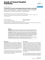
that were secured by threefold knots (Figure 1a). On every
corner of the defect, a k-wire with a thread guide was drilled
using the inside-out technique (Figure 1b). The k-wires that
carry guiding threads thread through the guide of the k-wire,
and the loops of the graft were pulled (inside-out) through the
femoral bone (Figure 1c). The threefold knots act as anchors
that seize within the subchondral bone and thus securely fix
the graft in the defect (Figure 1d).
BioSeed
®
-C was implanted in focal degenerative cartilage
defects of knees that showed radiological signs of osteoar-
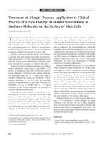
thritic degeneration (Figure 2). Applying the Kellgren-Law-
rence score to the preoperatively performed radiographs
showed that 24 patients had osteoarthritis with a Kellgren-
Lawrence score of 2 with constriction of the joint space (Fig-
ure 2a, black arrowhead) or a score of 3 with constriction and
formation of osteophytes (Figure 2b, white arrowheads). Nine-
teen out of twenty-four patients gave consent to clinical follow-
up conducted 4 years after implantation of the graft. Postop-
eratively, no clinical signs of persistent knee joint infection or
allergic reactions were evident. Knee joint effusion or swelling
was reported by 9 patients. Symptoms of temporary blocking
were observed in 4 out of 19 patients. None of the patients
acquired potentially graft-related autoimmune disorders or
signs of hypersensitivity. There were no signs of malignant
transformation, migration of chondrocytes, poisoning, toxicity,
organ failure, hepatic or renal disorders, or reproductive
defects or teratogenic effects. There were no signs of loosen-
ing, debonding, or ablation of the graft. Minimal asymptomatic
cartilage hypertrophic changes (<125%) were found in 3
patients, and abnormal cartilage growth was not evident. Nine
patients were subjected to second-look arthroscopy due to
symptoms like persistent grinding, catching, pain, or swelling.
The newly formed repair tissue showed good integration and
bonding as well as a visible contrast in color to the surround-
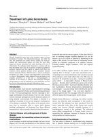
Figure 1
Arthrotomic implantation of BioSeed
®
-CArthrotomic implantation of BioSeed
®
-C. (a) BioSeed
®
-C was armed
in each corner with resorbable threads secured by threefold knots. (b)
In every corner of the defect, k-wires were drilled using the inside-out
technique. (c) Guiding threads were pulled through the femoral bone
using the k-wires, and the knots were guided into the subchondral
bone. (d) The knots serve as anchors, seizing the subchondral bone
and securely fixing the graft.
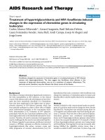
Figure 2
Radiographs of patients with focal degenerative cartilage defects prior to treatment with autologous chondrocyte grafts (BioSeed
®
-C)Radiographs of patients with focal degenerative cartilage defects prior
to treatment with autologous chondrocyte grafts (BioSeed
®
-C). (a)
This patient showed a Kellgren-Lawrence score of 2, with narrowing of
the joint space (black arrowhead). (b) This patient showed a Kellgren-
Lawrence score of 3, with narrowing of the joint space and osteophytes
(white arrowheads).
Available online />Page 5 of 11
(page number not for citation purposes)
ing tissue. In 1 patient, multiple lesions scattered across the
defect site and the retropatellar cartilage were observed. In 2
patients, a new cartilage lesion in the surroundings of the
transplanted area was detected and treated with abrasion
chondroplasty. In two other patients with persistent pain, the
ACI procedure failed and a total knee endoprosthesis was
implanted 4 years after implantation of the graft.
Clinical evaluation of surgical results four years after
implantation of BioSeed
®
-C
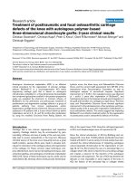
As assessed by the Lysholm score, statistically significant
improvements (P < 0.05) were observed as early as 6 months
after implantation of BioSeed
®
-C (Figure 3). Compared with
preoperative findings, the median Lysholm score significantly
improved (P < 0.05), increasing from 55.0 to 89.0 in patients
with focal osteoarthritic degeneration 4 years after implanta-
tion of the graft. The clinical outcome 4 years after implantation
of BioSeed
®
-C in osteoarthritic focal defects was evaluated
using the IKDC subjective knee evaluation score and the ICRS
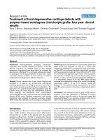
score (Figure 4). The IKDC score (Figure 4a) showed signifi-
cant improvement 1 year (P = 0.0068) and 4 years (P =
0.0017) postoperatively compared with the preoperative situ-
ation. The mean score increased from 49.0 to 70.1 after 4
years. In addition, the ICRS score improved significantly (P <
0.05) over the whole study period from 4.0 preoperatively to
2.0 at 4-year follow-up (Figure 4b).
The KOOS describes the patient's view about his knee and
associated problems (Figure 5). At 6-month, 1-year, and 4-
year follow-up, the patients' status improved significantly (P <
0.05) compared with preoperative findings. The median
scores increased in the subclasses pain (69 to 89), activities
of daily living (72 to 96), and knee-related quality of life (25 to
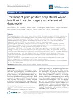
Figure 3
Clinical outcome after four years as evaluated by the Lysholm scoreClinical outcome after four years as evaluated by the Lysholm score.
Statistical analysis of the clinical outcome as assessed by the Lysholm
score was performed using analysis of variance on ranks (P <
0.00001) and subsequently the all-pairwise comparison according to
Dunn's method. Scores are presented as the median, with the ends of
the boxes defining the 25th and 75th percentiles and error bars defin-
ing the 10th and 90th percentiles. Where indicated (asterisks), differ-
ences were statistically significant (P < 0.05) compared with the
preoperative situation.
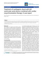
Figure 4
Clinical outcome after four years as evaluated by the International Knee Documentation Committee (IKDC) and International Cartilage Repair Society (ICRS) scoresClinical outcome after four years as evaluated by the International Knee
Documentation Committee (IKDC) and International Cartilage Repair
Society (ICRS) scores. (a) Statistical analysis of the clinical outcome
as assessed by the IKDC subjective knee evaluation score was per-
formed using analysis of variance (P < 0.007) and subsequently the t
test. Scores are presented as the mean, with error bars defining stand-
ard deviation. (b) The ICRS scores were statistically analyzed by using
analysis of variance on ranks (P < 0.000001) and subsequently the all-
pairwise comparison according to Dunn's method. Scores are pre-
sented as the median, with the ends of the boxes defining the 25th and
75th percentiles and error bars defining the 10th and 90th percentiles.
Where indicated (asterisks), differences were statistically significant (P
< 0.05) compared with the preoperative situation.
Arthritis Research & Therapy Vol 11 No 2 Kreuz et al.
Page 6 of 11
(page number not for citation purposes)
56) 4 years after implantation of the graft. Patients showed a
significant improvement in the subclass sports and recreation
(10 to 65) 4 years after implantation, whereas the subclass
symptoms showed no significant improvement (P > 0.05) of
the score (71 to 82). Both patients who needed total knee
replacement after 4 years did not improve in the scores over
the study period (P < 0.05). After 48 months, the Lysholm
score was 38.5 (24 to 53) points and the KOOS subclasses
symptoms and activity of daily life were 41 (38 to 44) points
each and significantly worse compared with the results of the
other patients (P < 0.05).
Magnetic resonance imaging four years after
transplantation of BioSeed
®
-C
Two patients had to undergo revision surgery and received a
total knee endoprosthesis. Therefore, 17 out of 19 patients
were analyzed by MRI 4 years after implantation of BioSeed
®
-
C. Patients with degenerative cartilage defects treated with
BioSeed
®
-C showed moderate to complete filling of the
defects (Figure 6). Eleven patients showed a complete filling
of the defect with cartilage repair tissue (Figure 6a). In 5
patients, the defects were filled more than 50% (Figure 6b),
and 1 patient showed a defect fill of less than 50% (Figure 6c).
The transosseous drill holes were still visible (Figure 6a,c,
white arrowheads). Representative MRIs as assessed preop-
eratively and at 4-year follow-up (Figure 7) show that cartilage
defects at the medial femoral condyle (Figure 7a,b) and patel-
lar defects (Figure 7c,d) were completely filled with cartilage
repair tissue after transplantation of the graft. A detailed MRI
analysis according to Henderson and Kreuz is given in Table
2. The cartilage signal in 16 out of 17 defects was normal or
showed slight alterations in the intensity. In one defect, the sig-
nal was hyperintense throughout large areas of the repair tis-
sue. Strong to moderate subchondral edema was evident in 6
patients, and 11 out of 17 patients showed no or mild edema.
Five patients showed moderate to strong signs of knee joint
effusion. No to mild knee joint effusion was evident in 12 out
of 17 patients treated with BioSeed
®
-C at 4-year follow-up.
Both patients who sustained a total knee endoprosthesis after
4 years had the last control MRI after 12 months. The defect
area was partially filled (<50%) with a hyperintensive repair tis-
sue and showed a concomitant moderate subchondral edema.
Discussion
The autologous gel-polymer cartilage graft, BioSeed
®
-C, uses
the well-known regenerative capacity of autologous chondro-
cytes, gel-like matrices for promoting tissue formation, and the
initial mechanical stability of resorbable polymer scaffolds for
cartilage repair [39]. The assembly of chondrocytes in poly-
mer-based scaffolds ensures the even distribution of a high
number of vital chondrocytes within the graft and has been
shown to allow the production of cartilage grafts that develop
toward hyaline cartilage in vitro [40]. In particular, the embed-
ding of culture-expanded chondrocytes in fibrin and polymer-
based scaffolds made of polyglycolic acid or copolymers of
polyglycolic and polylactic acid initiates chondrocyte re-differ-
entiation in vitro and allows for formation of cartilage matrix in
vivo after implantation of the graft [41,42]. The formation of
cartilage repair tissue after implantation of the polymer-based
graft made of non- and cryo-preserved chondrocytes has been
shown in the rabbit joint defect model [43] as well as in a large
animal horse model. In Haflinger horses, full-thickness carti-
lage defects of the fetlock joint were treated with the autolo-
gous polymer-based cartilage graft, and formation of a hyaline-
like cartilage repair tissue as well as firm bonding of the graft
to the adjacent healthy cartilage and to the subchondral bone
tissue were evident 1 year after implantation [44]. Since the
chondrocytes are embedded in and protected by the fibrin-
Figure 5
Clinical outcome after four years as evaluated by the Knee injury and Osteoarthritis Outcome Score (KOOS)Clinical outcome after four years as evaluated by the Knee injury and Osteoarthritis Outcome Score (KOOS). The KOOS is presented as a mean
value, and error bars define standard deviation. Where indicated (asterisks), differences were statistically significant (P < 0.05) compared with the
preoperative situation as assessed by the Mann-Whitney rank sum test. ADL, activities of daily living; QoL, quality of life; Sports/Rec, sports and
recreation.
Available online />Page 7 of 11
(page number not for citation purposes)
polymer matrix, BioSeed
®
-C allows for easy handling of the
graft during surgery, avoids the use of cover materials like peri-
osteum or collagen sheets, needs no healthy cartilage rim sur-
rounding the defect, and ensures arthroscopical implantation
and secure fixation [34]. In particular, the secure fixation of car-
tilage grafts is of importance to avoid transplant loosening,
debonding of the graft or ablation, and in turn clinical compli-
cations and re-operations. As assessed in recent biomechan-
ical studies, stable second-generation cartilage grafts for ACI
like BioSeed
®
-C allow good anchoring of the graft in the
defect by fibrin gluing, chondral or transosseous suturing, and
resorbable pin fixation [45-47]. From the cellular point of view,
particularly in an osteoarthritic environment, joint homeostasis
and the composition of the synovial fluid may be of special
importance in cartilage repair. In a goat model, it has been
shown that defects showed better repair when the lesion was
covered immediately with periosteum compared with defects
that were left untreated before transplantation [48]. Using a
chick limb bud assay, synovial fluid from patients with acute
traumatic cartilage defects has been shown to stimulate chon-
drogenesis, but synovial fluid from patients with chronic trau-
matic defects predominantly inhibited chondrogenic
development [49]. In contrast, synovial fluid obtained from
patients suffering from trauma, osteoarthritis, or inflammatory
rheumatoid arthritis stimulated the synthesis of proteoglycans
in a bovine model, with osteoarthritis and trauma synovial fluid
showing a markedly increased synthesis compared with rheu-
matoid arthritis [50]. In a rabbit model, it has been shown that
synovial fluid promotes and enhances cartilage tissue devel-
opment from perichondrium [51]. Although the influence of
synovial fluid or components of the fluid on cartilage repair
remains unclear and further studies are needed, it is evident
that an altered joint environment may influence tissue regener-
ation. Cartilage repair should be performed as early as possi-
ble and an inflammatory environment should be avoided.
From the clinical point of view, the use of autologous chondro-
cytes in suspension like in first-generation ACI has been
shown to be effective for the repair of localized traumatic
defects [10,11,52]. Second-generation cartilage grafts using
scaffolds based on collagen or hyaluronan for stabilizing autol-
ogous chondrocytes are considered to be technically more
attractive than first-generation ACI and have been shown to be
clinically as effective as 'classical' ACI [22,24]. In a recent
report of the treatment of traumatic and degenerative defects
[26] and in this case series, we demonstrated the safety and
effectiveness of the autologous cartilage gel-polymer graft
BioSeed
®
-C for the treatment of challenging defects like large
focal degenerative full-thickness cartilage lesions of the knee.
After 1-year follow-up, mean scores increased significantly
between 30% and 51% compared with the preoperative situ-
ation, depending on which score was analyzed. These good
results lasted and showed significant improvement of the
mean scores between 34% and 55% 4 years after implanta-
tion of the graft. This indicates a significant decrease in the
patients' pain and knee instabilities during activity as well as a
significant increase in patients' quality of life. The implantation
of BioSeed
®
-C in focal osteoarthritic defects showed signifi-
cant improvement in the KOOS subclasses pain, activity of
daily living, and quality of life 1 year and 4 years after implanta-
tion of the graft. Obviously, the efficacy of the polymer-based
autologous cartilage graft BioSeed
®
-C for the repair of oste-
oarthritic cartilage defects is shown by the improvement of
clinical scores, by patients' pain and quality of life as well as by
the good filling of the defects with repair tissue as assessed
by MRI. This is of special relevance since the BioSeed
®
-C-
mediated cartilage repair was achieved in a challenging
patient cohort with clinically osteoarthritic symptoms and focal
cartilage degeneration 1 and 4 years after implantation of the
graft. Interestingly, the status of the patient 2 years after ACI is
considered an important indicator for the future outcome. Dur-
ing this time, most of the complications of cell-based cartilage
repair as well as improvement in clinical scores and subjective
patient satisfaction were found. On the other hand, indicators
of a worse outcome like multiple surgical procedures, higher
age, and large defects correspond to findings published by
Figure 6
Magnetic resonance imaging (MRI) four years after implantation of BioSeed
®
-CMagnetic resonance imaging (MRI) four years after implantation of BioSeed
®
-C. (a) Out of 17 patients, 11 patients, including this one, showed com-
plete filling of the defect four years after implantation of BioSeed
®
-C. (b) Five patients, including this one, showed more than 50% defect filling but
not complete defect filling. (c) One patient showed less than 50% defect filling (black arrowhead). The repair tissue gives a slightly altered MRI sig-
nal, and transosseous drill holes are still evident (white arrowheads).
Arthritis Research & Therapy Vol 11 No 2 Kreuz et al.
Page 8 of 11
(page number not for citation purposes)
others [24,53]. In general, for measuring and evaluating the
clinical outcome of a given treatment strategy, patients' satis-
faction and improvement are most important and are best
assessed by well-established clinical outcome scores. From
the scientific point of view, additional detailed questions arise
regarding measurable parameters like the morphology and
quality of the formed repair tissue, chondrocyte viability and
distribution within the regenerative tissue as well as defect fill-
ing and graft integration. These issues can be addressed by
non-invasive MRI techniques as shown here or by dGEMRIC
(delayed gadolinium-enhanced MRI of cartilage) [54] as well
as by minimally invasive histological evaluation [13,26,55].
These techniques may offer measurable insights in clinical out-
come and value of the graft and may open avenues for devel-
oping precise indicators for the clinical outcome and
prognosis. However, for a comprehensive evaluation of a
regenerative cartilage repair therapy, both approaches are
mandatory, giving clinical scores for patients' satisfaction and
improvement as well as MRI and/or histological analysis for
repair tissue evaluation.
After implantation of BioSeed
®
-C, 9 out of 19 patients were
subjected to second-look arthroscopy due to symptoms like
grinding, catching, pain, or swelling. This re-intervention rate,
though for diagnostic purposes, is relatively high and may be
related to this challenging patient cohort with a considerable
number of concomitant surgeries like high tibial osteotomy and
anterior cruciate ligament reconstruction. However, only 4 out
of the 19 patients (21%) had to undergo revision surgery or re-
operation. This is in concordance with other studies reporting
rates of revision surgery of between 0% [56] and 25% [20].
Two patients were treated with an abrasion chondroplasty for
newly formed cartilage lesions beside the transplanted area.
Two additional patients out of the 19 patients received a total
knee endoprosthesis and were classified as 'graft failure',
although there is no evidence that the prosthesis was indi-
cated because of a failure of BioSeed
®
-C. This failure rate of
10% corresponds to earlier findings describing rates of failure
in ACI with other implants of between 5% [12] and 13% [20].
These patients with focal degenerative defects treated with
the chondrocyte graft were young and in the mid to long term
may have no treatment option other than total knee arthro-
plasty. Alternate treatment strategies such as ACI may lead to
several re-interventions and dissatisfied patients but may post-
pone total knee arthrosplasty as possibly the only option for
these patients and are therefore considered a good treatment
for focal degenerative cartilage defects. However, long-term
studies are needed to evaluate whether cell-based cartilage
grafts prevent total joint replacement in osteoarthritis. After 6
months, there was no difference in the scores of these 2
patients with an endoprosthesis and the rest of the study
group. After 12 months, these two patients with a 'graft failure'
had not yet improved and revealed worse results compared
with the other patients. After 12 months or after 48 months,
neither of the 2 patients reached 55 points in the Lysholm
score or 45 points in the IKDC score, indicating that there
might be a threshold for long-term graft survival and success-
ful tissue regeneration. Even if graft regeneration takes a long
time (from 2 to 3 years after surgery), a continuous improve-
ment should be detected 6 to 12 months after surgery. In this
context, the lack of clinical improvement, combined with insuf-
ficient MRI results, may be signs of long-term graft failure. In
general, re-operations and graft failure after implantation of
chondrocytes in a first-generation ACI procedure are caused
by problems associated with the periosteal flap [12,18,57].
This inherent technical disadvantage of the original ACI proce-
dure does not occur when using second-generation ACI grafts
like BioSeed
®
-C that are void of any cover materials. In addi-
tion, the easy and secure fixation of BioSeed
®
-C along with
the lack of any covering may reduce the operating time and
may result in a less invasive procedure since there is no need
to harvest periosteum from the tibia. Limitations of the study
are the small number of patients and the lack of a control
group. Since this observational case report study was initiated
first to gain insights in the safety and effectiveness of treating
consecutive cartilage defects with BioSeed
®
-C, the study is
Figure 7
Magnetic resonance imaging (MRI) before and four years after implan-tation of BioSeed
®
-CMagnetic resonance imaging (MRI) before and four years after implan-
tation of BioSeed
®
-C. (a) Preoperative MRI shows a cartilage defect
(encircled) at the medial femoral condyle. (b) After four years, MRI doc-
umented complete filling of the defect. Preoperatively, MRI shows a
patellar (c) cartilage defect (encircled) that was completely filled after
implantation of the graft as assessed by MRI at four years (d).
Available online />Page 9 of 11
(page number not for citation purposes)
further limited by the lack of predefined primary outcome
goals. The study was not performed in a randomized control-
led manner in comparison with an appropriate control group or
with an alternate treatment option like microfracturing. Further-
more, in young patients, degeneration of cartilage can be
observed after trauma whereas well-defined osteoarthritis
occurs predominantly in older patients. Therefore, first- or sec-
ond-generation ACI will not be recommended unrestrictedly
for the treatment of focal osteoarthritic cartilage defects. How-
ever, this is the first study showing long-term results using the
second-generation ACI graft BioSeed
®
-C in patients with
osteoarthritic and/or degenerative changes of the knee. The
study presents a continuous objective patient evaluation
including clinical scores and control MRI over the course of a
period of 4 years.
Conclusions
This extensive case report shows promising results after
implantation of the second-generation autologous cartilage
graft, BioSeed
®
-C, for the treatment of focal degenerative car-
tilage defects of the knee. Clinical evaluation 4 years after
implantation showed that the treatment of focal osteoarthritic
defects with BioSeed
®
-C leads to significant improvement of
the patients' condition as documented by reliable clinical out-
come scores and by cartilage regeneration as well as articular
resurfacing as assessed by MRI. The good clinical results
found 1 year after implantation of BioSeed
®
-C lasted and
remained stable for at least 4 years. Nevertheless, further long-
term studies are needed to evaluate whether cell-based carti-
lage grafts prevent total joint replacement in osteoarthritis.
Competing interests
CK is an employee of TransTissue Technologies GmbH, which
is a subsidiary of BioTissue Technologies GmbH (Freiburg,
Germany). BioTissue Technologies GmbH produces and dis-
tributes BioSeed
®
-C. CE works as a consultant for BioTissue
Technologies GmbH. All other authors declare that they have
no competing interests.
Authors' contributions
PCK helped to carry out the data evaluation, to draft the man-
uscript, and to perform the patient data collection and partici-
pated in data evaluation. CK helped to carry out the data
evaluation and to draft the manuscript. SM and CO helped to
perform the patient data collection and participated in data
evaluation. CE conceived the study, participated in its design
and coordination, performed the surgical procedures, and was
involved in the patient data collection and interpretation. All
authors read and approved the final manuscript.
Acknowledgements
None of the authors received funding for this study, for the study design,
for the collection, analysis and interpretation of data, or for preparing the
manuscript. No funding body had a role in the decision to submit the
manuscript for publication. Information or clinical photographs relating
to individual patients were made anonymous. Consent for the study was
obtained from each patient.
References
1. Curl WW, Krome J, Gordon ES, Rushing J, Smith BP, Poehling
GG: Cartilage injuries: a review of 31,516 knee arthroscopies.
Arthroscopy 1997, 13:456-460.
2. Hjelle K, Solheim E, Strand T, Muri R, Brittberg M: Articular carti-
lage defects in 1,000 knee arthroscopies. Arthroscopy 2002,
18:730-734.
3. Widuchowski W, Widuchowski J, Trzaska T: Articular cartilage
defects: study of 25,124 knee arthroscopies. Knee 2007,
14:177-182.
4. Brittberg M, Lindahl A, Nilsson A, Ohlsson C, Isaksson O, Peter-
son L: Treatment of deep cartilage defects in the knee with
autologous chondrocyte transplantation. N Engl J Med 1994,
331:889-895.
5. Hubbard MJ: Articular debridement versus washout for degen-
eration of the medial femoral condyle. A five-year study. J
Bone Joint Surg Br 1996, 78:217-219.
6. Matsusue Y, Yamamuro T, Hama H: Arthroscopic multiple oste-
ochondral transplantation to the chondral defect in the knee
associated with anterior cruciate ligament disruption. Arthros-
copy 1993, 9:318-321.
7. Steadman JR, Rodkey WG, Rodrigo JJ: Microfracture: surgical
technique and rehabilitation to treat chondral defects. Clin
Orthop Relat Res 2001, 391:S362-369.
8. Kreuz PC, Steinwachs MR, Erggelet C, Krause SJ, Konrad G, Uhl
M, Sudkamp N: Results after microfracture of full-thickness
chondral defects in different compartments in the knee. Oste-
oarthritis Cartilage 2006, 14:1119-1125.
9. Minas T: Autologous chondrocyte implantation in the arthritic
knee. Orthopedics 2003, 26:945-947.
10. Browne JE, Anderson AF, Arciero R, Mandelbaum B, Moseley JB
Jr, Micheli LJ, Fu F, Erggelet C: Clinical outcome of autologous
Table 2
Magnetic resonance imaging analysis four years after transplantation of the graft in focal degenerative cartilage defects
Characteristic Magnetic resonance imaging score
1 (normal) 2 (mild) 3 (moderate) 4 (abnormal)
Defect filling 11 (complete filling), including 3 with minimal
hypertrophic changes (<125%)
5 (>50%) 1 (<50%) 0 (no filling)
Signal intensity 8 (normal) 8 (some hyperintense areas) 1 (hyperintense areas) 0 (no signal)
Subchondral edema 4 (no edema) 7 (mild edema) 5 (moderate edema) 1 (distinct edema)
Effusion 1 (no effusion) 11 (mild effusion) 4 (moderate effusion) 1 (distinct effusion)
Arthritis Research & Therapy Vol 11 No 2 Kreuz et al.
Page 10 of 11
(page number not for citation purposes)
chondrocyte implantation at 5 years in US subjects. Clin
Orthop Relat Res 2005, 436:237-245.
11. Knutsen G, Drogset JO, Engebretsen L, Grontvedt T, Isaksen V,
Ludvigsen TC, Roberts S, Solheim E, Strand T, Johansen O: A ran-
domized trial comparing autologous chondrocyte implanta-
tion with microfracture. Findings at five years. J Bone Joint
Surg Am 2007, 89:2105-2112.
12. Peterson L, Minas T, Brittberg M, Nilsson A, Sjogren-Jansson E,
Lindahl A: Two- to 9-year outcome after autologous chondro-
cyte transplantation of the knee. Clin Orthop Relat Res 2000,
374:212-234.
13. Henderson I, Francisco R, Oakes B, Cameron J: Autologous
chondrocyte implantation for treatment of focal chondral
defects of the knee – a clinical, arthroscopic, MRI and histo-
logic evaluation at 2 years. Knee 2005, 12:209-216.
14. Magnussen RA, Dunn WR, Carey JL, Spindler KP: Treatment of
focal articular cartilage defects in the knee: a systematic
review. Clin Orthop Relat Res 2008, 466:952-962.
15. Wasiak J, Clar C, Villanueva E: Autologous cartilage implanta-
tion for full thickness articular cartilage defects of the knee.
Cochrane Database Syst Rev 2006, 3:CD003323.
16. Brittberg M: Autologous chondrocyte implantation – technique
and long-term follow-up. Injury 2008, 39:S40-49.
17. Driesang IM, Hunziker EB: Delamination rates of tissue flaps
used in articular cartilage repair. J Orthop Res 2000,
18:909-911.
18. Micheli LJ, Browne JE, Erggelet C, Fu F, Mandelbaum B, Moseley
JB, Zurakowski D: Autologous chondrocyte implantation of the
knee: multicenter experience and minimum 3-year follow-up.
Clin J Sport Med 2001, 11:223-228.
19. Kreuz PC, Steinwachs M, Erggelet C, Krause SJ, Ossendorf C,
Maier D, Ghanem N, Uhl M, Haag M: Classification of graft
hypertrophy after autologous chondrocyte implantation of full-
thickness chondral defects in the knee. Osteoarthritis Cartilage
2007, 15:1339-1347.
20. Minas T: Autologous chondrocyte implantation for focal chon-
dral defects of the knee. Clin Orthop Relat Res 2001,
391:S349-361.
21. Gooding CR, Bartlett W, Bentley G, Skinner JA, Carrington R,
Flanagan A: A prospective, randomised study comparing two
techniques of autologous chondrocyte implantation for osteo-
chondral defects in the knee: Periosteum covered versus type
I/III collagen covered. Knee
2006, 13:203-210.
22. Marcacci M, Berruto M, Brocchetta D, Delcogliano A, Ghinelli D,
Gobbi A, Kon E, Pederzini L, Rosa D, Sacchetti GL, Stefani G,
Zanasi S: Articular cartilage engineering with Hyalograft C: 3-
year clinical results. Clin Orthop Relat Res 2005, 435:96-105.
23. Nehrer S, Domayer S, Dorotka R, Schatz K, Bindreiter U, Kotz R:
Three-year clinical outcome after chondrocyte transplantation
using a hyaluronan matrix for cartilage repair. Eur J Radiol
2006, 57:3-8.
24. Bartlett W, Skinner JA, Gooding CR, Carrington RW, Flanagan
AM, Briggs TW, Bentley G: Autologous chondrocyte implanta-
tion versus matrix-induced autologous chondrocyte implanta-
tion for osteochondral defects of the knee: a prospective,
randomised study. J Bone Joint Surg Br 2005, 87:640-645.
25. Behrens P, Bitter T, Kurz B, Russlies M: Matrix-associated autol-
ogous chondrocyte transplantation/implantation (MACT/
MACI) – 5-year follow-up. Knee 2006, 13:194-202.
26. Ossendorf C, Kaps C, Kreuz PC, Burmester GR, Sittinger M,
Erggelet C: Treatment of posttraumatic and focal osteoarthritic
cartilage defects of the knee with autologous polymer-based
three-dimensional chondrocyte grafts: two year clinical
results. Arthritis Res Ther 2007, 9:R41.
27. Scharstuhl A, Schewe B, Benz K, Gaissmaier C, Buhring HJ,
Stoop R: Chondrogenic potential of human adult mesenchy-
mal stem cells is independent of age or osteoarthritis etiology.
Stem Cells 2007, 25:3244-3251.
28. Stoop R, Albrecht D, Gaissmaier C, Fritz J, Felka T, Rudert M,
Aicher WK: Comparison of marker gene expression in
chondrocytes from patients receiving autologous chondrocyte
transplantation versus osteoarthritis patients. Arthritis Res
Ther 2007, 9:R60.
29. Yang KG, Saris DB, Geuze RE, van Rijen MH, Helm YJ van der,
Verbout AJ, Creemers LB, Dhert WJ: Altered in vitro chondro-
genic properties of chondrocytes harvested from unaffected
cartilage in osteoarthritic joints. Osteoarthritis Cartilage 2006,
14:561-570.
30. Bae DK, Yoon KH, Song SJ: Cartilage healing after microfrac-
ture in osteoarthritic knees. Arthroscopy 2006, 22:
367-374.
31. Hollander AP, Dickinson SC, Sims TJ, Brun P, Cortivo R, Kon E,
Marcacci M, Zanasi S, Borrione A, De Luca C, Pavesio A, Soranzo
C, Abatangelo G: Maturation of tissue engineered cartilage
implanted in injured and osteoarthritic human knees. Tissue
Eng 2006, 12:1787-1798.
32. Kellgren JH, Lawrence JS: Radiological assessment of osteo-
arthrosis. Ann Rheum Dis 1957, 16:494-502.
33. Outerbridge RE: The etiology of chondromalacia patellae. J
Bone Joint Surg Br 1961, 43-B:752-757.
34. Erggelet C, Sittinger M, Lahm A: The arthroscopic implantation
of autologous chondrocytes for the treatment of full-thickness
cartilage defects of the knee joint. Arthroscopy 2003,
19:108-110.
35. Lysholm J, Gillquist J: Evaluation of knee ligament surgery
results with special emphasis on use of a scoring scale. Am J
Sports Med 1982, 10:150-154.
36. Roos EM, Roos HP, Lohmander LS, Ekdahl C, Beynnon BD: Knee
Injury and Osteoarthritis Outcome Score (KOOS) – develop-
ment of a self-administered outcome measure. J Orthop
Sports Phys Ther 1998, 28:88-96.
37. Irrgang JJ, Anderson AF, Boland AL, Harner CD, Kurosaka M,
Neyret P, Richmond JC, Shelborne KD: Development and valida-
tion of the international knee documentation committee sub-
jective knee form. Am J Sports Med 2001, 29:600-613.
38. International Cartilage Repair Society [ti
lage.org]
39. Sittinger M, Bujia J, Minuth WW, Hammer C, Burmester GR: Engi-
neering of cartilage tissue using bioresorbable polymer carri-
ers in perfusion culture. Biomaterials 1994, 15:451-456.
40. Bujia J, Sittinger M, Minuth WW, Hammer C, Burmester G, Kasten-
bauer E: Engineering of cartilage tissue using bioresorbable
polymer fleeces and perfusion culture. Acta Otolaryngol 1995,
115:307-310.
41. Endres M, Neumann K, Schroder SE, Vetterlein S, Morawietz L,
Ringe J, Sittinger M, Kaps C: Human polymer-based cartilage
grafts for the regeneration of articular cartilage defects. Tissue
Cell 2007, 39:293-301.
42. Kaps C, Frauenschuh S, Endres M, Ringe J, Haisch A, Lauber J,
Buer J, Krenn V, Haupl T, Burmester GR, Sittinger M: Gene
expression profiling of human articular cartilage grafts gener-
ated by tissue engineering. Biomaterials 2006, 27:3617-3630.
43. Perka C, Sittinger M, Schultz O, Spitzer RS, Schlenzka D, Burm-
ester GR: Tissue engineered cartilage repair using cryopre-
served and noncryopreserved chondrocytes. Clin Orthop Relat
Res 2000, 378:245-254.
44. Barnewitz D, Endres M, Kruger I, Becker A, Zimmermann J, Wilke
I, Ringe J, Sittinger M, Kaps C: Treatment of articular cartilage
defects in horses with polymer-based cartilage tissue engi-
neering grafts. Biomaterials 2006, 27:2882-2889.
45. Zelle S, Zantop T, Schanz S, Petersen W: Arthroscopic tech-
niques for the fixation of a three-dimensional scaffold for
autologous chondrocyte transplantation: structural properties
in an in vitro model. Arthroscopy 2007, 23:1073-1078.
46. Knecht S, Erggelet C, Endres M, Sittinger M, Kaps C, Stussi E:
Mechanical testing of fixation techniques for scaffold-based
tissue-engineered grafts. J Biomed Mater Res B Appl Biomater
2007, 83:50-57.
47. Drobnic M, Radosavljevic D, Ravnik D, Pavlovcic V, Hribernik M:
Comparison of four techniques for the fixation of a collagen
scaffold in the human cadaveric knee. Osteoarthritis Cartilage
2006, 14:337-344.
48. Saris DB, Dhert WJ, Verbout AJ: Joint homeostasis. The dis-
crepancy between old and fresh defects in cartilage repair. J
Bone Joint Surg Br 2003, 85:1067-1076.
49. Rodrigo JJ, Steadman JR, Syftestad G, Benton H, Silliman J:
Effects of human knee synovial fluid on chondrogenesis in
vitro. Am J Knee Surg 1995, 8:124-129.
50. Neidel J, Schulze M: Value of synovial analysis for prognosis of
matrix synthesis of transplanted chondrocytes. Orthopade
2000, 29:158-163.
51. Skoog V, Widenfalk B, Ohlsen L, Wasteson A: The effect of
growth factors and synovial fluid on chondrogenesis in peri-
Available online />Page 11 of 11
(page number not for citation purposes)
chondrium. Scand J Plast Reconstr Surg Hand Surg 1990,
24:89-95.
52. Fu FH, Zurakowski D, Browne JE, Mandelbaum B, Erggelet C,
Moseley JB Jr, Anderson AF, Micheli LJ: Autologous chondrocyte
implantation versus debridement for treatment of full-thick-
ness chondral defects of the knee: an observational cohort
study with 3-year follow-up. Am J Sports Med 2005,
33:1658-1666.
53. Peterson L, Brittberg M, Kiviranta I, Akerlund EL, Lindahl A: Autol-
ogous chondrocyte transplantation. Biomechanics and long-
term durability. Am J Sports Med 2002, 30:2-12.
54. Welsch GH, Mamisch TC, Hughes T, Zilkens C, Quirbach S,
Scheffler K, Kraff O, Schweitzer ME, Szomolanyi P, Trattnig S: In
vivo biochemical 7.0 Tesla magnetic resonance: preliminary
results of dGEMRIC, zonal T2, and T2* mapping of articular
cartilage. Invest Radiol 2008, 43:619-626.
55. Saris DB, Vanlauwe J, Victor J, Haspl M, Bohnsack M, Fortems Y,
Vandekerckhove B, Almqvist KF, Claes T, Handelberg F, Lagae K,
Bauwhede J van der, Vandenneucker H, Yang KG, Jelic M, Ver-
donk R, Veulemans N, Bellemans J, Luyten FP: Characterized
chondrocyte implantation results in better structural repair
when treating symptomatic cartilage defects of the knee in a
randomized controlled trial versus microfracture. Am J Sports
Med 2008, 36:235-246.
56. Loehnert J, Ruhnau K, Gossen A, Bernsmann K, Wiese M: Autol-
ogous chondrocyte transplantation (ACT) in knee joints. First
clinical results. Arthroskopie 1999, 12:34-42.
57. Minas T, Peterson L: Advanced techniques in autologous
chondrocyte transplantation. Clin Sports Med 1999, 18:13-44.